appendix - springer978-1-60327-407-4/1.pdf · is usuall y any need, especiall y if you memoriz e...
TRANSCRIPT

Appendix Acid-Base Made Easy
Some Definitions
LOGARITHM (TO THE BASE 10)
A logarithm is an exponent. It is a number that, if used as an exponent to 10, will give the number whose log it is.
EXAMPLES
103=1000 log 1000 = 3 10"3 = 0.001 log 0.001 = - 3
1003 = 2 log 2 = 0.3 10-o.3 = i/2 = o.5 log 0.5 = -0.3
Remember, log N + log M = log N x M, log N - log M = log N/M, log 10"N = log 1/10N, log VN = 1/2 log N. (See also A Homemade Log Table at the end of this section.)
P H
pH is the negative logarithm of the hydrogen ion concentration (i.e., pH = -log [H+] or 10"pH [H+]). (Note concentration is always molar.)
EXAMPLES
[H+] = 0.1M 0.1 = 10^ pH=l [H+] = 0.0001M 0.0001 = 10 "4 pH = 4 [[H+] = 0.5Af 0.5 = 5xlO"1 pH = -(0.7-1) = 0.3 [[H+] = 0.0002M 0.0002 = 2 x 10 ~ 4 pH = - (0.3 - 4) = 3.7 [H+] = 5 x 10"9M pH = - (0.7 - 9) = 8.3
ACID
Acid is any compound that can lose a hydrogen ion.
EXAMPLES
CH3COOH, CH3CH2OH, CH3NH3+, NH4
+
Note "strong acids," such as HCl, really do not fit this definition, see under Conjugate Base.
BASE
Base is any compound than can gain a hydrogen ion.
365
366 Passonneau and Lowry
EXAMPLES
CH3COO-, CH3CH2O-, CH3NH2, NH3
CONJUGATE BASE (OR ACID)
This is the base (or acid) in the following general equation:
acidM>ase + H+ [Eq.A-l]
i.e., every acid has its conjugate base (and every base has its conjugate acid). Strong acids and bases, such as HCl and NaOH, really do not fit this formulation. HCl in solution has already lost its H+ (i.e., it is completely dissociated and can be written H+C1"). NaOH is really Na+OH~ and the real base is OH", which qualifies because it can gain a H+:
OH + H * ^ H 2 0 [Eq.A-2]
BUFFER
Buffer is a mixture of a conjugate acid and base. It is a buffer (i.e., a pH buffer), because if H+ is added most of H+ will combine with the base. Eq. (A-l)], lessening the increase in H+ concentration that would otherwise result. Similarly, if H+ is removed from such a mixture, more will be formed from the acid [Eq. (A-1)], lessening the decrease in H+ concentration that would otherwise result. P.Ks AND THE HENDERSON-HASSELBALCH EQUATION
The more readily an acid loses H+, the stronger an acid it is. This strength is measured by its dissociation constant
[base][H+] [B][H+] K= —7—rrr— = rA1 [Eq. A-3]
[acid] [A] L M J When the acid is 50% dissociated, base and acid are equal and K = [H+]. For example, the dissociation constant of acetic acid is 2.5 x 10~5M. Therefore, in a mixture of equal molar concentrations of acetic acid and sodium acetate [H+] = 2.5 x 10"5M or pH = - (0.4 - 5) = 4.6.
Similarly, the dissociation constant of NH4+ is 5 x 10"10. Therefore, in
a mixture of equal molar concentrations of ammonium hydroxide (NH3 • H20) and NH/Cl", [H+] = 5 x 10"10 or pH = - (0.7-10) = 9.3.
Because of this relationship between AT and [H+], and by analogy with pH, the negative log o/K is calledpK. It is convenient to replace K and [H+] in Eq. [A-3] with pH and pK by taking the log of each side.
Appendix 367
logK = log 1 1 1 0 0 = log 111+ log [H+] r c
[A] [A] ^ - A"41
rearranging:
-log[H1 = -logK + logMorpH = pK + log[Bl r F A _ [A] [A] m' A ° J
This is the famous Henderson-Hasselbalch equation. This equation was originally written:
pH = pK + log [SaUl [ E q A-6] [Acid]
This was before Bronsted proposed the definition of acids and bases given above. Equation [A-6] is harder to use than Eq. [A-5], and is not quite exact.
From the pK of an acid and the Henderson-Hasselbalch equation, it is easy to calculate the pH for any ratio of acid to base.
Base/acid 100 1/100 20 1/20 10 1/10
PH pK + 2 pK-2 pK+ 1.3 ptf-1.3 pK+l pK-l
Base/acid 2 1/2 1 200 5000 10,000
PH pK + 0.3 pK-03 pK pK+23 ptf+3.7 pK+4
It is easy to make this calculation in your head, as accurately as there is usually any need, especially if you memorize the log of 2 and 1.25 (0.3 and0.1).(SeeAHomemade Log Table fefow>.)Youcanevenforgetwhether it is base/acid or acid/base in the H-H equation if you simply remember that, if base predominates, the pH is on the basic side of the pK, and if acid predominates, the pH is on the acid side of the pK.
However, if you prefer, Table 1 makes the calculation for you and gives the percentage of the buffer that is in the basic form. Notice it takes three times more acid or base to make a pH change of 0.1 unit near the pK than it does 1 pH unit away. At 1.3 units from pK, a 1% shift from acid to base makes a 0.1 unit pH change. Thus, the useful pH range is limited to about 1 pH unit on either side of the pK.
368 Passonneau and Lowry
Table 1 Base:Acid Ratios to Give Desired pH
pH pK+ 1.0 pK+0.9 pK+O.S pK + 0.1 pK+0.6 pK+0.5 p£+0.4 pK+03 pAT+0.2 pK+O.l p£+0.0
MAKING
Base acid 10 8 6.3 5 4 3.2 2.5 2 1.6 1.25 1.0
A BUFFER
Base as % ofA + B
91 89 86 83 80 76 71 67 61 56 50
pH p*-1.0 pK-0.9 ptf-0.8 ptf-0.7 pK-0.6 pK-0.5 pK-OA pAT-0.3 pK-0.2 pAT-0.1 pAT-0.0
Base acid
~~10 8 6.3 5 4 3.2 2.5 2 1.6 1.25 1.0
Base as % ofA + B
9 11 14 17 20 24 29 33 39 44 50
A buffer mixture can be made by either (a) mixing the free acid or base with its salt, or (b) adding a calculated amount of strong acid or strong base to the free base or free acid. For example, a pH 4.6 buffer can be made by making the solution 50 mM in acetic acid and 50 mM in sodium acetate, or it can be made by making the solution 100 mMin acetic acid and 50 mM in NaOH. This ignores the fact that some H+ had to be produced (10"4 6M) from the acetic acid. Therefore, the base (acetate) is in fact slightly greater than the added sodium or NaOH. In this case, the difference is only 1 part in 2000. Only below pH 3 and above pH 11, and with low buffer concentrations, does this ever become significant.
MIXTURE OF Two OR MORE ACID-BASE PAIRS
The H-H equation must apply simultaneously to all acid-base pairs in a given solution, i.e.:
[Bi] [B2] pH = pK! + log ^ = pK2 + log p ^ etc. [Eq. A-7]
Take as an example a mixture of acetic acid, acetate (pAT=4.6), Tris+, and Tris base (pK = 8.1). At pH 7:
log [BJ/tAJ = log [Ac]/[HAc] = 2.4 = log 500/1 and A g ]
log [BJ/[AJ = log [Tris]/[Tris*] = -1.1 = log 1/12.5
Appendix 369
DISSOCIATION CONSTANT OF H20
There is one acid that must not be forgotten; this is H20. It is a very weak acid; its pK is 15.7. That means that:
rtrifOHl = 10157 M and pH = 15.7 + log FOH1 [Eq. A-9] [H20] [H201
Because the concentration of H20 is practically constant in most aqueous solutions (55.5M), it has become customary to merge its concentration with the dissociation constant:
[H1[OH] = 55.5 x 10157 = 10"14 [Eq. A-10]
In the logarithmic form, this becomes:
pH + pOH = 14 or pOH = 14 - pH ^ A*nJ
where pOH is the negative log of [OH-]. PH OF FREE ACIDS AND BASES
The pH of a weak acid (or base) in the absence of added conjugate base (or acid) is easily calculated in all practical situations. When an acid is dissolved in H20, A<-» B + H+ (i.e., B = H+). Equation [A-3] rearranged becomes [B][H+] =£[A], or in this case [H+]2 = K(A)9 and [H+] = V#[A], or:
pH = l/2(pK-log[A]) [Eq.A-12]
For example, O.IM acetic acid has pH = 1/2(4.6 + 1) = 2.8. A 0.01M solution (-log [A] = 2) would have a pH of 3.3. The pK of NH4
+ is 9.3. A 50 mM solution of NH4C1 has pH = 1/2(9.3 + 1.3) = 5.3. The pH of a IM solution would be 4.65.
When a free base is dissolved in H20, it removes H+, forming hydroxyl ion: B + H 2 O H A + OH". Here [A] = [OH"] = 10"14/[H+] (see above). Equation [A-3] rearranged becomes [H+]/[A] = K/[B], Substituting for [A]:
[H*]2/10-14 =K/[B], [H*]2 = 1014 K/[B], and [H+] = Vl014K/[B], or
pH= 1/2(14 + pK + log [B]) ^ A"13l
For example, 0.2M Tris base has pH = l/2( 14 + 8.1 - 0.7) = 10.7. A 0. IM sodium acetate solution has pH = 1/2(14 + 4.6 - 1) = 8.8. (An easy way to remember Eqs. [A-12] and [A-13] is that an acid has a pH halfway
370 Passonneau and Lowry
between its ptfand the pH it would have if it were a strong acid, and a base has a pH halfway between its pK and the pH it would have if it were a strong base.)
Equations [A-12] and [A-13] are not strictly true, because they ignore the decrease in A or in B required to form H+ in one case and OH" in the other. The difference is exceedingly small, except with low concentrations of acids having pKs below 3 or bases with pKs over 11.
SOME COMPLICATIONS WITH RKS
We have been talking about pKs and dissociation constants as though they were invariant. Actually, they are affected by temperature and salt concentration (ionic strength). Increasing temperature and increasing ionic strength increase the dissociation constants (decrease the pKs). Tables of pKs are often calculated for 25° for "infinite dilution" (zero ionic strength). An observed pK is therefore, always lower than these "true" pKs, and should properly be called an apparent pK and written pK.
Ionic strength has a much greater effect if the dissociation involves a separation of charges than if it does not. Consider the three dissociations:
RNH3+^RNH2+H+
[Re. A-l] RCOOH % RCOO + H*
RP02H % RP022 + H+
Ionic strength will have little effect in the first case, a moderate effect in the second, and a large effect in the third. (Increasing ionic strength by increasing the dielectric constant of the solution, shields the charges from each other, making it easier for them to come apart.)
As examples, the true pK of acetic acid is given as 4.76, but a 0.1M acetate buffer has a plC near 4.6. The pK2 of a phosphate buffer is 7.2, a 0. IM phosphate buffer has a pKj of 6.8, and in a IM phosphate buffer the pK2' is around 6. (The pK3 is even more sensitive to ionic strength.) True pKs are given in Table 2, and in some cases, ionic strength effects may need to be taken into account.
There is one final complication in regard to pKs. We have been considering NH4
+, for example, as an acid with pK = 9.3 (i.e., a dissociation constant of 5 x 10~10. Unfortunately, in preBronsted days, NH3 was regarded in solution as NH4OH, which dissociated as NH4OH <-> NH4
+ +
Appendix 371
OH". Measurements of OH" concentration indicated a dissociation constant of 2 x 10"5; or pK=4.7. To distinguish these, one is written pKa (for the acid dissociation constant); the other is written pKb (for the "base dissociation constant"). It is clear that, since pOH + pH = 14, pKa + pKb
= 14. pKbs are still to be found in handbooks, often written simply as pK, and it is not always clear whether p£a or pKb is meant.
A HOMEMADE LOG TABLE
All the logarithms needed for practical acid-base aspects of most analytical work can be carried in your head, or figured out quicker than you can look up a log table. All you need to remember is that the log of 2=0.3 (actually 0.30103) and the log of 1.25 = 0.1 (actually 0.0969). Knowing the log of 2, it is clear that log 4 = 0.6 and log 8 = 0.9. Similarly, log 5 = 0.7 (i.e., log 10 - log 2), and log 2.5 = 0.4 (i.e., log 5 - log 2). We are only missing 0.2,0.5, and 0.8, which we obtain as the following tabulation:
log 1 = 0.0 log 1.25 = 0.1 (memory) log 1.6 = 0.2 (log 2-log 1.25)
log 2 = 0.3 (memory) log 2.5 = 0.4 (log 10-log 4) log 3.2 = 0.5 (log4-log 1.25)
log 4 = 0.6 (log 2 +log 2) log 5 = 0.7 (log 10-log 2)
log 6.3 = 0.8 Gog 5 +log 1.25) log 8 = 0.9 (log 4 +log 2)
log 10 =1.0
If you need to be closer than 0.1 pH unit, you can interpolate from this, e.g., log 9 is close to 0.95 (0.954). This list and the Henderson-Hasselbalch equation, both in your head, will save time and, more importantly, avoid blunders. STATISTICAL SHORTCUTS
This section is aimed at the "casual statistician" (Snedecor). Rarely, for his purposes, does the situation require or justify elaborate statistical treatment.
We are concerned here only with means, standard deviations, standard errors (of the mean), and standard errors of differences.
First, the orthodox calculations: Standard deviation (St. dev.) = Vsum of dev2 ^Eq A . 1 4 j
n - 1

372 Passonneau and Lowry
where dev is the individual deviation from the means and n is the number of samples.
[Eq. A-15]
Standard error (of the mean) (SE or SEM) = Vsum of dcvT=st dev n(n - 1) Vn
i - T [Eq.A-16] Standard error of the difference (SED) = vSEj2 + SE/
There is a longer way of calculating SED, but it has no clear advantage. Now the shortcuts.
1. For most purposes, calculation of SE to within 10% is all that is worthwhile. For example: 110 ± 10 (SE) says that there is a 1 out of 3 chance that the true mean lies outside of the range 90-110. What virtue could there possibly be in calculating that the SE is actually 10.3?
2. Conversely, calculation of the mean to closer than 10% of the SE is seldom worthwhile. In the previous example, what possible use would there be in calculating that the average is actually 110.4? If, however, the standard error was 1 instead of 10, it might be useful to calculate the mean more closely, say ± 0.1.
3. An approximate value for the SE is: SE = range of values ^ A"17]
n For example, if there are 10 values ranging from 80 to 120, SE = 40/10 = 4. This gives a surprisingly close estimate of the SE (usually within 15%). It is a valuable check for errors in more exact calculations. (Note, however, that with n > 12 or 13, this gives erroneous small estimates of SE).
4. Similarly, because SE = SDNn:
st. dev. = SEVn = range [Eq. A-18] Vn
5. For more orthodox calculations of means, SEs, and SDs, individual values and individual deviations from the mean can usually be rounded off with great saving of time. What guidelines are there to safe rounding off?
It is usually completely safe to round off individual values to within 10% of the range. For example, there are 10 numbers ranging from 32.2 to 43.6. The range is 11.4. Therefore, the numbers can be rounded off to the nearest whole number. No number will be changed by more than 0.5. This cannot increase the range by more than twice 0.5 or 1 (i.e., 9%).
Appendix 373
Therefore, this will not increase the SE or the SD by more than 9%, as the above shortcut method for calculating these statistics indicates.
Rounding off to within 10% of the range will have a completely negligible effect on the mean. Thus, in the above case, the average number is changed by only 0.25, and changes are randomized plus and minus.
Table 2 Dissociation Constants for Acids and Bases Used in Making Buffers
Acid or base
Oxalic acid, Kj P-glycerophosphoric, Kt
Histidine, K, Maleic, Kt
Phosphoric, Kt
Aspartic, Kj Glutamic, K, Arsenic, K, Lysine, Kx
Glycine, Kt
Phthalic, Kt
Tartaric, IQ Citric, Kj Fumaric, Kt
Glycylglycine, Kj Malic, Kj Formic Aspartic, K2
Lactic Barbituric Glutamic, K2
Tartaric, K2
Succinic, Kt
Oxalic, K2
Fumaric, K2
Citric, K2
Acetic Malic, K2
Benzoic Phthalic, K2
Succinic, K2
Maleic, K2
Pyrophosphoric, K3
Uric Citric, K3
Histidine, K2
pK> 1.21 1.47 1.73 1.83 1.96 2.14 2.14 2.25 2.25 2.35 2.92 3.00 3.05 3.10 3.10 3.40 3.76 3.87 3.87 4.00 4.09 4.13 4.13 4.22 4.53
4.75 4.76 5.00 5.20 5.57 5.57 5.75
5.77 5.83 6.00 6.10
Acid or base
4-Phenylimidazole MES** P-glycerophosphoric, K2
Cacodylic
2-Phenylimidazole 4-Hydroxyimidazole Arsenic, K2
Imidazole Phosphoric, K2
BESW
MOPSc
2,4,6-Collidine TES** HEPESeA
4-Methyimidazole Triethanolamine 2-Methyimidazole Glycylglycine, K2
Tri/ Tricine*' Pyrophosphoric, K2
Hydrazine, K2
2,4-Dimethylimidazole 2-Amino-2-methyl-1.3
propanediol Lysine Histidine, K3
Ammonium hydroxide Glutamic acid, K3
Glycine, K2
Aspartic acid, K3
2-Amino-2-methyl-l -propanol
Carbonic acid, K2
Lysine, K3
Arsenic, K3
Phosphoric, K3
PKa 6.13 6.15 6.19 6.19
6.53 6.53 6.77 7.07 7.12 7.15 7.2 7.40 7.5 7.55 7.65 7.9 8.00 8.07 8.1 8.15 8.22 8.23 8.48
8.78 8.92 9.14 9.23 9.43 9.75 9.75
9.90 10.36 10.54 11.6 12.24
aMES, 2-(N-morpholino)ethanesulfonic acid. *BES, tyN-fcw(2-hydroxyethyl)-2-aminoethanesulfonic acid. cMOPS, 3-N-(Morpholino)propanesulfonic acid. *TES, N-fris(hydroxymethyl)methyl-2-aminosulfonic acid. 'HEPES, N-2-hydroxyethylpiperazine-W-2-ethanesulfonic acid. fTris, rrw(hydroxymethyl)aminoethane. *Tricine, AT-rrw(hydroxymethyl)methylglycine. *At 20°, Good et al., 1966. 'Good, 1962. 3 4
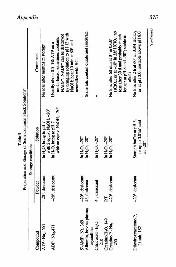
Appendix 375
O U
O
a 'o CO
CO
§
o
I
•I a, &
I .5
I 1
•I O O
5 §
3 3
5C o
P II
§ § . S
^ * 2
£ € • » • *
£ £M3
C/3
•8 1 o * O
T3 C * t?
!Z
1
$ cs
a 55
1
I
1 o
I CO
ll p b7 3
8
en .5 rf & <t>
a, •§ I
8
o
tsiCjl
do X X c cs
n i a
o o ^7 do WW a a
3
OS
m <
m T3
w 3
£5 S3
.S <D W-i
B CO
8 1-H
o d o fe a, CM 9 U i O
1
c3
•8 O
cu <L> c 2 § £ >> x
1 >% 5
CM
00 * - H
<-T
1 J
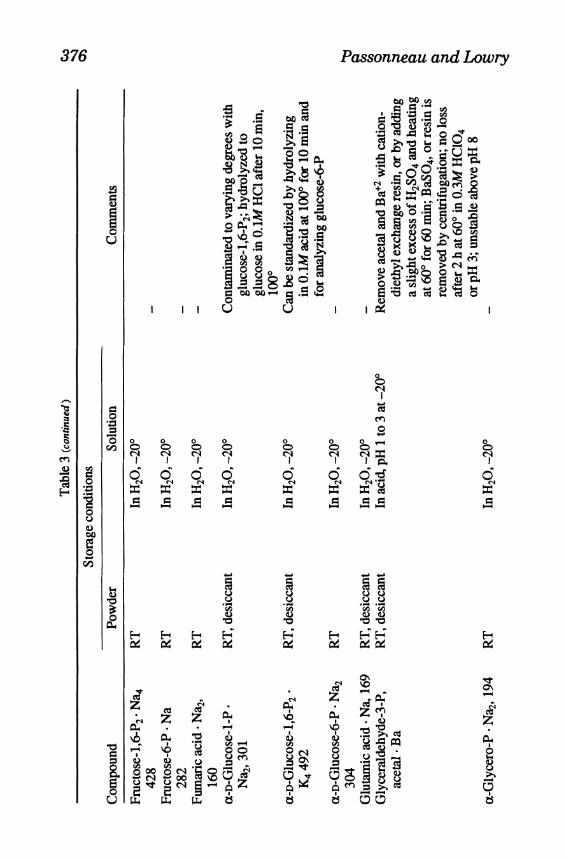
376 Passonneau and Lowry
g
O U
I I
60 2 ? 2 e C , H
I i I "3
«f .a 8 is
§ S 8 J
gftfe 2
° r f
§53
I* S o „. * S J2
• 8 -> cs 2 «-a a 8*a
5
§
"2
i en
§ -a a
, o CO
o
1 o
I CO
5
o O
6
£
o
d n
J3
*
$ G
O
d
a
d
a
o 7 « o 2
&Z
5 ^
o
°: a
1-8
H H 0d c*
£ * £ O. \6 &<
ruct
ose-
1 42
8 ru
ctos
e-6
282
PL, Oi
H Pd
i #
•a
is a ,—• u-
§ 8
• *N C/5
H C*
CL. i *-H
& 8 9
5 i Q i a
o cn
1
g 8
• *N
1 H tf
• vq i-H*
&
9 8
H cd
z a.
i VO
y 8 3 3
I
8
§ £ 8 8 11 H H Pd &
i • N
a, 1
69
de-3
-P,
luta
mic
aci
c ly
cera
ldeh
y ac
etal
• B
a
O O
H &
1 i
5 i 8

Appendix 377
i <
!> 2? 2 c ^3 x Q -tf 5 © D
i i i i i •8 C
a I CO
8
o o O
7 9, s ^
*
3 c
b
00
d
s
9, s
<3 P
90, WW .<= s
O U
O 00
m 3 O
I .4 S S r i U
I
3
& g g
8 1 b T
^ S 5 8
a ; * <?
c DO
8 >>
5
£ , & ! in * 1-5 & g |
w c s ^ w o .2 ^ H
en
i 0 \
8
3 f *
S CCL
00
o
I «
CO.
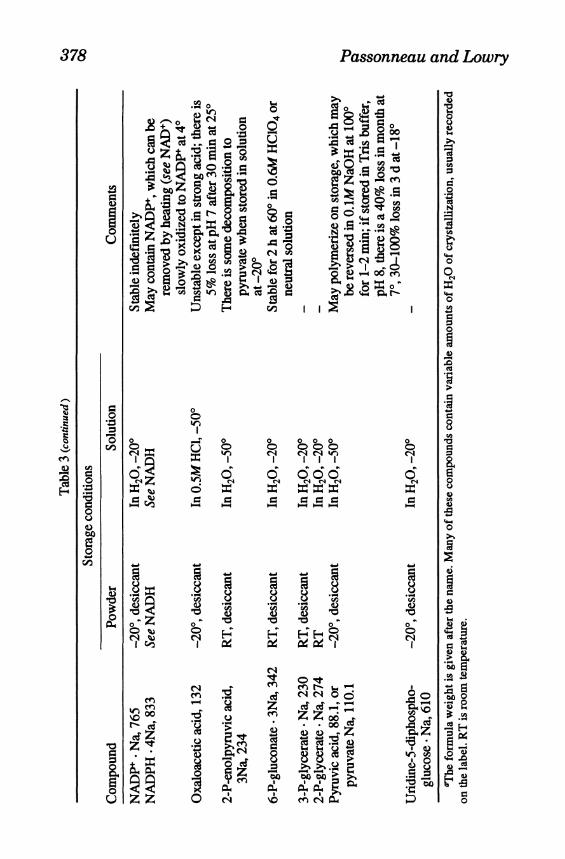
378 Passonneau and Lowry
g
O
U
o -a o
CO
o
!
I CO
I
1 !
en
3".
8 I •8 O
en
•8
1 8 8 I 1
u
g e s
i en
a.
t I
set e £
0 O
0 X
d c
h
SB c
o O
7 i
O X a
5k ^ %s o o o cscs *-> i i i
o o o M « N
X X X a a a
8 8 •8 -8 •8 H tf
CS
en «r 2 en
£ c3
i
hhes Od tf T
O Tt __ (^ h- v< H
CS CS o o G^ d i-H ^ H
Z Z od * • • oo >5 0> D _r Z
'cer
at
'cer
at
ivat
e
d a? a
i
• SO
cu a. en cs
a> *d o O T
i
o f i « 1
*n .1 T3
*c D
o
£ a> CA
o
1 n

Appendix 379
ll
is
a
|£
*-< f-H ir> o O ^
8 $ vo en en oo oo CO NO v o OS ON
m ON
8* o oo r̂ cs oo cs i-i m
VO VO r^mcSi -H»r>vo'^ ' i -HOsr^»n
«o o oo i - H ^ o r - o o a v m o c s ^ H m ' ^ o o t ^ v o i o r ^ o o o
O i-i
O N V O O o o v o i r i O o o r ^ o o o o m o s v o o s o o r ^ T f » r i c s 3
60
.3
1 •11
B0
.5 <2 3
< f t - 2' 5 ^ c/3 P S ,
X X X X X
Passonneau and Lowry
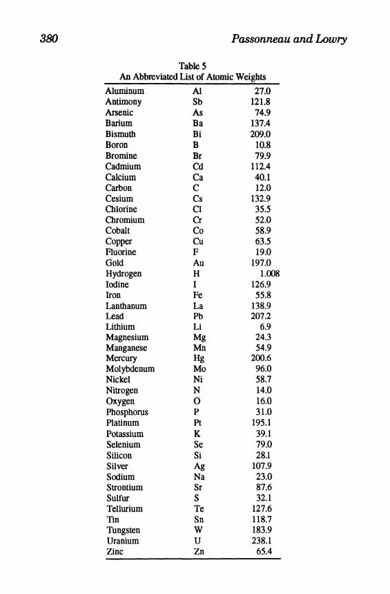
TableS An Abbreviated List of Atomic Weights
Aluminum Antimony Arsenic Barium Bismuth Boron Bromine Cadmium Calcium Carbon Cesium Chlorine Chromium Cobalt Copper Fluorine Gold Hydrogen Iodine Iron Lanthanum Lead Lithium Magnesium Manganese Mercury Molybdenum Nickel Nitrogen Oxygen Phosphorus Platinum Potassium Selenium Silicon Silver Sodium Strontium Sulfur Tellurium Tin Tungsten Uranium Zinc
Al Sb As Ba Bi B Br Cd Ca C Cs CI Cr Co Cu F Au H I Fe La Pb Li Mg Mn Hg Mo Ni N 0 P Pt K Se Si Ag Na Sr S Te Sn W U Zn
27.0 121.8 74.9
137.4 209.0
10.8 79.9
112.4 40.1 12.0
132.9 35.5 52.0 58.9 63.5 19.0
197.0 l.(X
126.9 55.8
138.9 207.2
6.9 24.3 54.9
200.6 96.0 58.7 14.0 16.0 31.0
195.1 39.1 79.0 28.1
107.9 23.0 87.6 32.1
127.6 118.7 183.9 238.1
65.4
References
Chapter 1 1. Negelein, E. and Haas, E. (1935) Uber die Wirkungweise des Zwischen-
ferments. Biochem. Z. 282,206-220. 2. Greengard, P. (1956) Determination of intermediary metabolites by enzymic
fluorimetry. Nature (London) 178,632-643. 3. Kaplan, N. O., Colowick, S. P., and Barnes, C. C. (1951) Effect of alkali on
diphosphopyridine nucleotide. 7. Biol Chem. 191,461-472. 4. Lowry, O. H., Roberts, N. R., and Kapphahn, J. I. (1957) The fluorometric
measurement of pyridine nucleotides. /. Biol. Chem. 224,1047-1064. 5. Lowry, O. H. and Carter, J. G. (1974) Stabilizing the alkali-generated fluores
cent derivatives of NAD and NADP. Anal Biochem. 59,639-642. 6. Lowry, O. H., Passonneau, J. V., and Rock, M. K. (1961) The stability of
pyridine nucleotides. J. Biol. Chem. 236,2756-2759. 7. Burch, H. B., Bradley, M. E., and Lowry, O. H. (1967) The measurement of
triphosphopyridine nucleotide and the role of hemoglobin in producing erroneous triphosphopyridine nucleotide values. /. Biol. Chem. 242,4546-4554.
Chapter 3 1. Levy, M. (1936) Studies on enzymatic histochemistry XVII. A micro kjeldahl
estimation. C. R. Trav. Lab. Carlsberg, Ser. Chim. 248,101-110. 2. Peters, J. P. and Van Slyke, D. D. (1932) Quantitative Clinical Chemistry, vol.
2 (Williams & Wilkins, Baltimore, MD), p. 4.
Chapter 4 1. Richter, D. and Dawson, R. M. C. (1948) Brain metabolism in emotional ex
citement and in sleep. Amer. J. Physiol. 154,73-79. 2. Wollenberger, A., Ristau, O., and Schoffa, G. (1960) Eine einfache technik
der extrem schnellen abkiihling grosserer gewebestucke. Pflueger's Arch. Gesamte Physiol. Menschen Tiere. 270, 399^*12.
3. Kerr, S. E. and Ghantus, M. (1937) The carbohydrate metabolism of the brain. III. On the origin of lactic acid. J. Biol. Chem. 117,217-225.
4. Ferrendelli, J. A., Gay, M. H., Sedgwick, W. G., and Chang, M. M. (1972) Quick-freezing of the murine CNS: Comparison of regional cooling rates and metabolite levels when using liquid nitrogen or Freon-12. J. Neurochem. 19, 979-987.
5. Nelson, S. R., Lowry, O. H., and Passonneau, J. V. (1966) in Head Injury Conference Proceedings (Caveness, W. F. and Walker, A. E., eds.), Lippincott, Philadelphia, PA, p. 444.
6. Bessey, O. H., Lowry, O. H., and Love, R. H. (1949) The fluorometric measurement of the nucleotides of riboflavin and their concentration in tissues. J. Biol. Chem. 180,755-769.
381
382 Passonneau and Lowry
7. Burch, H. B., Bradley, M. E., and Lowry, O. H. (1967) The measurement of triphosphopyridine nucleotide and the role of hemoglobin in producing erroneous triphosphopyridine nucleotide values. J. Biol Chem. 242,4546-4554.
Chapter 5 1. Cha, S. and Cha, C.-J. M. (1965) Kinetics of cyclic enzyme systems. Mol
Pharmacol 1,178,179. 2. Hintz, C. S., Chi, M. M.-Y., and Lowry, O.H. (1980) Correcting potential de
fect in an enymatic cycle for NADP. Anal Biochem. 128,186-190. 3. Lowry, O. H„ Passonneau, J. V., Schulz, D. W., and Rock, M. K. (1961) The
measurement of pyridine nucleotides by enzymatic cycling. J. Biol Chem. 236,2659-2756.
4. Chi, M. M.-Y., Lowry, C. V., and Lowry, O. H. (1978) An improved enzymatic cycle for nicotinamide adenine dinucleotide phosphate. Anal Biochem. 89,119-129.
5. Kato, T., Berger, S. J., Carter, J. A., and Lowry, 0. H. (1973) An enzymatic cycling method for nicotinamide-adenine dinucleotide with malic and alcohol dehydrogenases. Anal Biochem. 53,86-97
6. Breckenridge, B. McL. (1964) The measurement of cyclic adenlyate in tissues. ProcNatl Acad. Sci. USA 52,1580-1586..
7. Goldberg, N. D., Dietz, S. B., and O'Toole, A. G. (1969) Cyclic guanosine 3\5' monophosphate in mammalian tissues and urine. J. Biol. Chem. 224,4458-4466.
Chapter 6 1. Kornberg, A. and Pricer, W. E., Jr. (1951b) Enzymatic phosphorylation of
adenosine and 2,6-diaminopurine riboside. J. Biol Chem. 193,481-495. 2. Pfleiderer, G., Grein, L., and Wieland, T. (1955a) Specific determination of L-
alanine and L-glutamine by means of glutamic-pyruvic transaminase. Ann. Acad. Sci. Fenn. Ser. A2, 60, 381-388.
3. Pfleiderer, G., Gruber, W., and Wieland, T. (1955b) Eine enzymatische bestimmung der L-asparaginsaure. Biochem. Z. 326,446-450.
4. Kornberg, A. (1950) Reversible enzymatic synthesis of diphosphopyndine nucleotide and inorganic pyrophosphate. /. Biol. Chem. 182,779-793.
5. Moellering, H. and Gruber, G. (1966) Determination of citrate with citrate lyase. Anal. Biochem. 17, 369-376.
6. Thorn, W., Pfleiderer, G., Frowein, R. A., and Ross, I. (1955) Stoffwechselvor-gange im Gehirn bei akuter Anoxie, akuter Ischamie und in der Erholung. Pflueger'sArch. Gesamte Physiol. Menschen Tiere 261,334-360.
7. Slein, M. W. (1950) Phosphomannose isomerase. J. Biol. Chem. 186,753-761. 8. Slater, E. C. (1953) Spectrophotometric determination of fructose-1 ̂ -diphos
phate, hexose monophosphates, adenosinetriphosphate and adenosine-diphosphate. Biochem. J. 53,157-167.
9. Vishniac, W. and Ochoa, S. (1952) Fixation of carbon dioxide coupled to photochemical reduction of pyridine nucleotides by chloroplast preparations. J. Biol. Chem. 195, 75-96.
References 383
10. Slein, M. W., Cori, G. T., and Cori, C. F. (1950) A comparative study ofhexo-kinase from yeast and animal tissues. /. Biol. Chem. 186,763-780.
11. Paladini, A. C, Caputto, R., Leloir, L. F., Trucco, R. E., and Cardini, C. E. (1949) The enzymatic synthesis of glucose- 1,6-diphosphate. Arch. Biochem. 23,55-66.
12. Albers, R. W., Koval, G. McKahnn, G„ and Ricks, D. (1961) in Regional Neu-rochemistry (Kety, S. S. and Elkes, J., eds.), Pergamon, Oxford, p. 340.
13. Bublitz, C. and Kennedy, C. P. (1954) Synthesis of phosphatides in isolated mitochondria III. The enzymatic phosphorylation of glycerol. /. Biol Chem. 211,951-961.
14. Ochoa, S. (1948) Biosynthesis of tricarboxylic acids by carbon dioxide fixation. III. Enzymatic mechanisms. /. Biol. Chem. 174,133-157.
15. Noll, F. (1966) Methode zur Quantitativen Bestimmung von L(+)-Lactatmittels Lactat-Dehydrogenase und Glutamat-Pyruvate Transaminase. Biochem. Z. 346, 41-49.
16. Kubowitz, F. and Ott, P. (1943) Isoliering und Kristallisation eines Garungs-fermente aus Tumoren. Biochem. Z. 314,94-117.
17. Strominger, J. L., Maxwell, E. S., and Kalckar, H. M. (1957) Meth. Enzymol. 3,974.
18. Strominger, J. L. (1955) Enzymic synthesis of guanosine and cytidine triphosphates: A note of the nucleotide specificity of the pyruvate phosphokinase reaction. Biochim. Biophys. Acta 16,616,617.
19. Noda, L. and Kuby, S. A. (1963) Meth. Enzymol. 6,223. 20. Lowry, O. H., Schulz, D. W., and Passonneau, J. V. (1964) Effects of
adenylic acid on the kinetics of muscle phosphorylase a. J. Biol. Chem. 239, 1947-1953.
21. Barbehenn, E. K., Law, M. M-Y., Brown, J. G., and Lowry, O. H. (1976) Measurement of 5'adenylic acid by stimulation of phosphorylase a. Anal. Biochem. 70, 554-562.
22. Lienhard, G. E. and Secemski, 1.1. (1973) P1,P5-Di(adenosine-5')penta-phos-phate, a potent inhibitor of adenylate kinase. /. Biol. Chem. 248,1121-1123.
23. Lust, W. D„ Feussner, G. K., Barbehenn, E. K., and Passonneau, J. V. (1981) The enzymatic measurement of adenine nucleotides and P-creatine in picomole amounts. Anal. Biochem. 110,258-266.
24. Manchester, J. K., Chi,M. M.-Y., Carter, J., Pusateri, M.E.,McDougal,D. B., and Lowry, O. H. (1990) Measurement of 2-deoxyglucose and 2-deoxyglucose-6 phosphate in tissues. Anal. Biochem. 185,118-124.
25. Baranowski, T. (1963) in The Enzymes (Boyer, P. D., Lardy, H., and Myrback, K., eds.), rev. ed., vol. 7, Academic, New York, p. 63.
26. Velick, S. F. and Furfine, C. (1963) in The Enzymes (Boyer, P. D., Lardy, H., and Myrback, K., eds.), vol. 7, Academic, New York, p. 243.
27. Massey, V. and Alberty, R. A. (1964) Ionisation constants of fumarase. Biochim. Biophys. Acta 13,354-359.
28. Ray, W. J. Jr. and Roscelli, G. A. (1964) A kinetic study of the phosphogluco-mutase pathway. J. Biol. Chem. 239,1228-1236.
384 Passonneau and Lowry
29. Frieden, C (1959a) Glutamic dehydrogenase II. The effect of various nucleotides on the association-dissociation and kinetic properties. 7. Biol. Chem. 234, 815-820.
30. Frieden, C. (1959b) Glutamic dehydrogenase III. The order of substrate addition in the enzymatic reaction. J. Biol Chem. 234,2891-2896.
31. Matschinsky, F. M. (1964) Personal communication. 32. Passonneau, J. V. and Lauderdale, V. R. (1974) A comparison of three meth
ods of glycogen measurement in tissues. Anal Biochem. 60,405-412. 33. Lust, W. D., Passonneau, J. V., and Crites, S. K. (1975) The measurement of
glycogen in tissues by amylo-a-l,4-a-l,6-glucosidase after the destruction of preexisting glucose. Anal Biochem. 68,128-131.
34. Passonneau, J. V. and Rottenberg, D. A. (1973) An assessment of methods for measurement of glycogen synthetase activity including a new direct one-step assay. Anal. Biochem. 51,405-412.
35. de Azeredo, F. A. M., Feussner, G. K., Lust, W. D., and Passonneau, J. V. (1979) An enzymatic method for the measurement of GTP and GDP in biological samples. Anal Biochem. 95,512-519.
36. Kaufman, F. and Alivisatos, S. G. A. (1955) Purification and properties of the phosphorylating enzyme from spinach. /. Biol Chem. 216,141-152.
37. Cha, S. and Parks, J. E., Jr. (1964) Succinic thiokinase. II Kinetic studies; initial velocity, product inhibition, and effect of arsenate. J. Biol. Chem. 239, 1968-1977.
38. Olson, J. A. and Anfinsen, C. B. (1953) Kinetic and equilibrium studies on crystalline L-glutamic acid dehydrogenase. /. Biol. Chem. 202, 841-856.
39. Hintz, C. S., Chi, M. M.-Y., Fell, R. D., Ivy, J. L., Kaiser, K. K., Lowry, C. V., and Lowry, O. H. (1982) Metabolite changes in individual rat muscle fibers during stimulation. Am. J. Physiol. 242, C218-C228.
40. Krebs, H. A. (1953) Equilibria in transamination systems. Biochem. J. 54,82-86. 41. Fawaz, E. N., Roth, L., and Fawaz, G. (1966) The enzymatic estimation of
inorganic phosphate. Biochem. Z 344,212-214. 42. Pontremoli, S., de Flora, A., Grazi, E., Mangiarotta, G., Bonsignore, A., and
Horecker, B. L. (1961) Crystalline D-gluconate-6-phosphate dehydrogenase. J. Biol. Chem. 236,2975-2980.
43. Outlaw, W. H., Jr. and Lowry, O. H. (1979) Measurement of 10~7 to 10"12 mol of potassium by stimulation of pyruvate kinase. Anal Biochem. 92,370-374.
44. Cook, G. A., O'Brien, W. E., Wood, H. G., King, M. T., and Veech, R. L. (1978) A rapid, enzymatic assay for the measurement of inorganic pyrophosphate in animal tissues. Anal. Biochem. 91,557-565.
45. Jones, M. G. K., Outlaw, W. H., Jr., and Lowry, O. H. (1977) Enzymic assay of 10"7 to 10~14 moles of sucrose in plant tissues. Plant Physiol. 60, 379-383.
Chapter 7 1. Henriksson, J., Chi, M. M.-Y., Hintz, C S., Young, D. A., Kaiser, K. K, Salmons,
S., and Lowry, O. H. (1986) Chronic stimulation of mammalian muscle: Changes in enzymes of six metabolic pathways. Am. J. Physiol. 251, C614-C632.

References 385
2. Cole, B. R, Hays, A. E., Boylan, J. B., Burch, H. B., and Lowry, O. H. (1982) Distribution of enzymes of adenylate and guanylate metabolism in rat nephron. Am. 7. Physiol 243,F349-F355.
3. Chi, M. M.-Y., Hintz, C. S., Coyle, E. F., Martin, W. H. Ill, Ivy, J. L, Nemeth, P. M., Holloszy, J. O., and Lowry, O. H. (1983) Effects of detraining on enzmes of energy metabolism in individual human muscle fibers. Am. J. Physiol 244, C276-C287.
4. Chan, A. W. K., Perry, S. G., Burch, H. B., Fagioli, S., Alvey, T. R., and Lowry, O. H. (1979) Distribution of two aminotransferases and D-amino acid oxidase within the nephron of young and adult rats. 7. Histochem. Cytochem. 27,751-755.
5. Burch, H. B., Lowry,O. H., Kuhlman, A. M., Skerjance, J.,Diamant,E. J., Lowry, S. R., and Von Dippe, P. (1963) Changes in patterns of enzymes of carbohydrate metabolism in the developing rat liver. 7. Biol Chem. 238,2267-2273.
6. Burch H. B., Choi, S., Dence, C. N., Alvey, T. R., Cole, B. R., and Lowry, O. H. (1980) Metabolic of effects of fructose loads in different parts of the rat nephron. 7. Biol Chem. 255,8239-8244.
7. Passonneau, J. V., Lust, D. W., and Crites, S. K. (1977) Studies on the GABAergic system in astrocytoma and neuroblastoma cells in culture. Neurochem. Res. 2, 605-617.
8. Pusateri, M. E., Carter, J. G., Berger, S. J., and Lowry, O. H. (1984) Distribution of three enzymes of GABA metabolism in monkey retina. 7. Neurochem. 42, 1269-1273.
9. Chi, M. M.-Y., Manchester, J. K, Yang, V. C, Curato, A. D., Strickler, R. C, and Lowry, O. H. (1988) Contrast in levels of metabolic enzymes in human and mouse ova. Biol. ofReprod. 39,295-307.
10. Hintz, C. S., Turk, W. R., Cambon, N., Burch, H. B., Nemeth, P. M., and Lowry, O. H. (1985) A method for branched-chain amino acid aminotransfersse activities in microgram and nanogram tissue samples. Anal. Biochem. 146,418-422.
11. Burch, H. B., Cambon, N., and Lowry, O. H. (1985) Branched-chain amino acid aminotransferase along the rabbit and rat nephron. Kidney International 20,114-117.
12. Ichihara, A. and Koyama, A. (1966) Transaminase of branched chain amino acids. I. Branched chain amino acids a-ketoglutarate transaminase. 7. Biochem. (Tokyo) 59,160-169.
13. Taylor, R. T. and Jenkins, W. T. (1966) Leucine aminotransferase. I. Colorimetric assays. 7. Biol. Chem. 2A\, 4391-4395.
14. Lowry, C. V., Kimmey, J. S., Felder, S., Chi, M. M.-Y., Kaiser, K. K., Passonneau, P. N., Kirk, K. A., and Lowry, O. H. (1978) Enzyme patterns in single human muscle fibers. 7. Biol. Chem. 253,8269-8277.
15. Dietrich, W. D., Durham, D., Lowry, O. H., and Woolsey, T. A. (1981) Quantitative histochemical effects of whisker damage on single identified cortical barrels in the adult mouse. 7. Neurosci. 1,929-935.
16. Lowry, O. H., Berger, S. J., Carter, J. G., Chi, M. M.-Y., Manchester, J. K., Knor, J., and Pusateri, M. E. (1983) Diversity of metabolic patterns in human brain tumor enzymes of energy metabolism and related metabolites and cofactors. 7. Neurochem. 41,994-1010.

386 Passonneau and Lowry
17. Lowry, O. H., Berger, S. J., Chi, M. M-Y., Carter, J. G., Blackshaw, A., and Outlaw, W. (1977) Diversity of metabolic patterns in human brain tumors. I. High energy phosphate compounds and basic composition. 7. Neurochem. 29, 959-977.
18. Hintz, C S., Lowry, C. V., Kaiser, K., McKee, D., and Lowry, O. H. (1980) Enzyme levels in individuat rat muscle fibers. Am. J. Physiol 239, C58-C65.
19. Bass, A., Brdiczka, D., Eyer, S., Hofer, S., and Pette, D. (1969) Metabolic differentiation of distinct muscle types at the level of enzymatic organization. Eur. J. Biochem. 10,198-206.
20. Burch, H. B., Narins, R. G., Chu, C, FagioU, S., Choi, S., McCarthy, W., and Lowry, O. H. (1978) Distribution along the rat nephron of three enzymes of gluconeogenesis in acidosis and starvation. Am J. Physiol. 235, F246-F253.
21. Chi, M. M.-Y., Hintz, C. S., Henriksson, J., Salmons, S., HeUendahl, R. P., Park, I. L, Nemeth, P. M., and Lowry, O. H. (1986) Chronic stimulation of mammalian muscle: Enzyme changes in individual fibers. Am. J. Physiol 251, C633-C642.
22. Burch, H. B., Bross, T. E., Brooks, C. A., Cole, B. R., and Lowry, O. H. (1984) The distribution of six enzymes of oxidative metabolism along the rat nephron. /. Histochem. Cytochem. 32,731-736.
23. Kato, T. and Lowry, O. H. (1973) Enzymes of energy converting systems in individual mammalian nerve cell bodies. 7. Neurochem. 20,151-163.
24. Lowry, O. H., Roberts, N. R., and Lewis, C. (1956) The quantitive histochemistry of the retina. J. Biol. Chem. 220,879-892.
25. Lowry, O. H., Roberts, N. R., and Chang, M-L. W. (1956) The analysis of single cells. J. Biol. Chem. 222,97-107.
26. Kato, T. and Lowry, O. H. (1973) Distribution of enzymes between nucleus and cytoplasm of single nerve cell bodies. J. Biol. Chem. 248,2044-2048.
27. Curthoys, N. P. and Lowry, O. H. (1973) The distribution of glutaminase isoenzymes in the various structures of the nephron in normal, acidotic, and alka-lotic rat kidney. J. Biol. Chem. 248,162-168.
28. Curthoys, N. P. and Kuhlenschmidt, T. (1975) Phosphate-independent glutaminase from rat kidney: partial purification and identity with gamma-glutamyltrans-petidase. J. Biol. Chem. 250,2099-2105.
29. Lowry, O. H., Shulz, D. W., and Passonneau, J. V. (1964) Effects of adenylic acid on kinetics of muscle phosphorylase a. J. Biol. Chem. 253, 8269-8277.
30. Hsieh, B., Chi, M. M.-Y., Knor, J., and Lowry, O. H. (1979) Enzymes of glycogen metabolism and related metabolites in preimplantation mouse embryos. Dev. Biol. 72,342-349.
31. Henry, C. G. and Lowry, O. H. (1985) Enzyme and metabolites of glycogen metabolism in canine cardiac Puikinje fibers. Am. J. Physiol. 248, H599-H605.
32. Passonneau, J. V. and Rottenberg, D. A. (1973) An assessment of methods for measurement of glycogen synthetase activity including a new direct one-step assay. Anal. Biochem. 51,528-541.
33. Berger, S. J., DeVries, G. W., Carter, J. G., Schulz, D. W., Passonneau, P. N., Lowry, O. H., and Ferrendelli, J. A. (1980) The distribution of the components of the cyclic GMP cycle in retina. J. Biol. Chem. 255,3128-3135.

References 387
34. Cole, B. R., Hays, A. E., Boylan, J. B., Burch, H. B., and Lowry, O. H. (1982) Distribution of enzymes of adenylate and guanylate metabolism in rat nephron. Am. J. Physiol 243, F349-355.
35. Teutsch, H. F. and Lowry, O. H. (1982) Sex specific regional differences in hepatic glucokinase activity. Biochem. Biophys. Res. Comm. 106,533-538.
36. Seltzer, J. L. and McDougal, D. B., Jr. (1975) Enzyme levels in chick embryo heart and brain from 1-21 days of development. Develop. Biol. 42,95-105.
37. Burch, H. B., Kuhlman, A. M., Skerjance, J., and Lowry, O. H. (1971) Changes in patterns of enzymes of carbohydrate metabolism in the developing rat kidney. Pediatrics 47,199-296.
38. Holowach, J., Kauffman, F., Beossi, M. G., Thomas, C, and McDougal, D. B. Jr. (1968) The effects of a thiamine antagonist, pyrithiamine, on levels of selected metabolic intermediates and on activities of thiamine-dependent enzymes in brain and liver. JNeurochem. 15,621-631.
39. Mourad, N., and Parks, R. E., Jr. (1966) Erythrocytic nucleoside diphosphokinase II. Isolation and kinetics. J. Biol Chem. 241,271-278.
40. Goffeau, A., Pedersen, P. L., and Lehninger, A. L. (1967) The kinetics and inhibition of the adenosine diphosphate adenosine triphosphate exchange catalyzed by a purified mitochondrial nucleoside dij*osphokinase./.Bw/.C/^m. 242,1845-1853.
41. Lowry, O. H. and Passonneau, J. V. (1966) Kinetic evidence for multiple binding sites on phosphofructokinase. J. Biol. Chem. 241,2268-2279.
42. Kahana, E. E., Lowry, O. H., Schulz, D. W., Passonneau, J. V., and Crawford, E. J. (1961) The kinetics of phosphoglucoisomerase. J. Biol Chem. 235,2178-2184.
43. Lowry, O. H., Roberts, N. R., Schulz, D. W., Clow, J. E., and Clark, J. R. (1961) Quantitative histochemistry of retina. II. Enzymes of glucose metabolism. /. Biol. Chem. 236,2813-2820.
44. Cornell, N. W., Leadbetter, M., and Veech R. L. (1979) /. Biol. Chem. 254, 6627-6527.
45. Lowry, O. H. and Passonneau, J. V. (1964) The relationships between substrates and enzymes of glycolysis in brain. J. Biol. Chem. 239,31-42.
46. Pitts, F. N., Jr. and Quick, C. (1965) Brain succinic semialdehyde dehydrogenase. I. Assay and distribution. J. Neurochem. 12,893-900.
47. Middleton, B. (1973) The oxoacyl-coenzyme A thiolases of animal tissues. Biochem. J. 132,717-730.
48. Corder, C. N., Berger, M. L., and Lowry, O. H. (1974) Quantitative histochemistry of uridine diphosphoglucose-6-pyrophosphatase and uridine diphospho-glucose-pyrophosphorylase in developing rat kidney. J. Histochem. Cytochem. 22,1034-1038.
Chapter 9 1. Linderstrom-Lang, K., Holter, H., and Ohlsen, A. S. (1935) Studies on enzy
matic histochemistry XIII. The distribution of enzymes in the stomach of pigs. Compt.-Rend. Lab. Carlsberg, Serie Chim. 20,66-125.
2. Glick, D. (1961,1963) Quantitive Chemical Techniques ofHisto- and Cytochemistry, vols. 1 and 2 (Wiley [Interscience]), New York.
388 Passonneau and Lowry
3. Cole, B. R., Boylan, J. G., Bross, T. E., Burch, H. B., and Lowry, O. H. (1988) Progressive enzyme changes within anatomically defined segments of rat nephron: Demonstration of a new technique. / Histochem Cytochem 36,285-289.
4. Barbehenn, E. K., Wales, R. G., and Lowry, O. H. (1974) The explanation for the blockade of glycolysis in early mouse embryos. Proc. Natl Acad. ScL USA 71,1056-1060.
5. Chi, M. M.-Y., Manchester, J. K., Yang, V. C, Curato, A. D., Strickler, R. C, and Lowry, O. H. (1988) Contrast in levels of metabolic enzymes in human and mouse ova. Biol ofReprod. 39,295-307
6. Outlaw, W. H., Jr. and Lowry, O. H. (1977) Organic acid and potassium accumulation in guard cells during stomatal opening. Proc. Natl. Acad Sci. USA 74,4434-4438.
7. Wenger, B. (1955) Personal communication.. 8. Ess6n, B. Jansson, E., Henriksson, J., Taylor, A. W., and Saltin, B. (1975)
Metabolic characteristics of fibre types in human skeletal muscle. Acta Physiol Scand. 95,153-165.
9. Lowry, C. V., Kimmey, J. S., Felder, S., Chi, M. M.-Y., Kaiser, K. K., Passonneau, P. N., Kirk, K. A., and Lowry, O. H. (1978) Enzyme patterns in single human muscle fibers. /. Biol Chem. 253, 8269-8277.
10. Hintz, C. S., Lowiy, C. V., Kaiser, K. K., McKee, D., and Lowry, O. H. Enzyme levels in individual rat muscle fibers. Am. J. Physiol 239, C58-C65.
Chapter 12 1. Lowry, O. H. (1963) The chemical study of single neurons. Harvey Lect. 58,
1-19. 2. Matschinsky, F. M., Passonneau, J. V., and Lowry, O. H. (1968a) Quantitative
histochemical analysis of glycolytic intermediates and cofactors with an oil well technique. J. Histochem. Cytochem. 16,29-39.
3. Matschinsky, F. M., Rutherford, C. R., and Ellerman, J. E. (1968) Accumulation of citrate in pancreatic islets of obese hyperglycemic mice. Biochem. Biophys. Res. Commun. 33,855-862.
Appendix Good, N. E. (1962) Uncoupling of the Hill reaction from photophosphorylation by
anions. Arch. Biochem. Biophys. 96,653-661. Good, N. E., Winger, G. D., Winter, W., Connolly, T. N., Izawa, S., and Singh, R.
M-M. (1966) Hydrogen ion buffers for biological research. Biochemistry 5, 467-477.
Preface Bergmeyer, H. V. (1970) in Methods of Enzymatic Analysis, 2nd ed. (Verlag Chemie,
Weinheim).

Index
A Acid, definition, 365 Acids, normality of common,
table, 379 ADP, measurement of, 110-114
fluorometric 0.1-8 nmol, 113 kinetics of analytic enzymes, 113 spectrophotometric 15-120
nmol, 112 ADP, effect on glutamate
dehydrogenase, 161,169 removal from ATP, 131
AMP, measurement of, 111-117 catalytic method, with phospho-
rylase a Method II, 114-117 cycling 1-10 fmol, 115 fluorometric direct 50-500 pmol, 115
indirect 4-40 pmol, 115 kinetics of phosphorylasea, 116 precautions in tissue
measurement, 116 spectrophotometric, 114
direct measurement with pyruvate kinase and lactate dehydrogenase Method I, 111-114
fluorometric 0.1-4 nmol, 113 spectrophotometric
15-120 nmol, 112 AMP, effect on phosphorylase a
kinetics, 202, 265
presence in and removal from NADH, 113
presence in NADP*, 116 ATP-ADP cycle, 103-107
conversion of AMP to ATP, 104 cycling reagent for, 104 effect of time, temperature, and
enzyme concentration, 105 indicator reagent for, 104 kinetics of, 107 procedure with NADP cycle
supplement, 1-10 fmol, 106 sample procedure, 1-10 pmol,
105 total adenylate measurement,
2-10 pmol, 106 ATP, measurement with NADPH
as product, 121-126 cycling, 1-10 pmol, 123
0.2-1 pmol, 124 20-100 fmol, 125
fluorometric 0.1-10 nmol, 122
enzyme contamination problems, 122
kinetics of hexokinase, 125 spectrophotometric 15-100
nmol, 121 ATP, removal from ADP, 123 Adenylate deaminase (EC 3.5.4.6),
measurement of enzyme activity, 231-233
389
390 Index
fluorometric, direct 2-8 nmol product, 232
indirect, 0.2-1 nmol product, 232 spectrophotometry, 20-90 nmol
product, 231 Adenylate kinase, contamination in
adenylate measurement, 126
Adenylate kinase (EC 2.7.4.3), measurement of enzyme activity, 233-234
fluorometric, 1-10 nmol product, 234
spectrophotometric, 50-100 nmol product, 233
Adenylates, measurement of with luminescence, 126-128
ATP, 0.1-80 pmol, 126 myokinase, inhibition of by Ap5 A,
127 total adenylate measurement, 128
Alanine, measurement of, 117-118 fluorometric 0.1-8 nmol, 118 kinetics of alanine transaminase,
118 spectrophotometric 15-120
nmol, 117 Alanine transaminase (EC 2.6.1.2),
measurement of enzyme activity, 234-235
fluorometer, indirect assay, 0.2-10 nmol product, 235
Aldolase (EC 4.1.2.13), measurement of enzyme activity, 235-237
fluorometric, direct assay, 1-8 nmol product, 236
fluorometric, indirect assay, 0.1-5 nmol product, 236
spectrophotometric, 50-100 nmol product, 236
Alkaline fluorescence of pyridine nucleotides, see specific nucleotide
y-Aminobutyrate transaminase (EC 2.6.1.0)
cycling assay, 5-25 pmol product, 238
fluorometric, 1-10 nmol product, 238
measurement of enzyme activity, 237-239
spectrophotometric 30-150 nmol product, 237
y-Aminobutyric acid, measurement of, 154-157
cycling, 1-10 pmol, 156 fluorometric, direct assay,
3-10 pmol, 155 2-step assay, 1-10 pmol, 155 strong alkali method,
1O-100 pmol, 155 kinetics of "Gabase," 156 spectrophotometric, 15-200
nmol, 155 Amytal (amobarbital), inhibitor of
NADH oxidation, 262, 269,281,292,312
Analytical problems and suggestions for remedies, see Chapter 8
Ascorbic acid, as prevention of NADH oxidation, 16,264,291
Aspartate, measurement of, 118-121 cycling, 5-25 pmol, 120
0.1-5 pmol, 120 fluorometric 0.1-8 nmol, 119 kinetics of aspartate
transaminase, 119 spectrophotometric 15-120
nmol, 119
Index 391
Aspartate transaminase (EC 2.6.1.1), measurement of enzyme activity, 239-240
cycling, 10-50 pmol product, 240 fluorometric, direct, 1-10 nmol
product, 240 indirect, 0.1-1 nmol product, 240 spectrophotometric,
20-100 nmol product, 239 Atomic weights, abbreviated list,
380
B Balances, see also quartz-fiber
fishpole balance, 337 balance calibration, 349
colorometric, 349 fluorometric, 349 with frozen-dried tissue, 350
balance case, 344 balance fibers, 342 balance pans, 343 cleaning of, 351 lighting of, 347 linearity of, 349 mounting the fiber, 346
Base/acid ratio for buffers, table, 367, 368
Bases, normality of commonly used, 379
Branched chain amino acid aminotransferase (EC 2.6.1.42) measurement of enzyme activity, 241-242
fluorometric, indirect assay 0.1-1 nmol product, 241
Buffers, definition, 366 how to calculate composition of,
368 table of, 374
Carnitine acetyltransferase (EC 2.3.1.7), measurement of enzyme activity, 242-243
fluorometric indirect assay, 0.5-10 nmol product, 243
Citrate, measurement of, 128-130 fluorometric 1-8 nmol, 129 kinetics of citrate lyase, 129 spectrophotometric 50-120
nmol, 128 Citrate synthase (EC 4.1.3.7),
measurement of enzyme activity , 244-245
cycling assay, 2-20 pmol product, 245
fluorometric indirect assay, 0.3-6 nmol product, 244
Conjugate base (or acid), 366 Creatine, meaurement of, 130-132
fluorometric 1-8 nmol, 131 kineticsof creatine kinasereaction,
132 spectrophotometric 15-120 nmol,
130 side reactions of creatine kinase
(ATPase), 131 Creatine phosphate, see P-creatine Creatine phosphokinase (EC 2.7.3.2),
measurement of enzyme activity, 246-248
fluorometric, direct assay, 1-10 nmol product, 246
indirect assay, 1-10 nmol product, 247
special requirements for muscle, 247
spectrophotometric, 50-100 nmol product, 246
Cycling, enzymatic, principles of, 85

392 Index
kinetics of, 87 specific first-order rate constant,
82 Cycling, specific procedures,
see specific cycles or assays
D Defatting of frozen dried tissue
samples, 335 2-Deoxyglucose and 2-deoxy-
glucose-6-phosphate, measurement of , 132-134
fluorometric 2-10 nmol total, 134 kinetics of glucose-6-phosphate
dehydrogenase, 132 spectrophotometric 15-100nmol,
133 Design and development of new
methods, 317 P1 ,P5-di(adenosine-5')penta-
phosphate, adenylate kinase inhibitor, 117,127
Dihydroxyacetone phosphate, measurement of with
glycero-P dehydrogenase, Method 1,134-138
cycling assay, 0.5-10pmol, 136 0.1-.5pmol, 137
fluorometric assay 1-8 nmol, 135 indirect 10-100 pmol, 136
kinetics of glycero-P dehydrogenase, 135,137
special precautions, 137 measurement of with glyceralde-
hyde 3-P dehydrogenase Method II, see Fructose-1,6-bisphosphate Method II
Dissection of frozen-dried tissue sections, 329
histological identification, 334
Dissociation constant, definition for acid or base, 366
of acid andbases for buffers, table, 374
of water, 369 Dithiothreitol, to reduce glutathione,
139,141,162,202,314 Dry loading of tissue sections
for analysis, 354 Drying of frozen tissue under
vacuum, 326
E Enzyme activities, measurement of,
see specific enzyme, 229-305 Enzyme activity, unit of, 229 Enzyme stability at high dilution,
353 Evacuation of frozen-dried tissue
sections, 329
F Fluorescence of pyridine nucleotides,
4,5 conversion by alkali to fluorescent
products, 10 effect of enzymes on, 6 effect of pH on, 6 enhancement of, 6,11 filters for measurement, 9-10 increased fluorescence in alkali,
12-13 interfering substances, 12 quenching of, 7
Huorometry fluorometers, 20-21 pyridine nucleotides, useful range, 5 quenching, 5 reference standards, 8
Freezing of tissues for histochemistry, 322 for metabolites, 72-73

Index 393
Fructose, measurement of, 138-139 fluorometric 0.1-10 nmol, 139 kinetics of hexokinase for fructose,
139 spectrophotometric 15-200 nmol,
138 Fructose-1,6-bisphosphatase
(EC 3.1.3.11) measurement of enzyme activity, 248-250
cycling assay, 5-15 pmol product, 249
fluorometric direct assay, 5-10 nmol product, 249
indirect assay, 5-10 nmol product, 249
kinetics of enzyme, 250 Fructose-1,6-bisphosphate,
measurement of with aldolase and glycero-P
dehydrogenase Method I, 147-148
fluorometric 0.05-4 nmol, 147 kinetics of auxiliary enzymes,
148 spectrophotometric 8-60 nmol,
147 aldolase and glyceraldehyde-P
dehydrogenase Method II, 148-152
cycling, 2-20 pmol, 150 0.1-1 pmol, 151
fluorometric 0.1-10 nmol, 150 kinetics of auxiliary enzymes
151 spectrophotometric
8-100 nmol, 149 Fructose-6-phosphate, measurement
of with glucose-6-P dehydrogenase
and P-glucoisomerase Method 1,140-141
fluorometric 0.1-10 nmol, 141 kinetics of P-glucoisomerase
141 spectrophotometric 15-200
nmol, 140 P-fructokinase,Methodn, 142-144
cycling, 10-40 ftnol, 143 fluorometric 0.1-5 nmol, 143 spectrophotometric
8-100 nmol, 142 Fructose-6-phosphate and mannose-
6-phosphate measurement of, 144-146
cycling, 0.1-1 pmol, 145 fluorometric 0.1-10 nmol, 145 kinetics of P-glucoisomerase, 146 kinetics of P-mannoisomerase,
146 spectrophotometric 15-200 nmol,
146 Fumarase(EC3.1.3.1 l)measurement
of enzyme activity, 250-252
cycling, 50-150 pmol product, 251
fluorometric, indirect assay, 3-10 nmol product, 251
spectrophotometric, 60-150 nmol product, 251
Fumarate, meaurement of, 152-154 fluorometric, direct assay
0.1-10 nmol, 153 fluorometric, 2-step assay, 153 kinetics of fumarase, 154 spectrophotometric 15-200 nmol,
152
GTP-GDP cycle, 107-110 comments on cycle, 109 cycling reagent for, 108
394 Index
effects of time, temperature, and enzyme concentration, Table 9,108
indicator reagent for, 109 Glucose, measurement of, 157-160
cycling, 1-10 pmol, 158 10-50 fmol, 159
destruction with alkali, 177 fluorometric, 0.1-10 nmol, 157 kinetics of hexokinase, 159 spectrophotometric, 15-200nmol,
159 Glucose-1-P, measurement of,
160-162 fluorometric, 0.1-10 nmol, 161 kinetics of P-glucomutase, 161 spectrophotometric, 15-200nmol,
160 Glucose-1,6-bisphosphate,
measurement of, 165-167 fluorometric, 2-8 pmol, 166 kinetics of P-glucomutase, 166 modification for 10 fmol, 166
Glucose-6-phosphatase (EC 3.1.3.9) measurement of enzyme activity, 252-253
cycling, 10-100 pmol product, 253 fluorometric indirect assay,
1-10 nmol product, 253 spectrophotometric, 20-100 nmol
product, 253 tissue homogenate preparation for
assay, 252 Glucose-6-phosphate, measurement
of, 162-164 cycling, 1-10 pmol, 162
0.1-1 pmol, 164 10-50 fmol, 164
fluorometric, 0.1-10 nmol, 162 kinetics of glucose-6-P
dehydrogenase, 164
spectrophotometric, 15-200nmol, 162
Glucose-6-P dehydrogenase (EC 1.1.1.49),
measurement of enzyme activity, 254-256
cycling, 5-25 pmol product, 255 fluorometric direct assay,
1-10 nmol product, 255 spectrophotometric, 50-150nmol
product, 255 Glutamate, measurement of, 167-170
cycling, 2-10 pmol, 168 0.5-2.5 pmol, 168
fluorometric, 0.1-10 nmol, 168 kinetics of glutamate
dehydrogenase, 169 spectrophotometric, 15-200nmol,
167 Glutamate decarboxylase
(EC 4.1.1.15) measurement of enzyme activity,
256-258 cycling, 0.3-1.2 nmol product, 257 fluorometric indirect, 1.8-10pmol
product, 257 spectrophotometric, 30-100 nmol
product, 257 Glutamate dehydrogenase
(EC 1.4.1.3) measurement of enzyme activity,
258-259 cycling, 3-6 pmol product, 259 fluorometric indirect, 1-3 nmol
product, 259 spectrophotometric, 25-150 nmol
product, 258 Glutaminase (EC 3.5.1.2)
measurement of enzyme activity, 260-261
cycling, 25-100 pmol product, 261
Index 395
fluorometric indirect, 1-10 nmol product, 261
spectrophotometry, 25-100 nmol product, 260
Glutamine, measurement of, 170-171 cycling, 0.2-1.5 pmol, 171 fluorometric, 1-10 nmol, 170
Glycerol, measurement of, 171-172 fluorometric, 0.1-10 nmol, 172 spectrophotometry 15-200
nmol, 172 oc-Glycerophosphate, measurement of
Methodlwitha-glycero-Pdehydro-genaseandhydrazine, 172-174
fluorometric, 0.2-10nmol, 173 kinetics of reaction, 173 spectrophotometry,
15-200 nmol, 173 Method II with triose-P isomerase
and glyceraldehyde-3-P dehydrogenase, 174-176
fluorometric direct 0.1-5 nmol, 175
kinetics of glyceraldehyde-3-P dehydrogenase, 176
spectrophotometric, 8-100 nmol, 175
oc-Glycero-3-P dehydrogenase (EC 1.1.1.8)
measurement of enzyme activity, 262-263
cycling, 10-100 pmol product, 264 modification for muscle tissue, 264
fluorometric, direct 2-8 nmol product, 262 indirect 0.1-3 nmol product, 263
spectrophotometric, 20-80 nmol product, 262
Glycogen, measurement of, 177-179 cycling, l-10pmolglucosyl units,
178 fluorometric direct 0.1-10
glucosyl units, 178 spectrophotometric 15-200 nmol
glucosyl units, 177 Glycogen phosphorylase (EC 2.4.1.1)
measurement of enzyme activity, 264-267
cycling 10-50 pmol product, 266 modification for muscle tissue, 267
fluorometric direct 1-10 nmol product, 266 indirect 1-5 nmol product, 266
spectrophotometric 15-75 nmol product, 265
tissuepreparationforaandhforms, 265
Glycogen synthase (EC 2.4.1.11), measurement of enzyme activity, 268-270
cycling, 10-100 pmol product, 270
fluorometric indirect, 1-8 nmol product, 269
tissue preparation for i anddforms, 268
Guanine nucleotides, measurement of, 179-183
cycling, 1-3 pmol, 182 fluorometric 0.1-10 nmol,
180 kinetics of succinyl Co A
synthetase and pyruvate kinase, 181
removal of ADP as a contaminant, 181
396 Index
Guanine phophoribosyl transferase, see hypoxanthine phosphoribosyltransferase
Guanylate kinase (EC 2.7.4.8) measurement of enzyme activity, 270-272
cycling 0.3-2 pmol product, 271
fluorometric 2.5-5 nmol product, 271
spectrophotometry 25-100 nmol product, 270
H Hair-points for frozen-dried tissue
sections, 333 Henderson-Hasselbalch equation,
366 Hexokinase (EC 2.7.1.1)
measurement of enzyme activity, 272-274
cycling 1-10 pmol product, 273 fluorometric direct 1.5-10 nmol
product, 273 indirect 1-10 nmol product, 273 spectrophotometric 50-150 nmol
product, 173 Hydrazine as trapping agent for
oc-keto acid, see assays for P-pyruvate, lactate, glycerol, a-glycero-P, malate
p-Hydroxyacyl coenzyme A dehydrogenase (EC 2.4.2.8)
measurement of enzyme activity, 274-276
cycling 20-100 pmol product, 275 fluorometric indirect 0.1-3 nmol
product, 275 spectrophotometric 20-150 nmol
product, 274
Hypoxanthine (guanine) phosphoribosyl tranferase (EC 2.4.2.8) measurement of enzyme activity, 276-277
fluorometric indirect 0.5-5 nmol product, 276
I International unit of enzyme activity,
229 Isocitrate, measurement of, 183-184
fluorometric direct 0.1-10 nmol, 184
kinetics of isocitrate dehydrogenase, 184
spectrophotometric 15-200 nmol, 183
Isocitrate dehydrogenase (NAD) (EC 1.1.1.41)
meaurement of enzyme activity, 277-278
fluorometric 1-10 nmol product, 277
Isocitrate dehydrogenase (NADP) (EC 1.1.1.42)
measurement of enzyme activity, 278-279
cycling 2-10pmol product, 278 fluorometricindirect5-20nmol
product, 278
K Katal, unit of enzyme activity, 229 Ketoacid CoA transferase
(EC 2.8.3.5) measurement of enzyme activity,
279-280 cycling, 2-25 pmol product,
280 fluorometricindirect2-10nmol
product, 280

Index 397
a-Ketoglutarate, measurement of, 185-188
cycling, 1-10 pmol, 186 equilibrium of glutamate
dehydrogenase reaction, 186 fluorometric direct 2-8 nmol, 185
indirect 0.2-1 nmol, 185 kinetics of reaction, 187 spectrophotometric 15-200nmol,
185 a-Ketoglutarate dehydrogenase
(EC 1.2.4.2) measurement of enzyme activity,
280-281 fluorometric direct 1.5-10 nmol
product, 281 Kinetics, enzyme
factors affecting, 48-49 first order, 29-32 first order rate constant, 24, 33 half-time, definition, 24-25 Michaelis-Menten equation, 31 mixed zero-order and first order,
33-35 one-step reaction, one substrate,
29-32 one-step reaction, two substrates,
35-41 "random" order of addition,
37-38 "cooperative addition," 38-41 "ordered addition," 41
reversible first-order, 25 relaxation time, 25 second order rate constant, 38 second order reactions, 41-43 two-step reactions, 43-48
both steps first order, 45 first step zero order, second step
first order, 43 lag time, 45
Kinetics, nonenzyme, 23-28 first order reactions, 24-27 order of reaction, 23 second order reactions, 27-28
Kinetics of enzymes for assays of metabolites and enzyme actvities, see specific assay
Knives for frozen-dried tissue microdissection, 330-332
L Lactate, measurement of
with glutamate-pyruvate transaminase, Method 1,188-193
contamination problems, 192 cycling 1-10 pmol, 191
0.1-1 pmol, 191 2O-100 fmol, 191
fluorometric direct 0.2-10 nmol, 190 indirect 0.1-10 nmol, 190
kinetics of glutamate-pyruvate transaminase, 193
kinetics oflactatedehydrogenase (beef heart), 192
kinetics of overall reaction, 192 spectrophotometric 15-200
nmol, 189 withhydrazine, Method n, 193-195
fluorometric 0.5-10 nmol, 194 spectrophotometric 15-200
nmol, 194 Lactate dehydrogenase (EC 1.1.1.27)
measurement of enzyme activity
Method I cycling 50-500 pmol product, 282
fluorometric indirect 0.1-5 nmol product, 282
spectrophotometric 20-100 nmol product, 281

398 Index
Method II fluorometricindirect2-10nmol
product, 283 Log table, homemade, 371 Logarithms (to base 10), 365
M Malate, measurement of
Method I with aspartate transaminase, 195-197
fluorometric 0.1-10 nmol, 196 kineticsofmalatedehydrogenase
and aspartate transaminase, 196
spectrophotometric 15-200 nmol, 195
MethodH withhydrazine, 198-199 fluorometric 0.1-10 pmol, 198 spectrophotometric 15-200
nmol, 198 Malate dehydrogenase (EC 1.1.1.37)
measurement of enzyme activity
Method I cycling 5-250 pmol product, 285 fluorometric direct 2-10 nmol
product, 284 indirect 0.2-2 nmol product, 284
spectrophotometric 60-150 nmol product, 284
Method II fluorometricindirect2-10nmol
product, 286 Malate dehydrogenase
(decarboxylating) (BC1.1.1.40)measurementof enzyme activity, 287
cycling 2 pmol product, 287 fluorometric 5-10 pmol product,
287
Michaelis constant, 31 Modification of methods
enzyme measurement methods, 309-312
metabolite measurement methods, 312
Mounting tissues for sectioning, 323
N NAD cycle, 96-97
blank problems, 101-103 cycling reagent for, 97 effects of pH and temperature
on kinetics, 100 effects of time, temperature and
enzyme concentration, Table 7,99
indicator reagent for, 98 kinetic of cycling enzymes, 100 maximum cycling rate, 101 modification of, 98 removal of NAD from enzymes, 97 sample procedure, 0.2-2 pmol, 98
10-100 fmol, 99 NAD+
conversion to alkaline fluorescent product, 10
stabilization with imidazole, 11 destruction with weak alkali, 16
effect of temperature, 17 preparation of solutions, 19
standardizations, 19 stability in acid, 16
NADH conversion to alkaline fluorescent
product, 12 stabilization with imidazole, 11
degradation product inhibitory to enzymes, 16
destruction in acid, 3,13-14 fluorescence of, 4

Index 399
enhancement of fluoresence by enzymes-oc-glycero-P dehydrogenase, 147,173,176 lactate dehydrogenase, 118,120 malate dehydrogenase, 119
oxidation in small volumes, 16 preparation of solutions, 19
standardization of, 20 spectral absorption, 4 stability in storage, 15
NADP, cycle I, 88-96 double cycling, 94
sample procedure, 0.2-2 fmol, 95
effect of time, temperature, enzyme activity, 90-91
heat inactivation, precaution, 91-92
indicator reagent for, 90 reagent, 89-90 sampleprocedure, 0.1-1 pmole,91
0.01-.lpmol,93 sensitivity extension by reduction
in volume, 93 spectrophotometric procedure,
92 table for cycling rates, 89 useful cycling conditions for
fluorometric procedures fluorometric in 0.1 mL,
Table 2,91 spectrophotometric in 0.1 mL,
Table 3,93 fluorometric in 10 ^L, Table 4,
94 fluorometric in 2 |iL, Table 5,
94 NADP, cycle II, 96
yield with useful cycling procedures, Table 6,96
NADP+ conversion to alkaline
fluorescent product, 10 stabilization withimidazole, 11
destruction with alkali, 3,16 as an analytical step, 17 effect of temperature, 17
preparation of solutions, 19 standardization of, 19
NADPH conversion to alkaline fluorescent
product, 12 stabilization withimidazole, 11
destruction in acid, 3,13-14 fluorescence of, 4 spectral absorption, 4 stability in storage, 15 enhancement of, 6
Nicotinamide, inhibitor of NAD+
destruction, 237,264, 311
Nucleoside diphosphokinase (EC 2.7.4.6)
measurement of enzyme activity, 288-289
fluorometric indirect 0.1-10 nmol product, 289
Nucleotide triphosphates, see Total nucleotide triphosphates
O Oil well technique, 355-362
addition of sample/reagent, 360-361
cleaning of oil well racks,357 composition of racks, 355 heating and cooling of racks,
357 microscope and stage, 358 oil composition, 356 oil well pipets, 52, 61, 358
400 Index
Oxaloacetate, measurement of 199-200
fluorometric 0.1-8 nmol, 199 kinetics of malate dehydrogenase,
200 spectrophotometric 15-120 nmol,
199 Oxaloacetate, stability of,
97,199, 284
pH of free acids and bases, 369 pK, see dissociation constant Peroxide, hydrogen
conversionofpyridine nucleotides to fluorescent product,
seeNADH,NADPH destructionof a-keto acids, 112,169,
238,254, 269,290, 377 oxidation of NADH, 167
Phosphate, measurementpf, 200-203 cycling, 1-10 pmol, 201 fluorometric 1-10 nmol, 201
indirect, 1-10 nmol, 201 kinetics of phosphorylase a,
202-203 kinetics of overall reaction, 203 spectrophotometric 15-200nmol,
200 Phosphocreatine
measurement with pyridine nucleotides, 121-126
cycling 1-10 pmol, 123 cycling 0.2-1 pmol, 124 cycling 20-100 fmol, 125 fluorometric, 122 kinetics of creatine kinase,
126 spectrophotometric, 121
measurement with luminescence, 0.1-80 pmol, 126
Phosphoenolpyruvate carboxykinase (EC 4.1.1.32)
measurement of enzyme activity, 290-291
cycling 5-50 pmol product, 291 fluorometric indirect 2-8 nmol
product, 290 Phosphofructokinase (EC 2.7.1.11)
measurement of enzyme activity, 292-293
fluorometric direct 1-10 nmol product, 293 indirect 0.1-5 nmol product, 293
spectrophotometric 60-200 nmol product, 292
Phosphoglucoisomerase (EC 5.3.1.9) measurement of enzyme activity,
292-294 fluorometric direct 2-10 nmol
product, 294 indirect 100-500 pmol product, 294
spectrophotometric 25-150 nmol product, 294
Phosphoglucomutase (EC 2.7.5.1) measurement of enzyme
activity, 294-296 fluorometric direct 2-10 nmol
product, 295 indirect 3-10 nmol product, 295 indirect 0.2-2 nmol product, 296
spectrophotometric 60-150 nmol product, 295
6-Phosphogluconate, measurement of, 203-205
fluorometric direct 0.1-10 nmol, 204
kinetics of 6-phosphogluconate

Index 401
dehydrogenase, 204 spectrophotometric 15-200 nmol,
203 6-Phosphogluconate dehydrogenase
(EC 1.1.1.43) measurement of enzyme activity,
296-297 cycling 10-50 pmol product, 296 fluorometric 1-10 nmol
product, 297 spectrophotometric 50-150
nmol product, 297 3-Phosphoglycerate, measurement of
Method I by NADH oxidation, 205-207
fluorometric 0.2-8 nmol, 206 kinetics of P-glycerate kinase,
206 kinetics of glyceraldehyde-3-P
dehydrogenase, 206 spectrophotometric 15-200
nmol, 205 Method II indirect by NADH
formation, 207-209 fluorometric 0.1-10 nmol, 208
3-Pho^*K)glyca-atekinase(EC2.7.1.3.) measurement of enzyme activity,
298-299 fluorometric 5-10 nmol
product, 298 spectrophotometric 100-200
nmol product, 298 Phosphorylase, see Glycogen
phosphorylase P-pyruvate, measurement of, 209-211
fluorometric 0.1-8 nmol, 210 kinetics of pyruvate kinase, 211 spectrophotometric 15-200nmol,
209 trapping of preformed pyruvate
with hydrazine, 210
Pipeting errors in, 56 role of surface tension, 51-54 small pipets and tubes, 55 technique, 54-55
Pipets calibration of, 62-66
colormetric, 63-65 gravimetric, 63 volumetric, 65-66
cleaning of, 66-68 construction of, 57-62
glass, 58-61 quartz, 61
oil well, 61 properties of, 57-58 racks for, 68-69 rinsing, 56-57 smaller pipets, 61-62 storage of, 68
Potassium, measurement of, 211-215
blank problems, potassium contamination, 214
cycling 10-100 pmol, 213 1-10 pmol, 213
fluorometric direct 10-100 nmol, 212
indirect 1-10 nmol, 212 for tissue sections 0.2-2 nmol,
212 Problems in enzyme activity
analysis, 316-317 Problems in metabolite analysis,
314-315 Pyrophosphate, measurement of
Method I with UDPG pyrophosphorylase, 215-217
cycling 0.05-0.25 pmol, 217 fluorometric 0.1-9 nmol, 216

402
kinetics of UDPG pyrophosphorylase, 217
spectrophotometry 15-200nmol, 216
Method II with PPrdependent fructose-6-P-kinase,218-219
fluorometric 0.1-4 nmol, 218 spectrophotometry 15-120nmol,
218 Pyruvate, measurement of, 219-222
cycling 1-5 pmol, 221 0.1-.5pmol,221
fluorometric direct 0.1-8 nmol, 220
indirect 10-100 pmol, 220 kinetics of beef heart lactate
dehydrogenase, 222 spectrophotometry 15-120 nmol,
222 Pyruvate kinase (EC 2.7.1.40)
measurement of enzyme activity, 299-301
activation by fructose-1,6-bisphosphate, 300
fluorometric direct 1-5 nmol product, 300
indirect 0.1-5 nmol product, 300
spectrophotometric 50-150 nmol product, 300
Q Quartz fiber fishpole balance,
see Chapter 11 Quartz fibers, selection of for
balances, 339 sensitivity of, formula, 339
Quartz pipets, see Kpets Quinine for fluorometry standards,
8 reference solutions, 8
Index
S Sample carriers, for frozen-dried
tissue, 334 Sample volume, determination of, 336 Schiffs base, formed by
glyceraldehyde-3-P, 149 Sectioning of frozen tissues, 325 Sensitivity, methods to increase
analytical, 313 Simplification of assays, 307
elimination of steps, 307 reduction in scale, 308
Static, control by radioactive source 330,333,335,347,348,359
Statistical shortcuts, 371-373 standard error of the mean, 372 standard deviation, 371
Stock solutions, preparation and storage, table, 375-378
Succinate semialdehyde dehydrogenase (EC 1.2.1.24)
measurement of enzyme activity, 301-302
cycling 10-100 pmol product, 301
fluorometric 1-10 nmol product, 301
Sucrose, measurement of, 223-225 cycling 1-10 pmol, 224
0.1-1 pmol, 224 10-100 fmol, 225
fluorometric direct 0.1-5 nmol, 223 indirect 0.01-1 nmol, 224
spectrophotometric 8-100 nmol, 223
T Thiolase (EC 2.3.1.9) measurement
of enzyme activity, 302-303

Index 403
cycling 15-60 pmol product, 303 fluorometric indirect 2-5 nmol
product, 302 Tissue extracts, fluorescence of,
77-78 charcoal treatment of, 78 Horosil treatment of, 78 quenching of fluorescence,
77,135,210 removal of fluorescence, 77-78
Tissue, freezing of, see freezing Tissue preparation
extracts for metabolite measurement, 74-78
methanol-acid extracts, 76 neutralization of, 75-76 perchloric acid extract, 74-76
extracts for pyridine nucleotide measurement, 78-81
effect of hemoglobin on, 78-79 oxidized pyridine nucleotides,
79 reduced pyridine nucleotide, 79 total pyridine nucleotides, 79
homogenates for enzyme measurements, 71-72
storage of frozen tissue, 73-74 Tissue sections, 321-327
freezing, 322 mammalian ova, special
preparation, 328 storage of sections, 327 mounting of tissues for section,
323 Total nucleotide triphosphates,
measurement of, 225-227 fluorometric 0.05-4 nmol, 226 kinetic of P-fructokinase, 227 spectrophotometric 8-60 nmol,
226 Troubleshooting
in metabolite assays, 313 blanks, 315 mixing, 315
in enzyme assays, 316 specificity, 316 proportionality, 316
T\ibe racks, 68-69
U Unit of enzyme activity, see
International enzyme unit Uridine 5'diphosphoglucose,
measurement of, 227-228 fluorometric direct 0.05-5 nmol,
227 kinetics of uridine diphosphoglu-
cose dehydrogenase, 228 spectrophotometric 8-100 nmol,
227 Uridinediphosphoglucose
pyrophosphorylase (EC 2.7.7.9)
measurement of enzyme activity, 304-305
cycling 15-50 pmol product, 305
fluorometric 5-10 nmol product, 304
W Weighing tissue sections, 347
coirectionforabsorptionof gas and moisture, 349