appi.ajp.2011.10121780
TRANSCRIPT

8/11/2019 appi.ajp.2011.10121780
http://slidepdf.com/reader/full/appiajp201110121780 1/9
Article
Am J Psychiatry 169:4, April 2012 ajp.psychiatryonline.org 415
(11). Conversely, structural characteristics and functionalreactivity in the dorsal anterior cingulate cortex, an appar-ent functional homologue to the rat prelimbic cortex, arelikely critical for maintaining a balance between fear ex-pression and inhibition.
Many of the rodent studies mentioned above show thatfunctional variation in the infralimbic and prelimbic cor-tices is associated with variation in conditioned freezing.
Human neuroimaging studies show substantial individualvariation in the activation of the amygdala, the ventrome-dial prefrontal cortex, and the dorsal anterior cingulatecortex during fear extinction. Individual differences inthe activation of these brain regions correlate with psy-chophysiological indices of fear expression (9, 12). More-over, the thickness of the ventromedial prefrontal cortexand the dorsal anterior cingulate cortex is associated withpsychophysiological response during fear extinction (13).These data suggest that there are biological factors that in-uence the ability to express and control fear that predateexposure to fear-inducing stimuli.
(Am J Psychiatry 2012; 169:415–423)
Resting Amygdala and Medial PrefrontalMetabolism Predicts Functional Activation of the
Fear Extinction Circuit
Clas Linnman, Ph.D.
Mohamed A. Zeidan, B.A.
Sharon C. Furtak, Ph.D.
Roger K. Pitman, M.D.
Gregory J. Quirk, Ph.D.
Mohammed R. Milad, Ph.D.
Objective: Individual differences in aperson’s ability to control fear have beenlinked to activation in the dorsal anteriorcingulate cortex, the ventromedial pre-frontal cortex, and the amygdala. Thisstudy investigated whether functionalvariance in this network can be predictedby resting metabolism in these same re-gions.
Method: The authors measured restingbrain metabolism in healthy volunteerswith positron emission tomography us-ing [ 18 F]uorodeoxyglucose. This was fol-lowed by a 2-day fear conditioning andextinction training paradigm using func-tional MRI to measure brain activationduring fear extinction and recall. The au-thors used skin conductance response toindex conditioned responding, and theyused resting metabolism in the amygdala,the dorsal anterior cingulate cortex, andthe ventromedial prefrontal cortex to pre-
dict responses during fear extinction andextinction recall.
Results: During extinction training, rest-ing amygdala metabolism positively pre-dicted activation in the ventromedial pre-frontal cortex and negatively predictedactivation in the dorsal anterior cingulatecortex. In contrast, during extinction re-call, resting amygdala metabolism nega-tively predicted activation in the ventro-medial prefrontal cortex and positivelypredicted activation in the dorsal anterior
cingulate cortex. In addition, resting me-tabolism in the dorsal anterior cingulatecortex predicted fear expression (as mea-sured by skin conductance response) dur-ing extinction recall.
Conclusions: Resting brain metabolismpredicted neuronal reactivity and skinconductance changes associated with therecall of the fear extinction memory.
M uch has been learned about the brain networkimplicated in processing fear learning and its expression.Fear conditioning and extinction are believed to inducesynaptic plasticity in the amygdala. Amygdala GABA-ergicneurotransmission modulates both acquisition of fear(1) and its extinction consolidation (2), suggesting thatthis structure has a critical function in both fear learningand its extinction. In addition to the amygdala, fear ex-
tinction and extinction recall engage the infralimbic cor-tex in rodents (3, 4), whereas the more dorsal prelimbiccortex is associated with fear expression (5). In humans,increased activation of the amygdala to conditioned andextinguished cues has been reported in several neuroim-aging studies (6). Structural characteristics and functionalreactivity in the ventromedial prefrontal cortex, the func-tional homologue to the rat infralimbic cortex, are associ-ated with fear inhibition (7) and extinction recall (8–10).In support of this, high structural integrity of the whitematter tracts between the amygdala and the ventromedialprefrontal cortex is associated with low levels of anxiety

8/11/2019 appi.ajp.2011.10121780
http://slidepdf.com/reader/full/appiajp201110121780 2/9
416 ajp.psychiatryonline.org Am J Psychiatry 169:4, April 2012
PREDICTING ACTIVATION OF THE FEAR EXTINCTION CIRCUIT
ticipants (eight men). After participants were given a completedescription of the study and its procedures, written informedconsent was obtained in accordance with the Partners Health-Care System Human Research Committee requirements. Datafrom this cohort have not been reported in any previous publica-tions.
Fear Conditioning, Extinction, and Recall
The experimental protocol was identical to that of previousstudies described by Milad et al. (9). Briey, subjects were ha-bituated to all task images (not to be confused with MR images)before conditioning, during which no shock was delivered. Dur-ing fear conditioning, the participants were shown an image ofa room (e.g., an ofce) containing an unlit lamp. The lamp wasthen lit for 6 seconds to one of three colors (blue, red, or yellow;see Figure S1A in the data supplement) during 32 trials. Two of thecolors were followed by a highly annoying but not painful shockto the ngers at a 62.5% partial reinforcement schedule, whereas16 trials of the third color (nonreinforced conditioned stimulus) were not followed by a shock. The intertrial interval, with no dis-played images, varied between 12 and 18 seconds. No shocks were administered during the subsequent phases of the experi-ment. Shortly thereafter, during extinction training, the partici-pants were shown images of a second room (e.g., a library) with alamp that lit for 32 trials. Only one of the two reinforced cues (i.e.,colors) was extinguished (extinguished conditioned stimulus, 16trials), whereas the other was not presented (nonextinguishedconditioned stimulus). Sixteen trials of the nonreinforced cue were also presented. On the second day of the experiment, wetested extinction recall. Participants were shown images of the li-brary with the light turning on for 32 tr ials. Eight trials each of theextinguished and nonextinguished cues and 16 trials of the non-reinforced cue were presented. We measured skin conductanceresponses to the stimulus presentations throughout the fMRI ex-periment; see the Method section and Figure S1 in the online datasupplement for details.
Data Preprocessing, Analysis, and StatisticalInferences
During all phases of the experiment, fMRI was performed witha Trio 3-T whole-body MRI system (Siemens Medical Systems, Is-elin, N.J.) equipped for echo planar imaging with a 32-channelhead coil. We used SPM8 (www.l.ion.ucl.ac.uk) to process PETand MRI data (for additional details on both fMRI and PET datacollection, see the material in the online data supplement). ForPET analysis, each participant’s static FDG image was coregis-tered to individual high-resolution structural MR images. Afterspatial normalization of the structural images to the SPM8 In-ternational Consortium for Brain Mapping Montreal Neurologi-cal Institute (MNI) template, we applied the same normalizationparameters to the PET images, which were then smoothed (8 mmfull width at half maximum) and global mean normalized to 50.
Functional images were realigned, corrected for slice timing,coregistered with the structural image, normalized into MNIspace using parameters obtained from the structural normal-ization process, and nally smoothed (8 mm full width at halfmaximum). After preprocessing, we modeled each participant’sfunctional time series using a general linear model with regres-sors signifying the stimulus onsets and durations. Based on ourprevious studies (9, 13, 25–27), during extinction training, the ex-tinguished cue was divided into the rst four cue presentations(early extinction: reective of recalling the fear memory), thesubsequent eight cue presentations (mid extinction), and the lastfour cue presentations (late extinction: to capture the neural cor-relates of completed extinction). The context, the nonreinforcedcue, and the cue offsets were also modeled. During extinctionrecall, separate regressors modeled the rst four (early: reective
Much regional cerebral activity seen in functional MRI(fMRI) experiments is disregarded (14) because the meth-od relies on contrasting blood-oxygen-level-dependent(BOLD) signals between conditions. Neural reactivity in-duced by cue presentation, typically measured with theBOLD signal, accounts for only 1%–10% of the brain’s en-ergy consumption, a small portion of total neuronal ac-
tivity (15–17). Positron emission tomography (PET) using[18F]uorodeoxyglucose (FDG) provides a stable (18) es-timate of total, as opposed to cue-induced, brain activityby quantifying regional cerebral resting metabolism (19,20), which closely correlates with neuronal signaling (21).Higher metabolism in the amygdala and the dorsal ante-rior cingulate and lower resting metabolism in the ventro-medial prefrontal cortex have been related to anxiety (22–24). One possibility is that functional brain reactivity maydepend, to some extent, on resting regional brain activity.If so, resting activity assessed with PET should predict sub-sequent functional reactivity assessed with fMRI, speci-
cally during fear extinction and extinction recall tasks.To test this hypothesis, resting brain activity (metabo-
lism) was measured using FDG PET at rest. Within 5 daysof the PET scan, the same participants underwent a 2-dayfear conditioning and extinction protocol in the fMRIscanner (see Figure S1A in the data supplement that ac-companies the online edition of this article) to measurebrain reactivity during fear extinction and its recall (9). We used skin conductance responses as an index of con-ditioned fear responses. Data analysis focused on threea priori regions of interest based on their involvement infear conditioning and extinction: the amygdala, the ven-
tromedial prefrontal cortex, and the dorsal anterior cin-gulate cortex. We hypothesized that 1) resting metabolism in the amyg-
dala, the ventromedial prefrontal cortex, and the dorsalanterior cingulate cortex would predict fear response asmeasured by skin conductance; 2) resting metabolism inthe amygdala would predict functional activations in theventromedial prefrontal cortex and in the dorsal anteriorcingulate cortex during extinction training and recall; 3)metabolism in the ventromedial prefrontal cortex and inthe dorsal anterior cingulate cortex would positively andnegatively predict, respectively, extinction recall; and 4)given that functional activation in the ventromedial pre-frontal cortex is associated with fear reduction, whereasactivation in the dorsal anterior cingulate cortex is associ-ated with fear expression, the metabolism ratio betweenthese two regions would predict functional activation ofthe extinction network during learning and recall (9).
Method
Participants We recruited 21 right-handed healthy volunteers (11 men) be-
tween 21 and 40 years old (mean=26 years [SD=5]) from the localcommunity. MRI data from three participants were excluded be-cause of excessive movement, yielding a nal sample of 18 par-

8/11/2019 appi.ajp.2011.10121780
http://slidepdf.com/reader/full/appiajp201110121780 3/9
Am J Psychiatry 169:4, April 2012 ajp.psychiatryonline.org 417
LINNMAN, ZEIDAN, FURTAK, ET AL.
metabolism measurements were also entered in linear regressionmodels to investigate their respective inuences on BOLD reactiv-ity during extinction training and extinction recall.
Control Brain Region We conducted an additional analysis to examine whether me-
tabolism in the primary visual cortex (dened as Brodmann’s area17 in the Anatomical Automatic Labeling atlas [29]) predicted itsown BOLD reactivity during the presentation of visual stimuli rel-ative to baseline (with no visual stimuli) during extinction recall.
Results
Skin Conductance Responses During ExtinctionTraining and Recall
During extinction training, a signicant decline inskin conductance response was observed from the rstfour early extinguished cue trials to the last four late ex-tinguished cue trials (see Figure S1B in the online datasupplement), indicating that extinction learning had oc-curred. During extinction recall the next day, skin conduc-tance responses to the extinguished cue were signicantlylower than responses to the nonextinguished cue, indicat-ing the recall (retention) of extinction learning (see FigureS1B, right, in the online data supplement).
fMRI Responses During Extinction Training andRecall
As stated previously, extinction training was dividedinto an early phase (reecting recall of the acquired fearassociation) and a late phase (reecting extinction learn-ing). We observed amygdala deactivation from the earlyto late trials of the extinguished cue (MNI coordinates,
x=–26, y=2, z=–16; t=3.42, df=17, p=0.019, family-wise er-ror corrected), a nding that is consistent with reducedexpression of the conditioned fear response as extinc-tion proceeded. In contrast, we observed activation in theventromedial prefrontal cortex (x=10, y=48, z=–8; t=3.37,df=17, p=0.022, family-wise error corrected; Figure 1) fromearly to late trials of the extinguished cue, consistent withits role in extinction learning. During extinction recall thenext day, contrasting BOLD responses between the earlyextinguished and nonextinguished cue trials revealed ac-tivation in the ventromedial prefrontal cortex (x=10, y=42,z=–18; t=3.43, df=17, p=0.018, family-wise error corrected;Figure 1). This replicates earlier ndings (8, 9) and is con-sistent with the role of the ventromedial prefrontal cortexin the recall of the previous day’s extinction training. Sig-nicant activations and deactivations observed outsidethe a priori regions of interest are reported in Table S1 inthe online data supplement. An additional analysis exam-ined the responsiveness of the ventromedial prefrontalcortex during extinction recall to the early extinguishedand nonextinguished cue trials relative to the xationbaseline. The results indicated that relative to baseline, theventromedial prefrontal cortex was activated by the extin-guished cue but deactivated by the nonextinguished cue(see Figure S2 in the online data supplement).
of extinction recall) extinguished and nonextinguished cues, thesubsequent eight (late: reective of reextinction) extinguishedand nonextinguished cues, the context, the nonreinforced cue(the cue not paired with shock during conditioning), and the cueoffsets. Signal drift, biorhythms, and motion artifacts were mod-eled using high-pass temporal ltering (128 seconds), an autore-gressive AR-1 model, and six motion parameters (x, y, z, roll, pitch,and yaw). We identied the activated voxels in each experimen-tal phase using a statistical model containing a boxcar functionrepresenting the contrasts of interest, convolved with the SPM8canonical hemodynamic response function.
First-level contrast images were obtained for each participantand then modeled at the second level using a mixed linear model with subject and task factors as independent variables. To test forextinction processes, we contrasted early trials to late trials of thepreviously reinforced cue (extinguished conditioned stimulus) asdened above. To test for extinction recall the next day, we con-trasted early extinguished cue trials with early nonextinguishedcue trials while controlling for order effects with the adjacenttrials of the nonreinforced cue. The rationale for trial grouping was based on our previous human studies (9, 13, 25–28) and theanimal literature (3), which showed that ventromedial prefrontalcortex involvement in extinction recall is only evident during the
early phase of extinction recall.Statistical Inferences
For all analyses, clusters in the resulting SPM map that ex-ceeded 10 voxels and were signicant at p<0.05 (family-wise errorcorrected) were considered signicant. Given the results of ourprevious studies in fear extinction and of studies conducted byother laboratories, we had strong unidirectional a priori hypoth-eses with respect to the functional reactivity of the ventromedialprefrontal cortex, the dorsal anterior cingulate cortex, and theamygdala. These regions of interest were the same as those usedin the extraction of metabolic measurements from PET data (seebelow), with the exception of sphere size. Briey, the left and rightamygdala were dened according to the Anatomical AutomaticLabeling atlas (29). The ventromedial prefrontal cortex was de-ned using an 8-mm sphere centered at MNI coordinates x=5, y=35, z=–13, which was selected based on our previous study ofcortical thickness correlates of extinction memory in the ventro-medial prefrontal cortex (15). The dorsal anterior cingulate cortex was dened by an 8-mm sphere centered at MNI coordinates x=2, y=22, z=29, which was selected based on our previous study ofthe involvement of the dorsal anterior cingulate cortex in condi-tioned fear (11). Within these a priori regions, clusters survivingsmall-volume correction (8-mm radius sphere, approximately2,100 mm 3) at p<0.05 (family-wise error corrected) were also con-sidered signicant.
Metabolism as a Predictor of BOLD fMRI and SkinConductance Responses
We were interested in the roles of the amygdala, the ventrome-dial prefrontal cortex, and the dorsal anterior cingulate cortex inthe retention of extinction memory. Individual FDG metabolismmeasurements were calculated from four predened regions ofinterest: the amygdala bilaterally, the ventromedial prefrontal cor-tex, and the dorsal anterior cingulate cortex, dened as they werefor the fMRI regions of interest above, except that for the dorsalanterior cingulate and ventromedial prefrontal cortex, the regions were 5-mm radius spheres. We calculated the average metabolismin these regions and the ventromedial prefrontal cortex/dorsalanterior cingulate cortex metabolism ratio for each individual. Tocorrelate resting metabolism with fear during extinction learn-ing and recall, we calculated skin conductance indices (see theMethod section in the online data supplement) and entered theminto a linear regression model with the FDG PET images. Resting

8/11/2019 appi.ajp.2011.10121780
http://slidepdf.com/reader/full/appiajp201110121780 4/9
418 ajp.psychiatryonline.org Am J Psychiatry 169:4, April 2012
PREDICTING ACTIVATION OF THE FEAR EXTINCTION CIRCUIT
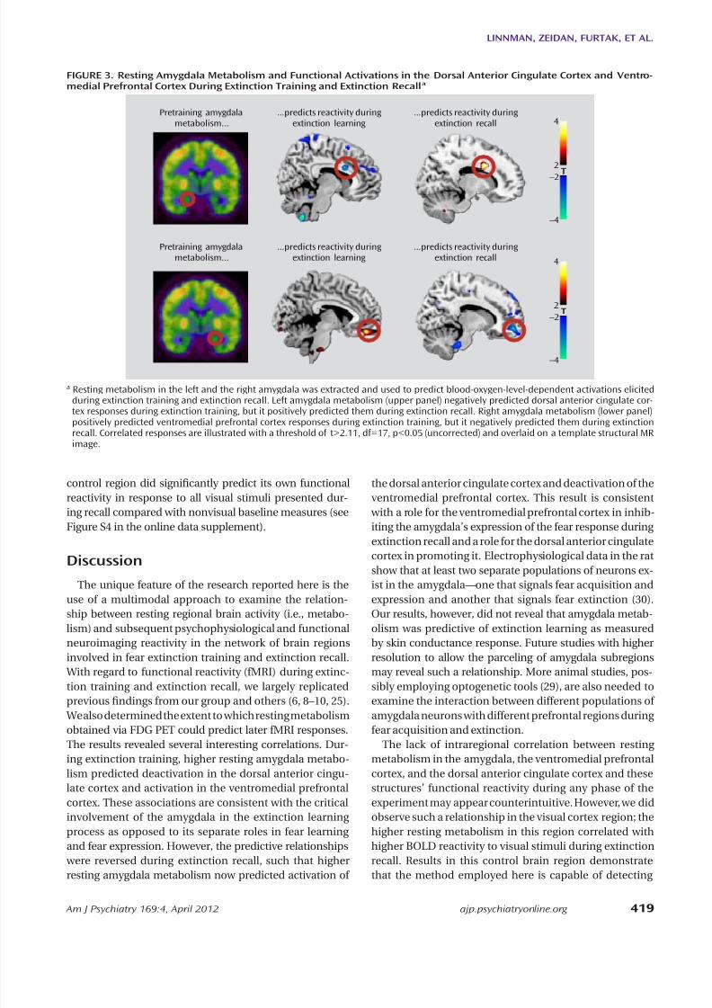
p=0.032, family-wise error corrected; r=0.73), whereas rightamygdala metabolism negatively predicted ventrome-dial prefrontal cortex activation (x=14, y=56, z=–10; t=3.86,df=17, p=0.021, family-wise error corrected; r=–0.69) (Fig-
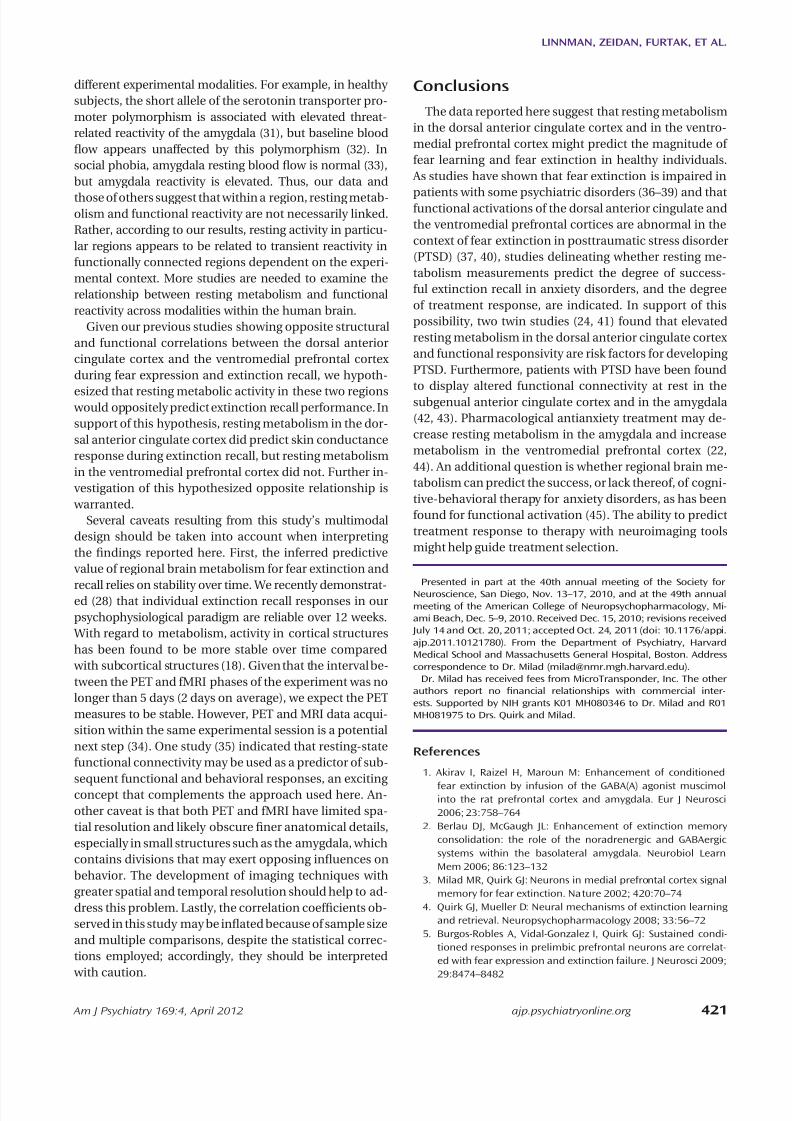
ure 3). Scatterplots of the observed correlations are pre-sented in Figure S3 in the online data supplement. Themetabolism ratio of the ventromedial prefrontal cortex tothe dorsal anterior cingulate cortex positively predictedpregenual anterior cingulate activation (x=–4, y=42, z=4;t=3.67, df=17, p=0.035, family-wise error corrected; r=0.68)(Figure 5); the rostral anterior cingulate cortex is a com-ponent of the ventromedial prefrontal cortex. Neither theventromedial prefrontal cortex nor the dorsal anteriorcingulate cortex metabolism alone predicted functionalactivations during extinction recall, including activationsin their own regions; the latter also applied to the amyg-dala. In contrast, metabolism in the primary visual cortex
Regional Brain Metabolism as a Predictor of SkinConductance Responses
In none of our a priori regions of interest did regionalmetabolism predict skin conductance responses duringextinction learning. During extinction recall, however,dorsal anterior cingulate cortex metabolism positivelypredicted skin conductance response (x=8, y=32, z=26;t=5.21, df=17, p=0.015, family-wise error corrected, r=0.80;Figure 2), indicative of poorer recall of fear extinction.
Regional Brain Metabolism as a Predictor of BOLD Activations
We examined the association between resting amygdalametabolism and subsequent BOLD reactivity in the con-trast between early and late trials of the extinguished cueduring extinction training. We observed that left amygdalametabolism negatively predicted activation in the dorsalanterior cingulate cortex (x=8, y=14, z=26; t=3.74, df=17,p=0.035, family-wise error corrected; r=–0.68), whereasright amygdala metabolism positively predicted activa-tion in the ventromedial prefrontal cortex (x=4, y=48,z=–18; t=3.87, df=17, p=0.020, family-wise error correct-ed; r=0.70) (Figure 3). Moreover, ventromedial prefrontal
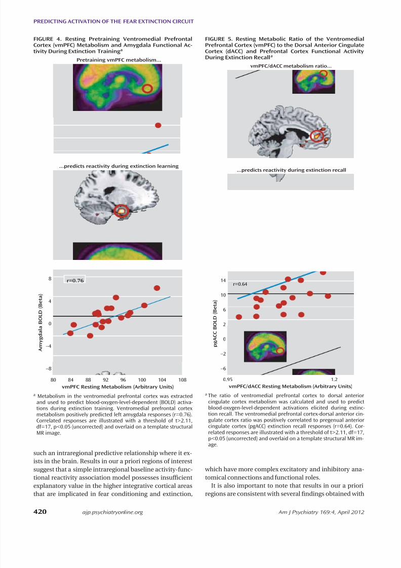
cortex metabolism positively predicted activation in theleft amygdala (x=–22, y=–4, z=–26; t=3.80, df=17, p=0.035,family-wise error corrected; r=0.69) (Figure 4). Neithermetabolism in the dorsal anterior cingulate cortex nor themetabolism ratio of the ventromedial prefrontal cortex tothe dorsal anterior cingulate cortex signicantly predictedBOLD responses during extinction training.
We then examined regional metabolism in relation toBOLD reactivity during extinction recall in the contrastbetween early extinguished and nonextinguished cue tri-als. In marked contrast to extinction training, left amygdalametabolism now positively predicted dorsal anterior cin-gulate cortex activation (x=16, y=12, z=30; t=4.27, df=17,
FIGURE 1. Blood-Oxygen-Level-Dependent (BOLD) Activa-tions During Extinction Training and Extinction Recall a
BOLD signal duringextinction learning
BOLD signal duringextinction recall
CS+late > CS+early CS+E > CS+N
T –4 –2 2 4
a The BOLD responses during extinction training (left) represent acti-vations to the conditioned stimulus (CS) during late extinction con-trasted with early extinction. The recall contrast (right) representsactivations to the extinguished cue (E) contrasted with activationsto the nonextinguished cue (N). Activation maps are illustratedwith a threshold of t>2.11, df=17, p<0.05 (uncorrected) and over-laid on a template structural MR image.
FIGURE 2. Resting Pretraining Metabolism in the DorsalAnterior Cingulate Cortex (dACC) Predicts Differential SkinConductance Responses During Extinction Recall a
S q u a r e
R o o t
S k i n C o n
d u c t a n
c e
R e s p o n s e s
C S +
E v s
C S
–
dACC Resting Metabolism (Arbitrary Units)
1
0
–1
74 78 82 86 90 94
T –4 –2 2 4
r=0.80
a Whole brain correlation map showing that the dorsal anteriorcingulate cortex metabolism was positively correlated with skinconductance responses to the extinguished cue (E) (minus the con-ditioned stimulus [CS]; r=0.80), indicative of spontaneously recov-ered fear. Correlated responses are illustrated with a threshold oft>2.11, df=17, p<0.05 (uncorrected) and overlaid on the averagemetabolism positron emission tomography maps from the study.
8/11/2019 appi.ajp.2011.10121780
http://slidepdf.com/reader/full/appiajp201110121780 5/9
Am J Psychiatry 169:4, April 2012 ajp.psychiatryonline.org 419
LINNMAN, ZEIDAN, FURTAK, ET AL.
the dorsal anterior cingulate cortex and deactivation of theventromedial prefrontal cortex. This result is consistent with a role for the ventromedial prefrontal cortex in inhib-
iting the amygdala’s expression of the fear response duringextinction recall and a role for the dorsal anterior cingulatecortex in promoting it. Electrophysiological data in the ratshow that at least two separate populations of neurons ex-ist in the amygdala—one that signals fear acquisition andexpression and another that signals fear extinction (30).Our results, however, did not reveal that amygdala metab-olism was predictive of extinction learning as measuredby skin conductance response. Future studies with higherresolution to allow the parceling of amygdala subregionsmay reveal such a relationship. More animal studies, pos-sibly employing optogenetic tools (29), are also needed to
examine the interaction between different populations ofamygdala neurons with different prefrontal regions duringfear acquisition and extinction.
The lack of intraregional correlation between restingmetabolism in the amygdala, the ventromedial prefrontalcortex, and the dorsal anterior cingulate cortex and thesestructures’ functional reactivity during any phase of theexperiment may appear counterintuitive. However, we didobserve such a relationship in the visual cortex region; thehigher resting metabolism in this region correlated withhigher BOLD reactivity to visual stimuli during extinctionrecall. Results in this control brain region demonstratethat the method employed here is capable of detecting
control region did signicantly predict its own functionalreactivity in response to all visual stimuli presented dur-ing recall compared with nonvisual baseline measures (see
Figure S4 in the online data supplement).
Discussion
The unique feature of the research reported here is theuse of a multimodal approach to examine the relation-ship between resting regional brain activity (i.e., metabo-lism) and subsequent psychophysiological and functionalneuroimaging reactivity in the network of brain regionsinvolved in fear extinction training and extinction recall. With regard to functional reactivity (fMRI) during extinc-tion training and extinction recall, we largely replicated
previous ndings from our group and others (6, 8–10, 25). We also determined the extent to which resting metabolismobtained via FDG PET could predict later fMRI responses.The results revealed several interesting correlations. Dur-ing extinction training, higher resting amygdala metabo-lism predicted deactivation in the dorsal anterior cingu-late cortex and activation in the ventromedial prefrontalcortex. These associations are consistent with the criticalinvolvement of the amygdala in the extinction learningprocess as opposed to its separate roles in fear learningand fear expression. However, the predictive relationships were reversed during extinction recall, such that higherresting amygdala metabolism now predicted activation of
FIGURE 3. Resting Amygdala Metabolism and Functional Activations in the Dorsal Anterior Cingulate Cortex and Ventro-medial Prefrontal Cortex During Extinction Training and Extinction Recall a
Pretraining amygdalametabolism...
...predicts reactivity duringextinction learning
...predicts reactivity duringextinction recall
Pretraining amygdalametabolism...
...predicts reactivity duringextinction learning
...predicts reactivity duringextinction recall
R
R
L
L
T
–4
–2
2
4
T
–4
–22
4
a Resting metabolism in the left and the right amygdala was extracted and used to predict blood-oxygen-level-dependent activations elicitedduring extinction training and extinction recall. Left amygdala metabolism (upper panel) negatively predicted dorsal anterior cingulate cor-tex responses during extinction training, but it positively predicted them during extinction recall. Right amygdala metabolism (lower panel)positively predicted ventromedial prefrontal cortex responses during extinction training, but it negatively predicted them during extinctionrecall. Correlated responses are illustrated with a threshold of t>2.11, df=17, p<0.05 (uncorrected) and overlaid on a template structural MRimage.
8/11/2019 appi.ajp.2011.10121780
http://slidepdf.com/reader/full/appiajp201110121780 6/9
420 ajp.psychiatryonline.org Am J Psychiatry 169:4, April 2012
PREDICTING ACTIVATION OF THE FEAR EXTINCTION CIRCUIT
which have more complex excitatory and inhibitory ana-tomical connections and functional roles.
It is also important to note that results in our a prioriregions are consistent with several ndings obtained with
such an intraregional predictive relationship where it ex-ists in the brain. Results in our a priori regions of interestsuggest that a simple intraregional baseline activity-func-tional reactivity association model possesses insufcientexplanatory value in the higher integrative cortical areasthat are implicated in fear conditioning and extinction,
FIGURE 4. Resting Pretraining Ventromedial PrefrontalCortex (vmPFC) Metabolism and Amygdala Functional Ac-tivity During Extinction Training a
A m y g d a l a
B O L D ( B e t a
)
vmPFC Resting Metabolism (Arbitrary Units)
8
4
–4
0
–8
80 84 88 92 96 100 104 108
...predicts reactivity during extinction learning
Pretraining vmPFC metabolism...
r=0.76
a
Metabolism in the ventromedial prefrontal cortex was extractedand used to predict blood-oxygen-level-dependent (BOLD) activa-tions during extinction training. Ventromedial prefrontal cortexmetabolism positively predicted left amygdala responses (r=0.76).Correlated responses are illustrated with a threshold of t>2.11,df=17, p<0.05 (uncorrected) and overlaid on a template structuralMR image.
FIGURE 5. Resting Metabolic Ratio of the VentromedialPrefrontal Cortex (vmPFC) to the Dorsal Anterior CingulateCortex (dACC) and Prefrontal Cortex Functional ActivityDuring Extinction Recall a
p g
A C C B O L D
( B e t a )
vmPFC/dACC Resting Metabolism (Arbitrary Units)
14
6
2
10
–2
0
–6
0.95 1.2
...predicts reactivity during extinction recall
vmPFC/dACC metabolism ratio...
r=0.64
a
The ratio of ventromedial prefrontal cortex to dorsal anteriorcingulate cortex metabolism was calculated and used to predictblood-oxygen-level-dependent activations elicited during extinc-tion recall. The ventromedial prefrontal cortex-dorsal anterior cin-gulate cortex ratio was positively correlated to pregenual anteriorcingulate cortex (pgACC) extinction recall responses (r=0.64). Cor-related responses are illustrated with a threshold of t>2.11, df=17,p<0.05 (uncorrected) and overlaid on a template structural MR im-age.
8/11/2019 appi.ajp.2011.10121780
http://slidepdf.com/reader/full/appiajp201110121780 7/9
Am J Psychiatry 169:4, April 2012 ajp.psychiatryonline.org 421
LINNMAN, ZEIDAN, FURTAK, ET AL.
ConclusionsThe data reported here suggest that resting metabolism
in the dorsal anterior cingulate cortex and in the ventro-medial prefrontal cortex might predict the magnitude offear learning and fear extinction in healthy individuals. As studies have shown that fear extinction is impaired in
patients with some psychiatric disorders (36–39) and thatfunctional activations of the dorsal anterior cingulate andthe ventromedial prefrontal cortices are abnormal in thecontext of fear extinction in posttraumatic stress disorder(PTSD) (37, 40), studies delineating whether resting me-tabolism measurements predict the degree of success-ful extinction recall in anxiety disorders, and the degreeof treatment response, are indicated. In support of thispossibility, two twin studies (24, 41) found that elevatedresting metabolism in the dorsal anterior cingulate cortexand functional responsivity are risk factors for developingPTSD. Furthermore, patients with PTSD have been foundto display altered functional connectivity at rest in thesubgenual anterior cingulate cortex and in the amygdala(42, 43). Pharmacological antianxiety treatment may de-crease resting metabolism in the amygdala and increasemetabolism in the ventromedial prefrontal cortex (22,44). An additional question is whether regional brain me-tabolism can predict the success, or lack thereof, of cogni-tive-behavioral therapy for anxiety disorders, as has beenfound for functional activation (45). The ability to predicttreatment response to therapy with neuroimaging toolsmight help guide treatment selection.
Presented in part at the 40th annual meeting of the Society forNeuroscience, San Diego, Nov. 13–17, 2010, and at the 49th annualmeeting of the American College of Neuropsychopharmacology, Mi-ami Beach, Dec. 5–9, 2010. Received Dec. 15, 2010; revisions received
July 14 and Oct. 20, 2011; accepted Oct. 24, 2011 (doi: 10.1176/appi.ajp.2011.10121780). From the Department of Psychiatry, HarvardMedical School and Massachusetts General Hospital, Boston. Addresscorrespondence to Dr. Milad ([email protected]).
Dr. Milad has received fees from MicroTransponder, Inc. The otherauthors report no nancial relationships with commercial inter-ests. Supported by NIH grants K01 MH080346 to Dr. Milad and R01MH081975 to Drs. Quirk and Milad.
References
1. Akirav I, Raizel H, Maroun M: Enhancement of conditioned
fear extinction by infusion of the GABA(A) agonist muscimolinto the rat prefrontal cortex and amygdala. Eur J Neurosci2006; 23:758–764
2. Berlau DJ, McGaugh JL: Enhancement of extinction memoryconsolidation: the role of the noradrenergic and GABAergicsystems within the basolateral amygdala. Neurobiol LearnMem 2006; 86:123–132
3. Milad MR, Quirk GJ: Neurons in medial prefrontal cortex signalmemory for fear extinction. Nature 2002; 420:70–74
4. Quirk GJ, Mueller D: Neural mechanisms of extinction learningand retrieval. Neuropsychopharmacology 2008; 33:56–72
5. Burgos-Robles A, Vidal-Gonzalez I, Quirk GJ: Sustained condi-tioned responses in prelimbic prefrontal neurons are correlat-ed with fear expression and extinction failure. J Neurosci 2009;29:8474–8482
different experimental modalities. For example, in healthysubjects, the short allele of the serotonin transporter pro-moter polymorphism is associated with elevated threat-related reactivity of the amygdala (31), but baseline bloodow appears unaffected by this polymorphism (32). Insocial phobia, amygdala resting blood ow is normal (33),but amygdala reactivity is elevated. Thus, our data and
those of others suggest that within a region, resting metab-olism and functional reactivity are not necessarily linked.Rather, according to our results, resting activity in particu-lar regions appears to be related to transient reactivity infunctionally connected regions dependent on the experi-mental context. More studies are needed to examine therelationship between resting metabolism and functionalreactivity across modalities within the human brain.
Given our previous studies showing opposite structuraland functional correlations between the dorsal anteriorcingulate cortex and the ventromedial prefrontal cortexduring fear expression and extinction recall, we hypoth-
esized that resting metabolic activity in these two regions would oppositely predict extinction recall performance. Insupport of this hypothesis, resting metabolism in the dor-sal anterior cingulate cortex did predict skin conductanceresponse during extinction recall, but resting metabolismin the ventromedial prefrontal cortex did not. Further in-vestigation of this hypothesized opposite relationship is warranted.
Several caveats resulting from this study’s multimodaldesign should be taken into account when interpretingthe ndings reported here. First, the inferred predictivevalue of regional brain metabolism for fear extinction and
recall relies on stability over time. We recently demonstrat-ed (28) that individual extinction recall responses in ourpsychophysiological paradigm are reliable over 12 weeks. With regard to metabolism, activity in cortical structureshas been found to be more stable over time compared with subcortical structures (18). Given that the interval be-tween the PET and fMRI phases of the experiment was nolonger than 5 days (2 days on average), we expect the PETmeasures to be stable. However, PET and MRI data acqui-sition within the same experimental session is a potentialnext step (34). One study (35) indicated that resting-statefunctional connectivity may be used as a predictor of sub-sequent functional and behavioral responses, an excitingconcept that complements the approach used here. An-other caveat is that both PET and fMRI have limited spa-tial resolution and likely obscure ner anatomical details,especially in small structures such as the amygdala, whichcontains divisions that may exert opposing inuences onbehavior. The development of imaging techniques withgreater spatial and temporal resolution should help to ad-dress this problem. Lastly, the correlation coefcients ob-served in this study may be inated because of sample sizeand multiple comparisons, despite the statistical correc-tions employed; accordingly, they should be interpreted with caution.

8/11/2019 appi.ajp.2011.10121780
http://slidepdf.com/reader/full/appiajp201110121780 8/9
422 ajp.psychiatryonline.org Am J Psychiatry 169:4, April 2012
PREDICTING ACTIVATION OF THE FEAR EXTINCTION CIRCUIT
25. Milad MR, Pitman RK, Ellis CB, Gold AL, Shin LM, Lasko NB,Zeidan MA, Handwerger K, Orr SP, Rauch SL: Neurobiologicalbasis of failure to recall extinction memory in posttraumaticstress disorder. Biol Psychiatry 2009; 66:1075–1082
26. Milad MR, Orr SP, Pitman RK, Rauch SL: Context modulationof memory for fear extinction in humans. Psychophysiology2005; 42:456–464
27. Milad MR, Orr SP, Lasko NB, Chang Y, Rauch SL, Pitman RK:Presence and acquired origin of reduced recall for fear ex-tinction in PTSD: results of a twin study. J Psychiatr Res 2008;42:515–520
28. Zeidan MA, Lebron-Milad K, Thompson-Hollands J, Im JJ,Dougherty DD, Holt DJ, Orr SP, Milad MR: Test-retest reliabil-ity during fear acquisition and fear extinction in humans. CNSNeurosci Ther (Epub ahead of print, Feb 16, 2011)
29. Tye KM, Prakash R, Kim SY, Fenno LE, Grosenick L, Zarabi H,Thompson KR, Gradinaru V, Ramakrishnan C, Deisseroth K:Amygdala circuitry mediating reversible and bidirectional con-trol of anxiety. Nature 2011; 471:358–362
30. Likhtik E, Popa D, Apergis-Schoute J, Fidacaro GA, Pare D:Amygdala intercalated neurons are required for expression offear extinction. Nature 2008; 454:642–645
31. Caspi A, Hariri AR, Holmes A, Uher R, Moftt TE: Genetic sensi-
tivity to the environment: the case of the serotonin transportergene and its implications for studying complex diseases andtraits. Am J Psychiatry 2010; 167:509–527
32. Viviani R, Sim EJ, Lo H, Beschoner P, Osterfeld N, Maier C,Seeringer A, Godoy AL, Rosa A, Comas D, Kirchheiner J: Base-line brain perfusion and the serotonin transporter promoterpolymorphism. Biol Psychiatry 2010; 67:317–322
33. Furmark T, Henningsson S, Appel L, Ahs F, Linnman C, PissiotaA, Faria V, Oreland L, Bani M, Pich EM, Eriksson E, FredriksonM: Genotype over-diagnosis in amygdala responsiveness: affec-tive processing in social anxiety disorder. J Psychiatry Neurosci2009; 34:30–40
34. Judenhofer MS, Wehrl HF, Newport DF, Catana C, Siegel SB,Becker M, Thielscher A, Kneilling M, Lichy MP, Eichner M, Klin-gel K, Reischl G, Widmaier S, Röcken M, Nutt RE, Machulla HJ,Uludag K, Cherry SR, Claussen CD, Pichler BJ: SimultaneousPET-MRI: a new approach for functional and morphologicalimaging. Nat Med 2008; 14:459-465
35. Ploner M, Lee MC, Wiech K, Bingel U, Tracey I: Prestimulusfunctional connectivity determines pain perception in hu-mans. Proc Natl Acad Sci USA 2010; 107:355–360
36. Holt DJ, Lebron-Milad K, Milad MR, Rauch SL, Pitman RK, OrrSP, Cassidy BS, Walsh JP, Goff DC: Extinction memory is im-paired in schizophrenia. Biol Psychiatry 2009; 65:455–463
37. Milad MR, Pitman RK, Ellis CB, Gold AB, Shin LM, Lasko NB,Zeidan MA, Handwerger K, Orr SP, Rauch SL: Neurobiologicalbasis for failure to recall extinction memory in posttraumaticstress disorder. Biol Psychiatry 2009; 66:1075–1082
38. Orr SP, Metzger LJ, Lasko NB, Macklin ML, Peri T, Pitman RK:
De novo conditioning in trauma-exposed individuals with andwithout posttraumatic stress disorder. J Abnorm Psychol 2000;109:290–298
39. Jovanovic T, Norrholm SD, Blanding NQ, Davis M, Duncan E,Bradley B, Ressler KJ: Impaired fear inhibition is a biomarkerof PTSD but not depression. Depress Anxiety 2010; 27:244–251
40. Rougemont-Bucking A, Linnman C, Zefro TA, Zeidan MA,Lebron-Milad K, Rodriguez-Romaguera J, Rauch SL, Pitman RK,Milad MR: Altered processing of contextual information dur-ing fear extinction in PTSD: an fMRI study. CNS Neurosci Ther2011; 17:227–236
41. Shin LM, Bush G, Milad MR, Lasko NB, Brohawn KH, Hughes KC,Macklin ML, Gold AL, Karpf RD, Orr SP, Rauch SL, Pitman RK:Exaggerated activation of dorsal anterior cingulate cortex dur-
6. Sehlmeyer C, Schoning S, Zwitserlood P, Peiderer B, Kircher T,Arolt V, Konrad C: Human fear conditioning and extinction inneuroimaging: a systematic review. PLoS One 2009; 4:e5865
7. Nili U, Goldberg H, Weizman A, Dudai Y: Fear thou not: activityof frontal and temporal circuits in moments of real-life cour-age. Neuron 2010; 66:949–962
8. Kalisch R, Korenfeld E, Stephan KE, Weiskopf N, Seymour B,Dolan RJ: Context-dependent human extinction memory ismediated by a ventromedial prefrontal and hippocampal net-work. J Neurosci 2006; 26:9503–9511
9. Milad MR, Wright CI, Orr SP, Pitman RK, Quirk GJ, Rauch SL:Recall of fear extinction in humans activates the ventromedialprefrontal cortex and hippocampus in concert. Biol Psychiatry2007; 62:446–454
10. Phelps EA, Delgado MR, Nearing KI, Ledoux JE: Extinctionlearning in humans: role of the amygdala and vmPFC. Neuron2004; 43:897–905
11. Kim MJ, Whalen PJ: The structural integrity of an amygdala-prefrontal pathway predicts trait anxiety. J Neurosci 2009;29:11614–11618
12. Indovina I, Robbins TW, Nunez-Elizalde AO, Dunn BD, BishopSJ: Fear-conditioning mechanisms associated with trait vulner-ability to anxiety in humans. Neuron 2011; 69:563–571
13. Milad MR, Quinn BT, Pitman RK, Orr SP, Fischl B, Rauch SL:Thickness of ventromedial prefrontal cortex in humans is cor-related with extinction memory. Proc Natl Acad Sci USA 2005;102:10706–10711
14. Shulman RG, Rothman DL, Behar KL, Hyder F: Energetic basisof brain activity: implications for neuroimaging. Trends Neuro-sci 2004; 27:489–495
15. Gusnard DA, Raichle ME, Raichle ME: Searching for a baseline:functional imaging and the resting human brain. Nat Rev Neu-rosci 2001; 2:685–694
16. Raichle ME, Mintun MA: Brain work and brain imaging. AnnuRev Neurosci 2006; 29:449–476
17. Buzsaki G, Kaila K, Raichle M: Inhibition and brain work. Neu-ron 2007; 56:771–783
18. Schaefer SM, Abercrombie HC, Lindgren KA, Larson CL, WardRT, Oakes TR, Holden JE, Perlman SB, Turski PA, Davidson RJ:Six-month test-retest reliability of MRI-dened PET measuresof regional cerebral glucose metabolic rate in selected subcor-tical structures. Hum Brain Mapp 2000; 10:1–9
19. Phelps ME, Huang SC, Hoffman EJ, Selin C, Sokoloff L, Kuhl DE:Tomographic measurement of local cerebral glucose meta-bolic rate in humans with (F-18)2-uoro-2-deoxy-D-glucose:validation of method. Ann Neurol 1979; 6:371–388
20. Reivich M, Kuhl D, Wolf A, Greenberg J, Phelps M, Ido T, CasellaV, Fowler J, Hoffman E, Alavi A, Som P, Sokoloff L: The [18F]uorodeoxyglucose method for the measurement of local cer-ebral glucose utilization in man. Circ Res 1979; 44:127–137
21. Sokoloff L: Relation between physiological function and en-ergy metabolism in the central nervous system. J Neurochem
1977; 29:13–2622. Evans KC, Simon NM, Dougherty DD, Hoge EA, Worthington JJ,Chow C, Kaufman RE, Gold AL, Fischman AJ, Pollack MH, RauchSL: A PET study of tiagabine treatment implicates ventral me-dial prefrontal cortex in generalized social anxiety disorder.Neuropsychopharmacology 2009; 34:390–398
23. Oler JA, Fox AS, Shelton SE, Rogers J, Dyer TD, Davidson RJ,Shelledy W, Oakes TR, Blangero J, Kalin NH: Amygdalar andhippocampal substrates of anxious temperament differ intheir heritability. Nature 2010; 466:864–868
24. Shin LM, Lasko NB, Macklin ML, Karpf RD, Milad MR, Orr SP,Goetz JM, Fischman AJ, Rauch SL, Pitman RK: Resting metabolicactivity in the cingulate cortex and vulnerability to posttrau-matic stress disorder. Arch Gen Psychiatry 2009; 66:1099–1107

8/11/2019 appi.ajp.2011.10121780
http://slidepdf.com/reader/full/appiajp201110121780 9/9
Am J Psychiatry 169:4, April 2012 ajp.psychiatryonline.org 423
LINNMAN, ZEIDAN, FURTAK, ET AL.
44. Schmidt ME, Andrews RD, van der Ark P, Brown T, MannaertE, Steckler T, de Hoon J, Van Laere K: Dose-dependent effectsof the CRF(1) receptor antagonist R317573 on regional brainactivity in healthy male subjects. Psychopharmacology 2010;208:109–119
45. Bryant RA, Felmingham K, Kemp A, Das P, Hughes G, Peduto A,Williams L: Amygdala and ventral anterior cingulate activationpredicts treatment response to cognitive behavior therapy forpost-traumatic stress disorder. Psychol Med 2008; 38:555–561
ing cognitive interference: a monozygotic twin study of post-traumatic stress disorder. Am J Psychiatry 2011; 168:979–985
42. Bluhm RL, Williamson PC, Osuch EA, Frewen PA, Stevens TK,Boksman K, Neufeld RW, Théberge J, Lanius RA: Alterationsin default network connectivity in posttraumatic stress disor-der related to early-life trauma. J Psychiatry Neurosci 2009;34:187–194
43. Lanius RA, Bluhm RL, Coupland NJ, Hegadoren KM, Rowe B,Theberge J, Neufeld RW, Williamson PC, Brimson M: Defaultmode network connectivity as a predictor of post-traumaticstress disorder symptom severity in acutely traumatized sub-
jects. Acta Psychiatr Scand 2010; 121:33–40