application of laparoscopy in upper urinary tract surgery
TRANSCRIPT

1
Application of Laparoscopy in Upper Urinary Tract
Surgery
By
Dr. Fariborz Bagheri, MD, FEBU
Doctoral (PhD) Thesis
Medical School, University of Pécs, Hungary
Head of the Doctoral School: Prof. Dr. Gabor Kovacs
PhD Program Leader: Dr. Gabor Jancso
Thesis Supervisor: Prof. Dr. Laszlo Farkas
Honorary Advisor: Prof. Dr. Günter Janetschek
Pecs 2016

2
“Few procedures provide the urologist with more
satisfaction than those that preserve renal function”
(Benjamin Abeshouse, 1950)
If we want to see into the future, we need only look to the
past

3
CONTENTS
ABBREVIATIONS and ACRONYMS
Page
4
1 INTRODUCTION 5
1.1 The changing face of renal cell carcinoma 5
1.1.1 Epidemiology of renal cell cancer 5
1.1.2 New genetic classification system 6
1.1.3 TNM-G classification and staging 8
1.1.4 Anatomic classification system 10
1.2 OPEN OR LAPAROSCOPIC SURGERY 14
1.3 LAPAROSCOPIC SURGERY FOR T1 KIDNEY CANCER 16
1.4 THE EFFECT OF INTRAOPERATIVE ISCHEMIA 18
1.4.1 Mechanism of ischemia-reperfusion injury 18
1.4.2 Repair of the tubular system 20
2. AIM OF THE STUDY 22
3. OPERATIVETECHNIQUES & POST-OP FUNCTIONAL EVAL. 22
3.1 LAPAROS. PARTIAL NEPHRECTOMY IN COLD ISCHEMIA 22
3.1.1 Background 22
3.1.2 Patients and laparoscopic approach 23
3.1.3 Discussion 27
3.1.4 Conclusion 29
3.2. LAPAROS. PARTIAL NEPHRECTOMY IN WARM ISCHEMIA 30
3.2.1 Introduction 30
3.2.2 Patients and methods 31
3.2.3 Renal scintigraphy 32
3.2.4 Assessment of total and partial differential renal function 32
3.2.5 Patient selection and statistical evaluations 34
3.2.6 Patients operated with laparoscopic radical nephrectomy 36
3.2.7 Results 36
3.2.8 Evaluation of the renal function based on serum creatinine 38
3.2.9 Evaluation of the renal function based on estimated GFR 39
3.2.10 Impact of parenchymal volume reduction on the kidney function 41
3.2.11 Discussion 43
3.2.12 Conclusion 48
3.3 TECHNICAL MODIFICATIONS OF LAPAROSCOPIC SURGERY 49
3.3.1 Laparos. partial adrenalectomy for recurrent pheochromocytoma 49
3.3.2 New tool for laparoscopic antegrade ureteral stenting 50
3.3.3 Ligation of a wide renal vein during laparoscopic nephrectomy 53
4. THESIS RELATED NEW OBSERVATIONS 56
5. REFERENCES 57
6. PUBLICATIONS AND PRESENTATIONS 62
7. ACKNOWLEDGEMENTS 66

4
ABBREVIATIONS AND ACRONYMS
99m
Tc-DMSA 99m
Technetium-Dimercaptosuccinic Acid……………..
AKI Acute kidney injury........................................................
AMP Adenosine Monophosphate…………………………….
ANOVA Analysis of Variance…………………………………..
ATN Acute Tubular Necrosis………………………………..
ATP Adenosine Triphosphate………………………………..
BMI Body Mass Index (kg/m2)……………………………….
CKD-EPI Chronic Kidney Disease Epidemiology Collaboration…
CT Computerized Tomography.............................................
EAU European Association of Urology………………………
eGFR Estimated Glomerular Filtration Rate………………….
GFR Glomerular Filtration Rate……………………………..
IR Ischemia-Reperfusion………………………………..…
LN Laparoscopic Nephrectomy……………………………
LPN Laparoscopic Partial Nephrectomy……………………
LRN Laparoscopic Radical Nephrectomy…………………….
MAG 3 Mercapto-Acetyltriglycine……………………………..
NSS Nephron-Sparing Surgery………………………………
OPN Open Partial Nephrectomy………………………........
P-DRF Partial Differential Renal Function……………………
PN Partial Nephrectomy…………………………………..
RCC Renal Cell Carcinoma………………………….……..
RF Renal Function………………………………….……..
RN Radical Nephrectomy……………………………........
ROI Region Of Interest……………………………………...
sCr Serum Creatinine…………….…………………………
T-DRF Total Differential Renal Function………………………
US Ultrasound………………………………………...........
WI Warm Ischemia………………………………………….
WIT Warm Ischemia Time……………………………….…..
5
1. INTRODUCTION
1.1. THE CHANGING FACE OF RENAL CELL CARCINOMA
1.1.1. Epidemiology of renal cell cancer.
Renal cell carcinoma (RCC) makes up approximately 2-3% of all cancers and
accounts for 90% of all malignancies of the kidney. It is the 9th
most common cancer
in men and 14th
in women. However, the incidence of RCC varies worldwide (Ferlay
J et al. 2012)1. It is more common in industrialized countries where it is ranked in
both sexes as 11th
most common tumor while in developing countries it is ranked as
21st place. Although the male-to-female ratio differs significantly for each type of
renal cell tumor and in different age groups, generally there is a 2:1 predominance of
men over women, with peak incidence occurring between 60 and 70 years of age
(Hew MN et al. 2012)2.
Through the past decades there has been a steady increase in number of clinically
recognized RCC in industrialized countries.
The growing number of incidentally detected RCC cannot be explained exclusively
by extensive use of modern imaging techniques. Several other factors related to life
style of modern industrialized countries may also play a role in the increasing rate of
RCC. The exact etiology of RCC, with exception of hereditary cases, is still
unknown. It is estimated that smoking accounts for around one third of the RCC
cases (Cumberbatch MG et al. 2016)3. In addition to smoking, which doubles the
risk for RCC, obesity, hypertension and an unhealthy diet are also associated with
the tumor development (Chow W-H et al. 2010)4.
Through the past decades the mortality rate has stabilized or even declined in most
European countries.
In spite of the increasing number of clinically detected RCC, the mortality rate due
to RCC has stabilized or even declined in European countries and the USA (Levi et
al. 2008)5. The widespread use of imaging technique resulted in the detection of
RCC that are smaller and of lower stage (Lightfoot et al. 2000)6. The number of
incidentally detected T1a and also T1b tumours is increasing in operation statistics
of most urological centers. These tumours metastasize less often compared with
higher stage tumours.

6
1.1.2. New genetic classification system
RCC is not considered as a single pathological entity, but different types
characterized by specific genetic alteration and natural history (Kovacs, 1993)7. The
new knowledge was discussed at a conference held in Heidelberg and published as
the Heidelberg Classification of Renal Cell Tumors (Kovacs et al. 1997)8. Each type
of tumour displays a combination of chromosomal-genomic alterations of diagnostic
impact (Table 1). The new classification system is now adapted by the WHO. The
diagnosis of a specific type of RCC itself has a strong impact on the prognosis of
tumors as the 5 years tumour specific survival indicates (Table 1).
Table 1. Some pertinent genetic data on distinct types of renal cell tumours.
Tumor Frequency
(%)
Genomic alterations* Gene mutations 5 years
survival (%)
cRCC 65-70 -3p, +5q, -6q, -8p, -9p, -14q VHL (50%)
PBRM1 (35%)
~60
pRCC 10-15 +7, +17, +8, +12, +16, +20, MET (5%) ~90
chRCC 5-7 -1, -2, -6, -10, -13, -17, -21 p53 (25%) ~95
RO 5-7 -1, -14q or t(11q:?) or none ? 100
ucRCC 5-7 ? ? ~20
CDC <1 ? ? 30
MTSCC <1 -1, -4, -6, -9, -13, -14, -15, -
22
? 100
MA <1 none ? 100
cRCC-conventional RCC, pRCC-papillary RCC, chRCC-chromophobe RCC, RO-
renal oncocytoma, ucRCC-unclassified RCC, CDC-collecting duct carcinoma,
MTSCC-mucinous tubular and spindle cell carcinoma, MA-metanephric adenoma.
*Genomic alterations: „-„ = loss, „+” = gain, t = translocation; VHL – von Hippel
Lindau gene; MET – Met tyrosin kinase receptor. (Table 1. is kindly provided by
Professor Gyula Kovacs).

7
Based on the characteristic cell morphology and growth pattern, 4 main types of
renal cell tumours can be distinguished (Figure 1). The most common conventional
RCC (also called clear cell RCC, which is a misleading name as every fourth of this
genetically defined tumour does not display clear but eosinophilic cytoplasm). In
spite of overlapping phenotype, the histological diagnosis can be made by
experienced pathologist on haematoxilin-eosin stained slides in the vast majority of
cases. However, in some cases a genetic analysis is necessary to detect the tumour
type specific alterations and to achive a correct diagnosis, especially for the
differential diagnosis of benign renal oncocytoma and malignant chromophobe
RCC (Table 1).
Figure 1. Characteristic histological picture of four main types of renal cell
tumours. A, Conventional RCC composed of trabecular arranged clear cells with
empty cytoplasm and picnotic nuclei. B, Papillary renal cell carcinoma composed of
medium sized eosinophilic cells with foamy cells in papillary stalks. C,
Chromophobe renal cell carcinoma with large pale cells, double nuclei arranged in
large epithelial sheets. D, Renal oncocytoma showing large nests of strong
eosinophilic cells embedded in oedematous stroma. (Haematoxylin and eosin
staining, x400) (Kindly provided by Professzor Gyula Kovacs)
A
D C
B

8
1.1.3. TNM-G Classification and Staging: Estimating the outcome of the disease
In 2012, 84.000 patients were diagnosed with renal cell carcinoma (RCC) and
35,000 patients died due to disease in European countries (Ferlay et al. 2013)9.
Mortality of RCC correlates directly with the presence or postoperative development
of metastasis. Approximately 20-25% of the most common conventional RCC are
presented with advanced disease at the time of diagnosis and further 15-20% will
develop metastasis within the next 5 years (Zisman et al. 2002)10
.
When diagnosing cancer, the TNM Classification and stage of the tumor are
important parameters which direct the management and used to estimate the
outcome of disease (Table 2). The cancer specific survival rate is much higher in a
cohort of patients with lower stage of RCC. There is a good correlation between the
size of tumour and progression of disease. The latest classification divided T1
tumours into subgroup ≤ 4 cm (T1a) and another one > 4 cm and ≤ 7 cm in diameter
(T1b), the former having a better prognosis. Moreover, T2 tumours have also been
divided into two classes: T2a are tumours with a size > 7 and ≤ 10 cm, and T2b
tumours > 10 cm, but confined to the kidney. This classification may be crucial by
decision of carrying out a partial nephrectomy. The regional lymph node (N)
positivity is another parameter having an influence on the outcome of the disease.
The new classification system uses only one class. e.g. metastasis in one or more
lymph nodes. The metastasis (M) is the ultimate sign of tumour malinancy, leading
to patients’ death in the overwhelming majority of cases in spite of modern targeted
therapy.
Nuclear grading refers to the appearance of the cancer cells under the microscope.
The grade indicates how the cancer may behave, and is one of the important
prognostic factor of RCC. The four grade nuclear grading system is the most used
grading system for kidney cancer. However, recent studies showed by follow-up
studies, that patients with grade 1 and grade 2 tumours have a similar survival and
therefore can be evaluated together as a single grade. Doing so will result in the
three grade system with better estimation of the outcome of the disease.

9
Table 2. TNM Classification of kidney cancers
T: Primary tumour
Tx Primary tumour cannot be assessed
T0 No evidence of primary tumour
T1 Tumour ≤ 7 cm in greatest dimension, limited to the kidney
T1a Tumour ≤ 4 cm in greatest dimension, limited to the kidney
T1b Tumour > 4 cm but ≤ 7 cm in greatest dimension
T2 Tumour > 7 cm in greatest dimension, limited to the kidney
T2a Tumour > 7 cm but ≤ 10 cm in greatest dimension
T2b Tumours > 10 cm limited to the kidney
T3 Tumour extends into major veins or perinephric tissues but not into the
ipsilateral adrenal gland or beyond Gerota’s fascia
T3a
Tumour grossly extends into the renal vein or its segmental (muscle-containing)
branches, or invades perirenal and/or renal sinus fat (peripelvic), but not into the
adrenal gland and not beyond Gerota’s fascia
T3b Tumour grossly extends into the vena cava (VC) below the diaphragm
T3c Tumour grossly extends into vena cava above the diaphragm or invades the wall
of the VC
T4 Tumour invades beyond Gerota’s fascia (including contiguous extension into
the ipsilateral adrenal gland)
N: Regional lymph nodes
NX Regional LNs cannot be assessed
N0 No regional LN metastasis
N1 Regional LN metastasis
M: Distant metastasis
M0 No distant metastasis
M1 Distant metastasis
The results of TNM classification are grouped to determine the patient’s overall
survival changes (Table 3). The four tumor stages (Stage I-IV) are a combination of
local growth of tumor without or with invasive growth, the lymph node involvement
and distant metastasis. The vast majority of tumors in Stage I and II have a high
cancer specific survival, whereas stage IV tumors are in most cases fatal.

10
Table 3. TNM stage grouping of kidney cancer
Stage I T1 N0 M0
Stage II. T2 N0 M0
Stage III. T3 N0 M0
T1,T2,T3 N1 M0
Stage IV. T4 Any N M0
AnyT AnyN M1
Stage I tumours are the best candidates for laparoscopic partial nephrectomy (LPN),
but larger tumours confined to the kidney (Stage II) may also be removed by
laparoscopic radical nephrectomy (LRN). From the technical point of view, another
scoring system, the PADUA score, is more important for a laparoscopic urologist.
1.1.4. Anatomic classification system: The PADUA and RENAL nephrometry
scores
Because of the widespread use of imaging techniques such as ultrasonography and
computed tomography, a large number of small localized renal cell tumours are
detected incidentally. The vast majority of incidentally detected tumours are pT1a or
pT1b and confined to the kidney, e.g. Stage I or Stage II tumours. For such patients,
organ-sparing surgery is considered as the standard of care if applicable. There is no
difference regarding tumour recurrence and cancer specific survival of patients with
small renal tumours operated with open or laparoscopic radical or partial
nephrectomy. Therefore, for decision making of the operation technique other
parameters are necessary.
Objective anatomical classification systems, such as the Preoperative Aspects and
Dimensions Used for an Anatomical (PADUA) classification system, and the
Radius (tumor size as maximal diameter), Exophytic/endophytic properties of the
tumor, Nearness of tumor deepest portion to the collecting system or sinus,
Anterior/posterior descriptor and the Location relative to the polar line (R.E.N.A.L.)
nephrometry score and the C-index have been proposed, to standardise the
11
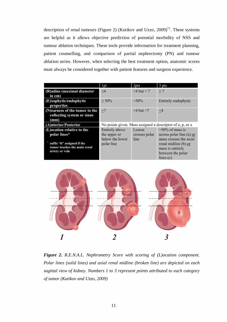
description of renal tumours (Figure 2) (Kutikov and Uzzo, 2009)11
. These systems
are helpful as it allows objective prediction of potential morbidity of NSS and
tumour ablation techniques. These tools provide information for treatment planning,
patient counselling, and comparison of partial nephrectomy (PN) and tumour
ablation series. However, when selecting the best treatment option, anatomic scores
must always be considered together with patient features and surgeon experience.
Figure 2. R.E.N.A.L. Nephrometry Score with scoring of (L)ocation component.
Polar lines (solid lines) and axial renal midline (broken line) are depicted on each
sagittal view of kidney. Numbers 1 to 3 represent points attributed to each category
of tumor (Kutikov and Uzzo, 2009)
12
The PADUA score is an important tool to predict possible risk of complications and
the length of ischemia time. It is important for selection of patients for open,
laparoscopic or robotic-assisted approaches. The size of tumour is one of the
important parameters for the choice of surgical treatment, e.g. partial or radical
nephrectomy. Small pT1a tumours are good candidates for NSS. However, in
selected patients larger tumours can be removed by partial nephrectomy applying
either open or laparoscopic surgery. Another important parameter of the scoring
system is the tumour location. The localisation of tumour within the kidney plays a
crucial role in the decision of the surgical treatment. Polar, rim or central location,
the relationship with renal sinus and collecting duct system, the deep position are
important parameters in the decision of total or partial nephrectomy. The
preoperative PADUA score is an independent predictor of the occurrence of
postoperative complications. A PADUA score of 10 or 8-9 means an approximately
30 and 15 fold risk for postoperative complications compared to PADUA score 6-7
which are reference scores of harad ratio (Figure 3) (Ficarra et al. 2009)12

13
Figure 3. (a) Longitudinal classification of the tumours; (b) margin location of the
tumours; (c) tumour relationship with renal sinus; (d) tumour relationship with
urinary collecting system; (e) tumour deepening into the parenchyma; (f) tumour
size classification.

14
1.2. OPEN OR LAPAROSCOPIC SURGERY FOR TREATMENT OF
KIDNEY CANCER
Surgery remains the only curative treatment for RCC. The objective of surgical
therapy is to excise the entire tumour with an adequate surgical margin. In 1969,
Robson and colleagues13
established radical nephrectomy (RN) as the “gold
standard” curative operation for localized RCC. In the past three decades, the
increased use of imaging modalities such as ultrasound (US) and computerized
tomography (CT) has led to increase the number of incidentally detected renal
masses6. Tumours detected by imaging techniques tend to be smaller, lower stage
lesions that are typically amenable to partial nephrectomy (PN). The main aim of
this surgical procedure is maximal preservation of unaffected renal parenchyma
without sacrificing cancer control. During the last several years, refinements in the
surgical technique of PN have made this procedure technically safe with acceptable
complication rates. Long-term outcome data indicate that open partial nephrectomy
(OPN) has cancer-free survival rates comparable to those of radical surgery with
better preservation of renal function (RF), reduced frequency of cardiovascular
events, and decreased overall mortality rate (Margreiter and Marbereger, 2010)14
.
According to European Association of Urology (EAU) guidelines 2016, with
grade of recommendation “A”, for T1a tumours, NSS is recommended and for T1b
tumours, NSS should be favoured over RN whenever feasible. RN is no longer the
gold standard treatment in these cases (Delakas et al. 2002)15
. Standard indications
for NSS are divided into the following categories: 1). Absolute – anatomical or
functional solitary kidney, and bilateral renal tumours. 2). Relative – functioning
opposite kidney is affected by a condition that might impair RF in the future, e.g.:
hereditary forms of RCC, diabetes mellitus, hypertension, autoimmune diseases,
stone formers, etc. 3). Elective – localized unilateral RCC with a healthy
contralateral kidney. Recently, cases of elective indications for NSS of RCC have
vastly increased. It has been proved that, in these cases, NSS for tumours limited in
diameter to 4 cm (pT1a) provides recurrence-free and long-term survival rates
similar to those observed after radical surgery (Becker et al. 2006)16
. For larger
tumours (pT1b), PN has demonstrated feasibility and oncological safety in carefully
selected patients (Patard et al. 2004)17
.

15
The impact of laparoscopy has increased rapidly within the last two decades, and as
a result, laparoscopic radical nephrectomy has become a recognized standard
surgical approach by the 2006 European Association of Urology guidelines. Parallel
to this evolution in technique, the indication for radical nephrectomy has also
changed to a great extent. By means of imaging techniques such as ultrasonography
and computed tomography scanning, an increasing number of small renal cancers
are being detected incidentally. For the majority of these tumors nephrectomy is an
over-treatment, and NSS has to be considered instead even if there is a normally
functioning contralateral kidney because oncologic results are as good as with
radical nephrectomy. Many surgeons are now confronted with the difficult situation
that they can offer radical nephrectomy by means of laparoscopy to remove large
tumors but are unable to perform laparoscopic NSS for the small ones. Thus, great
effort has been directed towards the development of reliable and reproducible
techniques for laparoscopic partial nephrectomy (LPN).
Open surgical partial nephrectomy is usually performed in ischemia to allow for
precise tumor excision and reconstruction of the renal parenchyma in a bloodless
field. Gill (Gill et al. 2002)18
was the first to show that these principles of open
surgery can be duplicated by means of laparoscopy. Ischemia time, however, is
critical for renal function which is traditionally restricted to a maximum of
30 minutes. Ischemia time can be increased substantially by cooling of the renal
parenchyma, which is easily induced during open surgery. When comparing
laparoscopy with open surgery, ischemia time is longer even in the most experienced
hands and hypothermia for protection of the renal function is difficult to achieve.
Several attempts have been made to overcome the aforementioned problems in
laparoscopic approaches. Direct excision of small and peripherally located tumors is
feasible without ischemia. Haemostasis can be achieved by bipolar coagulation,
ultrasonic scalpel, radiofrequency, microwave tissue coagulator and several other
devices. A variety of tissue sealants have also been used for this purpose. The main
problem with these techniques is not the haemostasis; however, because of ongoing
burning and charring of the tissues and continuing bleeding, it becomes impossible
to distinguish between normal parenchyma and tumor tissue so that a positive
16
surgical margin cannot be realized anymore. Therefore, this technique can be
recommended for highly selected cases only.
Several technical modifications of laparoscopic partial nephrectomy have resulted in
a substantial reduction of the time required for haemostasis and reconstruction of the
parenchyma. Knotting is the most time-consuming part of laparoscopic
reconstructive procedures. Therefore, all knots are replaced by clips. The large clips
(Hem-o-lok®) used for the approximation of the parenchyma have the additional
advantage that they avoid the suture cutting through the parenchyma when pressure
is applied. Thereby efficient haemostasis can be achieved.
1.3. LAPAROSCOPIC SURGERY FOR TREATMENT OF T1 KIDNEY
CANCER
Minimal invasive surgery, owing to its lower morbidity comparing to open surgery,
has reformed urologic surgical approach particularly in kidney surgery. Since its
introduction in 1990 by Clayman and colleagues (Clayman et al. 1991)19
,
laparoscopic nephrectomy (LN) for RCC has become an established surgical
procedure worldwide. Whether done retro-peritoneally or trans-peritoneally, the
laparoscopic approach must follow established open surgical oncological principles.
Long-term outcome data indicate that LRN has equivalent cancer-free survival rates
to those of open radical nephrectomy (Hemal et al. 2007)20
. Consequently, LRN has
become the standard of care for patients with T1 and T2 renal masses not treatable
by NSS (Burgess et al. 2007; Rosoff et al. 2009)21-22
.
Since the first laparoscopic partial nephrectomy (LPN) performed in 1992 by
Winfield and colleagues (Winfield et al. 1993)23
, in experienced hands and selected
patients, it has become an alternative to open partial nephrectomy (OPN). Generally,
during LPN, the renal ischemia time is longer than with OPN. Laparoscopic NSS
has a higher complication rate compared to open surgery. However, the oncological
outcome in available series with limited follow-up appears to be similar to the
outcome achieved with open nephron sparing surgery (Gill et al. 2007; Porpiglia et
al 2008)24-25
. The optimal indication for laparoscopic nephron sparing surgery is a
relatively small and peripheral renal tumour. With the ongoing advancements in
minimal invasive surgery for renal tumours, it is now feasible to perform LPN for
17
larger and more complicated renal masses in experienced centres (Albqami et al.
2005; Porpiglia et al. 2010)26-27
.
According to EAU guideline (2010), OPN was considered as the standard of care for
NSS and LPN was recommended to be performed by experienced surgeons. In EAU
guidelines (2016), with level of evidence “2b”, it has been modified as follow: PN
can be performed, either with an open, pure laparoscopic- or robot-assisted
approach, based on surgeon’s expertise and skills. As familiarity with laparoscopic
technique has grown in many centres, LPN application has also expanded
worldwide. Modifications to standard techniques have helped improve perioperative
characteristics and parameters, to levels comparable to open surgery (Eisenberg et al
2010)28
. However, LPN has many technical difficulties when comparing with OPN.
This is reflected in the learning curve. Operation of approximately 25 cases is
necessary for OPN whereas to achieve a good experience in LPN around 200
operations should be carried out (Porpiglia et al. 2008)25
.
Partial nephrectomy (PN) of either open or laparoscopic access can be divided into
two main categories: PN without ischemia and PN with ischemia. The former is
applicable in only selected cases of small peripheral tumors or with application of
distinctive instrumentations for haemostasis (Knudsen et al. 2010; Thompson et al.
2010)29-30
. Bloodless surgical field for optimal tumor excision can only be achieved
by establishing renal ischemia, which can be applied by either cold ischemia or
warm ischemia (WI). Renal ischemia can be global when the artery or the whole
pedicle is clamped or regional when renal parenchymal compression is used. Cold
ischemia is applied in cases where longer ischemic time is expected. Due to its
safety and easiness of application, global renal WI is most widely used in most
partial nephrectomies.
The interest in laparoscopic partial nephrectomy resulted in an urgent need for clear
data describing ischemic renal damage in relation to time. Exact evaluation of the
function of the operated kidney must be the basis of every study on that topic. This
statement sounds very simple. However, when studying the literature on that topic
one has to realize very quickly that not even this basic question has been answered
so far. How to evaluate unilateral renal function? Split renal function determined by

18
mercapto-acetyltriglycine (MAG 3) isotope clearance is the minimal requirement.
However, it cannot distinguish between loss of renal parenchymal volume due to
excision of healthy tissue together with the tumor and permanent ischemic damage
of the remaining tissue. Complete excision of a large tumor will result in a decrease
of the split renal function of the involved kidney. Therefore, additional parameters
are required to identify ischemic damage of the remaining tissue. Correlation of split
renal function with the volume of the kidney could solve the problem.
1.4. THE EFFECT OF INTRAOPERATIVE ISCHEMIA ON KIDNEY
FUNCTION
1.4.1. Mechanism of ischemia-reperfusion injury
During renal ischemia, hypoxia caused by cessation of renal blood flow, and finally
reperfusion caused by instant release of blood flow, trigger a complex series of
events that lead to tissue injury and acute tubular necrosis. The essential feature of
injury caused by ischemia and reperfusion (IR) is that the initial damage caused by
the ischemic insult is exacerbated by the reintroduction of blood flow to the relevant
area (Wein et al. 2007)31
. The sentinel biochemical event in renal ischemia is the
depletion of adenosine triphosphate (ATP), which is the major energy currency for
cellular work. ATP is metabolized to adenosine monophosphate (AMP). During
prolonged oxygen deprivation, AMP is further metabolized to the nucleosides
adenosine, inosine, and hypoxanthine. These compounds diffuse from the cell,
resulting in the loss of the substrate reservoir for ATP synthesis after reperfusion.
Furthermore, hypoxanthine becomes an important substrate in the development of
oxygen free radicals during the reperfusion period (Wein et al. 2007)31
. At the time
of reperfusion, xanthine oxidase plays a role in conversion of hypoxanthine to
xanthine. Xanthine is the major source of superoxide radical, which is ultimately
metabolized to hydrogen peroxide and hydroxyl radical that produce cellular injury.
This reaction takes about 30 minutes in the kidney (McCord ,1985)32
.
19
During prolonged ischemia, medullary hypoxia intensifies. Due to the high
metabolic requirement of the nephron structures located in the outer medulla are
most sensitive to injury. The straight portion of the proximal tubule sustains the most
severe injury. Other structures that sustain injury in this region include the medullary
thick ascending limb.
The arterial occlusion during surgery leads to a localized reduction in renal blood
flow which disproportionally decreased in the outer medulla due to arteriolar
vasoconstriction and local edema. IR leads to swelling of endothelial cells and
enhanced leukocyte-endothelium interaction and some leukocytes migrate into the
interstitial compartment. In rodent, endothelial cells loss their barrier function within
two hours after reperfusion. The early innate and also later the adaptive immune
responses contribute to the pathology of ischemic injury. During the early innate
response neutrophils, macrophages, natural killer cells accumulate at the ischemic
site and are active during the first days.
The reduced oxygen and nutrient delivery results in damage of epithelial cells
especially in the pars recta of proximal tubules, which cannot convert from oxidative
to glycolytic metabolism. Proximal tubular cells express toll-like receptors (TLR)
such as TLR2 and TLR4, which initiates proinflammatory response by releasing
cytokines and chemokines. Thus proximal tubular cells are not only victim the injury
but actively participate in the inflammatory response to IR damage of the kidney.
Kidney injury molecule-1 (KIM-1), a marker of proximal tubules injury accumulates
in the urine after ischemia indication a strong proximal tubules injury (Han et al.
2002)33
. Acute kidney injury also activates other genes including the neutrophil
gelatinase-associated lipocalin (NGAL) (Mishra et al. 2005)34
. Appearance of both
genes in the urine indicates a significant injury of proximal tubules.
Cells of the distal tubules are more resistant to hypoxia, oxidative injury, e.g.
ischemia-reperfusion. Cells of the distal nephron have greater capacity to shift from
oxidative to glycolytic metabolism during reduced oxygen supply. These cells
produce more anti apoptotic BCL-2 and also reparative growth factors than proximal
tubules and therefore minimize the cell death and ischemic injury (Gobe et al.

20
2007)35
. Crosstalk between distal tubular cells and proximal tubules may contribute
to the repair of the latter.
The interplay of these abnormalities forms the basis for the acute decrease in
glomerular filtration rate (GFR), which is the result of intrarenal vasoconstriction,
with a decrease in glomerular filtration pressure, tubular obstruction, transtubular
back leakage of the filtrate, and interstitial inflammation (Lameire and Vanholder,
2001)36
. Sublethal injury to tubular cells leads to irregularities in the cytoskeletal
organization of the tubule cells. After sublethal injury, the kidney has a remarkable
capacity for repair of normal structure and function.
1.4.2. Repair of the tubular system
The kidney can completely recover from short lasting (<30 minutes) ischemia.
Under normal circumstances human proximal tubular cells undergo a slow
regeneration replacing some damaged cells (Nadasdy et al. 1994)37
. The low rate of
cell death and replacement by mitotic tubular cells dramatically changes after
ischemia-reperfusion injury. Several cells loss the brush border and also the cell
polarity, undergo necrosis and apoptosis and cause luminal obstruction. Viable cells
with migration and stem cell like capacity replace the damaged cells, differentiate
into polarized epithelial cells and finish the regeneration (Figure 4). During this time
there is an increased mitotic activity in the kidney.

21
Figure. 4. Normal repair in ischemic acute kidney insufficiency. The current
understanding of tubular injury and repair after ischemic acute kidney injury (AKI).
With IR injury, the normally highly polar epithelial cell loses its polarity and brush
border with proteins mislocated on the cell membrane. With increasing time/severity
of ischemia, there is cell death by either necrosis or apoptosis. Some of the necrotic
debris is released into the lumen. Viable epithelial cells migrate and cover denuded
areas of the basement membrane. These cells undergo division and replace lost
cells. Ultimately, the cells go on to differentiate and reestablish the normal polarity
of the epithelium (from Yang et al. 2010)38
.
In conclusion, the kidney parenchyma has an enormous capacity for repair if the
ischemic time remains below a limited time. This time frame should be kept in
mind when carrying out open or laparoscopic partial nephrectomy.
22
2. AIM OF THE STUDY
1. To learn the upper urinary tract laparoscopy from international leading
urologists in the field, to overcome the learning phase and develop modifications of
standard laparoscopic techniques.
2. To design a study to answer some challenging questions in relation to the
impairment of renal function after partial nephrectomy:
a. What is the minimal renal ischemia time which can lead to kidney damage?
b. What is the maximum ischemia time which can be tolerated by the majority of
kidneys?
c. Are there other factors which may worsen the damage?
d. Are there renoprotective substances which can prolong ischemia time?
e. What is the impact of volume reduction on renal function outcome after partial
nephrectomy?
In line with these questions, in patients with small renal mass (pT1a) operated
with laparoscopic partial nephrectom under warm ischemia, for determination of
functional outcome, we designed a prospective randomized study to identify the
role of renal parenchymal volume reduction distinguished from the ischemia-
reperfusion injury.
3. OPERATIVE TECHNIQUES AND POSTOPERATIVE FUNCTIONAL
EVALUATIONS
3.1. LAPAROSCOPIC PARTIAL NEPHRECTOMY IN COLD ISCHEMIA
3.1.1. Background
Within a short period of time, laparoscopic radical nephrectomy for renal cell
carcinoma (RCC) became a standard of care (Chan et al. 2001)39
. Because of the
widespread use of imaging techniques such as ultrasonography and computed
23
tomography (CT), however, a large number of small localized renal tumors are
detected incidentally (Lightfoot et al. 2000)6. For such patients, organ-sparing
surgery has to be considered as an alternative to radical nephrectomy even if the
contralateral kidney functions normally (Herr, 1999)40
. In 2000, it was concluded by
Fergany et al41
that, owing to the encouraging reports about the tumor recurrence
and cancer specific survival in cases of NSS, it’s not unlikely that partial
nephrectomy may become more frequent than radical nephrectomy in the future. The
first laparoscopic partial nephrectomy on a porcine model was reported in 1993
(McDougal et al. 1998)42
. Many centers have since developed their techniques to
perform nephron sparing surgery by means of laparoscopy.
Günter Janetschek was one of the pioneers in performing and developing the
techniques of wedge resection for RCC by means of laparoscopy about two decades
ago (Janetschek et al. 1998)43
. However this technique was restricted to tumors 2 cm
or less as it was performed without renal ischemia. To duplicate all principles of
open surgery by means of laparoscopy, the technique was changed by introducing
renal ischemia and suture repair of the collecting system and renal parenchyma as
was described by Gill and colleagues (Gill et al. 2002)18
. Ischemia can be achieved
by temporary occluding the renal artery or artery and vein (en-bloc), the so-called
warm ischemia. The time available to do wedge resection and repair of collecting
system and parenchyma during warm ischemia is limited and the surgeon has to race
against the clock. Renal cooling during ischemia protects the kidney and offers the
surgeon extra time. The problem of renal cooling during ischemia when performing
laparoscopic wedge resection has not been solved yet. In 2003, we presented our
first experience with renal cooling during laparoscopic surgery for small RCC by
means of cold arterial perfusion.
3.1.2. Patients and laparoscopic approach
Between November 2001 and March 2003, laparoscopic partial nephrectomy in cold
ischemia was performed in 17 patients. During this period, no open NSS was done.
The indication was suspected RCC in 15 patients with a mean tumor size of 2.71 cm
(range: 1.5 - 4 cm), and a pyelonephretic lower pole due to recurrent stone disease in
2 patients. The metastatic work up was negative and all tumors were clinically T1a,
24
N0, M0. In all patients, preoperative angio-MRI was performed to visualize the renal
artery(s). Preoperative renal scintigraphy (DMSA) was done to have a baseline data
about the renal function for follow-up. Patients were consented for conversion to
laparoscopic radical nephrectomy or open partial nephrectomy if needed.
Preoperative preparation and anesthesia induction were done as usual. Placement of
an open tip ureteric catheter (usually a single pigtail catheter) was done under
fluoroscopy to be used later to check the integrity of the collecting system. Next, an
angiocatheter was passed into the main renal artery through a femoral puncture on
the ipsilateral side. This procedure was carried out by one of our interventional
radiologists. Then the patient was brought to 45-degree lateral decubitus position. In
this final position for laparoscopic surgery the angiocatheter was checked again and
advanced in the renal artery close to the origin of the segmental arteries if needed.
Port placement varied according to the tumor location.
The approach to the kidney was transperitoneal in 16 patients and retroperitoneal in
1 patient. In the first 2 patients the renal hilum was not dissected, and the renal artery
was occluded by a balloon integrated in the angiocatheter. However, since some
arterial bleeding occurred in the second patient, we shifted to use a tourniquet for
artery occlusion in the following 15 patients, which proved to be much safer. The
renal artery was secured and later on occluded using a tourniquet (5mm umbilical
tape and 10 Fr. silicon tube), which was placed as close to the origin of the artery as
possible. This tourniquet was handled through a separate 10 mm trocar in the lower
abdomen. We were prepared to occlude additional smaller arteries with laparoscopic
bulldog clamps, but this was never required. The renal vein was secured (distal to
the gonadal, lumbar and adrenal veins) but not occluded in 13 patients, using also
the umbilical tape. We stopped securing the renal vein as we found it was never
necessary.
Intravenous infusion of 200 cc of 20% mannitol was given 15 minutes before arterial
occlusion. After approaching the tumor, the fat overlying it was dissected and the
renal capsule around the tumor was incised by monopolar hook. Then cold ischemia
was started. This was achieved by occluding the renal artery, and perfusion of 1000
ml iced ringer lactate at 4°C at a rate of 50 ml/min through the angiocatheter. 100 ml
25
of 20% mannitol was added to each 1000 ml ringer lactate to achieve osmolality of
430 mOsm/ml to avoid parenchymal oedema. Renal temperature was continuously
monitored with a thermo probe residing in the parenchyma. When a parenchymal
temperature of 25°C was reached, perfusion was reduced to maintain a steady state.
The patient was warmed with warm air blanket (Bair Hugger, Augustine Medical
Inc. Eden Prairie, USA), and his temperature was continuously monitored. Tumor
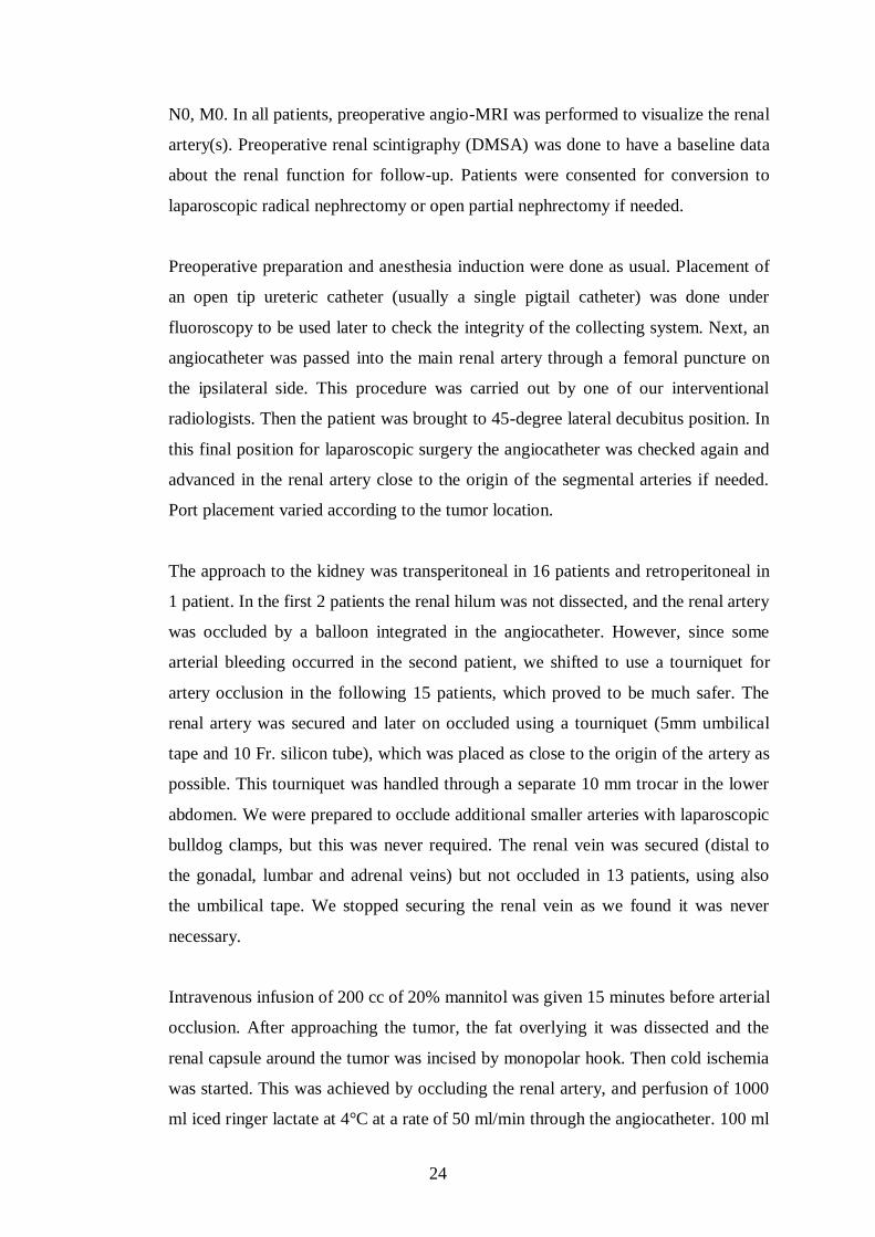
excision was performed in a bloodless field using scissors with no diathermy
(Figure. 5). A biopsy was taken from the tumor bed. The tumor and the overlying fat
were placed in separate organ bag, which were removed later. The integrity of the
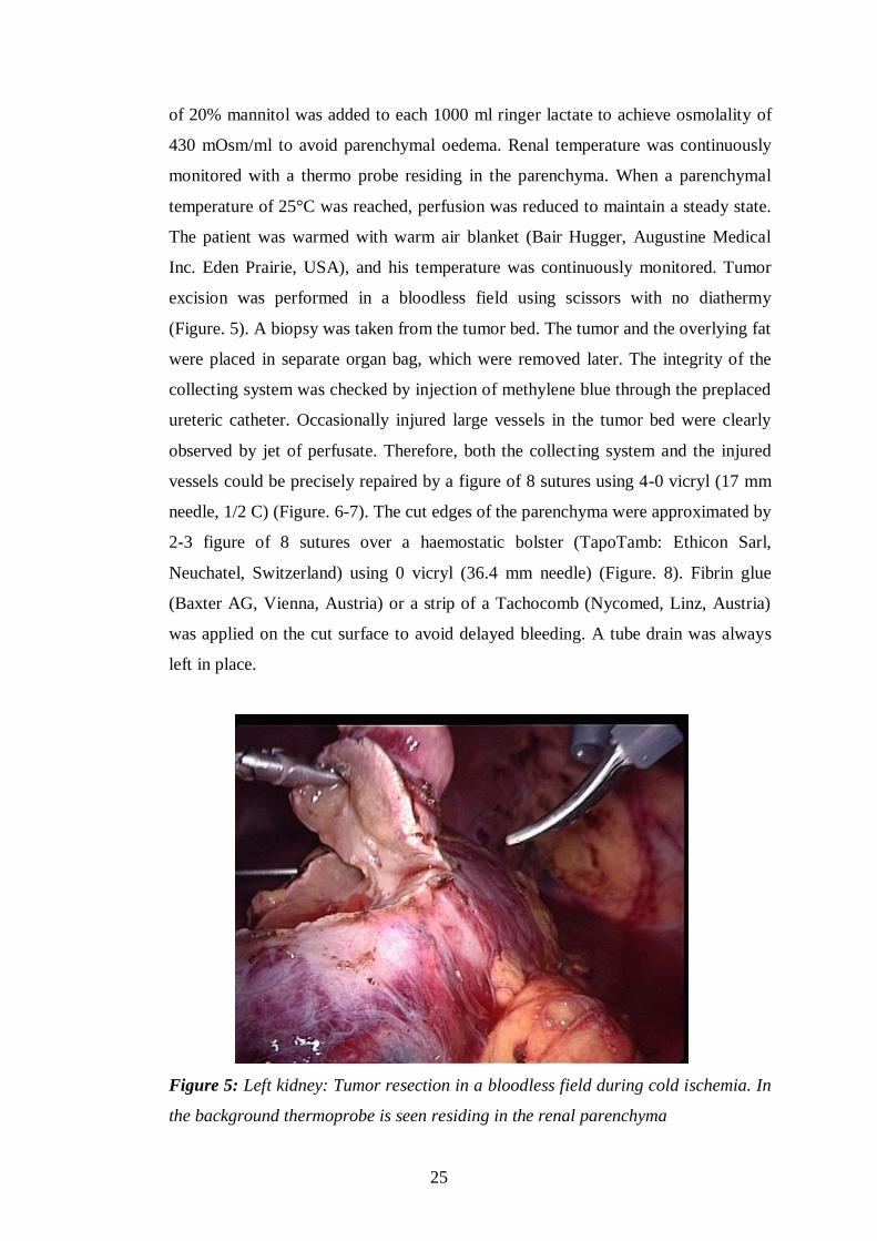
collecting system was checked by injection of methylene blue through the preplaced
ureteric catheter. Occasionally injured large vessels in the tumor bed were clearly
observed by jet of perfusate. Therefore, both the collecting system and the injured
vessels could be precisely repaired by a figure of 8 sutures using 4-0 vicryl (17 mm
needle, 1/2 C) (Figure. 6-7). The cut edges of the parenchyma were approximated by
2-3 figure of 8 sutures over a haemostatic bolster (TapoTamb: Ethicon Sarl,
Neuchatel, Switzerland) using 0 vicryl (36.4 mm needle) (Figure. 8). Fibrin glue
(Baxter AG, Vienna, Austria) or a strip of a Tachocomb (Nycomed, Linz, Austria)
was applied on the cut surface to avoid delayed bleeding. A tube drain was always
left in place.
Figure 5: Left kidney: Tumor resection in a bloodless field during cold ischemia. In
the background thermoprobe is seen residing in the renal parenchyma
26
A. B.
Figure 6. Left kidney. A, entry to collecting system is evidenced by methylene blue
leakage. B, suture repair of collecting system with jet of perfusate issuing from cut
vessel on tumor bed.
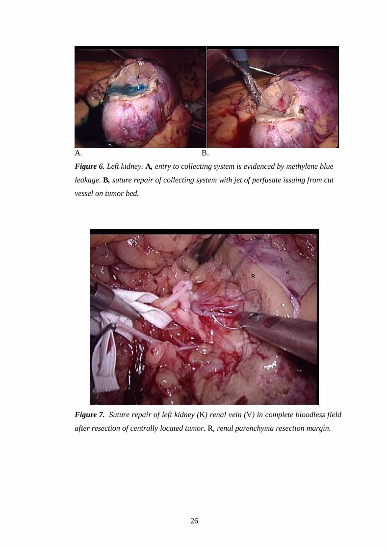
Figure 7. Suture repair of left kidney (K) renal vein (V) in complete bloodless field
after resection of centrally located tumor. R, renal parenchyma resection margin.

27
Figure 8. Left kidney after complete parenchymal repair with sutures over
hemostatic bolster.
3.1.3. Discussion
Laparoscopic partial nephrectomy with our technique could be performed
successfully in all patients with no conversion. The mean intraoperative blood loss
was 145 ml (30 – 650). In 2 patients we had high blood loss. In one of them it was
due to insufficient balloon occlusion and in the other one because of intermittent
failure of the perfusion pump, which resulted in venous backflow from the injured
renal vein. Only one patient required intraoperative blood transfusion. Mean total
ischemia time was 41 minutes. (27 - 101 min.). Entry to the collecting system
happened in 7 patients and was repaired intraoperatively. Repair of the renal vein,
segmental vein and artery was done in 2 patients, in whom the tumors were centrally
located very close to the hilum. Mean amount of perfusate was 1,600 ml (1,150-
2,800). Mean decrease of body temperature during cold perfusion was 0.66°C (0.5-
1.1). Mean operative time was 176 minutes (135-220). Urethral and ureteric
catheters were removed on the second postoperative day. Drainage was removed
when its output was less than 50ml/24hours. Mean hospital stay was 9.4 days (7- 14
days). Bleeding occurred in one case in the first postoperative day due to
28
parenchymal tear from the sutures. This was managed laparoscopically by bipolar
coagulation and application of a strip of Tachocomb (Nycomed, Linz, Austria). No
urinary fistula or urinoma were encountered. The histopathological examination
revealed RCC in 13 patients, angiomyolipoma in 2, and pyelonephretic renal tissue
in another 2. The resection margins were negative in 14 patients. In one patient,
negative margin was not described where the tumor was in direct contact with the
renal vein. During resection the vein was entered and repaired.
Postoperative renal function was evaluated in 8 patients. In 5 patients the reduction
in renal function was 1%, 1%, 2%, 3%, and 8%, respectively by renal scintigraphy
(DMSA). In the other 3 patients, CT scan showed undisturbed perfusion of the renal
parenchyma. We concluded that the reduction in renal function was most probably
attributed to reduction in the total renal volume after wedge resection.
Cold arterial perfusion is an old method, which however is not remembered anymore
nowadays. It was developed for open renal stone surgery by Marberger and
colleagues in 1978 (Marberger et al. 1978)44
and it is still used by vascular surgeons
to protect the kidney during the repair of complicated thoracoabdominal aneurysms
(Morishita et al 1999)45
. For these purposes, it has been proved to be effective and
safe. Since external renal cooling by slushed ice is easier to apply, this technique is
not used anymore for open kidney surgery.
Laparoscopic NSS without renal ischemia is feasible and can be achieved by step-
by-step resection and hemostasis without ischemia (Janethschek et al 1998; Jeschke
et al. 2001)43,46
. However, this technique has its drawbacks. It is restricted to small
tumors (2cm or less) in a favorable peripheral location. Hemostasis is slow and
tedious. The cut surface is continuously covered with blood and burned by the
extensive use of monopolar and bipolar coagulation, therefore distinction between
tumor and normal renal tissue is very difficult. This may compromise complete
tumor resection. Necrosis urinary fistula from the use of diathermy close to the
collecting system was also encountered. These were the reasons for us to replace this
technique by partial nephrectomy in ischemia.

29
During ischemia, the tissue is cut sharply in a bloodless field, avoiding the use of
diathermy. Normal renal tissue and tumor can be discriminated very precisely, so
that complete tumor resection can be continuously monitored. As in open surgery,
hemostasis relies on suturing the cut edges of the parenchyma on bolsters of
haemostatic materials (Gill et al. 2002)18
. A major restriction for this technique is the
time, as renal damage is expected if the ischemia time is longer than 30 minutes.
Therefore cooling of the kidney as in open surgery is required if ischemia time is
anticipated to last more than 30 minutes as in larger tumors and tumors in
unfavorable sites. In addition unexpected problems during surgery may extend the
time of operation.
In animal model, cooling has been performed by injecting cold fluid in the collecting
system (Landman et al. 2002)47
. Because of the relatively small surface of the
collecting system and its central location within the kidney, parenchymal cooling
may be insufficient. Contrary, cold arterial perfusion will directly cool the
parenchyma, which is well perfused. Because of the large surface area of the arterio-
venous system in the kidney, efficient heat exchange can be achieved. External renal
cooling with slushed ice during laparoscopic wedge resection has been described in
one patient (Gill et al. 2002)18
. This may become an alternative method for cold
ischemia in NSS.
During laparoscopic wedge resection, cold arterial perfusion has the advantage that
the laparoscopic procedure itself is not rendered more complicated than resection
without ischemia since all necessary maneuvers are performed prior to surgery.
Resection of the tumor and parenchymal repair can be done in a clear bloodless field
at leisure of time. During our initial experience we realized another advantage of our
technique. Since the whole renal vasculature from the renal artery to the vein is
completely filled with perfusate, repair of arterial and venous lesions becomes
feasible in a completely bloodless field. In addition jets of perfusate will help in
recognition of the exact site of the vascular injury. In two complicated centrally
located tumors, vascular lesions had to be repaired after removal of the tumor. These
procedures would certainly not have been possible without ischemia and cooling.

30
In our series we didn’t encounter any problem regarding the renal function or the
amount of perfusate used. This is probably because all the patients were with a
normal contralateral kidney and diuresis was induced prior, during and after
ischemia. However in a solitary kidney, one should be aware of the risk of volume
overload resulting from the non-excretion of the perfusate during temporary renal
ischemia. In this case excretion of the perfusate will depend on the fast recovery of
the kidney after ischemia.
3.1.4. Conclusion
Our initial experience of incorporating cold ischemia via arterial perfusion to
laparoscopic partial nephrectomy shows the feasibility and safety of this technique.
Injury to the arterial system has been observed neither in the literature nor in our
limited experience. This approach allows the duplication of the principles of the
open surgery and makes laparoscopic NSS for RCC and complex renal pathology
safe and reliable. It also allows for vascular repair during surgery of difficult central
tumors. We believe that our technique will make laparoscopic NSS safer for the less
experienced laparoscopist and will also increase the scope of indications for the
experienced one.
3.2. LAPAROSCOPIC PARTIAL NEPHRECTOMY IN WARM ISCHEMIA
3.2.1. Introduction
Partial nephrectomy (PN) has become a standard of care for treatment of small renal
masses. Hilar occlusion is commonly performed for a precise tumour resection and
renal reconstruction. The above surgical manoeuvre results in warm ischemia (WI)
of the remaining renal tissue and has been associated with ischemic-reperfusion
injury (RI) to the organ. Current evidence showed that the length of the warm
ischemia time (WIT) and the subsequent reperfusion injury may result in permanent
renal damage (Becker et al 2009; Simmons et al 2008)48-49
. Moreover, the resection
of the renal tumour and the suturing of the parenchyma resulted in additional
reduction of the functional renal tissue (Simmons et al 2012; Song et al 2011)50-51
.
31
Thus, two mechanisms of renal function damage during PN could be proposed.
Nevertheless, the importance of the mechanisms for the decline of the postoperative
renal function has not been investigated. The current prospective study evaluated the
split renal function and elucidated the role of renal parenchymal loss in patients with
small renal mass who were treated by LPN with WI.
3.2.2. Patients and methods
Small renal masses have been treated by LPN at our institutions since 2005. Thirty
five patients were enrolled in a prospective pilot study. Regional research ethics
committee approval was received and informed consent was obtained from all
patients. The procedures were performed by two experienced laparoscopists. The
exact location and dimensions of the tumour was identified by three-dimensional CT
scan prior to the operation. Only patients with a single exophytic mass of ≤ 4 cm in
diameter located in either lower or upper pole of the kidney with normal
contralateral kidney were enrolled.
All operations were performed by laparoscopic transperitoneal approach with en-
bloc hilar occlusion using a Rumel tourniquet. Two minutes before hilar occlusion,
0.5 gr/kg of 20% mannitol was infused. The surgical technique has been previously
described (Spaliviero and Gill, 2007)52
. Cold scissors were used for tumour
resection. Running sutures of 3/0 Vicryl were applied for collecting system closure
and haemostasis. In all cases, the parenchymal defect was filled with one or two
rolled Surgicel bolsters. Parenchymal reconstruction was achieved by running 0
Vicryl sutures secured at each parenchymal exit by a Hem-o-lok clip. The renal
pedicle was released only after tumour excision and completion of renorrhaphy. At
the time of hilar tourniquet release, 0.5 mg/kg of Furosemide injection was
administered.
After extraction of the specimen, the surrounding fat tissues were detached and
weight of the remaining resected mass was measured. The kidney was placed in its
anatomic position and the Gerota's fascia was closed.
The recorded parameters included the time for tumour resection, calyceal closure,
haemostatic sutures and the total WIT.

32
Serum creatinine (sCr) was recorded and estimated glomerular filtration rate (eGFR)
was calculated using chronic kidney disease epidemiology collaboration (CKD-EPI)
equation (Levey et al. 2009)53
.
The above measurements were performed preoperatively (baseline), 5-6 hours after
the surgery, on the 1st, 3
rd and 7
th postoperative days and at the end of 1
st, 3
rd, 6
th, and
12th postoperative months.
In this study, in addition, 15 randomized patients treated with laparoscopic radical
nephrectomy (LRN) were examined as a control group for our laboratory outcomes
and all the above parameters were registered in this group.
3.2.3. Renal scintigraphy
In order to distinguish the impact of parenchymal loss from WI effect on the
operated kidney, we planned a novel method of investigation with renal scan as
follow. All patients in LPN group underwent 99m Technetium-Dimercaptosuccinic
Acid (99mTc-DMSA) renal scintigraphy for the determination of split renal function
preoperatively and at the end of 1st, 3
rd, 6
th, and 12
th postoperative months.
99mTc-DMSA isotope is a static renal agent and allows accurate calculation of
differential renal function (DRF) (Kibar et al. 2003)54
. Since 99mTc-DMSA scan
provides relative functional percentage of the two kidneys and the contralateral
kidney served as a control for comparison after LPN, we selected patients with
solitary small polar mass (T1a), otherwise normal ipsilateral kidney, and normal
contralateral kidney. Such selections have resulted in a young cohort of patients with
mean age of 50.5 ± 11.9 years old in our study.
3.2.4. Assessment of total and partial differential renal function: A novel
approach.
Before the operation, all patients underwent radionuclide isotope examination
performed by 99mTc-DMSA. Renal scans were performed in supine position.
Individual kidney uptake and differential renal function (DRF) percentage of left-to-
right kidneys were determined by the Patlak-Rutland method (Rutland, 1996)55
. The
region of interest (ROI) of each kidney was determined with the use of an automated

33
computer program drawing the ROI around the whole kidney. For processing
purposes, all isotope results were saved in a computer program. This assessment
which is a customary method of evaluating a static renal scan was called a “Total-
DRF” (T-DRF) in our study (Figure 9A).
By this means, in the tumorous kidney, the postoperative decline in percentage ratio,
reflecting decrease in renal function, was considered as a consequence of both
factors: 1). IR injury caused by length of WIT. 2). The kidney parenchymal volume
reduction caused by removal of the tumour, and excision and suturing of the
surrounding healthy tissues.
In an attempt to distinguish the impact of each of these two factors, we introduced a
novel method which is referred as “Partial-DRF” (P-DRF). For this reason, in the
preoperative renal scans, the exact location of the tumour was determined and only
small polar masses (either upper or lower pole mass) were selected for the study. In
the tumorous kidney, a region in the tumour-free pole was selected and manually a
ROI was drawn in that pole. Identical ROI was selected in the same pole of the
contralateral kidney (Figure 9B). The same ROI drawing was used in all follow-up
studies of a given patient. Accordingly, P-DRF which reflects DRF of the intact pole
of the operated kidney, which is affected only by the IR injury, was compared with
the same pole on the contralateral kidney. The same processing was applied for all
patients in all isotope scan examinations. As a result, in the postoperative isotope
scans, with the P-DRF, we could compare an intact part of the operated kidney
which was impacted by WI but not affected by parenchymal volume reduction with
an identical segment of the normal contralateral kidney.
Any postoperative decline in the P-DRF of the operated kidney was considered as
the renal functional loss resulted from IR injury only.
All renal isotope tests were evaluated and reported by same specialist doctor in
nuclear medicine.

34
Figure 9: Renal scintigraphy is shown with an imaginary tumour in the lower pole
of one kidney (red circle). A: The ROI is selected (purple line) to demonstrate and
compare the T-DRF of both kidneys. B: The ROI is selected in the nontumorous pole
of the involved kidney and compared with the same ROI in the contralateral kidney.
3.2.5. Patient selection and statistical evaluations.
Any factor which could unpredictably influence on the WI consequence or the renal
function outcome was excluded from the statistical assessments. Accordingly, one
patient was excluded due to conversion to open partial nephrectomy assuming that
absence of pneumopreritoneum may give diverse renal functional outcome. One
patient was excluded due to continuous moderate bleeding from the resected site
during WI time. In this patient, we presume that WI was insufficient due to incessant
blood supply to the kidney. In one patient with two accessory renal arteries, we had to
clip one of the arteries for safe resection. In this case, we suppose that some renal
function deterioration may be as a result of arterial clipping so this patient was not
included for evaluation. In one case, five days after the operation, selective segmental
arterial embolization of the operated kidney was applied due to arteriovenous shunt.
This patient was also excluded owing to impact of embolization on the renal function
outcome. Three patients were disqualified for statistical analysis since they missed
more than one cycle of post operative follow-ups. Consequently, 28 patients were
enrolled in the final statistical analysis. Demographics of the 28 patients which were
included for analysis, and results of the operations are described in Table 4.

35
Table 4 – Patient’s demographics and operation results of LPN
Number of enrolled patients 35
Reasons for patient exclusion (number of patients) :
- Conversion to open partial nephrectomy
- Continuos moderate bleeding from the resected site during WIT
- One of two accessory arteries were clipped for safe resection
- Early postoperative bleeding and selective arterial embolization
- Missed more than one follow-up appointment
1
1
1
1
3
Number of patients included in statistical analysis 28
Male / female ratio 16 / 12
Patient’s age (years) 50.5 ± 11.9 (range: 23-74)
Body mass index 27.6 ± 4.3 (range: 19.7-39)
Right / left ratio 12 / 16
Tumor greatest dimension (mm) by CT Scan 26.4 ± 6.4 (range: 18-40)
PADUA classification of tumors 7 (range: 6-10)
Surgery indication:
- Relative
- Elective
- Absolute
17
11
0
Operative time (min) 145 ± 35 (range: 95-245)
Time used for tumor resection (min) 4.8 ± 1.5 (range: 2.5-10)
Time used for internal sutures (min) 9.6 ± 3.5 (range: 5-18)
Warm ischemia time (min) 22 ± 5.3 (range: 12-32)
Weight of the resected specimen (gm) 18 ± 9.1 (range: 6-40)
Histopathology results:
- RCC, Conventional type
- RCC, Chromophobe type
- RCC, Papillary type
- Oncocytoma
- Angiomyolipoma
19 (68%)
2 (7%)
4 (14%)
2 (7%)
1 (4%)
Positive Surgical Margins None
PADUA: Preoperative aspects and dimensions used for an anatomical classification
of renal tumours ; RCC : Renal cell carcinoma; WIT: Warm ischemia time
Statistical evaluations: The IBM SPSS version 20 (IBM Corp., Armonk, NY,
USA) was used for the calculations and statistical analysis. ANOVA and Pearson
product-moment correlation were calculated as deemed necessary. A p-value < 0.05
was considered statistically significant.
For further confirmation, a linear correlation coefficient was calculated for the
assessment of a possible correlation of the T-DRF decline to the WIT in the operated
kidney and to the mass of the resected specimen.

36
3.2.6. Patients operated with laparoscopic radical nephrectomy
In 15 patients with LRN selected for control group, the mean age was 61 ± 12 years
(range: 41-78). The male to female ratio was 11/4, and right to left involved kidney
ratio was 12/3. Their mean BMI was 29.7 ± 5.4 (range: 21.6-38.6), and the mean
operative time was 153 minutes (range: 100-220). The mean age, BMI and the mean
operative time were slightly higher than in the group of LPN.
3.2.7. Results
Twenty eight patients with small renal mass successfully underwent LPN and
completed one year follow up according to our protocol. Operation data and
histopathology results are summarized in Table 4. During operation, after hilar
unclamping, in six patients we observed mild bleeding from the resected margin,
which were resolved within 2-3 minutes by increasing pneumopreitoneum or
application of Surgicel. We didn’t have any significant bleeding, necessitate hilar re-
occlusion. In our selected patients for statistical evaluations, major intra- or
postoperative complication was not observed.
In the LPN group, the mean results of preoperative and postoperative renal function
measured by serum creatini (sCr), estimated glomerular filtration rate (eGFR, CKD-
EPI equation) as described by Levey and collegaues (Levey et al. 2009)53
and total
differential renal function (T-DRF) as well as partial differential renal function (P-
DRF) are summarized in Table 5.

37
Table 5. Mean estimations of kidney function before and after LPN surgery
Investigated parameter
and normal ranges
Pre- op
(baseline)
1 day
post-op
3 days
post- op
7 days
post- op
1 month
post- op
3 months
post- op
6 months
post- op
12 months
post- op
Mean of all
post-op results
after day 1
Serum Creatinine
(62-106 µmol/l)
71 ± 14
(44-94)
86 ± 22
(43-120)
82 ± 20
(42-124)
82 ± 16
(50-112)
80 ± 16
(43-103)
79 ± 17
(47-108)
80 ± 17
(40-98)
82 ± 16
(50-111) 81
(14% ↑)
eGFR (CKD-EPI)
(> 90 mL/min/1.73m2)
97 ± 17
(55-122)
81 ± 21
(44-114)
87 ± 20
(55-117)
85 ± 18
(60-106)
87 ± 18
(49-114)
90 ± 21
(48-124)
87 ± 20
(54-120)
86 ± 20
(48-116) 87
(10% ↓)
T-DRF of the involved
kidney (%)
49 ± 4
(43-58) Not done Not done Not done
42 ± 7
(24-52)
42 ± 7
(25-54)
41 ± 7
(25-52)
41 ± 7
(24-51)
42
(7% ↓)
P-DRF of non-tumorous
pole of involved kidney
(%)
50 ± 4
(44-59) Not done Not done Not done
47 ± 6
(37-57)
48 ± 5
(37-55)
46 ± 4
(35-52)
47 ± 4
(38-54) 47
(3% ↓)
Pre-Op: Preoperative; Post-op: Postoperative; eGFR: Estimated glomerular filtration rate; CKD-EPI: Chronic kidney disease
epidemiology collaboration; T-DRF: Total differential renal function; P-DRF: Partial differential renal function

38
3.2.8. Evaluation of the renal function based on serum creatinine.
The detailed mean values of these tests are shown in Table 5 for LPN and Figure 10
for both LPN and LRN patients. In Figure 10, the preoperative (time point 1) mean
values of serum creatinine as well as the mean values at the eight postoperative
check points is shown for both LPN and LRN groups. The two groups were
compared taking into consideration that in LRN group we had elder population (61 ±
12 years vs. 50.5 ± 11.9 years, p-value = 0.008), more male to female ratio (73% vs.
57%) and higher body mass index (BMI) (29.7 ± 5.4 vs. 27.6 ± 4.3, p-value = 0.18).
These data may explain the higher baseline (84 umol/l) serum creatinine in the LRN
group. It was a strong increase of serum creatinine level immediately after LRN with
a peak on the 3rd
post-op day. It declined slightly and remained constantly high (123
umol/l) until the end of first postoperative year. This reflects ~ 46% rise in sCr level
compared to the baseline in LRN patients. In contrary, in the LPN group, the serum
creatinine level was only slightly increased from 71 umol/l to 82 umol/l (~16%
increase) during the first three postoperative days and remained constant until the
end of observation.
1
1
40.00
50.00
60.00
70.00
80.00
90.00
100.00
110.00
120.00
130.00
140.00
150.00
160.00
1 2 3 4 5 6 7 8 9
Seru
m C
reati
nin
e (
µm
ol/l)
Various time points
Serum Creatinine results of LPN & LRN groups
LRN control group
LPN study group
Figure 10: The curves show the mean preoperative (time point 1) serum creatinine
and mean results of postoperative serum creatinin measured in patients with LRN

39
and LPN at different time intervals (point 2: 5-6 hours post-op; point 3: 1st post-op
day; point 4: 3rd
post-op day; point 5: 7th
post-op day; point 6: 1st post-op month;
point 7: 3rd
post-op month; point 8: 6th post-op month; point 9: 12
th post-op month)
3.2.9. Evaluation of the renal function based on estimated GFR.
As shown in Table 5 and Figure 11, the mean preoperative or baseline eGFR of
patients treated with LPN was 97 ± 17 (range: 55-122) which decreased to 81 ± 21
(range: 44-114) in the 1st postoperative day (p-value = 0.0069). This shows a 16%
decline in average eGFR which was the largest postoperative drop within one year
of follow up. Conventionally, we call it “transient-state” of kidney function
deterioration. In the 3rd
postoperative day, we observed a 7% recovery in the average
eGFR comparing to the 1st day. Although, due to the large deviations in the values,
this tendency toward recovery was statistically insignificant (p-value = 0.3821).
From the 3rd
postoperative day to end of the study in 12th
month, the average eGFR
remained roughly the same in both groups. In these time points of LPN group,
comparing the lowest with the highest values, which were in the 7th
postoperative
day and 3rd
month respectively, showed insignificant alteration in the eGFR (p-value
= 0.4483) (Figure 12). Accordingly, for simplicity, average of all postoperative
eGFR mean values after the transient-state (after the 1st day) were calculated and
considered as the “steady-state” of renal function deterioration. This was 87
ml/min/1.73 m2 in LPN patients which demonstrates ~10% decrease in renal
function comparing to the baseline (p-value = 0.0757).
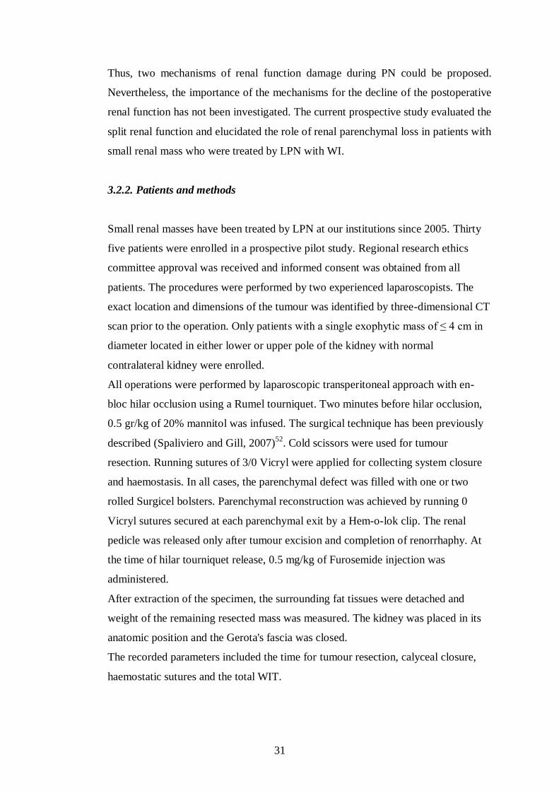
As shown in figure 13, the eGFR was lower for patients treated with LRN and
showed a decrease during the first three postoperative days and thereafter remained
at the same level. By this comparison, the difference in clinical parameter between
the two groups as indicated above also should be taken into account.
It is worth to highlight that in the LRN control group, the average decline in the
“steady state” of the kidney function was ~ 40% (81.34 declined to 48.36
ml/min/1.73 m2).
In a simple explanation, we can conclude that, by removal of one whole kidney,
nearly 40% of global kidney function declines, and by removal (resection) of one
part of a kidney, nearly 10% of the global kidney function decreases. Accordingly,

40
parenchymal volume reduction after partial nephrectomy has a very important
impact on outcome of kidney function and this should be certainly distinguished
from the impact of the IR injury caused by WI.
75
80
85
90
95
100
105
Pre- op(baseline)
1 dayPost-op
3 daysPost-op
7 daysPost-op
1 monthPost- op
3 monthsPost- op
6 monthsPost- op
12 monthsPost- op
eG
FR
( m
l/m
in/1
,73
m2
)
eGFR (CKD-EPI Equation) of LPN Patients
Figure 11. The graph shows the mean preoperative and postoperative eGFR values
in the studied time intervals for the LPN patients enrolled in the statistic evaluation.
R² = 0.0163
75
80
85
90
95
100
105
3 daysPost-op
7 daysPost-op
1 monthPost- op
3 monthsPost- op
6 monthsPost- op
12 monthsPost- op
eG
FR
( m
l/m
in/1
,73
m2
)
eGFR (CKD-EPI Equation) of LPN Patients
Figure 12. This graph shows same parameters as the previous graph (figure 11)
excluding the baseline and the 1st post-op values. It remarks the so called “steady-
state” of renal function after partial nephrectomy.
41
1
1
-
20.00
40.00
60.00
80.00
100.00
120.00
1 2 3 4 5 6 7 8 9
mL
/min
/1.7
3m
2
Various time points
Estimated GFR results of LPN & LRN groups
LPN study group
LRN control group
Figure 13. The curves show mean preoperative (time point 1) eGFR and mean
results of postoperative eGFR measured in patients with LPN and LRN at different
time intervals (point 2: 5-6 hours post-op; point 3: 1st post-op day; point 4: 3rd
post-op day; point 5: 7th post-op day; point 6: 1st post-op month; point 7: 3rd post-
op month; point 8: 6th post-op month; point 9: 12th post-op month)
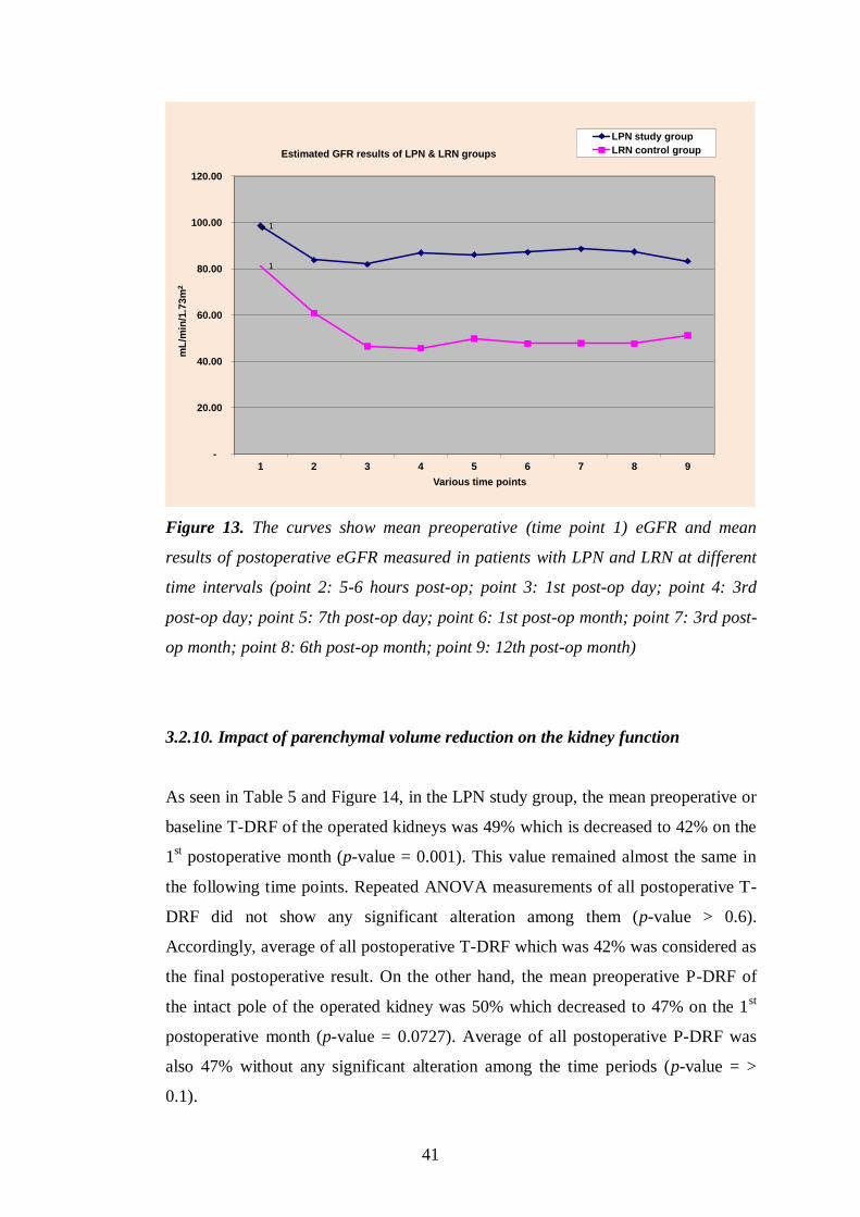
3.2.10. Impact of parenchymal volume reduction on the kidney function
As seen in Table 5 and Figure 14, in the LPN study group, the mean preoperative or
baseline T-DRF of the operated kidneys was 49% which is decreased to 42% on the
1st postoperative month (p-value = 0.001). This value remained almost the same in
the following time points. Repeated ANOVA measurements of all postoperative T-
DRF did not show any significant alteration among them (p-value > 0.6).
Accordingly, average of all postoperative T-DRF which was 42% was considered as
the final postoperative result. On the other hand, the mean preoperative P-DRF of
the intact pole of the operated kidney was 50% which decreased to 47% on the 1st
postoperative month (p-value = 0.0727). Average of all postoperative P-DRF was
also 47% without any significant alteration among the time periods (p-value = >
0.1).

42
35%
37%
39%
41%
43%
45%
47%
49%
51%
53%
55%
Pre- op(baseline)
1 monthPost- op
3 monthsPost- op
6 monthsPost- op
12 monthsPost- op
DR
F o
f th
e i
nvo
lved
kid
ney
Average of P-DRF and T-DRF of the operated kidney in relation to time line
P-DRF
T-DRF
Figure 14. The graph shows the mean decline of both P-DRF and T-DRF of the
operated kidney (LPN) in the studied time intervals.
Linear correlation coefficient was used to compare relationship of the T-DRF
decline in the operated kidney to the mass of the resected specimen (Figure 15).
We have also used linear correlation coefficient to compare relationship of the T-
DRF decline in the operated kidney to WIT (Figure 16). This showed a much
stronger correlation between T-DRF decline and the resected mass comparing to the
WI time (R2 = 0.7241 and p0.0837 respectively).

43
R² = 0.7241
0%
5%
10%
15%
20%
25%
5 15 25 35
Po
st-
op
. avera
ge d
ec
lin
e o
fT
-DR
F
Weight of resected specimen (gr.)
Correlation of T-DRF decline to resected mass
Figure 15. Comparing correlation of the T-DRF decline in the operated kidney to
the mass of the resected specimen
R² = 0.0837
0%
5%
10%
15%
20%
25%
10 15 20 25 30 35
Po
st-
op
. a
ve
rag
e d
ec
lin
e o
f T-D
RF
Warm Ischemia Time (min.)
Correlation of T-DRF decline to WIT
Figure 16. Comparing correlation of the T-DRF decline in the operated kidney to
WI time.

44
3.2.11. Discussion
In 1950, Benjamin Abeshouse wrote “Few procedures provide the urologist with
more satisfaction than those that preserve renal function”56
. While Dr. Abeshouse
may have practiced urology prior to the availability of the strong data we now
possess, his statement rings true to this day. On this principle, NSS has taken a
prominent position at the helm of the treatment of renal tumours. Likewise, there has
been continual progress toward resecting less and less renal parenchyma to preserve
more renal function without sacrificing any of oncological rules.
There are several factors determining the postoperative renal function: A. the
preoperative quality of renal function (underlying renal disease, limited glomerular
function, etc.); B. the quantity of renal parenchyma remained after operation; and C.
the warm ischemia time. The first factor can’t be modified by surgical technique; the
second is determined by the anatomical size and location of the tumour. The warm
ischemia time is influenced by the experience of the surgeon and the operation
technique applied.
We planned a prospective study in order to distinguish the impact of parenchymal
loss and effect of warm ischemia on the function of operated kidney. In our study,
99mTc-DMSA isotope was used which is a static renal agent and allows accurate
calculation of DRF (Kibar et al 2003)54
. This was measured preoperatively and
completed in different postoperative intervals in 28 patients with solitary small polar
renal mass and no any other abnormality in that kidney. Since 99m
Tc-DMSA scan
provides relative functional percentage of the two kidneys and the contralateral
kidney served as a control for comparison after LPN, we selected patients with
normal contralateral kidney. Such selections have resulted in a young cohort of
patients with mean age of 50.5 ± 11.9 years old in our study. The T-DRF was
measured in all isotope studies. Any postoperative decline in T-DRF in the operated
kidney was considered as a result of warm ischemia and ischemia-reperfusion injury
combined with parenchymal loss. In nearly all postoperative studies, mean decline
of T-DRF in the operated kidney was 7%. In order to distinguish the effect of warm
ischemia from the parenchymal loss, we introduced the so-called P-DRF in which a
region of interest was selected on nontumorous pole of the involved kidney and it
was compared with the same region in the contralateral kidney.

45
For this reason, we selected only patients with tumor mass of ≤ 4 cm in diameter
which were located on either upper or lower pole of the kidney so that the region
selected in the intact nontumorous pole of the operated kidney was not affected by
parenchymal loss and suturing. Any postoperative functional decline in this intact
pole of the operated kidney was considered to be as a result of warm ischemia and
ischemia-reperfusion injury only. In our study, mean postoperative decline in the P-
DRF of the operated kidney was only 3% which was found to be statistically
insignificant (p-value = 0.0727). In agreement with the previous studies, we believe
that within certain time limits of warm ischemia, which was 22 ± 5.3 minutes in our
study, the ischemia-reperfusion insult may be negligible or reversible. On the other
hand, deliberate parenchymal loss plays a major role in kidney function
deterioration.
Warm ischemia and the IR injury to the kidney have been considered for a long time
as the main factor related with postoperative renal function deterioration in patients
undergoing PN under WI30,48,57,58,59,60
. Several technically challenging techniques
have been introduced for the reduction of WI61,62
. Nevertheless, the impact of renal
parenchymal mass reduction was not distinguished from the effects of WI and IR in
the above literature. Parenchymal loss after PN occurs as a result of intentional
tumor excision, some normal parenchyma resection and suturing. Thus, the mass or
volume of the parenchymal loss should be considered and differentiated from IR
injury when evaluating the renal functional outcome after PN.
Few authors have studied the impact of parenchymal volume reduction on renal
function. In a large multicenter study cohort Shikanov and colleagues (Shikanov et
al. 2010)63
assessed the influence of renal ischemia on long-term global renal
function after LPN in patients with 2 functioning kidneys. They retrospectively
evaluated eGFR of 401 patients with median tumor size of 25 mm and normal
contralateral kidney who underwent LPN with median warm ischemia time of 29
minutes. Changes in early postoperative and nearly one year follow-up eGFR were
-16% and -11%, respectively. This result is nearly identical to our findings. The
percent change in last eGFR was worse in patients with larger tumors suggesting
that excising a larger lesion would result in more loss of renal parenchyma. Mir and
colleagues (Mir et al 2015)64
also reported that the preservation of the total renal

46
function after PN ranged between 88% and 91% which reflects approximately 10%
loss of the renal function (similar to our finding). Mir et al also showed that a higher
reduction of the eGFR in patients with larger tumors which could be attributed to the
excision of a larger lesions and the consequent greater loss of renal parenchyma.
This was documented also by Sharma and colleagues (Sharma et al 2008)65
in 21
patients with solitary kidney who underwent partial nephrectomy due to small renal
mass. Volume of the kidney was measured preoperatively and postoperatively by CT
scan and software with an automated segmentation algorithm. An average of 15%
parenchymal volume loss and 19.7% deterioration in kidney function were
documented in mid-term postoperative period. They concluded that the percent of
renal parenchymal volume loss was correlated with the percent loss in eGFR. It was
shown that volume loss had a more direct, predictable effect on ultimate eGFR than
ischemia time. A similar result was concluded by Simmons and colleagues
(Simmons et al. 2012)50
and also Song and colleagues (Song et al. 2009)66
. In the
later study, Diethylenetetramine pentaacetic acid (99m
Tc-DTPA) was used for
accurately estimating renal clearance and measuring eGFR by the kidney. They
measured changes in individual renal function and investigated factors determining
the degree of functional reduction in 65 patients who underwent LPN and 52
patients with open partial nephrectomy. In the entire cohort, renal volume reduction
was the most significant, independent prognosticator for eGFR reduction after
partial nephrectomy. Mir and colleagues (Mir et al. 2013)67
evaluated the role of
volume preservation by introducing computed tomography volumetric analysis using
free-hand scripting. The eGFR was measured by MAG3 in 92 patients who
underwent partial nephrectomy. The authors revealed that the ultimate renal function
after partial nephrectomy was primarily driven by parenchymal preservation with
ischemia playing a secondary role as long as it was within a limited time period.
Current literature has not concluded to the most important factor for the renal
function decline after PN and the contribution of WI to the postoperative renal
function has not been well documented64
. Some investigators advocated that the
parenchymal mass preservation was stronger correlated to the functional recovery in
comparison to the WI51,66
.
The current prospective study aimed in distinguishing the impact of parenchymal
loss from the WI effect on the operated kidney. The 99mTc-DMSA isotope was used

47
for the purpose due to the fact that it allows accurate calculation of DRF54
. The latter
parameter was measured preoperatively and in different postoperative intervals in 28
patients with solitary small polar tumours. Since 99mTc-DMSA scan provided
relative functional percentage of the two kidneys, the contralateral kidney served as
the control for the comparison after LPN. Consequently, only patients with normal
contralateral kidney were selected and a young patient population with mean age of
50.5 ± 11.9 years was eventually included in the study. Any postoperative decline in
the T-DRF of the operated kidney was considered as a result of WI and WI
combined with parenchymal loss. In nearly all postoperative studies, a mean decline
of 7% in the T-DRF was noted. In an attempt to distinguish the effect of WI from the
parenchymal loss, the P-DRF was introduced. A ROI was selected on the non-
tumorous pole of the involved kidney and was compared with the same ROI on the
contralateral kidney. Any interference of the excision area to the ROI was prevented
by including only patients with tumour mass of ≤ 4 cm in diameter located on either
upper or lower pole of the kidney. Any postoperative functional decline in this intact
pole of the operated kidney was considered to be as a result of WI only. The mean
postoperative decline in the P-DRF of the operated kidney was only 3% which was
found to be statistically insignificant (p-value = 0.072). In agreement with the
previous studies68
, it could be suggested that WI may result in negligible or
reversible renal damage within certain time limits of WIT such as a mean WIT of
the current study. In addition, the parenchymal loss seemed to play a more important
role in kidney function deterioration than WI. Considering the above, it could be
advocated that the LPN surgical technique could probably focus on the precise
tumour excision and suturing rather than to the minimization of WIT. Nevertheless,
additional studies are necessary for the confirmation of the above hypothesis.
Limitations of this study include the reliance on DRF and the use of the non-
operated kidney as a stable reference unit before and after the surgery. Any
postoperative compensatory hypertrophy of the contralateral kidney may result in a
false outcome of DRF. Takagi et al.69
showed that the compensatory hypertrophy of
the contralateral kidney after PN remained rather limited and less than 2.3% in most
cases. They concluded that the larger the excised volume of the kidney, the more
hypertrophy of the contralateral kidney was expected. The median tumour diameter
of the latter study was 3.5 cm and probably resulted in higher volume loss in
48
comparison to our series (median of 2.6 cm). Hence, we assume that the
compensatory hypertrophy may have been negligible in our study.
Another limitation of our study was the lack of stratification of the results according
to the length of the WIT or the tumour size. The parenchymal volume was never
measured and the current study could not provide information regarding the pre-and
postoperative changes in the volume of the renal parenchyma. Nevertheless, the
changes in the contour of the operated kidney may influence measurements of the
renal volume and the selection of ROIs out of the excision field for measurements
probably allowed for more reliable results. Moreover, the use of CT scans for the
evaluation of renal volume would expose the patient in additional radiation without
providing evidence that would significantly influence the results of the study.
3.2.12. Conclusion
In LPN, the parenchymal loss caused by the resection of the tumor and the
suturing of the surrounding normal tissues resulted in kidney function
deterioration which should be distinguished from WI effects. An average WIT of
22 minutes for a mean tumor diameter of 2.6 cm resulted in a 7% kidney function
decline. 4% could be attributed to the parenchymal loss and 3% to WI.
The ultimate renal function after partial nephrectomy is primarily driven by
parenchymal preservation with ischemia playing a secondary role as long as it is
within a limited time period. One of the major implications of our study is that
creating a bloodless filed by clamping the renal pedicle within certain time limits,
and consequently precision of excision and renorrhaphy, should be a primary
objective during any partial nephrectomy. This may result in more kidney function
preservation than putting all efforts to decrease WI time to zero while accepting all
potential complications.

49
3.3. TECHNICAL MODIFICATIONS OF LAPAROSCOPIC SURGERY
3.3.1. Laparoscopic partial adrenalectomy for recurrent pheochromocytoma
In 1998, we performed a transperitoneal laparoscopic bilateral partial adrenalectomy
in an 8-year-old boy with symptomatic familial pheochromocytoma, as a
manifestation of von Hippel-Lindau disease (VHL) (Radmayr et al. 2000)70
. The
symptoms recurred 53 months later, starting with hypertension, tachycardia and
headache. The 24-hour urinary excretion of catecholamines was elevated. MRI
revealed a 2.5 cm mass on the right adrenal remnant. After preoperative medications
with phenoxybenzamine and propranolol, we performed the second laparoscopic
transperitoneal partial adrenalectomy (Nambirijan et al. 2004)71
. The previous port
sites were used for placement of 3 trocars. Only the fourth trocar (5 mm) was
inserted through an additional incision. The only observed intraperitoneal adhesion
was at the inferior surface of the liver, which was fixed to the site of adrenal gland.
These adhesions were removed easily to approach the tumor. Intraoperative color
Doppler ultrasound was used to delineate the tumor. Considering that the tumor was
embedded to a great extent in the adrenal gland, the procedure proved to be
challenging. Dissection and hemostasis were achieved using monopolar scissors and
bipolar coagulation forceps. The vein from the tumour could be isolated and
transected preserving the adrenal vein. The tumour was removed completely sparing
the normal adrenal tissue. To avoid delayed bleeding, the cut surface was sealed
with fibrin glue. Operative time was 120 minutes. The blood loss was minimal and
drainage wasn’t needed. Postoperative course was uneventful. Histopathological
examination confirmed the diagnosis of pheochromocytoma. During follow-up,
blood pressure and catecholamine values returned to normal and no residual tumor
was observed.
Patients with VHL, as well as multiple endocrine neoplasia (MEN) type 2 disease,
are predisposed to recurrent bilateral adrenal pheochromocytoma. Partial
adrenalectomy is considered to be the first choice of treatment in these patients in
order to avoid a life-long steroid replacement therapy. In addition, in children
preserving the adrenocortical function is essential for normal child growth. The high
possibility of recurrence and also predisposition to other intraabdominal pathologies,
such as renal cell carcinoma and pancreatic islet cell tumors in VHL may necessitate

50
repeat surgical procedures. Therefore, any surgical procedure performed in these
patients should be as minimal as possible. Our experience with laparoscopy has
given rise to strong arguments over open surgery in dealing with the above–
mentioned diseases. These arguments are based on advantages, which could be
summarized as follows: 1. Minimal procedure, 2. Lower morbidity, 3. Less adhesion
formation, 4. Much easier repeat operation for recurrence(s). By this technique, not
only the recurrent tumour could be removed successfully but also the unaffected
adrenal gland was preserved for the second time.
3.3.2. New tool for laparoscopic antegrade ureteral stenting in repair of
circumcaval ureter and pyeloplasty operations.
Circumcaval ureter (CU) is a rare congenital anomaly of the inferior vena cava
(IVC). It forms as a result of posterior cardinal vein persisting as the renal segment
of the IVC during development. Because the posterior cardinal vein is located
ventral to the ureter, the ureter will become entrapped behind the vein. Extrinsic
compression or functional loss of the retrocaval ureteral segment may lead to
ureteral obstruction and hydronephrosis and subsequently to deterioration of kidney
function. Correction of such anomaly is recommended in specific cases. The
pathogenesis of the disease and also the indication of the operation are nearly similar
to that of uretero-pelvic junction (UPJ) stenosis. By development and progress of
laparoscopic technology, laparoscopic approach is considered as the standard of care
for such operation. One of the challenging and time-consuming parts of the
laparoscopic approach is intraoperative antegrade ureteral stenting. Accordingly,
several different methods have been studied and recommended for this purpose.
We have developed a new method for intraoperative antegrade ureteral stenting
(Bagheri et al. 2009)72
. The same instrument can be used in any type of laparoscopic
ureteric operations necessitating ureteral stenting. We have applied the new
technique for laparoscopic transperitoneal repair with transection and reanastomosis
of the ureter anterior to the IVC in patients with circumcaval ureters and in several
laparoscopic pyeloplasy and ureterotomy cases.
In one of the patients with circumcaval ureter, only a mild pelvic dilatation with
relatively slight ureteric narrowing was noticed. Therefore, the retrocaval ureteral
51
segment was preserved and the ureter vas transected at its distal dilated part lateral to
the IVC. The lower ureteral segment, together with the double coil stent, was
transpositioned to the front and lateral of the IVC. The ureteral stump had a normal
appearance and the patency of its lumen was confirmed by easy passage of an 8F
ureteral catheter. Spatulated end-to-end uretero-ureteral anastomosis was performed
using interrupted 4-0 Vicryl sutures.
In two patients, the rectrocaval segment especially the ascending course appeared to
be narrowed and aperistaltic. In these cases the ureter was transected medially to the
IVC to access the retrocaval segment. The upper stump had a tortuous appearance
with very narrow lumen. With peristaltic movements of the pelvis, urine jet was not
seen. Therefore that ureteral segment was resected until a normal ureteric lumen and
urine flow were visible.
For stenting, we attempted to avoid any retrograde procedure. We applied antegrade
stenting using a ureteral stenting cannula, which was developed by our group and
used routinely in laparoscopic pyeloplastic operation in our institute (Figure 17).
We developed two sets of ureteric stenting cannula which can be used for
intraoperative antegrade ureteric stenting. Each cannula has a length of 28cm to be
long enough to pass through the standard laparoscopic trocars and easily reach to the
ureteric stump. It is 3mm in diameter so can be easily inserted into any standard
trocar or directly into the abdomen by a small percutaneosu puncture. The lumen of
the cannulas can easily accept stents of up to 7 French.
The cannula in the upper portion of the picture has a mild curvature at the tip. This
can be inserted through any standard laparoscopic trocar. The tip of the cannula can
be directed to the lower ureteric stump to facilitate insertion of the stent or guidewire
into the ureter. The other two parts form a separate kit. The one with a sharp straight
tip works as a trocar. It is inserted into the straight cannula for percutaneous
puncture from any site of the abdomen. After insertion into the abdomen, the inner
trocar can be removed and the cannula can be used for stenting. At the top of each
cannula, appropriate sealing cap can be used for insertion of stent and pusher or a
guidewire without any leakage of CO2. After insertion of the stent into the distal
ureteric stump, the cannula can be removed. After the first few anastomotic sutures,
the upper part of the stent can be pushed into the upper ureteric stump or renal
pelvis.

52
Based on our experience, we recommend intraoperative antegrade ureteral stenting,
using the stenting cannula, which is fast, safe and easy to apply and additional
transureteral retrograde procedure can be avoided. Using antegrade ureteral stenting
has the advantage of having a dilated renal pelvis and intact upper ureter during the
surgery. These would provide a better preparation of the ureter and renal pelvis and
also gives a more accurate evaluation of the narrowed ureteral segment.
Figure 17. Two types of ureteral stenting cannula (28 cm long with 3-mm diameter)
were used for intraoperative ante-grade ureteral stenting. Cannula with mild
curvature at tip, which can be inserted through any trocar with diameter 10 mm
(upper). Ureteral stenting cannula formed of sharp, straight-tip trocar and cannula,
which can be inserted into abdomen from any direction (lower). At top of each
cannula is appropriate sealing cap used for insertion of double coil stent and its
pusher.
53
3.3.3. Ligation of a wide renal vein during laparoscopic nephrectomy: an effective
method
Laparoscopic nephrectomy has been refined to a point that is now considered as a
first option for radical nephrectomy with excellent results. The crucial step during
laparoscopic nephrectomy is the dissection and control of renal pedicle. Application
of the endovascular gastrointestinal anastomosis (GIA) stapler has been a standard
procedure to control renal vein during laparoscopic nephrectomy. The significant
complication due to device malfunctioning has led several surgeons to find another
secure method for renal vein ligation. Accordingly, the so called Hem-o-lok clips
were introduced. However, some renal veins may be so wide that cannot be
occluded safely by even the largest available clip. This fact inspired us to find a
simple, secure and cost-effective technique to ligate wide renal veins.
After renal pedicle dissection, the renal artery is occluded with Hem-o-lok clips and
divided when appropriate. A right angle dissector is passed posterior to the renal
vein. A 2-zero 70 cm monofilament suture is fed to the dissector, which is
withdrawn and pulled out. The other end of the suture is also grasped and drawn out
through the same trocar, so that the suture is placed around the vein. Extracorporally
one end of the suture is inserted into the convex side of a specially designed, round
eyed knot pusher (Figure 18). It is then grasped by a mosquito and fixed under
minimal tension by the assistant in the line of the trocar, while ensuring that there is
no kinking or twisting in whole length of the thread. The knot pusher is held by one
hand and the free end of the suture is held with the other hand to form a loop around
the fixed part, as in open surgery. By maintaining minimal tension on each end of
the thread, the loop is gently pushed down by the knot pusher to the level of the
renal vein and then slightly tightened. This maneuver can be repeated 3 to 5 times to
shrivel the vein. The whole procedure requires about 2 minutes and it can be easily
mastered even by an inexperienced laparoscopist.

54
Figure 18. Knot pusher with closed eye and curved tip used to push down standard
surgical knot through a 10 mm trocar.
This rapid and simple maneuver is important to shrink a vein of any diameter to
allow the safe application of clips. We preferred to use Hem-o-lok clips with a
locking mechanism, which provides a strong hold on the tissue and prevents clip
dislodgement. Although we have not experienced any dislodgement, we placed two
10 mm clips on the central side in addition to the ligature and 1 on the peripheral
side of the vein (Figure 19). This approach can be used to ligate the renal vein
without any morbidity or significant increase of operative time. The combined use of
suture tied extracorporally and clips with locking mechanism allows a safe, rapid
secure ligation and transection of a vein of any size and may replace the
endovascular GIA stapler, which may lead to complications.

55
Figre 19. A. right angle clamp is passed behind renal vein and fed with suture. B.
renal vein is shriveled by knots tied extracorporeally. C. vein is clipped with Hem-o-
lok clips. D. renal artery and vein after clipping is completed.

56
4. THESIS RELATED NEW OBSERVATIONS
In the past two decades, laparoscopic procedures have gained popularity among
urologists. However, many of the laparoscopic procedures require standardization so
all urologists can perform them safely and relatively easily. In our studies, we have
tried to standardize some of the upper urinary tract laparoscopic techniques such as
repair of circumcaval ureter, pyeloplasty, application of cold ischemia in partial
nephrectomy and concerns related to warm ischemia time in nephron-sparing
surgeries.
We have observed that in pyeloplasty and any type of upper urinary tract
reconstructive laparoscopic procedures, antegrade ureteral stenting with the ureteral
stenting cannula which is developed by our team facilitates the antegrade stenting
and its application is safe, easy and quick.
We have proved in a prospective study that in partial nephrectomy operations, loss
of renal parenchymal mass has an important impact on renal functional outcome. In
nephron-sparing laparoscopic surgeries with an average warm ischemia time of 22
minutes, we observed a 7 % kidney function decline of which, 4% contributed to the
parenchymal loss and only 3% to the ischemia. This research and findings can be of
beneficial in future studies to consider the following points:
1. In any partial nephrectomy surgery, intentional resection and suturing of the
renal parenchyma which results in loss of some renal mass, has a significant
impact on kidney function. This fact should be taken into consideration and it
should be distinguished from effects of warm ischemia time in future studies.
2. Probably, potentially unsafe and challenging techniques which advocate zero
ischemia time in partial nephrectomy should not be promoted among
urologists.
57
5. REFERENCES
1. Ferlay J, Soerjomataram I, Ervik M, Dikshit R, Eser S, Mathers C, Rebelo M, Parkin
DM, Forman D, Bray, F (2013). GLOBOCAN 2012 v1.0, Cancer Incidence and
Mortality Worldwide: IARC CancerBase No. 11 [Internet]. Lyon, France:
International Agency for Research on Cancer. Available from http://globocan.iarc.fr.
2. Hew MN, Zonneveld R, Kümmerlin IP, Opondo D, de la Rosette JJ, Laguna MP. Age
and gender related differences in renal cell carcinoma in a European cohort. J Urol.
2012 Jul;188(1):33-8.
3. Cumberbatch MG, Rota M, Catto JW, La Vecchia C. The Role of Tobacco Smoke in
Bladder and Kidney Carcinogenesis: A Comparison of Exposures and Meta-analysis
of Incidence and Mortality Risks. Eur Urol. 2016 Sep;70(3):458-66.
4. Chow W-H, Dong LM, Devesa SS. Epidemiology and risk factors for kidney cancer.
Nature reviews Urology. 2010;7(5):245-257.
5. Levi F, Ferlay J, Galeone C, et al. The changing pattern of kidney cancer incidence
and mortality in Europe. BJU Int 2008;101:949-958.
6. Lightfoot N, Conlon M, Kreiger N, Bissett R, Desai M, Warde P, Prichard HM.
Impact of noninvasive imaging on increased incidental detection of renal cell
carcinoma. Eur Urol. 2000 May;37(5):521-7.
7. Kovacs G. Molecular cytogenetics of renal cell tumors. Adv Cancer Res 1993;62:89-
124.
8. Kovacs G, Akhtar M, Beckwith BJ, et al. The Heidelberg classification of renal cell
tumours. J Pathol. 1997;183:131-133.
9. Ferlay J, Steliarova-Foucher E, Lortet-Tieulent J et al. Cancer incidence and mortality
patterns in Europe: estimates for 40 countries in 2012. Eur J Cancer 2013;49:1374-
1403.
10. Zisman A, Pantuck AJ, Wieder J et al. Risk group assessment and clinical outcome
algorithm to predict the natural history of patients with surgically resected renal cell
carcinoma. J Clin Oncol 2002;20:4559-4566.
11. Kutikov A, Uzzo RG. The R.E.N.A.L. Nephrometry Score: A Comprehensive
Standardized System for Quantitating Renal Tumor Size, Location and Depth. J Urol
2009;182:844–853.
12. Ficarra V, Novara G, Secco S, Macchi V, Porzionato A, De Caro R, Artibani W.
Preoperative aspects and dimensions used for an anatomical (PADUA) classification
of renal tumours in patients who are candidates for nephron-sparing surgery. Eur Urol.
2009 Nov;56(5):786-93.
13. Robson CJ, Churchill BM, Anderson W. The results of radical nephrectomy for renal
cell carcinoma. J Urol 1969;101:297-301.

58
14. Margreiter M, Marberger M. Current status of open partial nephrectomy. Curr Opin
Urol 2010;20:361-364.
15. Delakas D, Karyotis I, Daskalopoulos G, et al. Nephron-sparing surgery for localized
renal cell carcinoma with a normal contralateral kidney: a European three-center
experience. Urology 2002;60:998-1002.
16. Becker F, Siemer, S, Humke U, et al. Elective nephron-sparing surgery should become
standard treatment for small unilateral renal cell carcinoma: long-term survival data of
216 patients. Eur Urol 2006;49:308-313.
17. Patard JJ, Shvarts O, Lam JS, et al. Safety and efficacy of partial nephrectomy for all
T1 tumors based on an international multicenter experience. J Urol 2004;171:2181-
2185.
18. Gill IS, Desai MM, Kaouk JH, et al. Laparoscopic partial nephrectomy for renal
tumor: duplicating open surgical techniques. J Urol 2002;167:469-474.
19. Clayman RV, Kavoussi LR, Soper NJ, et al. Laparoscopic nephrectomy: initial case
report. J Urol 1991;146:278-282.
20. Hemal AK, Kumar A, Kumar R, et al. Laparoscopic versus open radical nephrectomy
for large renal tumors: a long-term prospective comparison. J Urol 2007;177:862-866.
21. Burgess NA, Koo BC, Calvert RC, et al. Randomized trial of laparoscopic vs open
nephrectomy. J Endourol 2007;21:610-613.
22. Rosoff JS, Raman JD, Sosa RE, et al. Laparoscopic radical nephrectomy for renal
masses 7 centimeters or larger. J Soc Laparoendoscopic Surg 2009;13:148-53.
23. Winfield HN, Donovan JF, Godet AS, et al. Laparoscopic partial nephrectomy: initial
case report for benign disease. J Endourol 1993;7:521-526.
24. Gill IS, Kavoussi LR, Lane BR, et al. Comparison of 1,800 laparoscopic and open
partial nephrectomies for single renal tumors. J Urol 2007;178:41-46.
25. Porpiglia F, Volpe A, Billia M, et al. Laparoscopic versus open partial nephrectomy:
analysis of the current literature. Eur Urol 2008;53:732-742.
26. Albqami N, Janetschek G. Laparoscopic partial nephrectomy. Curr Opin Urol
2005;15:306-311.
27. Porpiglia F, Fiori C, Piechaud T, et al. Laparoscopic partial nephrectomy for large
renal masses: results of a European survey. World J Urol 2010;28:525-529
28. Eisenberg MS, Brandina R, Gill IS. Current status of laparoscopic partial
nephrectomy. Curr Opin Urol 2010;20:365-370.
29. Knudsen BE, Chew BH, Tan AH, et al. Assessment of hydrodissection,
holmium:YAG laser vaporization of renal tissue, and both combined to facilitate
laparoscopic partial nephrectomy in porcine model. Urology 2010;75:1209-1212.

59
30. Thompson RH, Lane BR, Lohse CM, et al. Comparison of warm ischemia versus no
ischemia during partial nephrectomy on a solitary kidney. Eur Urol 2010;58:331-336.
31. Wein AJ, Kavoussi LR, Novick AC, et al. Partin A.W., Peters C.A. Campbell-Walsh
Urology. Philadelphia: Saunders Elsevier, 2007.
32. McCord JM. Oxygen-derived free radicals in postischemic tissue injury. N Engl J
Med 1985;312:159-163.
33. Han WK, Bailly V, Abichandani R et al. Kidney Injury Molecule-1 (KIM-1): a novel
biomarker for human renal proximal tubule injury. Kidney Int 2002;62:237-244.
34. Mishra J, Dent C, Tarabishu R et al. Neutrophil gelatinase-associated lipocalin
(NGAL) as a biomarker for acute renal injury after cardiac surgery. Lancet
2005;365:1231-1238.
35. Gobe G, Zhand XJ, Willgoss DA et al. Relationship between expression of Bcl-2
genes and growth factors in ischemic acute renal failure in the rat. J Am Soc Nephrol
2004;11:454-467.
36. Lameire N, Vanholder R. Pathophysiologic features and prevention of human and
experimental acute tubular necrosis. J Am Soc Nephrol 2001;12:20-28.
37. Nadasdy T, Laszik Z, Blick KE, et al. Tubular atrophy in the end-stage kidney: a
lectin and immunohistochemical study. Hum Pathol 1994;25:22-28.
38. Yang L, Besschetnova TY, Brooks CR et al. Epithelial cycle cell arrest in G2/M
mediates kidney fibrosis after injury. Nat Med 2010;26:535-543.
39. Chan DY, Cadeddu JA, Jarret TW, et al. Laparoscopic radical nephrectomy: cancer
control for renal cell carcinoma. J Urol 2001;166: 2095-2099.
40. Herr HW. Partial nephrectomy for unilateral renal cell carcinoma and a normal
contralateral kidney: 10-year follow-up. J Urol 1999;161:33-38.
41. Fergany, A. F., Hafez, K. S., and Novick, A. C.: Long- term results of nephron
sparing surgery for localized renal cell carcinoma: 10- year follow-up. J Urol
2000;163:442-448.
42. McDougal EM, Elbahnasy AM. Clayman RV. Laparoscopic wedge resection and
partial nephrectomy: the Washington University experience and review of the
literature. J Soc Laparoendosc Surg 1998;2:15-21.
43. Janetschek G, Daffner P, Peschel R, et al. Laparoscopic nephron sparing surgery for
small renal cell carcinoma. J Urol 1998;159:1152-1157.
44. Marberger M, Georgi M, Guenther R, et al. Simultaneous balloon occlusion of the
renal artery and hypothermic perfusion in situ surgery of the kidney. J Urol
1978;119:463-472.

60
45. Morishita K, Yokoyama H, Inoue S, et al. Selective visceral and renal perfusion in
thoracoabdominal aneurysm repair. Eur J Cardiothorac Surg, 1999;15:502-508.
46. Jeschke K, Peschel R, Wakonig J, et al. Laparoscopic nephron sparing surgery for
renal tumors. Urology 2001;58:688-672.
47. Landman J, Rehman J, Sundaram CP, et al. Renal hypothermia achieved by retrograde
intracavitary saline perfusion. J Endourol 2002;16:445-449.
48. Becker F, Van Poppel H, Hakenberg OW, Stief C, Gill I, Guazzoni G, Montorsi F,
Russo P, Stöckle M (2009) Assessing the impact of ischaemia time during partial
nephrectomy. Eur Urol 56:625-34.
49. Simmons MN, Schreiber MJ, Gill IS (2008) Surgical renal ischemia: a contemporary
overview. J Urol 180:19-30
50. Simmons MN, Hillyer SP, Lee BH, Fergany AF, Kaouk J, Campbell SC (2012)
Functional recovery after partial nephrectomy: effects of volume loss and ischemic
injury. J Urol 187:1667-1673.
51. Song C, Park S, Jeong IG, Hong JH, Park HK, Kim C-S, Ahn H (2011) Followup of
unilateral renal function after laparoscopic partial nephrectomy. J Urol 186:53-58.
52. Spaliviero M, Gill IS (2007) Laparoscopic partial nephrectomy. BJU international
99:1313-1328.
53. Levey AS, Stevens LA, Schmid CH, Zhang YL, Castro AF, Feldman HI, Kusek JW,
Eggers P, Van Lente F, Greene T, Coresh J, Ckd EPI (2009) A new equation to
estimate glomerular filtration rate. Annals of internal medicine 150:604-612.
54. Kibar M, Yapar Z, Noyan A, et al. Technetium-99m-N,N-ethylenedicysteine and Tc-
99m DMSA scintigraphy in the evaluation of renal parenchymal abnormalities in
children. Ann Nucl Med 2003;17:219-225.
55. Rutland MD: Origin of the Patlak-Rutland plot. Nucl Med Commun. 1996;17:441.
56. Abeshouse BS, Lerman S. Partial nephrectomy versus pyelolithotomy and
nephrolithotomy in the treatment of localized calculous disease of the kidney, with a
report of 17 partial nephrectomies. Int Abstr Surg. 1950;91(3):209–40.
57. Choi JD, Park JW, Lee SY, et al. Does prolonged warm ischemia after partial
nephrectomy under pneumoperitoneum cause irreversible damage to the affected
kidney? J Urol 2012;187:802-806.
58. Parekh DJ, Weinberg JM, Ercole B, et al. Tolerance of the human kidney to isolated
controlled ischemia. J Am Soc Nephrol. 2013;24:506-517.

61
59. Porpiglia F, Renard J, Billia M, et al. Is renal warm ischemia over 30 minutes during
laparoscopic partial nephrectomy possible? One-year results of a prospective study.
Eur Urol 2007;52:1170-1178.
60. Thomson RH, Lane BR, Lohse CM et al. Every minute counts when the renal hilum is
clamped during partial nephrectomy. Eur Urol 2010;58:340-345.
61. Gill IS, Patil MB, Abreu AL, et al. Zero ischemia anatomical partial nephrectomy: a
novel approach. J Urol 2012;187:807-814.
62. Nguyen MM, Gill IS. Halving ischemia time during laparoscopic partial nephrectomy.
J Urol 2008;179:627-632.
63. Shikanov S, Lifshitz D, Chan AA, et al. Impact of ischemia on renal function after
laparoscopic partial nephrectomy: a multicenter study. J Urol 2010;183:1714-1718.
64. Mir MC, Ercole C, Takagi T, Zhang Z, Velet L, Remer EM, Demirjian S, Campbell
SC (2015) Decline in renal function after partial nephrectomy: etiology and
prevention. J Urol 193:1889-1898
65. Sharma N, O'Hara J, Novick AC, et al. Correlation between loss of renal function and
loss of renal volume after partial nephrectomy for tumor in a solitary kidney. J Urol
2008;179:1284-1288.
66. Song C, Bang JK, Park HK, et al. Factors influencing renal function reduction after
partial nephrectomy. J Urol 2009;181:48-53.
67. Mir MC, Campbell RA, Sharma N, et al. Parenchymal volume preservation and
ischemia during partial nephrectomy: functional and volumetric analysis. Urology
2013;82:263-268.
68. Zargar H, Porpiglia F, Porter J, Quarto G, Perdona S, Bertolo R, Autorino R, Kaouk
JH. Achievement of trifecta in minimally invasive partial nephrectomy correlates with
functional preservation of operated kidney: a multi-institutional assessment using
MAG3 renal scan. World J Urol. 2016 Jul;34(7):925-31.
69. Takagi T, Mir MC, Sharma N, et al. Compensatory hypertrophy after partial and
radical nephrectomy in adults. J Urol 2014;192:1612-1619.
70. Radmayr CH, Neumann HPH, Bartsch G, et al. Laparoscopic partial adrenalectomy
for bilateral pheochromocytomas in a boy with von Hippel-Lindau disease: case
report. Eur Urol 2000;38:344-348.
71. Nambirajan T, Bagheri F, Abdelmaksoud A, et al. Laparoscopic partial adrenalectomy
for recurrent pheochromocytoma in a boy with von Hippel-Lindau disease. J
Laparoendosc Adv Surg Tech 2004;14:234–235.
72. Bagheri F, Pusztai C, Szántó A, et al. Laparoscopic repair of circumcaval ureter: one-
year follow-up of three patients and literature review. Urology 2009;74:148-153.

62
6. PUBLICATIONS AND PRESENTATIONS
Foreign language publications related to the thesis
Janetschek G, Bagheri F, Abdelmaksoud A, Biyani CS, Leeb K, Jeschke S. Ligation
of the renal vein during laparoscopic nephrectomy: An effective and reliable method
to replace vascular staplers. Journal of Urology 2003;170:1295-
IF: 3.297 cit: 37
Janetschek G, Abdelmaksoud A, Bagheri F, Al-Zahrani H, Leeb K, Gschwendtner
M. Laparoscopic partial nephrectomy in cold ischemia: renal artery perfusion.
Journal of Urology, 2004;171:68-
IF: 3.713 cit: 141
Nambirajan T, Bagheri F, Abdelmaksoud A, Leeb K, Neumann H, Graubner UB,
Janetschek G. Laparoscopic partial adrenalectomy for recurrent pheochromocytoma
in a boy with von Hippel-Lindau disease. Journal of Laparoendoscopic & Advanced
Surgical Techniques, 2004;14:
IF: 0.862 cit: 9
Abdelmaksoud A, Biyani CS, Bagheri F, Janetschek G. Laparoscopic approaches in
urology. BJU Int 2005;95:244-249.
IF: 2.247 cit:19
Bagheri F, Pusztai C, Szántó A, Holman E, Juhász Z, Beöthe T, Dániel B, Farkas L.
Laparoscopic repair of circumcaval ureter: one-year follow-up of three patients and
literature review. Urology 2009;74:148-153.
IF: 2.365 cit: 11
Bagheri F, Pusztai C, Farkas L, Kallidonis P, Burogany I, Szabo Z, Lantos J, Imre
M, Farkas N, Szanto A. Impact of parenchymal loss on renal function after
laparoscopic partial nephrectomy under warm ischemia. World J Urol
2016;34:1629-1634.
IF: 2.666 cit:0

63
Publications in Hungarian related to the thesis
Pusztai Csaba, Bagheri Fariborz, Jávorházy András, Bányai Dániel, Farkas Lászlò.
Korai hilus ellátás bal oldali laparoszkòpos radikális nephrectomia során. Magyar
urològia, 2008. /XX. Évfolyam / 2. szám
IF: 0
Pusztai Csaba, Bagheri Fariborz, Farkas Lászlò: Laparoszkòpos retroperitonealis
lymphadenectomia, korai tapasztalatok. Magyar urològia, 2008. /XX. Évfolyam / 2.
szám
IF: 0
Thesis related IF: 15.150
Thesis related citations: 217
Presentations related to the thesis
Janetschek G, Abdelmasoud A, Bagheri F, Al-Zahrani H, Leeb K, Gschwendt M.
Laparoscopic partial nephrectomy in cold ischemia: Renal artery perfusion. 21 st
World Congress on Endourology, September 21-24, 2003, Montreal, Canada
Janetschek G, Bagheri F, Abdelmasoud A, Biyani SH, Leeb K, Jeschke S. Ligation
of the renal vein during laparoscopic nephrectomy: An effective and reliable
method to replace vascular staplers. 21 st World Congress on Endourology,
September 21-24, 2003, Montreal, Canada
Bagheri F, Szanto A, Pusztai C, Holman E, Farkas L. Laparoscopic treatment of
retrocaval ureter: Our experience and literature review. 28th Congress of the Société
Internationale d’Urologie, Cape Town, Soth Africa. J Endourol 2007; 21:A-178-179.
Szekely J, Bagheri F, Villányi K, Pusztai C, Szántò A, Beöthe T, Farkas L.
Percutaneous nephropexy with U-tube nephrostomy: Long-term follow-up of an
alternative technique for treatment of symptomatic nephropstosis. 28th Congress of
the Société Internationale d’Urologie, Cape Town, Soth Africa

64
Bagheri F, Pusztai C, Sulecz I, Farkas L. Laparoscopic partial nephrectomy-
Ischemic renal damage in relation to time evaluated by split renal function in 15
patients. 23rd
Annual EAU Congress, Milan, Italy, 26-29 March 2008. J Urol 2008;
179: suppl 477.
Pusztai C, Bagheri F, Farkas L. Management of clinical stage I NSGCT based on
pathological stage: Role of laparoscopic retroperitoneal lymph node dissection
(RPLND). SIU World Uro-Oncology Update, November 19-22, 2008, Santiago
Bagheri F, Pusztai C, Domján Z, Buzogány I, Farkas L. Laparoscopic partial
nephrectomy in warm ischemia: Impact of parenchymal volume reduction on renal
function outcome. 26th Annual EAU Congress, Vienna, Austria, 18-22 March 2011.
Eur Urol Suppl 2011;10:83-83.
Bagheri F, Domján Z, Buzogány I, Pusztai C, Farkas L. Laparoscopic dismembered
pyeloplast: Tips and tricks. 26th Annual EAU Congress, Vienna, Austria, 18-22
March 2011. Eur Urol Suppl 2011;10:348-348.
Other publications
Hübler J, Bagheri F, Fábos Z: An unusual complication of transurethral resection:
Reflux into the vas deferens, seminal vesicles and epididymis. J Urol 1999;162:
1696-1699.
IF: 2.486 cit: 1
Djavan B, Fong YK, Harik M, Milani S, Reissigl A, Chaudry A, Anagnostou T,
Bagheri F, Waldert M, Kreuzer S, Fajkovic H, Marberger M: Longitudinal study of
men with mild symptoms of bladder outlet obstruction treated with watchful waiting
for four years. Urology 2004;64:1144-1148.
IF: 2.285 cit: 32
65
Djavan B, Fong YK, Chaudry A, Reissigl A, Anagnostou T, Bagheri F, Waldert M,
Fajkovic H, Marihart S, Harik M, Spaller S, Remzi M: Progression delay in men
with mild symptoms of bladder outlet obstruction: a comparative study of
phytotherapy and watchful waiting. World J Urol 2005;23:253–256.
IF: 2.286 cit: 10
Buzogany I, Bagheri F, Sule N, Magyarlaki T, Kalmar-Nagy K, Farkas L, Pajor G.
Association between carcinosarcoma and the transplanted kidney. Anticancer Res
2006;26:751-753.
IF: 1.479 cit: 2
Other presentations
Djavan B, Wammak R, Bagheri F, Zlotta A, Harik M, Kramer G, Chaudry A,
Hruby S, Waldert M, Mark I, Marberger M. Predictors of progression in men with
mild symptoms of bladder outlet obstruction. European Urology Supplements,
January 2002, Vol. 1, No.1, XVIIth Congress of the European Association of
Urology, Birmingham, UK.
Dobrovits M, Chaudry A, Anagnostou T, Bagheri F, Harik M, Marberger M,
Djavan B. A longitudinal prospective study of men with mild symptoms of boo
treated with watchful waiting over 4 years. European Urology Supplements,
February 2003, Vol.2, No.1, XVIIIth Congress of the European Association of
Urology, Madrid, Spain
Information Booklet: The Prostate and Its Related Diseases. The First Dubai
Prostate Disease Awreness Day, Dubai, 21, September 2011
Total number of publications: 12
Cumulative Impact Factor: 23.962
Citation: 263.

66
7. ACKNOWLEDGEMENTS
I wish to express sincere gratitude to Prof. Dr. Farkas László, my advisor and head
of Urology Department in Pécs at the time of my research, who assigned to me and
greatly supported me to implement laparoscopic techniques and accomplish my PhD
work in his department.
Many thanks to current acting head of Pécs Urology Department, Dr. Szántò Árpád
and all my colleagues and friends in the Department – doctors, operating theater
staff, anesthesiology team, nurses, administrative personnel, and lab members – for
their continuous help, patience, and availability in my operations and patients’ work
up.
Special thanks to head of the departments and colleagues in the following institutes
who were directly involved and maximally supportive in my work: Department of
Nuclear Medicine, Department of Research and Techniques, Diagnostic Center of
Pécs, Institute of Bioanalysis.
I would like to acknowledge the medical staff of University of Pécs, who gave me
the opportunity to complete my thesis in this University.
I am indebted to many of my colleagues and friends for their continued
encouragement, support, optimism and confidence in me in my whole urology career
and the PhD work.
Last, but not least, many thanks to my parents, my wife, my children, and Gabi
mama for their unconditional support and encouragement to pursue my interests,
even when the interests went beyond boundaries of language, field, and geography.
Thanks to God for everything.