application of the mckenzie system of mechanical diagnosis
TRANSCRIPT
Western University Western University
Scholarship@Western Scholarship@Western
Electronic Thesis and Dissertation Repository
3-7-2018 10:00 AM
Application Of The McKenzie System Of Mechanical Diagnosis Application Of The McKenzie System Of Mechanical Diagnosis
And Therapy In Patients With Shoulder Disorders And Therapy In Patients With Shoulder Disorders
Afshin Heidar Abady, The University of Western Ontario
Supervisor: Dr. Tom Overend, The University of Western Ontario
Joint Supervisor: Dr. Bert Chesworth, The University of Western Ontario
A thesis submitted in partial fulfillment of the requirements for the Doctor of Philosophy degree
in Health and Rehabilitation Sciences
© Afshin Heidar Abady 2018
Follow this and additional works at: https://ir.lib.uwo.ca/etd
Part of the Physical Therapy Commons, and the Physiotherapy Commons
Recommended Citation Recommended Citation Heidar Abady, Afshin, "Application Of The McKenzie System Of Mechanical Diagnosis And Therapy In Patients With Shoulder Disorders" (2018). Electronic Thesis and Dissertation Repository. 5320. https://ir.lib.uwo.ca/etd/5320
This Dissertation/Thesis is brought to you for free and open access by Scholarship@Western. It has been accepted for inclusion in Electronic Thesis and Dissertation Repository by an authorized administrator of Scholarship@Western. For more information, please contact [email protected].
i
Abstract
Shoulder pain is one of the leading causes of referrals to physiotherapy clinics. The
annual prevalence of shoulder complaints is about 100 to 160 per 1000 patients in general
population. Complexity of shoulder joint, and lack of uniformity of diagnostic labeling
commonly used in clinical practice, makes it difficult to make a precise diagnosis. In
addition, issues with reliability and validity exist for the shoulder Orthopedic Special
Tests (OSTs), making accurate diagnoses challenging.
The primary aim of this thesis was to investigate usefulness of the McKenzie system of
Mechanical Diagnosis and Therapy (MDT) in classifying and treating patients with
shoulder disorders. This thesis includes three research studies. The first study (chapter 2)
is a reliability study suggesting that the McKenzie system of MDT has very good inter-
examiner reliability in classifying patients with shoulder pain. The second study (chapter
3) has a specific focus on clinical application of the MDT system in patients with
shoulder pain through conducting a prospective longitudinal study. The primary objective
of this study was to determine whether patients’ pain and functional response to the
McKenzie system of MDT differs by MDT classification category at two and four weeks
following the start of MDT treatment. The study results suggest that classifying patients
with shoulder pain using the MDT system can impact treatment outcomes and the
frequency of discharge. When MDT-trained clinicians match the intervention to a
specific MDT classification, the outcome is aligned with the response expectation of the
classification. The third study (chapter 4) investigated the relationship between the results
of three shoulder OSTs (Hawkins-Kennedy, Speed’s test, and Empty Can) and the
McKenzie system of MDT classification to explore the possibility that MDT
classification of Derangement adversely affect the consistency of OSTs. The study results
suggest that, due to the rapidly changing nature of Derangement classification, there is
poorer agreement between the OSTs in patients with Derangement compared to patients
with Dysfunction classification. Thus, Derangement may be responsible for reducing the
overall agreement of commonly used OSTs. The thesis concludes with a discussion
ii
(chapter 5) of next steps towards comprehending usefulness of the MDT system in
management of patients with shoulder disorders.
Keywords
McKenzie; Mechanical Diagnosis and Therapy (MDT); Inter-examiner reliability;
Orthopedic Special Tests (OSTs); Shoulder.

iii
Co-Authorship Statement
Afshin Heidar Abady carried out the literature search and review, study design and
planning, data collection, statistical analysis, and interpretation / synthesis of results. He
took the lead role in the preparation of the manuscripts including the initial draft,
coordination of revisions, and submission. Richard Rosedale contributed to the literature
search and review, study design and planning, coordination of data collection,
interpretation of results, and manuscripts review and revision. Tom J Overend and Bert
M Chesworth contributed to study design and planning, interpretation of results, and
review and revision of the manuscripts. Michael A Rotondi supervised and guided the
statistical analysis, sample size calculation, interpretation of results and review and
revision of the manuscripts. All authors read and approved the final articles.
iv
Acknowledgments
The completion of this thesis would not have been possible without the support and
cooperation of my mentors, colleagues, family, and friends.
I would like to thank my supervisors Dr. Tom Overend and Dr. Bert Chesworth for
providing me with the opportunity to pursue my PhD at Western University. I can’t
express how grateful I am for their guidance, reassurance and encouragement throughout
the course of my program. Without their guidance and continual help this dissertation
would not have been possible.
My gratitude extends to my friend and advisory committee member, Mr. Richard
Rosedale, for his continual support, inspiration, exemplary technical advice, and for
connecting me with the team of McKenzie practitioners. I also would like to thank Dr.
Michael Rotondi, my advisory committee member for his great advice and guidance with
the design and conduct of my statistical analyses.
My PhD journey would not have been possible without such great support from this
amazing group of McKenzie practitioners who provided me with their invaluable
assistance with the patient recruitments and data collection:
Cora Aytona, PT Dip. MDT; Susan Bamberger, PT Dip. MDT; Yvonne Body, PT Dip.
MDT; Jane Borgehammar, PT Dip. MDT; Chris Chase, PT Dip. MDT; Colin Davies, PT
Dip. MDT; Jonathan Doulton, PT Dip. MDT; Gary Dykes, PT Dip. MDT; Keith
Fernandes, PT Cert. MDT; Anja Franz, PT Dip. MDT; Kim Greene, PT Dip. MDT; Nick
Hazledine, PT Dip. MDT; Steven Heffner, DC Dip. MDT; Scott Herbowy, PT Dip.
MDT; Josh Kidd, PT Dip. MDT; Melissa Kolski, PT Dip. MDT; Audrey Long, PT Dip.
MDT; Joe Maccio, PT Dip. MDT; Kristi Maguire, PT Dip. MDT; Brian McClenahan, PT
Dip. MDT; Mark Miller, PT Dip. MDT; Dave Oliver, PT Dip. MDT; Dave Pleva, PT
Dip. MDT; John Salituri, PT Cert. MDT; Allan Sawyer, PT Cert. MDT; Pete Wilde, PT
Dip. MDT; Allicia Wilson, PT Cert. MDT.

v
I also thank the great administrative staff at the Graduate Program of Health and
Rehabilitation Sciences and the School of Physical Therapy. Special thanks to Nancy
Inchley, Cathy Collins, Donna Beer, and Cheryl Harding.
My lovely mother, sister, and brothers deserve special thanks for their endless support,
encouragement and their unconditional love.
Last, but not least, I thank my best friend, sole mate and dear wife, Azadeh Fereidoni for
her love, encouragement, and exemplary patience throughout the course of this long
journey. Azy has always been my number one supporter - even when I was in doubt. I
can’t imagine life without loving her a bit more with every waking day.

vi
Table of Contents
Abstract ................................................................................................................................ i
Co-Authorship Statement................................................................................................... iii
Acknowledgments.............................................................................................................. iv
Table of Contents ............................................................................................................... vi
List of Tables ..................................................................................................................... ix
List of Figures ..................................................................................................................... x
List of Appendices ............................................................................................................. xi
Chapter 1 ............................................................................................................................. 1
1 General introduction and thesis outline ......................................................................... 1
1.1 Mechanical Diagnosis and Therapy (MDT) ........................................................... 1
1.2 MDT in extremities ................................................................................................. 2
1.2.1 The Shoulder ............................................................................................... 3
1.3 Limitations of conventional practice ...................................................................... 6
1.4 Thesis outline .......................................................................................................... 7
1.5 References ............................................................................................................. 10
Chapter 2 ........................................................................................................................... 19
2 Inter-examiner reliability of diplomats in the Mechanical Diagnosis and Therapy
system in assessing patients with shoulder pain .......................................................... 19
2.1 Introduction ........................................................................................................... 19
2.2 Method .................................................................................................................. 22
2.2.1 Design and procedure ............................................................................... 22
2.2.2 Sample size ............................................................................................... 23
2.2.3 Analysis..................................................................................................... 23
vii
2.3 Results ................................................................................................................... 23
2.4 Discussion ............................................................................................................. 26
2.5 References ............................................................................................................. 28
Chapter 3 ........................................................................................................................... 34
3 Application of the McKenzie system of Mechanical Diagnosis and Therapy
(MDT) in patients with shoulder pain; a prospective longitudinal study ..................... 34
3.1 Introduction ........................................................................................................... 34
3.2 Methodology ......................................................................................................... 37
3.2.1 Study design and setting ........................................................................... 37
3.2.2 Participants ................................................................................................ 37
3.2.3 Examination and classification ................................................................. 38
3.2.4 Intervention ............................................................................................... 38
3.2.5 Outcomes .................................................................................................. 39
3.2.6 Data Analysis ............................................................................................ 40
3.3 Results ................................................................................................................... 41
3.3.1 Main Analysis ........................................................................................... 44
3.4 Discussion ............................................................................................................. 48
3.5 References ............................................................................................................. 51
Chapter 4 ........................................................................................................................... 56
4 Consistency of commonly used orthopedic special tests of the shoulder when used
with the McKenzie system of Mechanical Diagnosis and Therapy ............................. 56
4.1 Introduction ........................................................................................................... 56
4.2 Methodology ......................................................................................................... 59
4.2.1 Study design and setting ........................................................................... 59
4.2.2 Participants ................................................................................................ 60

viii
4.2.3 Examination and classification ................................................................. 60
4.2.4 Intervention and outcomes ........................................................................ 61
4.2.5 Data analysis ............................................................................................. 62
4.3 Results ................................................................................................................... 63
4.4 Discussion ............................................................................................................. 66
4.5 References ............................................................................................................. 70
Chapter 5 ........................................................................................................................... 79
5 General Discussion and Future Direction .................................................................... 79
5.1 Overview of thesis ................................................................................................ 79
5.2 Implications of thesis findings on practice, and future research ........................... 80
5.3 Limitations ............................................................................................................ 83
5.4 Potential Bias ........................................................................................................ 84
5.5 Conclusion ............................................................................................................ 86
5.6 References ............................................................................................................. 87
Appendices ........................................................................................................................ 90
Curriculum Vitae ............................................................................................................ 108
ix
List of Tables
Table 2-1. Demographic information of the participating practitioners ........................... 24
Table 2-2. Frequency (%) of vignette classification by rater ............................................ 25
Table 2-3. Agreement findings by MDT classification across raters ............................... 25
Table 2-4. Agreement by MDT classification across raters and the actual MDT vignette
classification ..................................................................................................................... 25
Table 3-1. Distribution of the MDT classifications at baseline ........................................ 43
Table 3-2. Patient characteristics and primary outcome scores at baseline ...................... 44
Table 3-3. Baseline and follow-up primary outcome scores and results of analysis
comparing MDT classifications (values are means and standard deviations) .................. 45
Table 3-4. Contrasts between pairs of MDT classifications for main outcomes at primary
and secondary study target points ..................................................................................... 45
Table 4-1. Patient characteristics and outcome scores at baseline .................................... 64
Table 4-2. Agreement findings for Empty Can test by MDT classification ..................... 65
Table 4-3. Agreement findings for Hawkins-Kennedy test by MDT classification ......... 66
Table 4-4. Agreement findings for Speed test by MDT classification ............................. 66
x
List of Figures
Figure 3-1. Flow of patients and MDT classifications. Abbreviations: AD, Articular
Dysfunction; CD, Contractile Dysfunction; DER, Derangement; DYD, Dysfunction;
MDT, Mechanical Diagnosis and Therapy. ...................................................................... 42
Figure 3-2. Mean NPRS score from baseline to discharge in each MDT classification.
Abbreviations: DER, Derangement; DYS, Dysfunction; MDT, Mechanical Diagnosis and
Therapy; NPRS, Numeric Pain Rating Scale. ................................................................... 46
Figure 3-3. Mean UEFI score from baseline to discharge in each MDT classification.
Abbreviations: DER, Derangement; DYS, Dysfunction; MDT, Mechanical Diagnosis and
Therapy; UEFI, Upper Extremity Functional Index. ........................................................ 47
Figure 3-4. Frequency of discharge for MDT classifications at primary and secondary
target points. Abbreviations: DER, Derangement; DYS, Dysfunction; MDT, Mechanical
Diagnosis and Therapy. .................................................................................................... 48
Figure 4-1. Flow of patients and MDT classifications. Abbreviations: AD, Articular
Dysfunction; CD, Contractile Dysfunction; DER, Derangement; DYS, Dysfunction;
MDT, Mechanical Diagnosis and Therapy. ...................................................................... 63

xi
List of Appendices
Appendix A. MDT Extremity ‘OTHER’ Category .......................................................... 91
Appendix B. Research Ethics Approval-Study 1 .............................................................. 92
Appendix C. Research Ethics Approval-Study 2 & 3 ....................................................... 93
Appendix D. Numeric Pain Rating Scale (NPRS) ............................................................ 94
Appendix E. Upper Extremity Functional Index (UEFI) .................................................. 95
Appendix F. Sample Vignettes ......................................................................................... 96
1
Chapter 1
1 General introduction and thesis outline
1.1 Mechanical Diagnosis and Therapy (MDT)
The McKenzie system of Mechanical Diagnosis and Therapy (MDT) was initially
described in 1981, to introduce a new comprehensive approach to the classification and
management of low back pain.1 The system comprises both assessment and intervention
components. The MDT system uses a non-pathology specific mechanical syndrome
classification that is based on an assessment that includes the use of repeated movements
while symptoms are monitored.2 The primary objective of this assessment approach is to
obtain a pattern of symptomatic response introduced as “centralization”, which is defined
as the sequential and lasting abolition of all peripherally referred symptoms and
subsequent elimination of any residual spinal pain in response to a single direction of
repeated movements or sustained postures.1 The assessment may also reveal a
“directional preference” which is described as a particular direction of lumbosacral
movement or sustained posture that leads to centralization, reduction, or even abolition
of symptoms, while the patient’s limited range of spinal movement concurrently returns
to normal.3 A standardized McKenzie assessment form developed for this purpose is used
to record patient’s history, physical examination results and classification. Each
classification requires a different and individually tailored management approach.2
The overall objective of the MDT system is to enhance patient self-management
consisting of three fundamental phases: 1) patient education and demonstration about the
benefits of appropriate positions, and exercise on their symptoms, and the provocative
influence of the opposite movements and postures; 2) patient education on how to
maintain improvement in their symptoms; and 3) patient education on how to regain full
function to their lumbar spine without symptom recurrence.3
It is worth mentioning that many clinicians use the intervention component of the
McKenzie system alone (e.g. repeated or sustained flexion/extension exercises) without
2
going through the appropriate steps of the MDT assessment. It is appropriate in such
circumstances to introduce the intervention descriptively (e.g. repeated prone extension)
rather than identifying it as McKenzie exercises, that stands for a more comprehensive
assessment and matched intervention approach.3 This matter is very prominent taking
into consideration the frequency with which the MDT system has erroneously been
equated with that of extension exercises.3 This misconception is predominantly due to the
fact that the proportion of the patients who benefit from extension is so large.
There has been a growing body of literature on the application of the MDT system in
patients with spinal disorders. A series of systematic reviews support the efficacy of the
MDT system in the management of acute and chronic low back pain.4-10 The MDT
system has also demonstrated acceptable reliability3, 11-17 as well as diagnostic and
prognostic validity18-28 among experienced physiotherapists, when used with patients
with spinal disorders.
1.2 MDT in extremities
McKenzie’s original description1 indicates that MDT could also be applied to extremity
problems, and in his book on the application of MDT in the human extremities,29 there is
a detailed description of the clinical application.
According to McKenzie, extremity problems consist of the following syndromes:29
• Derangement, identified by the presence of a directional preference which will give a
rapid and lasting improvement in symptoms, in range of movement and in function;
• Articular Dysfunction, identified by intermittent pain consistently produced only at a
restricted end range of motion with no rapid change of symptoms or range;
• Contractile Dysfunction, identified by intermittent pain, consistently produced by
loading the musculo-tendinous unit, for instance, with an isometric contraction against
resistance;

3
• Postural Syndrome, identified by intermittent pain only produced by sustained loading,
with movements and activities being unaffected;
• OTHER subgroups are considered when none of the above syndrome patterns are
present. Each has a definition and specific criteria that together complete the
classification for all remaining presentations. Examples include Trauma, Peripheral
Nerve Entrapment and Inflammatory (Appendix A).29
When we started developing our study design in 2012, literature in this area was limited
to individual case studies, which generally revealed very good treatment responses.30-34
One survey of the prevalence, classification and preferred loading strategies for the use of
the MDT system in the extremities has also been published; demonstrating that 30
participating therapists were able to use the system to successfully classify all patients
with an extremity problem.35 Kelly and coworkers36 studied the inter-examiner reliability
of the MDT system in the extremities by conducting a pilot study with 11 patient
vignettes and three MDT trained practitioners. May and colleagues37 completed a follow-
up study using 25 patient vignettes and 93 MDT diploma therapists.
1.2.1 The Shoulder
The clinical application of the MDT classification system for the extremities has not been
investigated in any samples comprised exclusively of patients with shoulder pain. The
shoulder is one of the leading causes of referrals to physiotherapy clinics. The annual
prevalence of shoulder complaints is reported to be between 100 to 160 per 1000 patients
in the general population,38 and in some studies as high as 30% of the total referrals of
patients with musculoskeletal disorders, making it the third most common
musculoskeletal disorder after low back pain and neck pain.39 In addition, the complexity
of the shoulder joint, and lack of uniformity of diagnostic labeling40 commonly used in
clinical practice, makes it difficult to make a precise diagnosis of the underlying cause of
pain. In the shoulder joint, stability is sacrificed for mobility. The shoulder can move in
more than 16,000 positions, and it is predominantly called ‘the shoulder complex’
consisting of the acromioclavicular joint, the sternoclavicular joint, the scapulothoracic

4
articulation, and the glenohumeral joint.41,42 As the arm moves to elevation, movement
takes place in all the four joints, therefore, proper coordination must exist between
movements in all these joints in order to have smooth arm movements.41
The stability of the glenohumeral joint depends on both static and dynamic stabilizers.
The static stabilizers are structures such as the labrum, glenohumeral ligaments, the joint
capsule, capsular ligaments, and bony glenoid whereas dynamic stabilizers are the local
musculature (the rotator cuff and periarticular muscles).43 The greatest degree of the
shoulder motion occurs in the glenohumeral joint due to its ball and socket structure.40
The head of the humerus is considerably larger with respect to the glenoid fossa;
therefore, only 30% of the humeral head can contact the glenoid fossa at a given time.44
The bony glenoid is a shallow structure deepened by the glenoid labrum.45 The glenoid
and the labrum combine to make up a socket with a depth up to 9 millimeters.46 From a
theoretical viewpoint, all the above mentioned anatomical structures could potentially be
a source of shoulder pain. Pain can also arise from the cervical spine and it may originate
from the intervertebral disc, facet joints or nerve roots. However, there is a growing
recognition in the literature that the focus on identifying the specific pathoanatomic
source of pain has not resulted in satisfactory clinical diagnosis and subsequent
management; therefore, systems such as MDT use a non-pathoanatomical approach in
assessment and management of patients in both spinal and extremity disorders.
Pathoanatomic explanations for the response to MDT assessment and the classification of
Derangement Syndrome in the shoulder have not yet been forthcoming. However, the
spinal classification of Derangement has been described using the dynamic disc model
originally described by Robin McKenzie in the lumbar spine. Multiple cadaveric,47–49
discographic50 and MRI51 studies showed posterior transfer of nuclear content in response
to anterior disc loading associated with lumbar flexion, as well as the reversely directed
anterior nuclear migration in response to lumbar extension.52 Acknowledging that the
annulus has nociceptors in its outer third53 and has been recognized as a possible source
of low back pain,54 it seems that pain that aggravated with flexion may be due to an
5
increase in mechanical noxious stimuli on the posterior annulus resulting from both
annular tension and posterior migration of nuclear contents with lumbar flexion.1, 55
These findings support the McKenzie description whereby an offset load applied to the
disc in a symptom- and fissure-specific direction of spinal movement would apply a
reductive force or load onto displaced nuclear content, redirecting it back toward its more
physiologic central location. Such a reduction would require an intact, competent annulus
and a functioning hydrostatic mechanism.52 The symptom-generating annulus and/or
nerve root are consequently mechanically decompressed, resulting in a lessening of
nociceptive stimuli and the centralization of pain. The direction of spinal testing that
elicits this beneficial pain response is referred to as the patient’s “directional
preference”.52
If we speculated what possible structures in the shoulder might have a potential to act
similarly to what was described in the spine for the Derangement classification, we may
think of the labrum, or even the capsule. For example, it may be possible that the
symptomatic and mechanical response seen with the MDT Derangement classification
could be due to the capsule becoming temporarily entrapped in the joint causing pain and
movement loss.
The MDT classification of Contractile Dysfunction is clearly related to the shoulder’s
contractile structures, tendons or muscles. Hence pain is provoked by active and resisted
movements and the shoulder moves relatively pain free passively. So the same principles
of rehabilitating tendinopathies would be applicable to Contractile Dysfunctions,
appropriate loading being the key in the rehabilitation process.
Articular Dysfunction where pain is only provoked at end range of the joint movement,
actively or passively, would implicate passive joint structures. Ligamentous tissue and
the capsule would likely be the structures more commonly associated when either a
trauma or disuse has left these structures shortened and painful when stretched. The
remodeling process needed would be the repeated end-range stimulus in the painful

6
range. Recovery would be slow, but pain-free range should gradually be restored as the
capsule or ligaments are stimulated over a period of weeks and months.
1.3 Limitations of conventional practice
In general, developing a useful and comprehensive classification system for
musculoskeletal disorders has been a great challenge for practitioners and researchers. In
order to apply an appropriate treatment, the first step is to classify patients based on their
clinical presentation. That would decrease practice variation, and enhance the
effectiveness of treatment.41-42 A useful classification system would direct appropriate
treatment and predict outcomes.
Conventionally-used diagnostic tests grounded in anatomy and biomechanics provide
essential information, however such measures are not without shortcomings.43 For
instance, in one of the earliest studies of its kind, Boden and coworkers,44 reported that
16% of asymptomatic volunteers had meniscal abnormalities in their magnetic resonance
imaging (MRI) results consistent with a tear. The prevalence of MRI findings of a
meniscal tear increased from 13% in individuals younger than 45 years of age to 36% in
those older than 45.44 There are a significant number of similar MRI, x-ray, and
ultrasonographic screening studies conducted on the knee, hip, shoulder, and lumbar
spine that report the prevalence of incidental abnormal findings with diagnostic tests in
asymptomatic subjects. There are also reports that persons with, for instance, low back
pain have normal MRI.45-52 Therefore, despite the enormous amount of valuable
information that diagnostic tests provide, the high incidence of abnormal findings in
asymptomatic subjects should be taken into account when clinicians interpret their
results. It is crucial to correlate these findings with clinical findings before planning
therapy.
On the other hand, for clinical findings, commonly used orthopaedic special tests have
also demonstrated limited utility in informing diagnosis. In the shoulder joint in
particular, studies have revealed conflicting diagnostic performance for the majority of
7
orthopaedic tests used in the assessment of common shoulder disorders such as rotator
cuff disorders, superior labrum anterior-to-posterior (SLAP) lesions, etc.46, 53-68
Diagnostic labels for shoulder disorders such as adhesive capsulitis, frozen shoulder, and
impingement syndrome are used often in clinical practice and research. Two systematic
reviews have shown that criteria to define those labels were not uniform among the
randomized controlled trials (RCTs) included in the studies.40,69 Schellingerhout and
colleagues also reported that besides the lack of uniformity, the currently used labels have
only fair to moderate inter-observer reproducibility and in systematic reviews none of the
trials using a diagnostic label show a significant benefit of treatment.40 They strongly
suggested abolishing the use of these labels and directed future research towards
unlabeled population with general shoulder disorder. Furthermore, they proposed that
subgroups with a better prognosis and/or treatment outcome could then be identified
within this patient population. Preferably, these new subgroups will be based on common
characteristics that are valid and reproducible, to avoid the current problems with inter-
observer agreement.40
Taking into consideration the shortcomings of conventionally used examination
procedures, a growing body of opinion favors implementing a different approach than a
patho-anatomical model in the assessment and diagnosis of musculoskeletal disorders.
We believe that the McKenzie system of Mechanical Diagnosis and Therapy (MDT) is
one of the alternative methods that may fill the current care gap in the effective
assessment and diagnosis of musculoskeletal disorders (and the shoulder joint in
particular), leading practitioners toward better patient care.
1.4 Thesis outline
Lack of extensive supporting evidence on the application of the MDT system in the
extremities, in general, and particularly in patients with shoulder disorders inspired us to
focus our research project on the application of this method in patients with shoulder pain
being one of the leading causes of referrals to physiotherapists. Thus, the overall
objective of this thesis was to investigate the usefulness of the MDT system in patients
8
with shoulder problems. This study was conducted with three sets of experiments, the
results of which are presented as separate thesis chapters.
For a classification system to be of clinical use, it must have certain characteristics.70
First, different clinicians must be able to reliably classify patients into different
subgroups so that one can be certain that these subgroups actually exist. Second, it must
be verified that the classification system has clinical application in a significant
proportion of the patient population. Finally, the value of the classification system needs
to be determined by undertaking efficacy studies with and without classification.70 The
first feature requires reliability studies; the second feature, cross-sectional prevalence
studies; and the third feature, prospective cohort studies and randomized controlled
trials.70 Reliability is necessary to ensure consistent identification between clinicians.
However, if reliability were perfect but the classification system only applied to a small
proportion of all potential patients, its clinical use would be limited. For a system to be
clinically useful, it must be able to incorporate a substantial proportion of all potential
patients.71
As the first step, in the study reported in chapter 2 we conducted a reliability study
examining the inter-rater reliability of MDT trained practitioners in classifying patients
with shoulder disorders using clinical vignettes. The aim of this study was to investigate
the inter-examiner reliability of MDT-trained diploma therapists when classifying
patients with shoulder disorders. We hypothesized that the MDT system has good inter-
rater reliability when classifying patients with musculoskeletal shoulder disorders.
In chapter 3 we investigated the clinical application of the MDT system in patients with
shoulder pain using a prospective longitudinal cohort study. The primary aim of this
study was to investigate whether the response of pain and function to MDT treatment
differs by classification category. The secondary objectives were to describe the
frequency of discharge over time by MDT classification category, and determine the
proportion of shoulder patients appropriately classified using the MDT system.
9
In our final study, we described the consistency of three commonly used Orthopedic
Special Tests (OSTs) of the shoulder when used with the MDT classification. A common
observation by MDT clinicians indicates that the results of OSTs can change dependent
upon the MDT classification. The aim of this study was to examine whether the shoulder
MDT classification and subsequent treatment received affects the consistency of the
results of commonly used shoulder OSTs, in particular, to answer the question of whether
the occurrence of a shoulder Derangement interferes with the results of and hence skews
the interpretation of the OSTs. We hypothesized that there would be lower agreement
between the consecutive results of the OSTs in patients with shoulder Derangements
compared to patients with shoulder Articular or Contractile Dysfunctions over the course
of their treatment.
10
1.5 References
1. McKenzie RA. The lumbar spine. Mechanical diagnosis and therapy. Waikanae, New
Zealand: Spinal Publications; 1981.
2. May S. The McKenzie classification system in extremities: A reliability study using
McKenzie assessment forms and experienced clinicians. J Manipulative Physical
Ther. 2009;32:556-63.
3. May S, Donelson R. Evidence-informed management of chronic low back pain with
the McKenzie method. Spine J. 2008;8:134-41.
4. Clare HA, Adams R, Maher CG. A systematic review of efficacy of McKenzie
therapy for spinal pain. Aust J Physiother. 2004;50:209-16.
5. Cook C, Hegedus EJ, Ramey K. Physical therapy exercise intervention based on
classification using the patient response method: a systematic review of the literature.
J Man Manip Ther. 2005;13:152-62.
6. Machado LAC, de Souza MvS, Ferreira PH, Ferreira ML. The McKenzie method for
low back pain. A systematic review of the literature with a meta-analysis approach.
Spine. 2006;31:E254-62.
7. Hettinga DM, Jackson A, Klaber Moffett J, May S, Mercer C, Woby SR. A
systematic review and synthesis of higher quality evidence of the effectiveness of
exercise interventions for nonspecific low back pain of at least 6 weeks duration.
Phys Ther Rev. 2007;12:221-32.
8. Slade SC, Keating J. Unloaded movement facilitation exercise compared to no
exercise or alternative therapy on outcomes for people with non-specific chronic low
back pain: a systematic review. J Manipulative Physiol Ther. 2007;30:301-11.

11
9. Aina A, May S, Clare H. The centralization phenomenon of spinal symptoms—a
systematic review. Man Ther. 2004;9:134-43.
10. May S, Littlewood C, Bishop A. Reliability of procedures used in the physical
examination of non-specific low back pain: a systematic review. Aust J Physiother.
2006;52:91-102.
11. Razmjou H, Kramer JF, Yamada R. Intertester reliability of the McKenzie evaluation
in assessing patients with mechanical low back pain. J Orthop Sports Phys Ther.
2000;30:368–89.
12. Fritz JM, Delitto A, Vignovic M, Busse RG. Interrater reliability of judgments of the
centralisation phenomenon and status change during movement testing in patients
with low back pain. Arch Phys Med Rehabil. 2000;81:57–61.
13. Kilpikoski S, Airaksinen O, Kankaanpaa M, Leminen P, Videman T, Alen M.
Interexaminer reliability of low back pain assessment using the McKenzie method.
Spine. 2002;27:207–14.
14. Clare HA, PT, Adams R, Maher CG. Reliability of the McKenzie spinal pain
classification using patient assessment forms. Physiotherapy. 2004;90:114-9.
15. Clare HA, PT, Adams R, Maher CG. Reliability of McKenzie classification of
patients with cervical or lumbar pain. J Manipulative Physiol Ther. 2005;28:122-7.
16. Dionne CP, Bybee RF, Tomaka J. Inter-rater reliability of McKenzie assessment in
patients with neck pain. Physiotherapy. 2006;92:75-82.
17. Werneke MW, Hart DL, Deutscher D, Stratford PW. Clinician’s ability to identify
neck and low back interventions: an inter-rater chance corrected agreement pilot
study. J Man Manip Ther. 2011;19:172-81.
18. Dionne CP, Bybee RF, Tomaka J. Correspondence of diagnosis to initial treatment
for neck pain. Physiotherapy. 2007;93:62-8.
12
19. Long A. The centralisation phenomenon. Its usefulness as a predictor of outcome in
conservative treatment of chronic low back pain. Spine. 1995;20:2513–21.
20. Sufka A, Hauger B, Trenary M, et al. Centralisation of low back pain and perceived
functional outcome. J Orthop Sports Phys Ther. 1998;27:205–12.
21. Werneke M, Hart DL, Cook D. A descriptive study of the centralization phenomenon.
A prospective analysis. Spine. 1999;24:676–83.
22. Werneke M, Hart DL. Centralisation phenomenon as a prognostic factor for chronic
pain or disability. Spine. 2001;26:758–65.
23. Werneke M, Hart DL. Discriminant validity and relative precision for classifying
patients With nonspecific neck and back pain by anatomic pain patterns. Spine.
2003;28:161–6.
24. Wetzel FT, MD, Donelson R. The role of repeated end-range/pain response
assessment in the management of symptomatic lumbar discs. Spine J. 2003;3:146-54.
25. Berthelot JM, Delecrin J, Maugars Y, Passuti N. Contribution of centralization
phenomenon to the diagnosis, prognosis, and treatment of diskogenic low back pain.
Joint Bone Spine. 2007;74:319-23.
26. Long A, BSc, PT, May S, MScPT, Fung T, PhD. The comparative prognostic value
of directional preference and centralization: A useful tool for front-line clinicians? J
Man Manip Ther. 2008;16:248-54.
27. Werneke M, Hart DL, Resnik L, Stratford PW, Reyes A. Centralization: Prevalence
and effect on treatment outcomes using a standardized operational definition and
measurement method. J Orthop Sports Phys Ther. 2008;38(3):116-25.
28. Werneke M, Hart DL, Cutrone G, Oliver D, McGill M, Weinberg J, Grisby D,
Oswald W, Ward J. Association between directional preference and centralization in
patients With low back pain. J Orthop Sports Phys Ther. 2011;41(1):22-31.
13
29. McKenzie RA, May S. The Human Extremities: Mechanical Diagnosis and Therapy.
Waikanae, New Zealand: Spinal Publications, 2000.
30. Aina A, May S. A shoulder derangement. Man Ther. 2005;10:159-63.
31. Littlewood C, May S. A contractile dysfunction of the shoulder. Man Ther.
2007;12:80-3.
32. Shouta K, Hiroshi T, May S. Application of mechanical diagnosis and therapy to a
patient diagnosed with a de Quervain’s disease: A case study. J Hand Ther.
2009;22:278-84.
33. Littlewood C. Contractile dysfunction of the shoulder (rotator cuff tendinopathy): an
overview. J Man Manip Ther. 2012;20(4):209-13.
34. Krog C, May S. Derangement of the temporomandibular joint; a case study using
mechanical diagnosis and therapy. Man Ther. 2012;17(5):483-6.
35. May S, Rosedale R. A survey of the McKenzie classification system in the
extremities: Prevalence of mechanical syndromes and preferred loading strategies.
Phys Ther. 2012;92(9):1175-86.
36. Kelly E, May S, Ross J. The reliability of the McKenzie classification system using
extremity McKenzie assessment forms. Int J Mech Diagn Ther. 2008;3:3-6.
37. May S, Ross J. The McKenzie classification system in extremities: a reliability study
using McKenzie assessment forms and experienced clinicians. J Manip Physiol Ther.
2009 32(7):556-63.
38. Winters JC, Sobel JS, van der Windt DAWM, et al. NHG Standaard
Schouderklachten (versie 1999) Guidelines for shoulder Complaints of the Dutch
College of General Practitioners (version 1999). Huisarts en Wetenschap
1999;42:222e31.

14
39. Luime JJ, Koes BW, Hendriksen IJ, et al. Prevalence and incidence of shoulder pain
in the general population: a systematic review. Scand J Rheumatol. 2004;33:73–81.
40. Schellingerhout JM, Verhagen AP, Thomas S, Koes BW. Lack of uniformity in
diagnostic labeling of shoulder pain: Time for a different approach. Man Ther.
2008;13:478-83.
41. Sarrafian SK. Gross and functional anatomy of the shoulder, Clin Orthop Relat Res.
173:11, 1983.
42. Moseley JB Jr, Jobe FW, Pink M, et al. EMG analysis of the scapular muscles during
a shoulder rehabilitation program. Am J Sports Med. 20:128, 1992.
43. Terry GC, Hammon D, France P, et al. The stabilizing function of passive shoulder
restraints, Am J Sports Med. 19:26–34, 1991.
44. Kessell L. Clinical disorders of the shoulder, ed 2, Edinburgh, 1986, Churchill
Livingstone.
45. Moseley HP, Overgaard B. The anterior capsular mechanism in recurrent anterior
dislocations of the shoulder: morphological and clinical studies with special reference
to the glenoid labrum and glenohumeral ligaments, J Bone Joint Surg. Br. 44:913,
1962.
46. Bowen MK, Russell FW. Ligamentous control of shoulder stability based on selective
cutting and static translation experiments, Clin Sports Med. 10:757, 1991.
47. Krag M, Seroussi RE, Wilder DG, et al. Internal displacement distribution from in
vitro loading of human thoracic and lumbar spinal motion segments. Spine.
1987;12(10):1001–7.
48. Shah J, Hampson W, Jason M. The distribution of surface strain in the cadaveric
lumbar spine. J Bone Joint Surg. 1978;60B:246–51.

15
49. Shepherd J. In vitro study of segmental motion in the lumbar spine [abstract]. J Bone
Joint Surg 1995;77B(Suppl 2):161.
50. Schnebel B, Simmons J, Chowning J, Davidson R. A digitizing technique for the
study of movement of intradiscal dye in response to flexion and extension of the
lumbar spine. Spine. 1988;13(3):309–12.
51. Fennell A, Jones A, Hukins D. Migration of the nucleus pulposus within the
intervertebral disc during flexion and extension of the spine. Spine. 1996;21:2753–7.
52. Wetzel FT, Donelson R. The role of repeated end-range/pain response assessment in
the management of symptomatic lumbar discs. Spine J. 2003;3(2):146-54.
53. Yoshizawa H, O’Brien JP, Smith WT, et al. The neuropathology of intervertebral
discs removed for low back pain. J Pathol. 1980;132:95–104.
54. Kuslich S, Ulstrom C. The tissue origin of low back pain and sciatica: a report of pain
response to tissue stimulation during operations on the lumbar spine using local
anesthesia. Orthoped Clin North Am 1991;22(2):181–7.
55. Simunic D, Broom N, Robertson P. Biomechanical factors influencing nuclear
derangement of the intervertebral disc. Presented at the International Society for the
Study of the Lumbar Spine; April 11–14, 2000; Adelaide, Australia.
56. Guide to Physical Therapist Practice. 2nd ed. American Physical Therapy
Association. Phys Ther. 2001;81:9-746.
57. Koes BW, van Tulder MW, Thomas S. Diagnosis and treatment of low back pain.
BMI. 2006;332:1430-4.
58. Johnson D. Defending disablement. J Orthop Sports Phys Ther. 2001;31(5):265-7.
59. Boden BD, Davis DO, Dina TS, Stoller DW, Brown SD, Vailas JC, Labropoulos P. A
prospective and blind intervention of Magnetic Resonance Imaging of the knee.
16
Abnormal findings in asymptomatic subjects. Clin Orthop Relat Res. 1992;282:177-
85.
60. Beattie P. Lumbar magnetic resonance imaging and its role in the physical therapy
evaluation of people with low back pain. Orthop Phys Ther Practice. 1999;11(1):36-
37, 45.
61. Schibany N, Zehetgruber H, Kainberger F, Wuring C, Ba-Ssalamah A, Herneth AM,
Lang T, Gruber D, Breitenseher MJ. Rotator cuff tears in asymptomatic individuals: a
clinical and ultrasonographic screening study. European J Radiol. 2004;51:263-8.
62. Reilley P, Macleod I, MacfarlaneR, Windley J, Emery RJH. Dead men and
radiologists don’t lie: a review of cadaveric and radiological studies of rotator cuff
tear prevalence. Ann R Coll Surg Engl. 2006; 88:116-21.
63. Bedson J, Croft PR. The discordance between clinical and radiographic knee
osteoarthritis: a systematic search and summary of the literature. BMC Musculoskelet
Disord. 2008;9:116-26.
64. Englund M, Guermazi A, Gale D, Hunter DJ, Aliabadi P, Clancy M, Felson D.
Incidental meniscal findings on knee MRI in middle-aged and elderly persons. N
Engl J Med. 2008;359:1108-15.
65. Lin E. Magnetic resonance imaging of the knee: Clinical significance of common
findings. Curr Probl Diagn Radiol. 2010;39(4):152-9.
66. Yusuf E, Kortekaas MC, Watt I, Huizinga TWJ, Kloppenburg M. Do knee
abnormalities visualized on MRI explain knee pain in knee osteoarthritis? A
systematic review. Ann Rheum Dis. 2011;70(1):60-67. Epub 2010 Sep 9.
67. Silvis ML, Mosher TJ, Smetana BS, Chinchili VM, Flemming DJ, Walker EA, Black
KP. High prevalence of pelvic and hip magnetic resonance imaging findings in
asymptomatic collegiate and professional hockey players. Am J Sport Med.
2011;39(4):715-21.

17
68. Kuhn JE, Dunn WR, Ma B, Wright RW, Jones G, Spencer EE, Wolf B, Safran M,
Spinder KP, McCarty E, Kelly B, Holloway B. Interobserver agreement in the
classification of rotator cuff tears. Am J Sport Med. 2007;35(3):437-41.
69. Hegedus EJ, Goode A, Campbell S, Morin A, Tamaddoni M, Moorman CT. Physical
examination tests of the shoulder: a systematic review with meta-analysis of
individual tests. Br J Sports Med. 2008;42:80-92.
70. Hughes P, Taylor NF, Green RA. Most clinical tests cannot accurately diagnose
rotator cuff pathology: a systematic review. Aust J Physiother. 2008;54:159-70.
71. Powell JW, Huijbregts PA, Jensen R. Diagnostic utility of clinical tests for SLAP
lesions: A systematic literature review. J Man Manip Ther. 2008;16(5):E58-E79.
72. Walsworth MK, Doukas WC, Murphy KP, Mielcarek BJ, Michener LA. Reliability
and diagnostic accuracy of history and physical examination for diagnosing glenoid
lesions. Am J Sport Med. 2008;36(1):162-8.
73. Dessaur WA, Magarey ME. Diagnostic accuracy of clinical tests for superior labral
anterior posterior lesions: A systematic review. J Orthop Sports Phys Ther.
2008;38(6):341-52.
74. Walton DM, Sadi J. Identifying SLAP lesions: A meta-analysis of clinical tests and
exercise in clinical reasoning. PhysTher Sport. 2008;9:167-76.
75. Lewis JS. Rotator cuff tendinopathy/subacromial impingement syndrome: is it time
for a new method of assessment? Br J Sports Med. 2009;43:259-64.
76. Munro W, Healy R. The validity and accuracy of clinical tests used to detect labral
pathology of the shoulder- A systematic review. Man Ther. 2009;14:119-30.
77. Nomden JG, Slagers AJ, Bergman GJD, Winters JC, Kropmans TJB, Dijkstra PU.
Interobserver reliability of physical examination of shoulder girdle. Man Ther.
2009;14:152-9.

18
78. Beaudreuil J, Nizard R, Thomas T, Peyre M, Liotard JP, Boileau P, Marc T, Dromard
C, Steyer E, Bardin T, Orcel P, Walch G. Contribution of clinical tests to the
diagnosis of rotator cuff disease: A systematic review. Joint Bone Spine. 2009;76:15-
9.
79. Meserve BB, Cleland JA, Boucher TR. A meta-analysis examining clinical test utility
for assessing superior labral anterior posterior lesions. Am J Sport Med.
2009;37(11):2252-8.
80. McFarland EG, Garzon-Muvdi J, Jia X, Desai P, Petersen SA. Clinical and diagnostic
tests for shoulder disorders: A critical review. Br J Sports Med. 2010;44(5):328-32
81. May S, Chance-Larsen K, Littlewood C, Lomans D, Saad M. Reliability of physical
examination tests used in the assessment of patients with shoulder problems: a
systematic review. Physiotherapy. 2010;96:179-90.
82. Gadogan A, Laslett M, Hing W, McNair P, Williams M. Interexaminer reliability of
orthopaedic special yests used in the assessment of shoulder pain. Man Ther
2011;16:131-5.
83. Tucker S, Taylor NF, Green RA. Anatomical validity of the Hawkins-Kennedy test-A
pilot study. Man Ther 2011;16:399-402.
84. Green S, Glazier R, Forbes A. Systematic review of randomized controlled trials of
interventions for painful shoulder: Selection criteria, outcome assessment, and
efficacy. Br Med J. 1998;134:354-60.
85. Riddle DL. Classification and low back pain: a review of the literature and critical
analysis of selected syndromes. Phys Ther. 1998;78:708-37.
86. May S. Classification by McKenzie Mechanical Syndromes: A survey of McKenzie-
trained faculty. J Manip Ther. 2006;29:637-42.

19
Chapter 2
2 Inter-examiner reliability of diplomats in the Mechanical
Diagnosis and Therapy system in assessing patients with
shoulder pain1
2.1 Introduction
It is accepted that an accurate diagnosis is an important prerequisite for developing an
effective treatment strategy.1 Interventions are ideally targeted to a specific diagnosis;
hence, an incorrect diagnosis may well lead to inappropriate management of a
pathological condition and an increased likelihood for a poor treatment outcome. If the
procedures and tests used in an examination are not reliable and valid, an incorrect
diagnosis is the likely sequela.2 A key to accurate diagnosis is the reliability of the
diagnostic tests being used by the clinician. Inter-rater reliability has been defined as
“the extent to which examiners, using the same test on the same patients, agree on the
results of the test”.3
The literature has highlighted the fact that establishing an accurate diagnosis in patients
with shoulder pain is problematic.4-8 Many commonly used examination procedures and
orthopedic special tests for the shoulder lack reliability2,8 and validity.4,9-10 Additionally,
there is a growing body of evidence suggesting that the findings from imaging tests, such
as Ultrasound, Computed Tomography or Magnetic Resonance Imaging, should not be
relied upon entirely for clinical decision making, as the incidence of pathological findings
in clinically asymptomatic shoulders is significant.11-14 This clearly compromises the
clinician’s ability to make an accurate patho-anatomical diagnosis. As a result, there have
1 A version of this chapter has been published and is used with permission. Heidar Abady A,
Rosedale R, Overend TJ, Chesworth BM, Rotondi MA. Inter-examiner reliability of diplomats
in the Mechanical Diagnosis and Therapy system in assessing patients with shoulder pain. J
Man Manip Ther. 2014 Nov;22(4):199-205.
20
been calls for6,8 and the development of7-8,15-16 non-pathoanatomic shoulder subgroups so
that interventions can be more accurately matched to the patients who are classified
within a given subgroup.
One widely used non-pathoanatomical classification scheme is the Mechanical Diagnosis
and Therapy (MDT) system. It was initially introduced by Robin McKenzie in 1981 as a
new approach to the classification and management of patients with low back pain.17 He
later described application of this system to the cervical and thoracic spines.18 The MDT
system classifies patient presentations based on analyzing the symptomatic and
mechanical effect of different loading strategies, positions and postures.19 Each MDT
syndrome requires its own particular management approach.
A series of systematic reviews support the efficacy of the MDT system in the
management of acute and chronic low back pain.20-27 The MDT system for patients with
spinal disorders has also demonstrated acceptable reliability,28-34 as well as diagnostic and
prognostic validity,35-45 among experienced physiotherapists. McKenzie proposed that
this system of diagnosis and treatment could also be applied to extremity disorders.17
McKenzie’s book on the application of MDT to human extremities46 contains a detailed
explanation of its clinical application to patients with peripheral joint disorders.
According to McKenzie, patients with extremity disorders can be classified into the
following four syndromes.46
• Derangement syndrome: identified by a rapid response to a direction-specific
loading strategy, known as the directional preference. A lasting improvement in
symptoms, range of motion and enhanced function will be achieved once the directional
preference has been established and utilized.
• Articular dysfunction: distinguished by intermittent and consistent pain only
produced at a diminished end range with a slower response to specific tissue loading
strategy.
21
• Contractile dysfunction: distinguished by intermittent pain consistently produced,
but this time only when the musculo-tendinous unit is loaded, for instance, with an
isometric contraction against resistance.
• Postural syndrome: intermittent pain only produced by prolonged postures that,
once avoided, result in a return to a normal pain-free state. The remainder of the physical
examination is normal.
• OTHER: patients who cannot be classified under any of the mechanical
syndromes. Examples include trauma, articular structurally compromised, recent surgery
and chronic pain syndrome (Appendix A).
These categories allow for the full spectrum of musculoskeletal presentations to be
classified within the MDT system.
Use of MDT in the extremities has not been investigated to the same extent as it has in
the spine. Currently the scientific literature in this area has been limited to individual case
studies which generally reveal a very good treatment response.47-54 One survey of the
prevalence, classification and preferred loading strategies for the use of the MDT system
in the extremities has also been published; demonstrating that 30 participating therapists
were able to use the system to successfully classify all patients with an extremity
problem.16 A more recent pilot RCT study conducted on patients with rotator cuff
tendinopathy revealed comparable treatment outcomes in these patients using the MDT-
based, self-managed, loaded exercise program versus the usual physiotherapy program.55
The MDT classification system, when used on patients with spinal disorders, has
demonstrated acceptable inter-examiner reliability among trained physiotherapists.28-34 In
the extremities, Kelly et al.56 conducted a pilot study with 11 patient vignettes and three
MDT trained practitioners, including two credentialed and one diploma therapists. May et
al.19 continued with a follow-up study using 25 patient vignettes and 93 MDT diploma
therapists. However, the inter-examiner reliability of the MDT classification system for
the extremities has not been investigated in any samples comprised exclusively of

22
patients with shoulder disorders. The previous two studies included patients with a
variety of extremity joint disorders, with no secondary analysis exploring inter-examiner
reliability of the MDT system in any individual joint such as the shoulder. Only 7 out of
25 vignettes of the larger reliability study19 were shoulder cases (correspondence from
study author). The aim of our study was to investigate the inter-examiner reliability of
MDT-trained diploma therapists when classifying patients with shoulder disorders.
2.2 Method
2.2.1 Design and procedure
This was a two-phase study. In phase 1, a convenience sample of 11 MDT diploma
holders were recruited from a publicly available list of MDT practitioners registered with
the McKenzie Institute International who practice in Canada or the United States. They
were asked to create 54 anonymous written clinical vignettes based upon findings from
the initial assessment of previously treated patients with shoulder disorders. They were
directed to document the patients’ age in years, but ‘not transfer’ any identifying
information regarding their patients including their name, address, telephone, and date of
birth in order to maintain anonymity of the patients. The number of vignettes created for
each sub-classification was 11 derangements, 11 articular dysfunctions, 11 contractile
dysfunctions, 11 ‘spinal’ category, which represents patients with shoulder pain deemed
to be originating from the cervical spine, and 10 OTHER categories. Due to a very low
incidence of ‘postural syndrome’ in patients with extremity disorders16 a ‘spinal’
category was used as the fifth MDT subgroup for this study and the ‘postural’ subgroup
was assigned to the OTHER category. The ‘spinal’ category included patients with
complaints of shoulder pain who were determined to have pain originating from the neck;
this is commonly seen clinically and has been extensively reported in the literature.46, 52
The standard McKenzie extremity assessment form routinely utilized by MDT
practitioners was used to structure the clinical findings of the vignettes. In the event that a
clinician did not have any recent patients that would fit one specific MDT sub-
classification, the vignette was created based on the presentation of patients in that

23
subgroup from the past. Ethics approval for the study was obtained from the Health
Sciences Research Ethics Board of Western University (Appendix B).
In phase two, the 54 vignettes from phase 1 were used to examine inter-rater reliability.
These vignettes were sent to six MDT diploma holders who practice in Canada and the
United States who had no involvement with the first phase of the study. They were also
recruited from the publicly available list of MDT practitioners registered with the
McKenzie Institute International. Following informed consent, an explanation of the
study was provided and the clinicians were asked to review each vignette and identify the
MDT classification for each vignette from the following five subgroups: derangement,
articular dysfunction, contractile dysfunction, spinal and OTHER. All six clinicians were
blinded to the MDT classification represented by each vignette.
2.2.2 Sample size
A confidence interval (CI) approach for sample size estimation of Kappa was used.57
This method allows researchers to design their inter-examiner agreement study with any
number of outcomes and any number of examiners using a pre-specified level of
precision in the estimation of Kappa.57 Assuming a preliminary estimate of Kappa = 0.7,
with a 95% CI of 0.2, we determined that 54 vignettes were needed for six clinician
examiners (MDT diploma holders).
2.2.3 Analysis
The Kappa coefficient, standard error (SE), and raw percentage of agreement were
calculated across the six participating physiotherapists. Data were analyzed using the
MAGREE macro in Statistical Analysis System (SAS) version 9.3 for Windows. Kappa
values were interpreted using the traditional thresholds of: Less than 0.40= Poor; 0.41-
0.60= Moderate; 0.61-0.80= Good; and 0.81-1.00= Very Good.58
2.3 Results
Five physical therapists and one chiropractor who solely apply the MDT method when
treating their patients with extremity disorders were recruited to classify the clinical

24
vignettes. Demographic information provided by the participating practitioners is shown
in Table 2.1. Distribution of the MDT classification ratings of the clinicians, in addition
to the true classification of the vignettes is shown in Table 2.2.
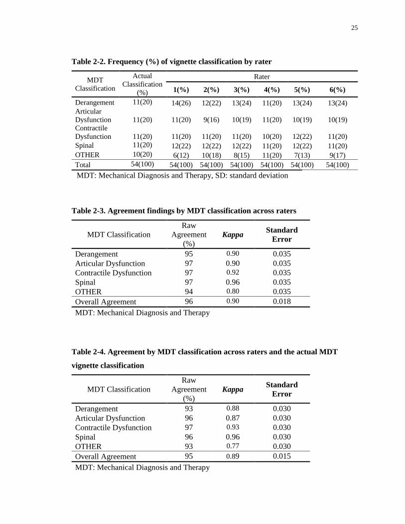
There was consensus among all 6 raters on the vignettes’ classification in 78% of the
vignettes (42 out of 54). The raw overall level of multi-rater agreement among the six
clinicians was 96%. The corresponding Kappa value was 0.90 (SE=0.018). The highest
level of chance-adjusted agreement was for the spinal category with Kappa=0.96; the
lowest level was for the OTHER category with Kappa=0.80. By factoring in the true
diagnoses of the vignettes in our analysis, the raw agreement and Kappa were 95% and
0.89, respectively. Values of agreement for each one of the MDT classifications are
shown in Tables 2.3 and 2.4.
Table 2-1. Demographic information of the participating practitioners
Variables Distribution
Number of raters 6
Age, mean (SD) (years) 51 (8.6)
Gender Female : 2
Male:4
Years in practice,
mean (SD) 25.7 (8)
Years since MDT diploma, mean (SD) 16 (4)
Proportion of extremity patients in caseload (n)
<25% : 2
25-50% : 4
Practice setting (n)
Private : 4
Hospital Outpatient: 1
Specialty Clinic : 1
MDT: Mechanical Diagnosis and Therapy, SD: standard deviation
25
Table 2-2. Frequency (%) of vignette classification by rater
MDT
Classification
Actual
Classification
(%)
Rater
1(%) 2(%) 3(%) 4(%) 5(%) 6(%)
Derangement 11(20) 14(26) 12(22) 13(24) 11(20) 13(24) 13(24)
Articular
Dysfunction
11(20) 11(20) 9(16) 10(19) 11(20) 10(19) 10(19)
Contractile
Dysfunction
11(20) 11(20) 11(20) 11(20) 10(20) 12(22) 11(20)
Spinal 11(20) 12(22) 12(22) 12(22) 11(20) 12(22) 11(20)
OTHER 10(20) 6(12) 10(18) 8(15) 11(20) 7(13) 9(17)
Total 54(100) 54(100) 54(100) 54(100) 54(100) 54(100) 54(100)
MDT: Mechanical Diagnosis and Therapy, SD: standard deviation
Table 2-3. Agreement findings by MDT classification across raters
MDT Classification
Raw
Agreement
(%)
Kappa Standard
Error
Derangement 95 0.90 0.035
Articular Dysfunction 97 0.90 0.035
Contractile Dysfunction 97 0.92 0.035
Spinal 97 0.96 0.035
OTHER 94 0.80 0.035
Overall Agreement 96 0.90 0.018
MDT: Mechanical Diagnosis and Therapy
Table 2-4. Agreement by MDT classification across raters and the actual MDT
vignette classification
MDT Classification
Raw
Agreement
(%)
Kappa Standard
Error
Derangement 93 0.88 0.030
Articular Dysfunction 96 0.87 0.030
Contractile Dysfunction 97 0.93 0.030
Spinal 96 0.96 0.030
OTHER 93 0.77 0.030
Overall Agreement 95 0.89 0.015
MDT: Mechanical Diagnosis and Therapy

26
2.4 Discussion
To our knowledge, this study is the first to address inter-examiner reliability of the MDT
system exclusively in patients with shoulder pain. The results support the findings of
previous reliability studies on the application of MDT in the extremities.19, 56 The
principal findings of our study suggest that experienced McKenzie practitioners have a
“very good” level of inter-examiner agreement when classifying patients with shoulder
pain using the MDT system. The highest level of agreement was for the ‘spinal’ category
with Kappa=0.96, and the lowest level of agreement was for the OTHER category with
Kappa=0.80. The relatively lower level of agreement for the OTHER category was
anticipated because multiple subcategories are included in this MDT classification. This
makes diagnosis more challenging particularly when the decision is solely based on
information collected in the initial assessment. A relatively higher level of agreement for
the ‘spinal’ category may be due to the presence of more identifying symptoms, such as
paraesthesia, reported in some of the vignettes, and also the presence of, in some cases, a
relatively quick response in the shoulder pain level of these patients by addressing their
cervical spine. By including the actual classification of the vignettes in our analysis, as
shown in Table 4, there is only a slight decline in both percent agreement and the Kappa
value. This slight decline could be due to the presence of insufficient clinical information
provided in the vignettes, as these were based only on the clinical information gathered in
the initial assessment session.
The results of our study on the shoulder generally reinforce the findings of previous
reliability studies in the spine and the extremities, suggesting that the MDT system is a
reliable method to classify patients with musculoskeletal shoulder disorders. Multiple
studies have been conducted on inter-examiner reliability of the MDT system in patients
with spinal disorders demonstrating an acceptable level of reliability among MDT
practitioners in classifying their patients.28-34 For instance, Razmjou et al.28 and
Kilpikoski et al.30 reported good inter-examiner reliability between two MDT trained
therapists in classifying patients with low back pain into MDT classifications
(Kappa=0.7). In another type of study using video and written clinical vignettes,

27
Werneke et al.34 reported substantial to almost perfect inter-rater agreement in identifying
treatment approaches for neck and low back disorders among MDT trained therapists.
There are only two studies addressing inter-examiner reliability of the MDT system for
patients with extremity disorders.19, 56 These two studies included a pilot study with 11
clinical vignettes56 and three therapists, and a follow up study with 25 clinical vignettes
and 93 MDT diploma holders.19 The pilot study showed “good” agreement with a Kappa
value of 0.7, and the follow up study revealed “very good” agreement with a Kappa value
of 0.83 (95% CI, 0.68-0.98). The clinical vignettes used for these studies were based on
patients with both upper and lower extremity disorders. There was little difference
between the reliability in upper (Kappa=0.85) and lower extremity (Kappa=0.80) cases.19
The major limitation of the current study was that only practitioners with an MDT
diploma, the highest level of MDT training, were included. This limits the
generalizability of the findings of this study, as the inter-rater agreement among
clinicians without this level of training may not be as high. Therefore, this study is a first
step when evaluating the reliability of using the MDT system to classify patients with
shoulder pain. Future studies should include practitioners with different levels of training
and experience so that the agreement findings are generalizable to a broader group of
practitioners. Another limitation of this study was using written vignettes instead of
having actual patients. The major concern in this regard, as stated by Werneke et al,34 is
the purification of the intervention being expressed in the vignettes, which may not
represent all aspects of clinical practice, making the diagnosis easier for the raters and
inflating the calculated Kappa value. One strength of using written vignettes is that this
approach eliminates the potential error created by inconsistent patient presentations
between raters. As an alternative, future studies could consider the use of real patients
instead of written vignettes in order to further establish reliability of the MDT system in
the extremities.
28
2.5 References
1. Dessaur WA, Magarey ME. Diagnostic accuracy of clinical tests for superior labral
anterior posterior lesions: a systematic review. J Orthop Sports Phys Ther.
2008;38(6):341-52.
2. De Winter AF, Jans MP, Scholten RJPM, Deville W,van Schaardenburg D, Bouter
LM. Diagnostic classification of shoulder disorders: interobserver agreement and
determinants of disagreement. Ann Rheum Dis. 1999;58(5):272-7.
3. Lucas N, Macaskill P, Irwig L, Moran R, Bogduk N. Reliability of physical
examination for diagnosis of myofascial trigger point. Clin J Pain. 2009;25:80-9.
4. Hegedus EJ, Goode A, Campbell S, Morin A, Tamaddoni M, Moorman CT. Physical
examination tests of the shoulder: a systematic review with meta-analysis of
individual tests. Br J Sports Med. 2008;42:80-92.
5. Hughes P, Taylor NF, Green RA. Most clinical tests cannot accurately diagnose
rotator cuff pathology: a systematic review. Aust J Physiother. 2008;54:159-70.
6. Schellingerhout JM, Verhagen AP, Thomas S, Koes BW. Lack of uniformity in
diagnostic labeling of shoulder pain: time for a different approach. Man Ther.
2008;13:478-83.
7. Lewis JS. Rotator cuff tendinopathy: a model for the continuum of pathology and
related management. Br J Sports Med. 2010;44(13):918-23.
8. May S, Chance-Larsen K, Littlewood C, Lomans D, Saad M. Reliability of physical
examination tests used in the assessment of patients with shoulder problems: a
systematic review. Physiotherapy. 2010;96:179-90.
9. Munro W, Healy R. The validity and accuracy of clinical tests used to detect labral
pathology of the shoulder - A systematic review. Man Ther. 2009;14:119-30.

29
10. Tucker S, Taylor NF, Green RA. Anatomical validity of the Hawkins-Kennedy test-
A pilot study. Man Ther. 2011;16:399-402.
11. Schibany N, Zehetgruber H, Kainberger F, et al. Rotator cuff tears in asymptomatic
individuals: a clinical and ultrasonographic screening study. Eur J Radiol.
2004;51(3):263-8.
12. Reilley P, MacLeod I, MacFarlane R, Windley J, Emery RJH. Dead men and
radiologists don’t lie: a review of cadaveric and radiological studies of rotator cuff
tear prevalence. Ann R Coll Surg Engl. 2006;88:116-21.
13. Yamaguchi K, Ditsios K, Middleton WD, Hildebolt CF, Galatz LM, Teefey SA. A
comparison of asymptomatic and symptomatic shoulders the demographic and
morphological features of rotator cuff disease. J Bone Joint Surg Am.
2006;88(8):1699-704.
14. Girish G, Lobo LG, Jacobson JA, Morag Y, Miller B, Jamadar DA. Ultrasound of the
shoulder: asymptomatic findings in men. Am J Roentgenol. 2011;197(4):W713-9.
15. CarterT, Hall H, McIntosh G, Murphy J, MacDougall J, Boyle C. Intertester
reliability of a classification system for shoulder pain. Physiotherapy. 2012;98(1):40-
6.
16. May S, Rosedale R. A survey of the McKenzie classification system in the
extremities: Prevalence of mechanical syndromes and preferred loading strategies.
Phys Ther. 2012;92(9):1175-86.
17. McKenzie RA. The lumbar spine: Mechanical Diagnosis and Therapy. Waikanae,
New Zealand: Spinal Publications; 1981.
18. McKenzie RA. The Cervical and Thoracic Spine: Mechanical Diagnosis and Therapy.
Waikanae, New Zealand: Spinal Publications; 1990.
30
19. May S, Ross J. The McKenzie classification system in extremities: a reliability study
using McKenzie assessment forms and experienced clinicians. J Manip Physiol Ther.
2009 32(7):556-63.
20. Clare HA, Adams R, Maher CG. A systematic review of efficacy of McKenzie
therapy for spinal pain. Aust J Physiother. 2004;50:209-16.
21. Cook C, Hegedus EJ, Ramey K. Physical therapy exercise intervention based on
classification using the patient response method: a systematic review of the literature.
J Man Manip Ther. 2005;13:152-62.
22. Machado LAC, de Souza MvS, Ferreira PH, Ferreira ML. The McKenzie method for
low back pain. A systematic review of the literature with a meta-analysis approach.
Spine. 2006;31:E254-62.
23. Hettinga DM, Jackson A, Klaber Moffett J, May S, Mercer C, Woby SR. A
systematic review and synthesis of higher quality evidence of the effectiveness of
exercise interventions for nonspecific low back pain of at least 6 weeks duration.
Phys Ther Rev. 2007;12:221-32.
24. Slade SC, Keating J. Unloaded movement facilitation exercise compared to no
exercise or alternative therapy on outcomes for people with non-specific chronic low
back pain: a systematic review. J Man Manip Ther. 2007;30:301-11.
25. May S, Donelson R. Evidence-informed management of chronic low back pain with
the McKenzie method. Spine J. 2008;8:134-41.
26. Aina A, May S, Clare H. The centralization phenomenon of spinal symptoms—a
systematic review. Man Ther. 2004;9:134-43.
27. May S, Littlewood C, Bishop A. Reliability of procedures used in the physical
examination of non-specific low back pain: a systematic review. Aust J Physiother.
2006;52:91-102.

31
28. Razmjou H, Kramer JF, Yamada R. Intertester reliability of the McKenzie evaluation
in assessing patients with mechanical low back pain. J Orthop Sports Phys Ther.
2000;30:368–89.
29. Fritz JM, Delitto A, Vignovic M, Busse RG. Interrater reliability of judgments of the
centralisation phenomenon and status change during movement testing in patients
with low back pain. Arch Phys Med Rehabil. 2000;81:57–61.
30. Kilpikoski S, Airaksinen O, Kankaanpaa M, Leminen P, Videman T, Alen M.
Interexaminer reliability of low back pain assessment using the McKenzie method.
Spine. 2002;27:207–14.
31. Clare HA, PT, Adams R, Maher CG. Reliability of the McKenzie spinal pain
classification using patient assessment forms. Physiotherapy. 2004;90:114-9.
32. Clare HA, PT, Adams R, Maher CG. Reliability of McKenzie classification of
patients with cervical or lumbar pain. J Manip Physiol Ther. 2005;28:122-7.
33. Dionne CP, Bybee RF, Tomaka J. Inter-rater reliability of McKenzie assessment in
patients with neck pain. Physiotherapy. 2006;92:75-82.
34. Werneke MW, Hart DL, Deutscher D, Stratford PW. Clinician’s ability to identify
neck and low back interventions: an inter-rater chance corrected agreement pilot
study. J Man Manip Ther. 2011;19:172-81.
35. Dionne CP, Bybee RF, Tomaka J. Correspondence of diagnosis to initial treatment
for neck pain. Physiotherapy. 2007;93:62-8.
36. Long A. The centralisation phenomenon. Its usefulness as a predictor of outcome in
conservative treatment of chronic low back pain. Spine. 1995;20:2513–21.
37. Sufka A, Hauger B, Trenary M, et al. Centralisation of low back pain and perceived
functional outcome. J Orthop Sports Phys Ther. 1998;27:205–12.

32
38. Werneke M, Hart DL, Cook D. A descriptive study of the centralization phenomenon.
A prospective analysis. Spine. 1999;24:676–83.
39. Werneke M, Hart DL. Centralisation phenomenon as a prognostic factor for chronic
pain or disability. Spine. 2001;26:758–65.
40. Werneke M, Hart DL. Discriminant validity and relative precision for classifying
patients with nonspecific neck and back pain by anatomic pain patterns. Spine.
2003;28:161–6.
41. Wetzel FT, MD, Donelson R. The role of repeated end-range/pain response
assessment in the management of symptomatic lumbar discs. Spine J. 2003;3:146-54.
42. Berthelot JM, Delecrin J, Maugars Y, Passuti N. Contribution of centralization
phenomenon to the diagnosis, prognosis, and treatment of diskogenic low back pain.
Joint Bone Spine. 2007;74:319-23.
43. Long A, May S, Fung T. The comparative prognostic value of directional preference
and centralization: a useful tool for front-line clinicians? J Man Manip Ther.
2008;16:248-54.
44. Werneke M, Hart DL, Resnik L, Stratford PW, Reyes A. Centralization: Prevalence
and effect on treatment outcomes using a standardized operational definition and
measurement method. J Orthop Sports Phys Ther. 2008;38(3):116-25.
45. Werneke M, Hart DL, Cutrone G, et al. Association between directional preference
and centralization in patients with low back pain. J Orthop Sports Phys Ther.
2011;41(1):22-31.
46. McKenzie RA, May S. The Human Extremities: Mechanical Diagnosis and Therapy.
Waikanae, New Zealand: Spinal Publications, 2000.
47. Aina A, May S. A shoulder derangement. Man Ther. 2005;10:159-63.

33
48. Littlewood C, May S. A contractile dysfunction of the shoulder. Man Ther.
2007;12:80-3.
49. Shouta K, Hiroshi T, May S. Application of mechanical diagnosis and therapy to a
patient diagnosed with a de Quervain’s disease: A case study. J Hand Ther.
2009;22:278-84.
50. Littlewood C. Contractile dysfunction of the shoulder (rotator cuff tendinopathy): an
overview. J Man Manip Ther. 2012;20(4):209-13.
51. Krog C, May S. Derangement of the temporomandibular joint; a case study using
mechanical diagnosis and therapy. Man Ther. 2012;17(5):483-6.
52. Menon A, May S. Shoulder pain: Differential diagnosis with mechanical diagnosis
and therapy extremity assessment - A case report. Man Ther. 2013;18(4):354-7.
53. Lynch G, May S. Directional preference at the knee: a case report using mechanical
diagnosis and therapy. J Man Manip Ther. 2013;21(1):60-6.
54. Kidd J. Treatment of shoulder pain utilizing mechanical diagnosis and therapy
principles. J Man Manip Ther. 2013;21(3):168-73.
55. Littlewood C, Malliaras P, Mawson S, May S, Walters SJ. Self-managed loaded
exercise versus usual physiotherapy treatment for rotator cuff tendinopathy: a pilot
randomised controlled trial. Physiotherapy. 2014;100(1):54-60.
56. Kelly E, May S, Ross J. The reliability of the McKenzie classification system using
extremity McKenzie assessment forms. Int J Mech Diagn Ther. 2008;3:3-6.
57. Rotondi MA, Donner A. A confidence interval approach to sample size estimation for
studies of interobserver agreement with multiple raters and outcome. J Clin
Epidemiol. 2012;65(7):778-84.
58. Streiner DL, Norman GR, Health measurement scales. A Practical Guide to Their
Development and Use. 3rd ed. Oxford: Oxford University Press; 2003.
34
Chapter 3
3 Application of the McKenzie system of Mechanical
Diagnosis and Therapy (MDT) in patients with shoulder
pain; a prospective longitudinal study2
3.1 Introduction
Shoulder pain is a common problem in the general population with reported rates ranging
from 100 to 160 per 1000 patients.1 Once present, shoulder symptoms have proven to be
persistent and recurrent, with 50% still unresolved after 18 months.2 It is thus not
surprising that shoulder pain is one of the leading causes of referrals to physiotherapy.1
The complexity of the shoulder joint, poor accuracy of shoulder clinical tests3-6 and the
lack of uniformity of diagnostic labeling7 make a precise diagnosis difficult to achieve.
Without a precise diagnosis, treatment is likely to be more arbitrary than targeted which
may contribute to the lack of efficacy for most interventions.8 This difficulty for
clinicians is compounded by the knowledge that many pathological findings revealed on
diagnostic tests such as Magnetic Resonance Imaging, x-rays, or ultrasound are
asymptomatic9-13 and so cannot be relied upon to make informed clinical decisions as to
the source of the pain.
The issue of uniformity and accuracy of diagnosis and treatment is an important concern
to address. These confounding factors have led to the call for and proposal of alternative
methods of assessment and classification.7, 14-15 Though some alternative classification
systems have been developed, their widespread use and acceptance among practitioners
has proven to be challenging. This may be due to their relatively recent introduction and a
dearth of research exploring their validity. If such a system was successfully embraced it
2 A version of this chapter has been published and is used with permission. Heidar Abady A,
Rosedale R, Chesworth BM, Rotondi MA, Overend TJ. Application of the McKenzie system of
Mechanical Diagnosis and Therapy (MDT) in patients with shoulder pain; a prospective
longitudinal study. J Man Manip Ther. DOI 10.1080/10669817.2017.1313929.
35
would reduce the variation in clinical practice amongst clinicians, and potentially lead to
an enhanced effect of treatment.16-17
The McKenzie system of Mechanical Diagnosis and Therapy (MDT) is one alternative
approach to the assessment, classification and treatment of musculoskeletal disorders.
The MDT system was initially described in 1981 with the introduction of a new approach
to the classification and management of back pain.18 It uses non-pathology specific
classifications that are based on a detailed history and a physical examination exploring
the effects of repeated movements, positions and loading strategies on symptoms and
motion.19 Each classification is matched to a different management approach.19
A series of systematic reviews support application of the MDT system in the
management of acute and chronic low back pain.20-27 McKenzie’s original description18
indicated that MDT could also be applied to extremity problems, the application of which
is outlined in his book on the human extremities.28 According to McKenzie, extremity
problems can be classified into the following syndromes and OTHER subgroups:28
• Derangement, identified by the presence of a directional preference which will give a
lasting positive change in symptoms, in range of movement and in function;
• Articular Dysfunction, identified by intermittent pain consistently produced at a
restricted end range with no rapid change of symptoms or range;
• Contractile Dysfunction, identified by intermittent pain, consistently produced by
loading the musculo-tendinous unit, for instance, with an isometric contraction against
resistance;
• Postural syndrome is only produced by sustained loading - the rest of the physical
examination would be normal;
• `OTHER subgroups are considered when none of the above syndrome patterns are
present. Each has a definition and criteria that together complete the classification for all

36
remaining presentations. Examples include Inflammatory, Trauma and Chronic Pain
Syndrome (Appendix A).
Despite the number of studies on the utility of the MDT system for spinal pain,20-32 there
is limited scientific literature about its application with extremity musculoskeletal
disorders. For shoulder disorders, only three case studies,33-35 and one case series36 have
been published. The prevalence of MDT syndromes in the extremities has been
investigated in a number of separate surveys37-38 and in a more recently conducted survey
by May and Rosedale.15 The latter showed that more than one third of patients with
extremity disorders were classified as Derangements. The authors suggested that if
further research shows the rapid treatment response of this subcategory in the extremities,
as it is proven to be in the spine, this would have a significant impact on the future
treatment of a major group of patients with extremity disorders. Our previous study
revealed substantial inter-rater agreement (Kappa=0.90) between MDT-trained experts
when classifying McKenzie upper extremity syndromes in vignettes of patients with
shoulder disorders.39 Therefore, the next logical step would be to investigate the
application of the MDT system in patients with shoulder problems.
The primary objective of this study was to determine if the response of pain and function
to MDT treatment differs by classification category at two and four weeks following the
start of physiotherapy treatment. The secondary objective was to describe the frequency
of discharge over time by MDT classification category.
We hypothesized that patients with Derangement classification would be discharged
earlier, and there would be a statistically significant treatment response in pain reduction,
and improved function compared to patients with shoulder Dysfunction at two weeks and
four weeks from their admission.

37
3.2 Methodology
3.2.1 Study design and setting
This study utilized a prospective longitudinal design. An international group of 15
licensed physiotherapists recruited and collected data from consecutive eligible patients
attending their clinic for rehabilitation of a shoulder problem. These study collaborators
were McKenzie Institute International diploma or credential holders who had greater than
one year of experience in using the MDT system with patients who complained of upper
extremity problems.
Instructions, consent forms, and data collection sheets were distributed to all the study
collaborators. In order to minimize bias, the collaborators had no awareness of the study
objectives and hypotheses. Completed data sheets were sent to the primary investigators
and stored in a password protected database. Patients’ baseline demographic and
historical variables were recorded including age, sex, hand dominance, physical demands
of job/daily activities, previous episodes and duration of symptoms. Ethics approval for
the study was obtained from the Health Sciences Research Ethics Board of Western
University (appendix C). Clinical data from a total of 105 patients were collected from
March 2013 to November 2014. Sample size was estimated to ensure a reasonable
number of cases across subcategories.
3.2.2 Participants
To be included in the study, patients were required to be over the age of 18, English
speaking, and have shoulder pain for which they were seeking physiotherapy
intervention. No specific shoulder diagnosis was required for inclusion. Patients were
excluded if they had a surgical procedure on their shoulder within six months prior to the
start of physical therapy treatment. No specific shoulder diagnoses were excluded as the
intent was to classify all patients presenting with shoulder pain using the MDT system.
38
3.2.3 Examination and classification
Patients were assessed and treated using the MDT method and principles. A “treatment-
as-usual” approach was followed. A standard MDT evaluation method was used for all
participants, and the patients’ diagnoses were classified according to the MDT system
utilized in the extremities. The patients were classified to one of the five following
subgroups: Derangement, Articular Dysfunction, Contractile Dysfunction, OTHER and
Spinal; the latter was included as patients referred with “shoulder pain” could eventually
be diagnosed as a condition originating from the cervical spine. Spinal classification is
believed to be a cervical spine Derangement and is anticipated to respond to treatment in
a similar manner as shoulder Derangement. OTHER refers to the patients who did not
meet the definition for any one of the above-mentioned classifications.
3.2.4 Intervention
Treatment ensued based on accepted procedures for each classification, and patients were
treated with individually matched exercises and the appropriate progression of forces
following the MDT method.28 The detailed intervention and progression of forces were
left to the discretion of the treating practitioners. There would have been multiple
individually tailored exercise programs based on each patient’s specific classification and
response to repeated movements; the patient classified as having a shoulder Derangement
with a directional preference for extension for example, would have been given repeated
end range extension exercises by the clinician. They would have been advised to perform
these exercises regularly, every one to three hours, in sets of 10-15 repetitions. They may
also have been advised to temporarily avoid certain exacerbating movements and
positions. If the patient improved, the intervention would remain unchanged; however, if
progress plateaued then the patient may be guided to apply more force, as long as more
force demonstrated a positive effect. Once resolution was well underway the patient
would be encouraged to resume all movements with confidence, but integrate the
directional preference movements into their daily routine. Those patients classified as
having an Articular Dysfunction would have been given repeated end range exercises in
the direction of the painful and limited movement, approximately 10 repetitions every

39
two to three hours. This would be performed until the movement became full and pain-
free and the patient felt confident to move freely in all directions. Those with Contractile
Dysfunctions would have been treated with a progressive resisted exercise regime in the
direction of the painful movement until the movement became pain-free with resistance
and full activity restored.
OTHER subgroups would have been managed depending on the particular subgroup. For
example, a patient with Chronic Pain Syndrome would be managed with pain education,
graded exposure to activity and the addressing of psychosocial barriers to recovery. If the
shoulder pain was classified as Spinal i.e. from a Cervical Derangement, the patient
would have been advised to perform repeated end range exercises in the directional
preference with the same details as outlined above for shoulder Derangements.
The patients were followed up until their discharge from physiotherapy, or after 4 weeks,
or 8 treatment sessions, whichever came first. The patients’ clinical information was
collected at the initial assessment, and the treatment effects were evaluated at primary
and secondary target points. The primary target point was the fifth treatment session, or
two weeks since the start of treatment, or discharge from physiotherapy treatment,
whichever came first. The secondary target point was the eighth treatment session, or
four weeks since the start of treatment, or discharge from physiotherapy treatment,
whichever came first.
3.2.5 Outcomes
Patients were monitored for change in the primary outcome measures used for the study
[the Upper Extremity Functional Index (UEFI) 40, and the Numeric Pain Rating Scale
(NPRS)41]. The UEFI is a patient-reported outcome measure consisting of 20 items that
capture a variety of upper extremity activities. Its purpose is to examine patients’ current
upper extremity functional status.40 Scores can vary from 0-80, with higher scores
indicating less functional limitation (i.e. better function).40 It has been shown to have
excellent test-retest reliability (intraclass correlation coefficient = 0.85-0.95), and internal
consistency (coefficent alpha) of 0.94.40,42 The minimal level of detectable change

40
(MDC) is 9 points,40 with a minimal clinically important difference (MCID) of 9-10
points.42
The NPRS is an 11-point scale with scores that can vary from 0 (no pain) to 10 (the worst
possible pain).41 It has been shown to have adequate test-retest reliability (r=0.63-0.92)
and excellent internal consistency (coefficient alpha = 0.84-0.98).43 The MDC for the
NPRS has been reported to be 2.5-3 in patients with shoulder and upper extremity
disorders,44-45 with a MCID of 2.17 reported in both surgical and non-surgical patients
with shoulder problems after 3-4 weeks of rehabilitation.46
Data on the primary outcomes were included in the analysis when they were available for
at least two out of three data collection points. In case a patient was discharged before
their third data collection point, the Last Observation Carried Forward (LOCF)
imputation method was utilized to fill in the missing score for the third data collection
point. The secondary outcome was the rate of discharge for each one of the MDT
classifications at both study target points.
3.2.6 Data Analysis
Descriptive statistics were calculated for the MDT classifications, patient characteristics
and the two primary outcome variables at baseline. The comparison for the primary
outcomes of pain and function was performed among the three major classifications of
Spinal, Derangement, and Dysfunction. As there were fewer patients in Articular and
Contractile Dysfunction classifications, the two sub-categories were merged to make up a
general classification of Dysfunction in order to have a more balanced sample size in
comparison to the Derangement and Spinal classifications. Both Articular and Contractile
Dysfunction are believed to demonstrate similar responses to treatment over time.
Depending on whether the compared variable was continuous or categorical, one-way
analysis of variance (ANOVA) or Chi square analysis was conducted to compare the
following baseline characteristics and potential confounding variables among the MDT
subcategories: NPRS and UEFI scores at baseline, age, sex, hand dominancy of the
affected shoulder, and duration of symptoms, history of previous episodes of same
41
condition, medication use, concurrent physiotherapy treatments received, and physical
demand of work/daily activities.
For the primary objective, a two-way mixed model ANOVA was conducted for the
primary outcomes of pain (measured by the NPRS) and function (measured by the UEFI)
to compare the interaction between MDT classifications (Spinal, Derangement, and
Dysfunction) and time (baseline, week 2, and week 4). When the sphericity assumption
was not met by our data, a Greenhouse-Geisser correction was used. In the presence of a
significant interaction between MDT classifications and time, one-way ANOVA and
planned pairwise comparisons were performed for each time point (baseline, week 2, and
week 4) to further investigate where the differences between the MDT classifications
actually existed. For the secondary objective frequency of discharge by MDT
classification and time was reported in percent. The SPSS version 20 (SPSS Inc,
Chicago, IL) was used for all data analyses.
3.3 Results
Between March 2013 and November 2014, 105 patients consented to participate in the
study and were recruited. The flow of patient recruitment and MDT classifications is
shown in Figure 3.1. Of the 105 patients recruited for the study, 12 patients subsequently
dropped out after their initial visits, for the following reasons: shoulder manipulation
performed by an orthopaedic surgeon (n=1); treatment sought in another clinic closer to
home (n=1); change in insurance coverage prompted treatment by another physiotherapy
clinic (n=1); treating practitioner took emergency leave of absence (n=2); travel out-of-
town for extended period of time (n=3); failure to return for follow up treatment after

42
initial visit (n=4).
Figure 3-1. Flow of patients and MDT classifications. Abbreviations: AD, Articular
Dysfunction; CD, Contractile Dysfunction; DER, Derangement; DYD, Dysfunction;
MDT, Mechanical Diagnosis and Therapy.
Of the 93 patients who completed the study, 11 patients had either a concurrent condition
of two MDT classifications, or were classified under the OTHER subgroups. These
patients were excluded, leaving 82 patients for the main analyses. In 63.4% of the cases,
the provisional diagnoses remained unchanged over the course of treatment. The
distribution of MDT classifications is shown in Table 3.1. Seventy-two percent of
participants (59 out of 82) had their data collected for all the three data collection points.
For the remaining 27% who were discharged prior to their third data collection point,
LOCF was utilized to fill in the missing data.

43
Table 3-1. Distribution of the MDT classifications at baseline
MDT
Classification
Frequency Percent May and Rosedale3
(%)
DER 35 37.6 42.5
AD 9 9.7 10.8
CD 11 11.8 11.7
Spinal 27 29.0
OTHER 35.0
DER with residual AD 2 2.2
DER with residual CD 1 1.1
Spinal with residual AD 2 2.2
Spinal with DER 1 1.1
Spinal with residual CD 1 1.1
OTHER 4 4.3
Total 93 100.0
Abbreviations: AD, Articular Dysfunction; CD, Contractile Dysfunction; DER,
Derangement; MDT, Mechanical Diagnosis and Therapy
There was no significant difference (P >0.05) among the three MDT classifications at
baseline for NPRS and UEFI scores, and other baseline characteristics (Table 3.2). Only
two patients in the Derangement group received concurrent treatments (a cold pack)
along with their MDT-directed treatments. The remaining patients received solely the
MDT-directed treatments, therefore, no comparison was conducted among the MDT
classifications for this variable.
3 May S, Rosedale R. A survey of the McKenzie classification system in the extremities:
Prevalence of mechanical syndromes and preferred loading strategies. Phys Ther.
2012;92(9):1175-86.
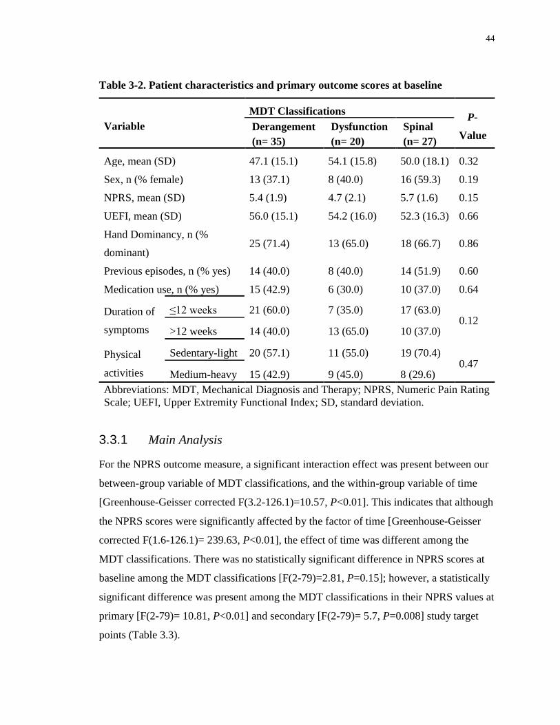
44
Table 3-2. Patient characteristics and primary outcome scores at baseline
Variable
MDT Classifications P-
Value Derangement
(n= 35)
Dysfunction
(n= 20)
Spinal
(n= 27)
Age, mean (SD) 47.1 (15.1) 54.1 (15.8) 50.0 (18.1) 0.32
Sex, n (% female) 13 (37.1) 8 (40.0) 16 (59.3) 0.19
NPRS, mean (SD) 5.4 (1.9) 4.7 (2.1) 5.7 (1.6) 0.15
UEFI, mean (SD) 56.0 (15.1) 54.2 (16.0) 52.3 (16.3) 0.66
Hand Dominancy, n (%
dominant) 25 (71.4) 13 (65.0) 18 (66.7) 0.86
Previous episodes, n (% yes) 14 (40.0) 8 (40.0) 14 (51.9) 0.60
Medication use, n (% yes) 15 (42.9) 6 (30.0) 10 (37.0) 0.64
Duration of
symptoms
≤12 weeks 21 (60.0) 7 (35.0) 17 (63.0) 0.12
>12 weeks 14 (40.0) 13 (65.0) 10 (37.0)
Physical
activities
Sedentary-light 20 (57.1) 11 (55.0) 19 (70.4) 0.47
Medium-heavy 15 (42.9) 9 (45.0) 8 (29.6)
Abbreviations: MDT, Mechanical Diagnosis and Therapy; NPRS, Numeric Pain Rating
Scale; UEFI, Upper Extremity Functional Index; SD, standard deviation.
3.3.1 Main Analysis
For the NPRS outcome measure, a significant interaction effect was present between our
between-group variable of MDT classifications, and the within-group variable of time
[Greenhouse-Geisser corrected F(3.2-126.1)=10.57, P<0.01]. This indicates that although
the NPRS scores were significantly affected by the factor of time [Greenhouse-Geisser
corrected F(1.6-126.1)= 239.63, P<0.01], the effect of time was different among the
MDT classifications. There was no statistically significant difference in NPRS scores at
baseline among the MDT classifications [F(2-79)=2.81, P=0.15]; however, a statistically
significant difference was present among the MDT classifications in their NPRS values at
primary [F(2-79)= 10.81, P<0.01] and secondary [F(2-79)= 5.7, P=0.008] study target
points (Table 3.3).

45
Table 3-3. Baseline and follow-up primary outcome scores and results of analysis
comparing MDT classifications (values are means and standard deviations)
Assessment
Time/Variable
MDT Classifications
P-Value Derangement
(n= 35)
Dysfunction
(n= 20)
Spinal
(n= 27)
Baseline
NPRS 5.4 (1.9) 4.7 (2.1) 5.7 (1.6) 0.15
UEFI 56.0 (15.1) 54.2 (16.0) 52.3 (16.3) 0.66
Week 2
NPRS 1.53 (1.71) 3.35 (1.87) 1.26 (1.32) <0.01
UEFI 72.89 (7.40) 59.30 (14.85) 72.81 (5.76) <0.01
Week 4
NPRS 0.86 (1.16) 1.77 (1.47) 0.68 (1.12) <0.01
UEFI 75.68 (5.47) 65.45 (16.07) 76.40 (4.18) <0.01
Abbreviations: MDT, Mechanical Diagnosis and Therapy; NPRS, Numeric Pain Rating
Scale; UEFI, Upper Extremity Functional Index.
The Derangement classification had significantly lower NPRS scores than the
Dysfunction group indicating pain reduction at week 2 (P<0.01) and week 4 (P=0.02).
The Spinal classification also had significantly lower NPRS scores in comparison to the
Dysfunction group at week 2 (P<0.01) and week 4 (P<0.01). Derangement and Spinal
classifications had no statistically significant difference in their NPRS scores at week 2
(P=0.49) and week 4 (P=0.56) (Table 3.4).
Table 3-4. Contrasts between pairs of MDT classifications for main outcomes at
primary and secondary study target points
Contrasts Value of Contrast* (SE) P-Value
Week 2 Week 4 Week 2 Week 4
NPRS
DER vs DYS -1.82 (0.51) -0.92 (0.38) <0.01 0.02
DER vs Spinal 0.27 (0.39) 0.17 (0.29) 0.49 0.56
DYS vs Spinal 2.09 (0.49) 1.09 (0.39) <0.01 <0.01
UEFI
DER vs DYS 13.58 (3.55) 10.24 (3.71) <0.01 0.01
DER vs Spinal 0.07 (1.67) -0.72 (1.22) 0.97 0.56
DYS vs Spinal - 13.51 (3.50) -10.96 (3.68) <0.01 <0.01
Abbreviations: DER, Derangement; DYS, Dysfunction; MDT, Mechanical Diagnosis
and Therapy; NPRS, Numeric Pain Rating Scale; NS, not significant; UEFI, Upper
Extremity Functional Index; SE, standard error. * Mean difference

46
The NPRS mean scores with 95% confidence intervals for each of the MDT
classifications are shown in Figure 3.2 as a function of time.
Figure 3-2. Mean NPRS score from baseline to discharge in each MDT
classification. Abbreviations: DER, Derangement; DYS, Dysfunction; MDT,
Mechanical Diagnosis and Therapy; NPRS, Numeric Pain Rating Scale.
For the UEFI outcome measure, a significant interaction effect was present between our
between-group variable of MDT classifications and the within-group variable of time
[Greenhouse-Geisser corrected F(2.31-91.08)=7.08, P< 0.01]. This indicates that
although the UEFI scores were affected by the factor of time [Greenhouse-Geisser
corrected F(1.15-91.08)=122.99, P<0.01], the effect of time was different among the
MDT classifications. There was no statistically significant difference in UEFI scores at
baseline among the MDT classifications [F(2-79)= 0.441, P=0.66]; however, a
statistically significant difference was present among the MDT classifications in their
UEFI values at primary [F(2-79)= 15.87, P<0.01] and secondary [F(2-79)= 10.47,
P<0.001] study target points (Table 3.3). The Derangement classification had
0
1
2
3
4
5
6
7
0.5 1.0 1.5 2.0 2.5 3.0 3.5
NP
RS
MEAN of NPRS with 95% Confidence Interval
DER DYS SPINAL
BASELINE WEEK2 WEEK4
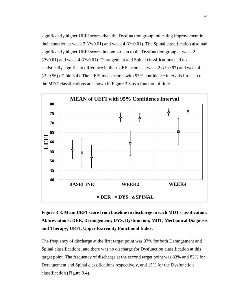
47
significantly higher UEFI scores than the Dysfunction group indicating improvement in
their function at week 2 (P<0.01) and week 4 (P=0.01). The Spinal classification also had
significantly higher UEFI scores in comparison to the Dysfunction group at week 2
(P<0.01) and week 4 (P<0.01). Derangement and Spinal classifications had no
statistically significant difference in their UEFI scores at week 2 (P=0.97) and week 4
(P=0.56) (Table 3.4). The UEFI mean scores with 95% confidence intervals for each of
the MDT classifications are shown in Figure 3.3 as a function of time.
Figure 3-3. Mean UEFI score from baseline to discharge in each MDT classification.
Abbreviations: DER, Derangement; DYS, Dysfunction; MDT, Mechanical Diagnosis
and Therapy; UEFI, Upper Extremity Functional Index.
The frequency of discharge at the first target point was 37% for both Derangement and
Spinal classifications, and there was no discharge for Dysfunction classification at this
target point. The frequency of discharge at the second target point was 83% and 82% for
Derangement and Spinal classifications respectively, and 15% for the Dysfunction
classification (Figure 3.4).
40
45
50
55
60
65
70
75
80
0.5 1.0 1.5 2.0 2.5 3.0 3.5
UE
FI
MEAN of UEFI with 95% Confidence Interval
DER DYS SPINAL
BASELINE WEEK2 WEEK4

48
Figure 3-4. Frequency of discharge for MDT classifications at primary and
secondary target points. Abbreviations: DER, Derangement; DYS, Dysfunction;
MDT, Mechanical Diagnosis and Therapy.
3.4 Discussion
To the best of our knowledge, this is the first study to address the clinical application of
the MDT system in patients with shoulder disorders. Over time, patients in the
Derangement and Spinal groups demonstrated very similar pain and function responses to
treatment and showed significantly greater improvement in comparison to patients with
Dysfunction. These treatment responses existed at both the primary and secondary study
target time points of week 2 and 4, respectively. Consistent with this, compared to
patients in the Dysfunction group, a high percentage of patients with Derangement and
Spinal classifications achieved their treatment goals relatively quickly and were
discharged from treatment at weeks 2 and 4. This highlights the point that the Spinal
extremity classification is in fact a cervical spine derangement and like the shoulder
Derangement, classification is anticipated to demonstrate a rapid treatment response.

49
Therefore, it appears in this non-randomized cohort that when MDT-trained clinicians
match the intervention to a specific MDT classification, the outcome is aligned with the
response expectation of the classification. Hence, shoulder Derangement or shoulder pain
that has a cervical Derangement will respond and resolve rapidly. Dysfunctions will
respond, but in a more graduated manner, achieving discharge status at a later point.
As shown in table 1, the distribution of the MDT classifications in our sample was
comparable to those reported by May and Rosedale.15 They did not look at Cervical
Derangement as a separate classification for patients with shoulder disorders, however it
is interesting to note that only 2% of the total upper and lower extremity patients were
classified with spinal problems in their survey. This contrasts dramatically with the 29%
of shoulder pain patients diagnosed with Cervical Derangements in this cohort. It is
possible that this reflects an increase in the recognition of Cervical Derangements as a
source of shoulder pain by MDT clinicians or that the study clinicians studied by May
and Rosedale effectively screened out the cervical spine in most of their extremity
patients.
There were several limitations to this study. First, due to the use of a “treatment-as-usual”
approach, it was not possible to have a pre-specified number of treatment sessions for
each one of our study participants. As a result, it is possible that the patients in each
category received a different number of treatment sessions, ultimately affecting treatment
outcome. However, our treating clinicians had no awareness of the study objectives,
suggesting they had little motivation to affect the outcome of each classification category
other than to treat the patient as best as they could, given the clinical findings and MDT
classification category. Secondly, exercise compliance was not investigated; therefore, it
is uncertain whether the inferior results of the Dysfunction patients resulted from poor
exercise compliance or the actual nature of the MDT classification. Third, there was no
treatment group assigned to a control condition or conventional physiotherapy
intervention removing the ability to compare MDT classification with other treatment
approaches. Fourth, a greater proportion of patients included in this study had a pain
duration of less than 12 weeks (Table 2). Therefore, it may be that most of these patients

50
would have recovered without any intervention. Fifth, there was also no randomization
because the MDT method was selected as the only method of intervention and the
patients were required to be treated within their respective MDT classification groups.
Finally, the treating physiotherapists were MDT-trained practitioners and the treatment
results may not be generalizable to other physiotherapists with less MDT training. As a
next step, randomized controlled trials are needed to compare the MDT system with
conventional treatment for patients with shoulder disorders.
Considering the well-described limitations of conventional patho-anatomic models for
diagnosis and treatment of patients with shoulder complaints,3-6, 10 the MDT system, may
be worthy of further investigation to fill the current gap in diagnosis and management of
patients with extremity problems. The encouraging aspect of the study results is that two-
thirds of our study participants (66.6%) were classified as either a shoulder Derangement
or a cervical Spinal Derangement. If further studies confirm that patients classified as
Derangements conform to their expected rapid response to tailored MDT treatments,
there is potential to significantly impact quality of life and health care utilization for a
majority of patients with shoulder problems.

51
3.5 References
1. Winters JC, Sobel JS, van der Windt DAWM, et al. NHG Standaard
Schouderklachten (versie 1999) Guidelines for Shoulder Complaints of the Dutch
College of General Practitioners (version 1999). Huisarts en Wetenschap.
1999;42:222e31.
2. Croft P, Pope D, Silman A. The clinical course of shoulder pain: prospective cohort
study in primary care. Primary Care Rheumatology Society Shoulder Study Group.
BMJ. 1996;313(7057):601-2.
3. Hughes P, Taylor NF, Green RA. Most clinical tests cannot accurately diagnose
rotator cuff pathology: a systematic review. Aust J Physiother. 2008;54:159-70.
4. Walton DM, Sadi J. Identifying SLAP lesions: A meta-analysis of clinical tests and
exercise in clinical reasoning. Phys Ther. Sport 2008;9:167-76.
5. Beaudreuil J, Nizard R, Thomas T, Peyre M, Liotard JP, Boileau P, Marc T, Dromard
C, Steyer E, Bardin T, Orcel P, Walch G. Contribution of clinical tests to the
diagnosis of rotator cuff disease: A systematic review. Joint Bone Spine. 2009;76:15-
9.
6. Gadogan A, Laslett M, Hing W, McNair P, Williams M. Inter-examiner reliability of
orthopaedic special tests used in the assessment of shoulder pain. Man Ther.
2011;16:131-135.
7. Schellingerhout JM, Verhagen AP, Thomas S, Koes BW. Lack of uniformity in
diagnostic labeling of shoulder pain: Time for a different approach. Man Ther.
2008;13:478-483.
8. Green S, Buchbinder R, Glazier R, Forbes A. Systematic review of randomised
controlled trials of interventions for painful shoulder: selection criteria, outcome
assessment, and efficacy. BMJ. 1998;316(7128):354-60.
52
9. Boden BD, Davis DO, Dina TS, Stoller DW, Brown SD, Vailas JC, Labropoulos P. A
prospective and blind intervention of Magnetic Resonance Imaging of the knee.
Abnormal findings in asymptomatic subjects. Clin Orthop Relat Res. 1992;282:177-
85.
10. Schibany N, Zehetgruber H, Kainberger F, Wuring C, Ba-Ssalamah A, Herneth AM,
Lang T, Gruber D, Breitenseher MJ. Rotator cuff tears in asymptomatic individuals:
aclinical and ultrasonographic screening study. Eur J Radiol. 2004;51:263-268.
11. Bedson J, Croft PR. The discordance between clinical and radiographic knee
osteoarthritis: a systematic search and summary of the literature. BMC Musculoskelet
Disord. 2008; 9:116.
12. Yusuf E, Kortekaas MC, Watt I, Huizinga TWJ, Kloppenburg M. Do knee
abnormalities visualized on MRI explain knee pain in knee osteoarthritis? A
systematic review. Ann Rheum Dis. 2011 Jan;70(1):60-67. Epub 2010 Sep.
13. Silvis ML, Mosher TJ, Smetana BS, Chinchili VM, Flemming DJ, Walker EA, Black
KP. High prevalence of pelvic and hip magnetic resonance imaging findings in
asymptomatic collegiate and professional hockey players. Am J Sport Med.
2011;39(4):715-21.
14. Lewis JS. Rotator cuff tendinopathy: a model for the continuum of pathology and
related management. Br J Sports Med. 2010;44(13):918–23.
15. May S, Rosedale R. A survey of the McKenzie classification system in the
extremities: Prevalence of mechanical syndromes and preferred loading strategies.
Phys Ther. 2012;92(9):1175-86.
16. Guide to physical therapist practice. 2nd ed. American Physical Therapy Association.
Phys Ther. 2001;81:9–746.
17. Koes BW, van Tulder MW, Thomas S. Diagnosis and treatment of low back pain.
BMI 2006;332:1430-1434.
53
18. McKenzie RA. The lumbar spine. Mechanical Diagnosis and Therapy. Waikanae,
New Zealand: Spinal Publications; 1981.
19. May S. The McKenzie classification system in Extremities: A reliability study using
McKenzie assessment forms and experienced clinicians. J Manipulative Physiol Ther.
2009;32(7):556-63).
20. Clare HA, Adams R, Maher CG. A systematic review of efficacy of McKenzie
therapy for spinal pain. Aust J Physiother. 2004;50:209-16.
21. May S, Littlewood C, Bishop A. Reliability of procedures used in the physical
examination of non-specific low back pain: a systematic review. Aust J Physiother.
2006;52:91-102.
22. Cook C, Hegedus EJ, Ramey K. Physical therapy exercise intervention based on
classification using the patient response method: a systematic review of the literature.
J Man Manip Ther. 2005;13:152-62.
23. Machado LAC, de Souza MvS, Ferreira PH, Ferreira ML. The McKenzie method for
low back pain. A systematic review of the literature with a meta-analysis approach.
Spine 2006;31:E254-62.
24. Hettinga DM, Jackson A, Klaber Moffett J, May S, Mercer C, Woby SR. A
systematic review and synthesis of higher quality evidence of the effectiveness of
exercise interventions for nonspecific low back pain of at least 6 weeks duration.
Phys Ther Rev. 2007;12:221-32.
25. Slade SC, Keating J. Unloaded movement facilitation exercise compared to no
exercise or alternative therapy on outcomes for people with non-specific chronic low
back pain: a systematic review. J Man Manip Ther. 2007;30:301-11.
26. May S, Donelson R. Evidence-informed management of chronic low back pain with
the McKenzie method. Spine J. 2008;8:134-41.
54
27. Aina A, May S, Clare H. The centralization phenomenon of spinal symptoms—a
systematic review. Man Ther. 2004;9:134-43.
28. McKenzie RA, May S. The Human Extremities: Mechanical Diagnosis and Therapy.
Waikanae, New Zealand: Spinal Publications, 2000.
29. Long A. The centralisation phenomenon. Its usefulness as a predictor of outcome in
conservative treatment of chronic low back pain. Spine. 1995;20:2513–21.
30. Sufka A, Hauger B, Trenary M, et al. Centralisation of low back pain and perceived
functional outcome. J Orthop Sports Phys Ther. 1998;27:205–12.
31. Long A, May S, Fung T. The Comparative Prognostic Value of Directional
Preference and Centralization: A Useful Tool for Front-Line Clinicians? J Man
Manip Ther. 2008;16:248-254.
32. Werneke M, Hart DL, Cutrone G, Oliver D, McGill M, Weinberg J, Grisby D,
Oswald W, Ward J. Association Between Directional Preference and Centralization in
Patients With Low Back Pain. J Orthop Sports Phys Ther. 2011;41(1):22-31.
33. Aina A, May S. A shoulder derangement. Man Ther. 2005;10:159-63.
34. Kidd JA. Treatment of shoulder pain utilizing mechanical diagnosis and therapy
principles. J Man Manip Ther. 2013;21:168-173.
35. Littlewood C, May S. A contractile dysfunction of the shoulder. Man Ther.
2007;12:80-3.
36. Aytona MC, Dudley K. Rapid resolution of chronic shoulder pain classified as
derangement using the McKenzie method: a case series. J Man Manip Ther.
2013;21:207-212.
37. Turner K. An audit of extremity patients. McKenzie Institute UK Newsletter.
2004;13(1): 2-25.

55
38. May S. Classification by McKenzie mechanical syndromes; Report on directional
preference and extremity patients. Int J Mech Diagn Ther. 2006;1(3):7-11.
39. Heidar Abady A, Rosedale R, Overend T, Chesworth B, Rotondi M. Inter-examiner
reliability of diplomats in the Mechanical Diagnosis and Therapy system in assessing
patients with shoulder pain. J Man Manip Ther. 2014 Nov; 22(4): 199–205.
40. Stratford PW, Binkley, JM, Stratford DM. Development and initial validation of the
upper extremity functional index. Physiother Can. 2001;53(4):259-267.
41. Jensen MP, Karoly P, Braver S. The measurement of clinical pain intensity: a
comparison of six methods. Pain. 1986;27(1):117–126.
42. Hefford C, Abbott JH, Arnold R, Baxter GD. The patient-specific functional scale:
validity, reliability, and responsiveness in patients with upper extremity
musculoskeletal problems. J Orthop Sports Phys Ther. 2012;42(2):56-65.
43. Jensen MP, McFarland CA. Increasing the reliability and validity of pain intensity
measurement in chronic pain patients. Pain. 1993;55(2):195-203.
44. Stratford PW, Spadoni G. The reliability, consistency, and clinical application of a
numeric pain rating scale. Physiotherapy Canada. 2001;53(2):88-114.
45. Mintken PE, Glynn P, Cleland JA. Psychometric properties of the shortened
disabilities of the Arm, Shoulder, and Hand Questionnaire (QuickDASH) and
Numeric Pain Rating Scale in patients with shoulder pain. J Shoulder Elbow Surg.
2009;18(6):920-6.
46. Michener LA, Snyder AR, Leggin BG. Responsiveness of the numeric pain rating
scale in patients with shoulder pain and the effect of surgical status. J Sport Rehabil.
2011;20(1):115-28.

56
Chapter 4
4 Consistency of commonly used orthopedic special tests of the
shoulder when used with the McKenzie system of
Mechanical Diagnosis and Therapy4
4.1 Introduction
Shoulder pain is one of the primary reasons for referral to physiotherapy with an annual
prevalence of 100 to 160 per 1000 patients in the general population.1 It has been shown
to be relentless and recurring, with half of all cases remaining unresolved after 18
months.2 Complexity of the shoulder, and absence of uniformity in diagnostic labeling3
hinder accurate diagnosis. This can have significant implications for conservative
management where ideally the diagnosis should directly guide clinical reasoning and
decision making.4-5 These diagnostic challenges may inadvertently lead to inappropriate
and perhaps more costly interventions.6
For physical examination of the shoulder, Orthopedic Special Tests (OSTs) are
commonly used7 and despite a heavy reliance on their use, demonstrate only limited
utility for informing diagnosis.3-4, 6, 8 Studies have revealed conflicting diagnostic
performance for the majority of OSTs used in the assessment of common shoulder
disorders such as rotator cuff pathology, sub-acromial impingement and superior labrum
anterior-to-posterior (SLAP) lesions.8-23 Considering the shortcomings of commonly used
OSTs, a growing body of opinion favours the implementation of an approach that is
different than a patho-anatomical based assessment and diagnosis of musculoskeletal
disorders.3,15,24 In principle, the use of a reliable form of classification should decrease
practice variation, and enhance the effectiveness of treatment by matching that
intervention to a specific subgroup.25-26 The McKenzie system of Mechanical Diagnosis
4 A version of this chapter has been published and is used with permission. Heidar Abady A,
Rosedale R, Chesworth BM, Rotondi MA, Overend TJ. Consistency of commonly used
orthopedic special tests of the shoulder when used with the McKenzie system of mechanical
diagnosis and therapy. Musculoskelet Sci Pract. DOI 10.1016/j.msksp.2017.10.001.

57
and Therapy (MDT) is one alternative method that has been proposed to assist the
clinician in formulating a classification that enables an appropriate management
strategy.27 The MDT system was initially described in 1981 as a new method for
classification and treatment of patients with back pain.28 The system uses a non-pathology
specific classification approach that consists of a thorough history and physical
examination monitoring the effects of repeated movements, sustained positions and
loading strategies on patients’ clinical presentations.29
Several systematic reviews show varying degrees of support for the utilization of the
MDT system when treating patients with acute and chronic low back pain.30-37 The MDT
system has also demonstrated acceptable reliability38-42 and varying degrees of validity43-
53 when used in patients with spinal disorders. A growing body of evidence supports the
application of the MDT system when treating patients with musculoskeletal disorders of
the extremity.27, 54-63 Although reliability varies considerably between different study
designs,64, 65 very good inter-examiner reliability has been reported specifically for the
shoulder.61
In the McKenzie system, extremity disorders include the following syndromes and
subgroups:66
• Derangement, identified by the presence of a directional preference which will give a
rapid and lasting improvement in symptoms, in range of movement and in function;
• Articular Dysfunction, identified by intermittent pain consistently produced only at a
restricted end range of motion with no rapid change of symptoms or range;
• Contractile Dysfunction, identified by intermittent pain, consistently produced by
loading the musculo-tendinous unit, for instance, with an isometric contraction against
resistance;
• Postural syndrome, identified by intermittent pain only produced by sustained loading,
with movements and activities being unaffected;

58
• OTHER subgroups are considered when none of the above syndrome patterns are
present. Each has a definition and specific criteria that together complete the
classification for all remaining presentations. Examples include Trauma, Peripheral
Nerve Entrapment and Inflammatory (Appendix A).
Although there are clear issues with the validity and clinical interpretation of OSTs, their
use is still widespread, with many clinicians continuing to utilize these tests as a basis for
diagnosis in shoulder disorders.7 One common observation by MDT clinicians and
reported in various case studies67-69 is that the results of OSTs can change depending
upon the MDT classification. For example, in one case study,68 the initial treatment of a
patient with a shoulder Derangement was reported to have an immediate effect on the
‘Empty can’ test, the ‘Lift off’ test and the ‘Hawkins-Kennedy’ test, with test results
shifting from positive to negative within the first session and remaining negative until
discharge. It is possible that the insights from this case may give one possible explanation
as to why these OSTs appear inherently unreliable and of questionable validity.
Derangement has a variable nature in terms of movement loss, direction of preference
and pain behavior. Hence, at times a patient may be experiencing severe symptoms,
considerable loss of motion and limited function; at other times the symptoms may be
milder, with greater range and better function. This may happen either naturally in
response to the patient’s daily movements and loading of the joint or in response to the
therapeutic intervention e.g. repeated end range movements in the directional preference.
The implication for OSTs when tested in the presence of Derangement is that at times,
when the Derangement is more severe they may test positive and at other times when the
Derangement is milder they may test negative. The OSTs are intended to gauge the
presence or absence of a particular pathology or diagnosis, however, in the presence of
Derangement, the OST results may be dependent upon the current behavior of the
Derangement rather than reflecting the specific pathology they are proposed to identify.
This can be particularly apparent when the Derangement is treated with directional
preference exercises, where it can be taken from a more painful and limited state to a
much less severe state in a short period of time. The classification of Derangement is
reported to be a prevalent cause of shoulder pain24, 27 as it is with other musculoskeletal

59
problems.24 Hence its presence could be a factor underlying the historic lack of accuracy
of the OSTs.
The aim of our study was to investigate, in patients with shoulder complaints, whether
MDT classifications and their subsequent treatment regime affects the agreement of
commonly used OSTs over time. To determine if shoulder Derangement interferes with
the results of OSTs, we hypothesized that over the course of treatment, there would be
lower agreement between consecutive OST results in patients with shoulder Derangement
compared to patients with shoulder Articular or Contractile Dysfunction. This would be
the first study to explore the consistency of OST results within the MDT classification
system of the shoulder.
4.2 Methodology
4.2.1 Study design and setting
This was a multi-centre prospective longitudinal study that ran concurrently with a study
that explored the clinical application of the MDT system in patients with shoulder
disorders.27 An international group of 15 McKenzie Institute International diploma and
credential holders recruited and collected data from consecutive patients visiting their
clinics for treatment of a shoulder problem. These study collaborators were licensed
physiotherapists with over one year of experience in applying the MDT system to
patients who presented with an upper extremity problem.
Instructions, consent forms and data collection sheets were distributed to the study
collaborators. To minimize bias, participating physiotherapists had no awareness of the
study objectives and hypotheses. In addition, different orthopedic clinicians who were
unaware of the patients’ MDT classifications performed and recorded the OST results.
The patients were followed up until their discharge from their treatment program, and the
completed data collection forms were sent to the primary investigator for analysis. Ethics
approval for the study was obtained from the Health Sciences Research Ethics Board of
Western University (Appendix C).
60
A confidence interval (CI) approach for sample size estimation of Kappa was used.70
Assuming a preliminary estimate of Kappa = 0.7, with a 95% CI of 0.2, we decided that
89 participants were needed for five MDT classifications to ensure a reasonable number
of cases across subcategories. Considering a 10% dropout rate, a total of 100 participants
was calculated to be a sufficient number for our primary outcome; however, by the time
the primary investigators received sufficient data from the study collaborators and
declared the end of the study, five additional patients were already recruited and their
data were collected. Therefore, clinical data for a total of 105 patients were collected
from March 2013 to November 2014.
4.2.2 Participants
To be included in the study, participants were required to be over the age of 18, English
speaking and with a shoulder disorder for which they were pursuing physiotherapy
intervention. No specific shoulder diagnosis was required for inclusion. Patients were
excluded if they had a surgical intervention on their shoulder within six months before
the beginning of their physiotherapy program. No specific shoulder diagnoses were
excluded, as one of the intentions of our concurrent study27 was to classify all patients
presenting with shoulder pain using the MDT system.
4.2.3 Examination and classification
A “treatment-as-usual” approach was utilized, and patients were assessed and treated
following MDT methods and principles. Patients were allocated to one of the following
five subgroups: Derangement, Articular Dysfunction, Contractile Dysfunction, OTHER
and Spinal; the latter was recognized as patients referred with “shoulder pain” but the
cervical spine was confirmed as the source of symptoms. Spinal classification was
accepted to be a cervical spine Derangement and was expected to demonstrate a similar
treatment response as shoulder Derangement when the cervical spine was treated.
OTHER subgroups included all patients who failed to meet the criteria for any one of the
previously described classifications.

61
4.2.4 Intervention and outcomes
Treatment followed recognized procedures for each MDT classification; patients were
treated with distinctively matched exercises and the relevant progression of forces were
pursued as per the MDT method.66 As there would have been numerous individualized
MDT exercise programs depending on each patient’s diagnosis and response to treatment,
the specific intervention and progression of forces were left to the discretion of the
treating practitioners.
Three commonly used OSTs documented in systematic reviews of shoulder tests10-12, 14, 15,
17, 18, 20 were utilized: Empty Can, Hawkins-Kennedy, and Speed’s. In the Empty Can test,
resistance is given to abduction in two different positions -- 90 degrees of arm abduction
with neutral (no) rotation, and 90 degrees of abduction with the shoulder medially rotated
and angled forward 30 degrees (empty can position), so that the patient’s thumb points
toward the floor in the plane of the scapula.71 Examiners look for weakness or pain,
which reflects a positive test.71 In the Hawkins-Kennedy test, with the elbow in 90
degrees of flexion, the examiner forward flexes the arm to 90 degrees then quickly
medially rotates the shoulder.72 As the indicator of a positive test, examiners look for a
sharp pain in the superior aspect of the shoulder.72 The Speed’s test consists of resisted
forward flexion of the arm while the elbow is fully extended and the patient’s forearm is
first supinated, and then pronated.72 A positive test induces increased tenderness in the
bicipital groove, particularly with the arm supinated.72
The treating practitioner classified the patients into one of the five MDT classifications.
To avoid any potential bias from the treating clinician, a second practitioner with
education and training in applying the above named OSTs, was blinded to the patients’
MDT classifications and administered the OSTs. The patients were followed up until
their discharge from physiotherapy, or after 4 weeks or 8 treatment sessions, whichever
came first. The patients’ clinical information was collected at the initial assessment, and
data on the OST results were collected at sessions 1, 3, 5 and 8, or at their discharge from
physiotherapy treatment, whichever came first.

62
4.2.5 Data analysis
Descriptive statistics were calculated for the MDT classifications, and patient
characteristics. Based on whether the compared variable was continuous or nominal, one-
way analysis of variance (ANOVA) or Chi square analysis was performed to compare the
following baseline characteristics and potential confounding factors among the MDT
classifications: Upper Extremity Functional Index (UEFI),73 and Numeric Pain Rating
Scale (NPRS)74 scores at baseline, age, sex, hand dominance of the affected shoulder,
duration of symptoms, the history of previous episodes with the same condition,
medication use and the physical demands of work/daily activities. There were fewer
participants in the Articular and Contractile Dysfunction categories and since both types
of Dysfunctions have significant similarities, such as their consistent response to
examination procedures and slower recovery time, the two groups were merged into a
single broad classification of Dysfunction. This allowed for a more equivalent sample
size in comparison to the Derangement and Spinal classifications. However, an additional
analysis was also conducted whereby the two Dysfunction classifications were analyzed
as separate groups.
The Kappa coefficient and standard error (SE) were calculated to determine the level of
agreement of OST results on repeated testing during treatment within each MDT
classification. Repeated OST test results were included in the analysis when they were
available for at least three out of four data collection points. The participants with less
than three sets of data were excluded from the main analysis. The MAGREE macro in
Statistical Analysis System (SAS) version 9.3 for Windows was used for data analysis.
Traditional thresholds of Kappa values were utilized for interpretation as follows: Less
than 0.40 = Poor; 0.41-0.60 = Moderate; 0.61-0.80 = Good; and 0.81-1.00 = Very
Good.75

63
4.3 Results
The flow of patient enrolment and MDT diagnoses is presented in Figure 4.1.
Figure 4-1. Flow of patients and MDT classifications. Abbreviations: AD, Articular
Dysfunction; CD, Contractile Dysfunction; DER, Derangement; DYS, Dysfunction;
MDT, Mechanical Diagnosis and Therapy.
Of the 105 patients enrolled in the study, 12 patients dropped out for the following
reasons: shoulder manipulation done by specialist (n=1); treatment continued in another
centre closer to patient (n=1); change in insurance coverage urged switching to another
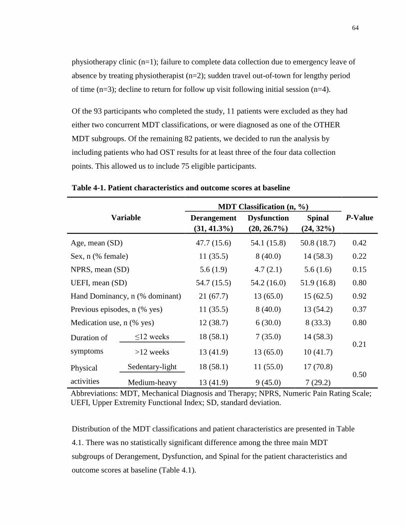
64
physiotherapy clinic (n=1); failure to complete data collection due to emergency leave of
absence by treating physiotherapist (n=2); sudden travel out-of-town for lengthy period
of time (n=3); decline to return for follow up visit following initial session (n=4).
Of the 93 participants who completed the study, 11 patients were excluded as they had
either two concurrent MDT classifications, or were diagnosed as one of the OTHER
MDT subgroups. Of the remaining 82 patients, we decided to run the analysis by
including patients who had OST results for at least three of the four data collection
points. This allowed us to include 75 eligible participants.
Table 4-1. Patient characteristics and outcome scores at baseline
Variable
MDT Classification (n, %)
P-Value Derangement
(31, 41.3%)
Dysfunction
(20, 26.7%)
Spinal
(24, 32%)
Age, mean (SD) 47.7 (15.6) 54.1 (15.8) 50.8 (18.7) 0.42
Sex, n (% female) 11 (35.5) 8 (40.0) 14 (58.3) 0.22
NPRS, mean (SD) 5.6 (1.9) 4.7 (2.1) 5.6 (1.6) 0.15
UEFI, mean (SD) 54.7 (15.5) 54.2 (16.0) 51.9 (16.8) 0.80
Hand Dominancy, n (% dominant) 21 (67.7) 13 (65.0) 15 (62.5) 0.92
Previous episodes, n (% yes) 11 (35.5) 8 (40.0) 13 (54.2) 0.37
Medication use, n (% yes) 12 (38.7) 6 (30.0) 8 (33.3) 0.80
Duration of
symptoms
≤12 weeks 18 (58.1) 7 (35.0) 14 (58.3) 0.21
>12 weeks 13 (41.9) 13 (65.0) 10 (41.7)
Physical
activities
Sedentary-light 18 (58.1) 11 (55.0) 17 (70.8) 0.50
Medium-heavy 13 (41.9) 9 (45.0) 7 (29.2)
Abbreviations: MDT, Mechanical Diagnosis and Therapy; NPRS, Numeric Pain Rating Scale;
UEFI, Upper Extremity Functional Index; SD, standard deviation.
Distribution of the MDT classifications and patient characteristics are presented in Table
4.1. There was no statistically significant difference among the three main MDT
subgroups of Derangement, Dysfunction, and Spinal for the patient characteristics and
outcome scores at baseline (Table 4.1).
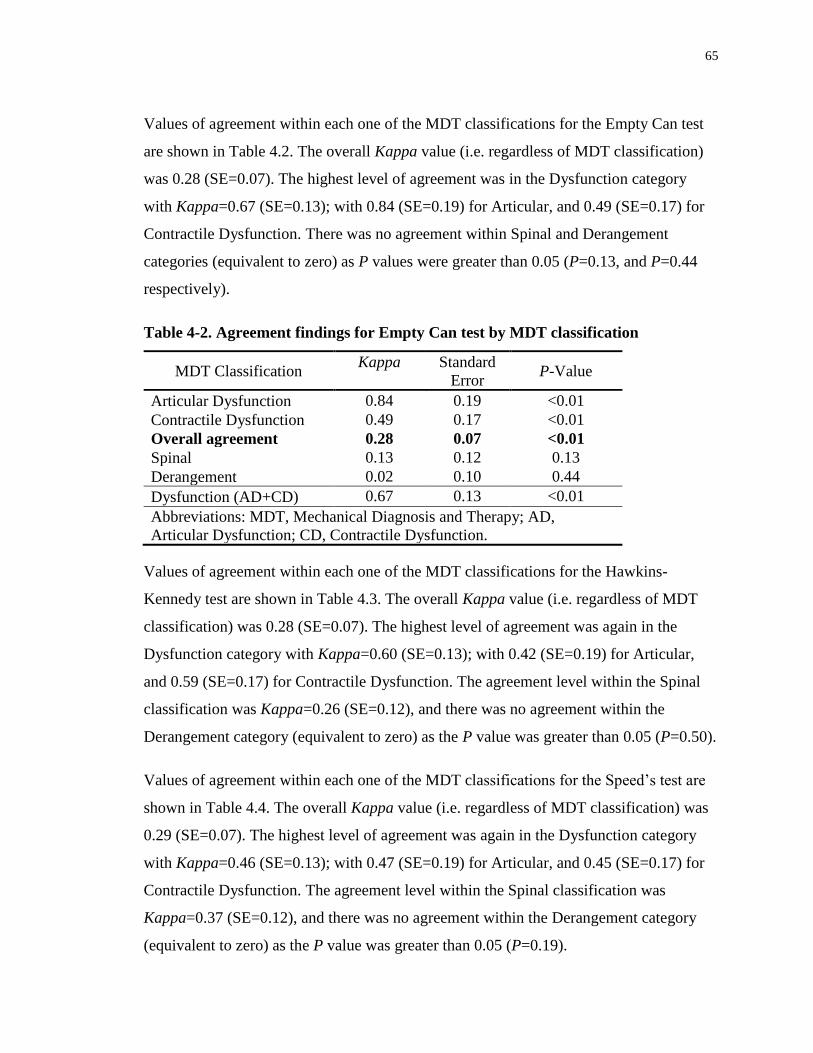
65
Values of agreement within each one of the MDT classifications for the Empty Can test
are shown in Table 4.2. The overall Kappa value (i.e. regardless of MDT classification)
was 0.28 (SE=0.07). The highest level of agreement was in the Dysfunction category
with Kappa=0.67 (SE=0.13); with 0.84 (SE=0.19) for Articular, and 0.49 (SE=0.17) for
Contractile Dysfunction. There was no agreement within Spinal and Derangement
categories (equivalent to zero) as P values were greater than 0.05 (P=0.13, and P=0.44
respectively).
Table 4-2. Agreement findings for Empty Can test by MDT classification
MDT Classification Kappa Standard
Error P-Value
Articular Dysfunction 0.84 0.19 <0.01
Contractile Dysfunction 0.49 0.17 <0.01
Overall agreement 0.28 0.07 <0.01
Spinal 0.13 0.12 0.13
Derangement 0.02 0.10 0.44
Dysfunction (AD+CD) 0.67 0.13 <0.01
Abbreviations: MDT, Mechanical Diagnosis and Therapy; AD,
Articular Dysfunction; CD, Contractile Dysfunction.
Values of agreement within each one of the MDT classifications for the Hawkins-
Kennedy test are shown in Table 4.3. The overall Kappa value (i.e. regardless of MDT
classification) was 0.28 (SE=0.07). The highest level of agreement was again in the
Dysfunction category with Kappa=0.60 (SE=0.13); with 0.42 (SE=0.19) for Articular,
and 0.59 (SE=0.17) for Contractile Dysfunction. The agreement level within the Spinal
classification was Kappa=0.26 (SE=0.12), and there was no agreement within the
Derangement category (equivalent to zero) as the P value was greater than 0.05 (P=0.50).
Values of agreement within each one of the MDT classifications for the Speed’s test are
shown in Table 4.4. The overall Kappa value (i.e. regardless of MDT classification) was
0.29 (SE=0.07). The highest level of agreement was again in the Dysfunction category
with Kappa=0.46 (SE=0.13); with 0.47 (SE=0.19) for Articular, and 0.45 (SE=0.17) for
Contractile Dysfunction. The agreement level within the Spinal classification was
Kappa=0.37 (SE=0.12), and there was no agreement within the Derangement category
(equivalent to zero) as the P value was greater than 0.05 (P=0.19).
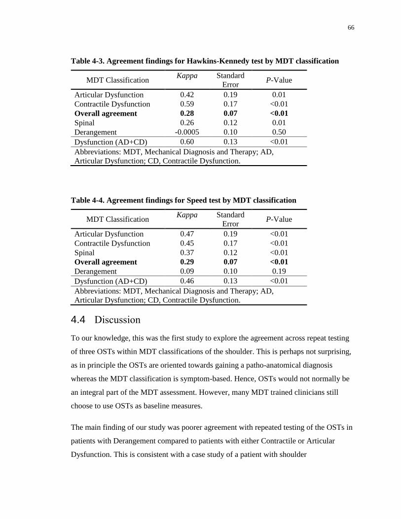
66
Table 4-3. Agreement findings for Hawkins-Kennedy test by MDT classification
MDT Classification Kappa Standard
Error P-Value
Articular Dysfunction 0.42 0.19 0.01
Contractile Dysfunction 0.59 0.17 <0.01
Overall agreement 0.28 0.07 <0.01
Spinal 0.26 0.12 0.01
Derangement -0.0005 0.10 0.50
Dysfunction (AD+CD) 0.60 0.13 <0.01
Abbreviations: MDT, Mechanical Diagnosis and Therapy; AD,
Articular Dysfunction; CD, Contractile Dysfunction.
Table 4-4. Agreement findings for Speed test by MDT classification
MDT Classification Kappa Standard
Error P-Value
Articular Dysfunction 0.47 0.19 <0.01
Contractile Dysfunction 0.45 0.17 <0.01
Spinal 0.37 0.12 <0.01
Overall agreement 0.29 0.07 <0.01
Derangement 0.09 0.10 0.19
Dysfunction (AD+CD) 0.46 0.13 <0.01
Abbreviations: MDT, Mechanical Diagnosis and Therapy; AD,
Articular Dysfunction; CD, Contractile Dysfunction.
4.4 Discussion
To our knowledge, this was the first study to explore the agreement across repeat testing
of three OSTs within MDT classifications of the shoulder. This is perhaps not surprising,
as in principle the OSTs are oriented towards gaining a patho-anatomical diagnosis
whereas the MDT classification is symptom-based. Hence, OSTs would not normally be
an integral part of the MDT assessment. However, many MDT trained clinicians still
choose to use OSTs as baseline measures.
The main finding of our study was poorer agreement with repeated testing of the OSTs in
patients with Derangement compared to patients with either Contractile or Articular
Dysfunction. This is consistent with a case study of a patient with shoulder

67
Derangement68 that reported test results for the Empty Can, Lift off, and Hawkins-
Kennedy tests during a standard MDT assessment and treatment protocol. These tests
changed from positive to negative during the initial treatment session and remained
negative until discharge. This inconsistency of the OSTs has been a frequent observation
by MDT practitioners among patients with Derangement. Specifically, what is noted is
that positive OSTs will often become negative as soon as the treatment process is
initiated, hence the assumption in these cases is that the tests were initially false positives
and not truly indicative of the patho-anatomical condition they were being used to
diagnose. In our study, inconsistent test results for OSTs performed in patients assigned
to the Derangement classification were revealed by poor agreement statistics across
repeat testing from the initial assessment through three to four treatment sessions. This
may be due to the variable and quickly changing nature of the Derangement classification
especially as it rapidly responds to intervention. Reproduction of these findings in
another cohort would provide confirmatory evidence that some OST results are impacted
by the nature of the MDT classification.
The overall agreement for Empty Can, Hawkins-Kennedy, and Speed’s tests were almost
identical with a Kappa=0.28 (SE=0.07) for Empty Can and Hawkins-Kennedy tests, and
a Kappa=0.29 (SE=0.07) for Speed’s test. However, as shown in tables 4.2 to 4.4 when
values for Derangement and Spinal (a cervical spine Derangement) were removed from
the analyses, the agreement level increased dramatically with Kappa values of 0.67
(SE=0.13), 0.60 (SE=0.13), and 0.46 (SE=0.13) for Empty Can, Hawkins-Kennedy, and
Speed’s tests respectively. Furthermore, P-values for the Derangement classification
were greater than 0.05 for all the three OSTs studied. The P-value was similarly greater
than 0.05 for the Spinal classification for the Empty Can test. This indicates that the
agreement was no greater than zero for the above listed analyses, while agreement varied
between moderate-to-good for either Dysfunction classification when the Derangement
and Spinal categories were eliminated from the analyses. In the case of Articular
Dysfunction for the Empty Can test, the agreement was the highest with Kappa= 0.84
(SE=0.19) which indicates a very good agreement.

68
The low agreement or no agreement with repeated testing of the OSTs in patients with
Derangement classification, including spinal Derangements, may be due to the variable
and quickly changing nature of the classification especially as it rapidly responds to
intervention. Therefore, the presence of Derangement may explain the poor consistency
recorded for the majority of the OSTs and was certainly responsible for reducing the
overall agreement in the OSTs used in this study. These results would give additional
support for the position taken that clinicians should not rely on these OSTs as diagnostic
and prognostic tools.3-4, 6, 15 However, there is a clear difference in their consistency in the
presence of a Derangement as compared to when Derangements were absent. A rationale
could be made for an initial MDT screening of shoulder patients to ensure that shoulder
and cervical Derangements have been ruled out before any other testing is performed.
This may then enhance the value of the OSTs and perhaps lead to their improved
diagnostic capability, if indeed a patho-anatomical diagnosis is still sought.
Alternatively, these OSTs could be used as baseline tests in the differentiation between
the MDT classifications of Derangement and Articular and Contractile Dysfunctions. If
the OSTs change from positive before, to negative after a repeated movement exam or
the initiation of treatment then this would be consistent with a Derangement being
present.
The major limitations of this study were as follows: As a “treatment-as-usual” approach
was followed, a pre-determined number of treatment sessions was not feasible for each
one of our patients. Thus, it is possible that the study participants received a variable
number of treatment sessions, potentially influencing treatment results. However, the
treating physiotherapists were unaware of the study objectives, minimizing any
inclination to influence the outcome of each classification category. In addition, a second
practitioner, blinded to the patients’ MDT classifications administered the OSTs to avoid
any potential bias from the treating clinician. A second limitation due to following a
“treatment as usual” approach was that some patients did not have their data available for
all four data collection points; therefore, analysis was done on data from three data
collection points to avoid weakening power of our analysis. A third limitation of the

69
study was that only three OSTs were evaluated in the study as it was not feasible to
include all the numerous OSTs used for shoulder assessment. Therefore, no
extrapolations is made to other OSTs not investigated in the current study. Finally, the
MDT method was followed; therefore, the study results may not be generalizable to other
methods of practice.
As a next step, future studies could investigate other OSTs utilized for shoulder
assessment, and use a pre-set and equal number of treatment sessions for all patients so
that data would be available for all data collection points. Due to the presence of a clear
pattern in our findings indicating that the Derangement classification could be the reason
for inconsistent OST results, further investigations are warranted on the OSTs utilized in
the assessment of other musculoskeletal disorders in both spinal and peripheral
conditions.
In conclusion, due to the ability of the Derangement classification to change rapidly, it
clearly has the capacity to compromise the reliability of OSTs potentially reducing their
clinical utility. Thus, being aware of this characteristic of Derangement prior to the use of
these shoulder OSTs could assist clinicians in their interpretation of the test results.

70
4.5 References
1. Winters JC, Sobel JS, van der Windt DAWM, et al. NHG Standaard
Schouderklachten (versie 1999) Guidelines for Shoulder Complaints of the Dutch
College of General Practitioners (version 1999). Huisarts en Wetenschap.
1999;42:222e31.
2. Croft P, Pope D, Silman A. The clinical course of shoulder pain: prospective cohort
study in primary care. Primary Care Rheumatology Society Shoulder Study Group.
BMJ. 1996;313(7057):601-2.
3. Schellingerhout JM, Verhagen AP, Thomas S, Koes BW. Lack of uniformity in
diagnostic labeling of shoulder pain: Time for a different approach. Man Ther.
2008;13:478-83.
4. Lange T, Matthijs O, Jain NB, Schmitt J, Lutzner J, Kopkow C. Reliability of specific
physical examination tests for the diagnosis of shoulder pathologies: a systematic
review and meta-analysis. Br J Sports Med. 2017;51(6):511-18.
5. McClure PW, Michener LA. Staged approach for rehabilitation classification:
Shoulder disorders (STAR-Shoulder). Phys Ther. 2015;95:791-800.
6. Cook C. The lost art of the clinical examination: an overemphasis on clinical special
tests. J Man Manip Ther. 2010;18 (1):3-4.
7. Sciascia AD, Spigelman T, Kibler WB, et al. Frequency of use of clinical shoulder
examination tests by experienced shoulder surgeons. J Athl Train. 2012;47:457-66.
8. Hegedus EJ, Goode A, Campbell S, Morin A, Tamaddoni M, Moorman CT. Physical
examination tests of the shoulder: a systematic review with meta-analysis of
individual tests. Br J Sports Med. 2008;42:80-92.

71
9. Kuhn JE, Dunn WR, Ma B, Wright RW, Jones G, Spencer EE, Wolf B, Safran M,
Spinder KP, McCarty E, Kelly B, Holloway B. Interobserver agreement in the
classification of rotator cuff tears. Am J Sport Med. 2007;35(3):437-41.
10. Dessaur WA, Magarey ME. Diagnostic accuracy of clinical tests for superior labral
anterior posterior lesions: A Systematic Review. J Orthop Sports Phys Ther.
2008;38(6):341-52.
11. Hughes P, Taylor NF, Green RA. Most clinical tests cannot accurately diagnose
rotator cuff pathology: a systematic review. Aust J Physiother. 2008;54:159-70.
12. Powell JW, Huijbregts PA, Jensen R. Diagnostic utility of clinical tests for SLAP
lesions: A systematic literature review. J Man Manip Ther. 2008;16(5):E58-E79.
13. Walsworth MK, Doukas WC, Murphy KP, Mielcarek BJ, Michener LA. Reliability
and diagnostic accuracy of history and physical examination for diagnosing glenoid.
Am J Sport Med. 2008;36(1):162-68.
14. Walton DM, Sadi J. Identifying SLAP lesions: A meta-analysis of clinical tests and
exercise in clinical reasoning. Phys Ther Sport. 2008;9:167-76.
15. Beaudreuil J, Nizard R, Thomas T, Peyre M, Liotard JP, Boileau P, Marc T, Dromard
C, Steyer E, Bardin T, Orcel P, Walch G. Contribution of clinical tests to the
diagnosis of rotator cuff disease: A systematic review. Joint Bone Spine 2009;76:15-
19.
16. Lewis JS. Rotator cuff tendinopathy/subacromial impingement syndrome: is it time
for a new method of assessment? Br J Sports Med. 2009;43:259-64.
17. Meserve BB, Cleland JA, Boucher TR. A meta-analysis examining clinical test utility
for assessing superior labral anterior posterior lesions. Am J Sport Med.
2009;37(11):2252-58.
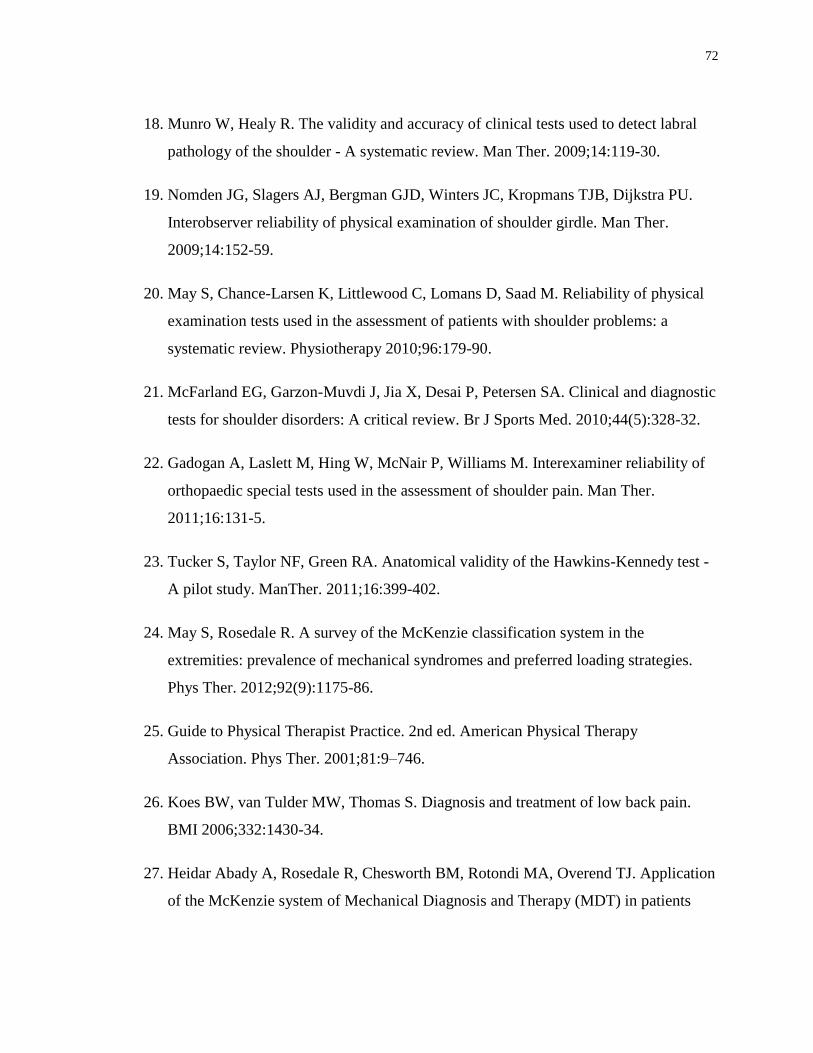
72
18. Munro W, Healy R. The validity and accuracy of clinical tests used to detect labral
pathology of the shoulder - A systematic review. Man Ther. 2009;14:119-30.
19. Nomden JG, Slagers AJ, Bergman GJD, Winters JC, Kropmans TJB, Dijkstra PU.
Interobserver reliability of physical examination of shoulder girdle. Man Ther.
2009;14:152-59.
20. May S, Chance-Larsen K, Littlewood C, Lomans D, Saad M. Reliability of physical
examination tests used in the assessment of patients with shoulder problems: a
systematic review. Physiotherapy 2010;96:179-90.
21. McFarland EG, Garzon-Muvdi J, Jia X, Desai P, Petersen SA. Clinical and diagnostic
tests for shoulder disorders: A critical review. Br J Sports Med. 2010;44(5):328-32.
22. Gadogan A, Laslett M, Hing W, McNair P, Williams M. Interexaminer reliability of
orthopaedic special tests used in the assessment of shoulder pain. Man Ther.
2011;16:131-5.
23. Tucker S, Taylor NF, Green RA. Anatomical validity of the Hawkins-Kennedy test -
A pilot study. ManTher. 2011;16:399-402.
24. May S, Rosedale R. A survey of the McKenzie classification system in the
extremities: prevalence of mechanical syndromes and preferred loading strategies.
Phys Ther. 2012;92(9):1175-86.
25. Guide to Physical Therapist Practice. 2nd ed. American Physical Therapy
Association. Phys Ther. 2001;81:9–746.
26. Koes BW, van Tulder MW, Thomas S. Diagnosis and treatment of low back pain.
BMI 2006;332:1430-34.
27. Heidar Abady A, Rosedale R, Chesworth BM, Rotondi MA, Overend TJ. Application
of the McKenzie system of Mechanical Diagnosis and Therapy (MDT) in patients

73
with shoulder pain; a prospective longitudinal study. J Man Manip Ther. 2017. DOI
10.1080/10669817.2017.1313929.
28. McKenzie RA. The lumbar spine. Mechanical Diagnosis and Therapy. Waikanae,
New Zealand: Spinal Publications; 1981.
29. May S. The McKenzie classification system in extremities: A reliability study using
McKenzie assessment forms and experienced clinicians. J Manip Physiol Ther.
2009;32(7):556-63.
30. Clare HA, Adams R, Maher CG. A systematic review of efficacy of McKenzie
therapy for spinal pain. Aust J Physiother. 2004;50:209-16.
31. Cook C, Hegedus EJ, Ramey K. Physical therapy exercise intervention based on
classification using the patient response method: a systematic review of the literature.
J Man Manip Ther. 2005;13:152-62.
32. Machado LAC, de Souza MvS, Ferreira PH, Ferreira ML. The McKenzie method for
low back pain. A systematic review of the literature with a meta-analysis approach.
Spine 2006;31:E254-62.
33. Hettinga DM, Jackson A, Klaber Moffett J, May S, Mercer C, Woby SR. A
systematic review and synthesis of higher quality evidence of the effectiveness of
exercise interventions for nonspecific low back pain of at least 6 weeks duration.
Phys Ther Rev. 2007;12:221-32.
34. Slade SC, Keating J. Unloaded movement facilitation exercise compared to no
exercise or alternative therapy on outcomes for people with non-specific chronic low
back pain: a systematic review. J Manip Physiol Ther. 2007;30:301-11.
35. May S, Donelson R. Evidence-informed management of chronic low back pain with
the McKenzie method. Spine J. 2008;8:134-41.

74
36. Aina A, May S, Clare H. The centralization phenomenon of spinal symptoms—a
systematic review. Man Ther. 2004;9:134-43.
37. May S, Littlewood C, Bishop A. Reliability of procedures used in the physical
examination of non-specific low back pain: a systematic review. Aust J Physiother.
2006;52:91-102.
38. Razmjou H, Kramer JF, Yamada R. Intertester reliability of the McKenzie evaluation
in assessing patients with mechanical low back pain. J Orthop Sports Phys Ther.
2000;30:368-89.
39. Kilpikoski S, Airaksinen O, Kankaanpaa M, Leminen P, Videman T, Alen M.
Interexaminer reliability of low back pain assessment using the McKenzie method.
Spine. 2002;27:207-14.
40. Clare HA, PT, Adams R, Maher CG. Reliability of McKenzie classification of
patients with cervical or lumbar pain. J Manip Physiol Ther. 2005;28:122-7.
41. Dionne CP, Bybee RF, Tomaka J. Inter-rater reliability of McKenzie assessment in
patients with neck pain. Physiotherapy. 2006;92:75-82.
42. Werneke M, Hart DL, Deutscher D, Stratford PW. Clinician’s ability to identify neck
and low back interventions: an inter-rater chance corrected agreement pilot study. J
Man Manip Ther. 2011;19:172-81.
43. Long A. The centralisation phenomenon. Its usefulness as a predictor of outcome in
conservative treatment of chronic low back pain. Spine. 1995;20:2513-21.
44. Sufka A, Hauger B, Trenary M, et al. Centralisation of low back pain and perceived
functional outcome. J Orthop Sports Phys Ther. 1998;27:205-12.
45. Werneke M, Hart DL, Cook D. A descriptive study of the centralization phenomenon.
A prospective analysis. Spine. 1999;24:676-83.
75
46. Werneke M, Hart DL. Centralisation phenomenon as a prognostic factor for chronic
pain or disability. Spine. 2001;26:758-65.
47. Wetzel FT, MD, Donelson R. The role of repeated end-range/pain response
assessment in the management of symptomatic lumbar discs. Spine J. 2003;3:146-54.
48. Berthelot JM, Delecrin J, Maugars Y, Passuti N. Contribution of centralization
phenomenon to the diagnosis, prognosis, and treatment of diskogenic low back pain.
Joint Bone Spine. 2007;74:319-23.
49. Long A, May S, Fung T. The comparative prognostic value of directional preference
and centralization: a useful tool for front-line clinicians? J Man Manip Ther.
2008;16:248-54.
50. Werneke M, Hart DL, Cutrone G, Oliver D, McGill M, Weinberg J, Grisby D,
Oswald W, Ward J. Association between directional preference and centralization in
patients with low back pain. J Orthop Sports Phys Ther. 2011;41(1):22-31.
51. Takasaki H, May S. Mechanical diagnosis and therapy has similar effects on pain and
disability as 'wait and see' and other approaches in people with neck pain: a
systematic review. J Physiother. 2014;60(2):78-84.
52. Petersen T, Larsen K, Nordsteen J, Olsen S, Fournier G, Jacobsen S. The McKenzie
method compared with manipulation when used adjunctive to information and advice
in low back pain patients presenting with centralization or peripheralization: a
randomized controlled trial. Spine, 2011;36(24):1999-2010.
53. Machado LA, Maher CG, Herbert RD, Clare H, McAuley JH The effectiveness of the
McKenzie method in addition to first-line care for acute low back pain: a randomized
controlled trial. BMC Med. 2010;8:10.
54. Aina A, May S. A shoulder derangement. Man Ther. 2005;10:159-63.

76
55. May S. Classification by McKenzie mechanical syndromes; Report on directional
preference and extremity patients. Int J Mech Diagn Ther. 2006;1(3):7-11.
56. Kelly E, May S, Ross J. The reliability of the McKenzie classification system using
extremity McKenzie assessment forms. Int J Mech Diagn Ther 2008;3:3-6.
57. May S, Ross J. The McKenzie classification system in the extremities: A reliability
study using McKenzie assessment forms and experienced clinicians. J Manipulative
Physiol Ther. 2009;32:556-63.
58. Shouta K, Hiroshi T, May S. Application of Mechanical Diagnosis and Therapy to a
patient diagnosed with a de Quervain’s disease: A case study. J Hand Ther.
2009;22:278-84.
59. Scott A, Huisman E, Khan K, Conservative treatment of chronic Achilles
tendinopathy. Can Med Assoc J. 2011;183.1159-65.
60. Littlewood C, Ashton J, Chance-Larsen K, May S, Sturrock B. Exercise for rotator
cuff tendinopathy: a systematic review. Physiotherapy. 2012;98:101-9.
61. Heidar Abady A, Rosedale R, Overend TJ, Chesworth BM, Rotondi MA. Inter-
examiner reliability of diplomats in the Mechanical Diagnosis and Therapy system in
assessing patients with shoulder pain. J Man Manip Ther. 2014;22(4):199–205.
62. Rosedale R, Rastogi R, May S, Chesworth BM, Filice F, Willis S, Howard J, Naudie
D, Robbins SM., Efficacy of exercise intervention as determined by the McKenzie
system of Mechanical Diagnosis and Therapy for knee osteoarthritis: a randomized
controlled trial., J Orth Sports Phys Ther. 2014;44:3:173-81.
63. Willis S, Rosedale R, Rastogi R, Robbins S, Inter-rater reliability of the McKenzie
System of Mechanical Diagnosis and Therapy in the examination of the knee. J Man
Manip Ther. 2017;25(2):83-90.
77
64. Takasaki H, Okuyama K, Rosedale R. Inter-examiner classification reliability of
Mechanical Diagnosis and Therapy for extremity problems - Systematic review.
Musculoskelet Sci Pract. 2017;27:78-84.
65. Takasaki H. Agreement of Mechanical Diagnosis and Therapy classification in
people with extremity conditions. Phys Ther. 2016;96(10):1525-53.
66. McKenzie RA, May S. The Human Extremities: Mechanical Diagnosis and Therapy.
Waikanae, New Zealand: Spinal Publications, 2000.
67. Kaneko S, Takasaki H, May S, Application of mechanical diagnosis and therapy to a
patient diagnosed with de Quervain's disease: a case study. J Hand Ther.
2009;22:278-84.
68. Kidd J. Treatment of shoulder pain utilizing mechanical diagnosis and therapy
principles. J Man Manip Ther. 2013;21(3):168-73.
69. Lynch G, May S, Directional preference at the knee: a case report using mechanical
diagnosis and therapy. J Man Manip Ther. 2013;21(1):60-6.
70. Rotondi MA, Donner A. A confidence interval approach to sample size estimation for
studies of interobserver agreement with multiple raters and outcome. J Clin
Epidemiol. 2012 Jul;65(7):778-84.
71. Cook CE, Hegedus EJ. Orthpedic Physical Examination Tests: An Evidence-Based
Approach. Upper Saddle River, NJ: Pearson Prentice Hal; 2008.
72. Magee DJ. Orthopedic Physical Assessment: 5th Edition. St. Louis, MO: Saunders
Elsevier; 2008.
73. Stratford PW, Binkley, JM, Stratford DM. Development and initial validation of the
upper extremity functional index. Physiother Can. 2001;53(4):259-67.
74. Jensen MP, Karoly P, Braver S. The measurement of clinical pain intensity: a
comparison of six methods. Pain. 1986;27(1):117-26.

78
75. Streiner DL, Norman GR, Health measurement scales. A Practical Guide to Their
Development and Use. 3rd ed. Oxford: Oxford University Press; 2003.

79
Chapter 5
5 General Discussion and Future Direction
5.1 Overview of thesis
The overall objective of this thesis was to examine the usefulness of the McKenzie
system of Mechanical Diagnosis and Therapy (MDT) in patients with extremity
musculoskeletal disorders, with a focus on the shoulder. Due to a high prevalence of
shoulder pain in the general population,1, 2 and to fill the current gap in assessment and
treatment of patients with shoulder problems, the population of interest in this thesis was
individuals with shoulder musculoskeletal disorders. The complexity of the shoulder, and
absence of uniformity of diagnostic labeling3 ordinarily used in clinical practice, makes it
a great challenge for practitioners to make a correct diagnosis of the underlying source of
pain. Without an accurate diagnosis, treatment is anticipated to be more arbitrary and this
may contribute to the lack of efficacy for most conventional interventions.4 To our
knowledge, studies included in this thesis were the first to investigate the application of
the MDT system exclusively in patients with shoulder disorders. The thesis assessed the
inter-examiner reliability of the MDT system when evaluating patients with shoulder pain
using clinical vignettes. We also examined the clinical application of the MDT system
when treating patients with shoulder disorders and provided insight into whether the
system is applicable for a significant proportion of these patients. Finally, this thesis also
evaluated the consistency of three commonly used Orthopedic Special Tests (OSTs) of
the shoulder when used with the MDT classification.
This thesis demonstrated a “very good” level of inter-examiner agreement between
McKenzie practitioners when classifying patients with shoulder pain using the MDT
system (kappa=0.90). The results support the findings of previous reliability studies on
the application of the MDT system in the extremities,5, 6 suggesting that the MDT system
is a reliable method for classifying patients with extremity musculoskeletal disorders.
In the next phase of this thesis the focus was on verifying whether the response of
pain and function to MDT treatment varies by MDT classification category over the
80
course of treatment. We also looked at the distribution of discharge frequency over
time for each MDT classification category in response to treatment. The results
suggested that the MDT classification in patients with shoulder pain can impact
treatment outcomes and the frequency of discharge. As hypothesized, Derangement
and Spinal categories had quicker responses to their treatments with a higher rate of
early discharge from treatment in comparison to patients in the articular or
contractile classifications. Thus, the treatment outcomes are aligned with the
response expectation of the MDT classification. The study also demonstrated that
the distribution of patients with Derangement and Spinal categories together make
up over two-third of patients with shoulder pain, reinforcing the importance of
quick response times for Derangement and Spinal categories.
As a final step, we explored whether the shoulder MDT classification and its subsequent
treatment affects the consistency of test results for three commonly used shoulder OSTs.
Our main interest was to determine whether the interpretation of the OSTs changes in the
Derangement category. The study revealed poor overall agreement for the Empty Can,
Hawkins-Kennedy and Speed’s tests; however, the agreement was moderate to very good
in patients with articular and contractile Dysfunctions with kappa ranging between 0.42
for the Hawkins-Kennedy test to 0.84 for the Empty Can test while there was no
agreement for any of the OSTs in patients from the shoulder Derangement category, and
for the Empty Can test in patients from the spinal Derangement category (P values >
0.05). The agreement was poor for the Hawkins-Kennedy and Speed’s tests in patients
with spinal classification with kappa values of 0.26 and 0.37 respectively. This poor
agreement may be due to the rapidly changing nature of patients in the Derangement
classifications. Thus, patients in the Derangement category were responsible for reducing
the overall agreement of the OSTs explaining the poor consistency for the OSTs.
5.2 Implications of thesis findings on practice, and future
research
In chapter 2, we found a “very good” level of agreement among the MDT practitioners in
classifying patients with shoulder disorders using clinical vignettes. The results reinforce

81
the findings of previous reliability studies conducted on clinical cases with spinal7-13 and
extremity5, 6 musculoskeletal disorders. There have been three additional reliability
studies on the extremities14-16 and one systematic review17 since the current thesis work.
All but one of the reliability studies on the extremities were vignette-based studies
suggesting a strong evidence for reliability of the MDT classification system in patients
with extremity disorders. Takasaki14 conducted an inter-examiner reliability study
including 33 patients. He reported that the inter-examiner agreement for provisional
MDT classification was “good” when the examiners were seeing the same patients
concurrently but “poor” when the patients were seen successively. The poor agreement
could be due to the fact that during the first examiner’s assessment, the response of the
clinical problem to the examination procedure may be altered and hence present
somewhat differently during the subsequent assessment. This can occur especially with
Derangement syndrome which is known to have rapid changes to end range movements
performed during an assessment. Thus there might be inconsistent patient presentations
between raters when they are rated successively, leading to “poor” agreement.
Having considered the available literature, there is strong evidence supporting inter-
examiner reliability of the MDT system when used with patients with extremity problems
suggesting that this system could be reliably used in classifying the extremity patient
population. Additional studies may be needed considering the use of real patients instead
of written vignettes to further establish reliability of the MDT system in patients with
musculoskeletal disorders of the extremities.
In chapter 3, we demonstrated when MDT-trained clinicians match the intervention to a
specific MDT classification, the outcome is aligned with the response expectation of the
classification. Patients in the Derangement and Spinal categories make up a great
majority of the patient population with shoulder disorders, and they showed very similar
pain and functional responses to treatment. We demonstrated significantly greater
improvement in comparison to patients with Dysfunction; therefore, compared to patients
with Dysfunction, a high percentage of patients with Derangement and Spinal
classifications achieved their treatment goals relatively quickly and were discharged from
82
treatment. This reinforces the point that the Spinal extremity classification is in fact a
cervical spine Derangement and like the shoulder Derangement, this classification is
anticipated to demonstrate a rapid treatment response. In comparison, Dysfunction
respond in a more graduated manner, achieving discharge status at a later point. The
results suggest that knowing the MDT classifications for patients with a shoulder problem
can provide clinicians with valuable information on prognosis, which is one of the key
questions patients have for their clinicians. To further investigate clinical application of
the MDT system in patients with shoulder disorders, randomized controlled trials are
needed to compare the MDT system with conventional treatment for this patient
population.
In chapter 4, we demonstrated that there is poorer agreement between the OSTs in
patients with Derangement, (including Spinal Derangement) compared to patients with
either Contractile or Articular Dysfunction. These Dysfunctions demonstrated acceptable
agreement. The lack of agreement for the OSTs in the Derangement classification may be
due to the variable and quickly changing nature of this category especially because it
rapidly responds to intervention. Therefore, the presence of Derangement may explain
the poor consistency documented for most OSTs. These results reinforce why clinicians
should be cautious when using these OSTs as diagnostic and prognostic tools.3, 18-20 There
was a clear difference in their consistency in the presence of a Derangement as compared
to when Derangements were not included in the agreement calculations. Therefore it may
be that an initial MDT screening of shoulder patients should be used to ensure that
shoulder and cervical Derangements have been ruled out before other testing is
performed. This may then enhance the value of the OSTs and perhaps lead to their
improved diagnostic capability, if indeed a patho-anatomical diagnosis is still sought.
Alternatively, these OSTs could be used as baseline tests to be used in the differentiation
between the MDT classifications of Derangement and Articular and Contractile
Dysfunctions. If the OSTs change from positive before, to negative after a repeated
movement exam or the initiation of treatment, then this would be consistent with a
Derangement being present. As a next step, future studies could further examine our
study objectives by including other OSTs utilized for shoulder evaluation.

83
5.3 Limitations
Although we addressed some current gaps in the literature, there are several limitations to
this thesis. The major limitation of the reliability study was that only MDT diplomats
were included. This reduces the generalizability of the findings of this study, suggesting
that the agreement among clinicians without this level of training may not be as high.
Future studies should include practitioners with different levels of education and
expertise so that the results could be more generalizable. Another limitation of this study
was using written vignettes as opposed to having actual patients. Although using written
vignettes can minimize the potential error generated by inconsistent patient presentations
between raters,13 the concern is the simplification of the intervention being demonstrated
in the vignettes.13 These vignettes may not represent all the complexities and subtleties of
clinical practice, making the diagnosis easier for the raters and inflating the calculated
kappa value.13 Future reliability studies could include real patients instead of written
vignettes to minimize these shortcomings.
The clinical application of the MDT system in the shoulder had the following limitations:
First, due to the use of a “treatment-as-usual” approach, it was not feasible to have a pre-
determined number of treatment sessions for each one of our study participants. Thus, it
is possible that the patients in each classification received a different number of treatment
sessions; this may have influenced the outcomes. However, the treating practitioners had
no awareness of the study objectives, and therefore would have been less likely to
influence the outcome associated with each MDT category. Rather, they treated each
patient as best as they could, considering the clinical presentation. Secondly, exercise
compliance was not closely monitored, thus it is uncertain whether the poorer results in
the Dysfunction group was because of poor exercise compliance or the typical nature of
this MDT classification. Third, and due to the nature of the study design, there was no
control group to receive conventional physiotherapy intervention eliminating the ability
to compare MDT system with other treatment approaches. Fourth, there was no
randomization because the MDT method was selected as the only method of intervention
and the patients were required to be treated within their respective MDT classification

84
groups. As a next step, well-designed randomized controlled trials are warranted to
compare the MDT system with other treatment approaches for patients with shoulder
pain.
The “treatment-as-usual” approach was also a limitation for our third study, however, as
indicated before, the treating physiotherapists were unaware of the study objectives and
therefore less likely to influence the results. A second limitation of this study also
resulted from following a “treatment as usual” approach. As some patients were
discharged from their treatment earlier than the final study target point, they did not have
their data available for all the four data collection points. Therefore, analysis was
conducted on data from three data collection points instead of four. The third limitation
of the study was that only three OSTs were included in the study as it would not be
feasible in a study to include all the numerous OSTs used for shoulder assessment. As a
next step, future studies could further explore other OSTs utilized for shoulder
assessment, and have a pre-set and equal number of treatment sessions for all patients so
that data would be available for all data collection points. Due to the presence of a clear
pattern in our findings indicating that the Derangement classification could be the reason
that the OSTs fail to meet the purpose they are used for, further investigations are
warranted on the OSTs utilized in the assessment of other musculoskeletal disorders in
both spinal and peripheral conditions.
5.4 Potential Bias
Bias is defined as any tendency which prevents unprejudiced consideration of a
question.24 In research, bias can take place at any stage of a project including study
design, data collection, as well as in the process of data analysis and publication.25 Pre-
trial biases may arise from a flawed study design, selection or channeling bias. In our
project, definition of outcome measures and study objectives were clearly defined and
measures were taken to blind study collaborators who collected data from their patients.
As all the participating practitioners were MDT trained, there could been a potential bias
among the practitioners towards inflating the effectiveness of the MDT system, however
as a measure to minimize such bias, a second practitioner different from the treating

85
practitioner, and blinded to the patients’ MDT classifications administered the OSTs to
avoid any potential bias from the treating clinician in registering the test results.
Selection bias was avoided by including all patients with shoulder pain who referred to
physical therapy intervention except for listed exclusion criteria such as recent trauma or
surgery, etc. Although there may seem to be channeling bias by including patients with
rapid response to treatment in Derangement compared to patients with Dysfunction,
however, all group allocation was conducted after a standard MDT assessment by the
treating clinicians having no knowledge of our study objectives.
As a source of bias during a trial, chronology bias occurs when historic controls are used
as a comparison group for patients undergoing an intervention.25 We chose a prospective
study design to avoid such bias. Transfer bias may occur when there is unequal patients
lost to follow-up among study groups. In our study, 12 patients were lost to follow-up
after their initial assessment; therefore, it was not possible to identify what proportion of
them were from different MDT classifications. As a result, these patients were excluded
from our analysis. Performance bias may occur when more experienced practitioners treat
a specific patient population whereas other classifications were treated by less
experienced therapist; however, in our studies consecutive sampling was conducted and
the study collaborators treated mixed number of all MDT classifications.
Bias after a trial's conclusion may take place during data analysis or publication.25
Confounding factors should be considered during analysis if there is a chance of
influencing the results. We looked at multiple confounding factors listed in table 3-2 that
might have had potential influence on the results, but there was no statistically significant
difference among the groups for the considered factors. Citation bias refers to the fact
that researchers and study sponsors are less likely to publish unfavorable results. We
published all our findings and also cited studies with conflicting findings on reliability of
the MDT system published by Takasaki.14,15

86
5.5 Conclusion
Considering the well-described limitations of conventional patho-anatomical models for
diagnosis and management of patients with shoulder complaints,21-23 the MDT system,
may be worthy of further investigation to fill the current gap in the diagnosis and
treatment of the musculoskeletal patient population. One of the promising aspects of our
study results is that two-thirds of our study participants were classified as either a
shoulder Derangement or a cervical Spinal Derangement clinically observed to have a
rapid response to MDT treatment. If future studies confirm that patients diagnosed as
Derangements conform to their expected rapid response to tailored MDT treatments,
there is potential to significantly impact MDT treatment outcomes for a majority of
patients with shoulder problems. In addition, with the effect of both shoulder and spinal
Derangement classification on the agreement of sequentially performed OSTs, it would
seem reasonable to account for this phenomenon in the orthopedic assessment process.
Due to the variable nature of patients in the Derangement category and their ability to
change rapidly, this category clearly has the potential to compromise the reliability of
OSTs and thus reduce their clinical utility. Therefore, screening for patients with
Derangement prior to the use of these three shoulder OSTs may contribute to their
diagnostic capability.

87
5.6 References
1. Winters JC, Sobel JS, van der Windt DAWM, et al. NHG Standaard
Schouderklachten (versie 1999) Guidelines for shoulder Complaints of the Dutch
College of General Practitioners (version 1999). Huisarts en Wetenschap
1999;42:222e31.
2. Luime JJ, Koes BW, Hendriksen IJ, et al. Prevalence and incidence of shoulder pain
in the general population: a systematic review. Scand J Rheumatol. 2004;33:73–81.
3. Schellingerhout JM, Verhagen AP, Thomas S, Koes BW. Lack of uniformity in
diagnostic labeling of shoulder pain: Time for a different approach. Man Ther.
2008;13:478-83.
4. Green S, Buchbinder R, Glazier R, Forbes A. Systematic review of randomised
controlled trials of interventions for painful shoulder: selection criteria, outcome
assessment, and efficacy. BMJ. 1998;316(7128):354-60.
5. May S, Ross J. The McKenzie classification system in extremities: a reliability study
using McKenzie assessment forms and experienced clinicians. J Manip Physiol Ther.
2009 32(7):556-63.
6. Kelly E, May S, Ross J. The reliability of the McKenzie classification system using
extremity McKenzie assessment forms. Int J Mech Diagn Ther. 2008;3:3-6.
7. Razmjou H, Kramer JF, Yamada R. Intertester reliability of the McKenzie evaluation
in assessing patients with mechanical low back pain. J Orthop Sports Phys Ther.
2000;30:368–89.
8. Fritz JM, Delitto A, Vignovic M, Busse RG. Interrater reliability of judgments of the
centralisation phenomenon and status change during movement testing in patients
with low back pain. Arch Phys Med Rehabil. 2000;81:57–61.

88
9. Kilpikoski S, Airaksinen O, Kankaanpaa M, Leminen P, Videman T, Alen M.
Interexaminer reliability of low back pain assessment using the McKenzie method.
Spine. 2002;27:207–14.
10. Clare HA, PT, Adams R, Maher CG. Reliability of the McKenzie spinal pain
classification using patient assessment forms. Physiotherapy. 2004;90:114-9.
11. Clare HA, PT, Adams R, Maher CG. Reliability of McKenzie classification of
patients with cervical or lumbar pain. J Manip Physiol Ther. 2005;28:122-7.
12. Dionne CP, Bybee RF, Tomaka J. Inter-rater reliability of McKenzie assessment in
patients with neck pain. Physiotherapy. 2006;92:75-82.
13. Werneke MW, Hart DL, Deutscher D, Stratford PW. Clinician’s ability to identify
neck and low back interventions: an inter-rater chance corrected agreement pilot
study. J Man Manip Ther. 2011;19:172-81.
14. Takasaki H. Agreement of Mechanical Diagnosis and Therapy classification in
patients with extremity conditions, Phys Ther. 2016;96(10):1525-32.
15. Takasaki H, Iwasada Y, May S. Attitude towards the use of MDT and reliability of
classification extremity problems by credentialed therapists. J Chiropr Med.
2016;14:32-8.
16. Willis S, Rosedale R, Rastogi R, Robbins SM. Inter-rater reliability of the McKenzie
system of Mechanical Diagnosis and Therapy in the examination of the knee. J Man
Manip Ther. 2017;25(2):83-90.
17. Takasaki H, Okuyama K, Rosedale R. Inter-examiner classification reliability of
Mechanical Diagnosis and Therapy for extremity problems - Systematic review.
Musculoskelet Sci Pract. 2017;27:78-84.
89
18. Lange T, Matthijs O, Jain NB, Schmitt J, Lutzner J, Kopkow C. Reliability of specific
physical examination tests for the diagnosis of shoulder pathologies: a systematic
review and meta-analysis. Br J Sports Med. 2017;51(6):511-8.
19. Cook C. The lost art of the clinical examination: an overemphasis on clinical special
tests. J Man Manip Ther. 2010;18 (1):3–4.
20. Beaudreuil J, Nizard R, Thomas T, Peyre M, Liotard JP, Boileau P, Marc T, Dromard
C, Steyer E, Bardin T, Orcel P, Walch G. Contribution of clinical tests to the
diagnosis of rotator cuff disease: A systematic review. Joint Bone Spine 2009;76:15-
9.
21. Hughes P, Taylor NF, Green RA. Most clinical tests cannot accurately diagnose
rotator cuff pathology: a systematic review. Aust J Physiother. 2008;54:159-70.
22. Gadogan A, Laslett M, Hing W, McNair P, Williams M. Inter-examiner reliability of
orthopaedic special tests used in the assessment of shoulder pain. Man Ther.
2011;16:131-5.
23. Schibany N, Zehetgruber H, Kainberger F, Wuring C, Ba-Ssalamah A, Herneth AM,
Lang T, Gruber D, Breitenseher MJ. Rotator cuff tears in asymptomatic individuals:
aclinical and ultrasonographic screening study. Eur J Radiol. 2004;51:263-8.
24. Dictionary.com; http://dictionary.reference.com/browse/bias.
25. Pannucci CJ, Wilkins EG. Identifying and avoiding bias in research. Plast Reconstr
Surg. 2010;126(2):619-25. doi: 10.1097/PRS.0b013e3181de24bc.

90
Appendices
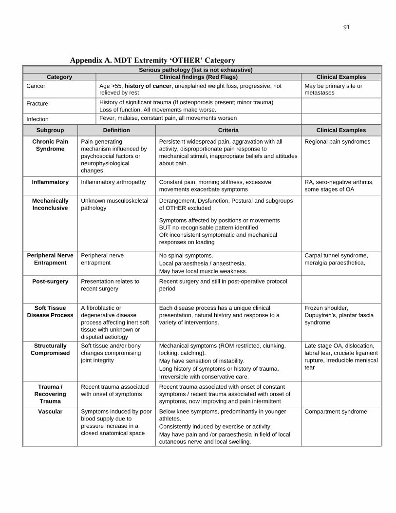
91
Appendix A. MDT Extremity ‘OTHER’ Category
Serious pathology (list is not exhaustive)
Category Clinical findings (Red Flags) Clinical Examples
Cancer Age >55, history of cancer, unexplained weight loss, progressive, not relieved by rest
May be primary site or metastases
Fracture History of significant trauma (If osteoporosis present; minor trauma)
Loss of function. All movements make worse.
Infection Fever, malaise, constant pain, all movements worsen
Subgroup Definition Criteria Clinical Examples
Chronic Pain
Syndrome
Pain-generating
mechanism influenced by
psychosocial factors or
neurophysiological
changes
Persistent widespread pain, aggravation with all
activity, disproportionate pain response to
mechanical stimuli, inappropriate beliefs and attitudes
about pain.
Regional pain syndromes
Inflammatory Inflammatory arthropathy Constant pain, morning stiffness, excessive
movements exacerbate symptoms
RA, sero-negative arthritis,
some stages of OA
Mechanically
Inconclusive
Unknown musculoskeletal
pathology
Derangement, Dysfunction, Postural and subgroups
of OTHER excluded
Symptoms affected by positions or movements
BUT no recognisable pattern identified
OR inconsistent symptomatic and mechanical
responses on loading
Peripheral Nerve
Entrapment
Peripheral nerve
entrapment
No spinal symptoms.
Local paraesthesia / anaesthesia.
May have local muscle weakness.
Carpal tunnel syndrome,
meralgia paraesthetica,
Post-surgery Presentation relates to
recent surgery
Recent surgery and still in post-operative protocol
period
Soft Tissue
Disease Process
A fibroblastic or
degenerative disease
process affecting inert soft
tissue with unknown or
disputed aetiology
Each disease process has a unique clinical
presentation, natural history and response to a
variety of interventions.
Frozen shoulder,
Dupuytren’s, plantar fascia
syndrome
Structurally
Compromised
Soft tissue and/or bony
changes compromising
joint integrity
Mechanical symptoms (ROM restricted, clunking,
locking, catching).
May have sensation of instability.
Long history of symptoms or history of trauma.
Irreversible with conservative care.
Late stage OA, dislocation,
labral tear, cruciate ligament
rupture, irreducible meniscal
tear
Trauma /
Recovering
Trauma
Recent trauma associated
with onset of symptoms
Recent trauma associated with onset of constant
symptoms / recent trauma associated with onset of
symptoms, now improving and pain intermittent
Vascular Symptoms induced by poor
blood supply due to
pressure increase in a
closed anatomical space
Below knee symptoms, predominantly in younger
athletes.
Consistently induced by exercise or activity.
May have pain and /or paraesthesia in field of local
cutaneous nerve and local swelling.
Compartment syndrome

92
Appendix B. Research Ethics Approval-Study 1

93
Appendix C. Research Ethics Approval-Study 2 & 3

94
Appendix D. Numeric Pain Rating Scale (NPRS)
What number on a scale of 0 to 10 would you give to your pain over the past 24 hours?
0 = No Pain
1-3 = Mild Pain (nagging, annoying, interfering little with ADLs)
4–6 = Moderate Pain (interferes significantly with ADLs)
7-10 = Severe Pain (disabling; unable to perform ADLs)
Reference
McCaffery, M., & Beebe, A. (1993). Pain: Clinical Manual for Nursing Practice.
Baltimore: V.V. Mosby Company.
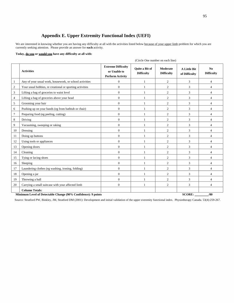
95
Appendix E. Upper Extremity Functional Index (UEFI)
We are interested in knowing whether you are having any difficulty at all with the activities listed below because of your upper limb problem for which you are
currently seeking attention. Please provide an answer for each activity.
Today, do you or would you have any difficulty at all with:
(Circle One number on each line)
Activities
Extreme Difficulty
or Unable to
Perform Activity
Quite a Bit of
Difficulty
Moderate
Difficulty
A Little Bit
of Difficulty
No
Difficulty
1 Any of your usual work, housework, or school activities 0 1 2 3 4
2 Your usual hobbies, re creational or sporting activities 0 1 2 3 4
3 Lifting a bag of groceries to waist level 0 1 2 3 4
4 Lifting a bag of groceries above your head 0 1 2 3 4
5 Grooming your hair 0 1 2 3 4
6 Pushing up on your hands (eg from bathtub or chair) 0 1 2 3 4
7 Preparing food (eg peeling, cutting) 0 1 2 3 4
8 Driving 0 1 2 3 4
9 Vacuuming, sweeping or raking 0 1 2 3 4
10 Dressing 0 1 2 3 4
11 Doing up buttons 0 1 2 3 4
12 Using tools or appliances 0 1 2 3 4
13 Opening doors 0 1 2 3 4
14 Cleaning 0 1 2 3 4
15 Tying or lacing shoes 0 1 2 3 4
16 Sleeping 0 1 2 3 4
17 Laundering clothes (eg washing, ironing, folding) 0 1 2 3 4
18 Opening a jar 0 1 2 3 4
19 Throwing a ball 0 1 2 3 4
20 Carrying a small suitcase with your affected limb 0 1 2 3 4
Column Totals:
Minimum Level of Detectable Change (90% Confidence): 9 points SCORE: /80
Source: Stratford PW, Binkley, JM, Stratford DM (2001): Development and initial validation of the upper extremity functional index. Physiotherapy Canada. 53(4):259-267.

96
Appendix F. Sample Vignettes
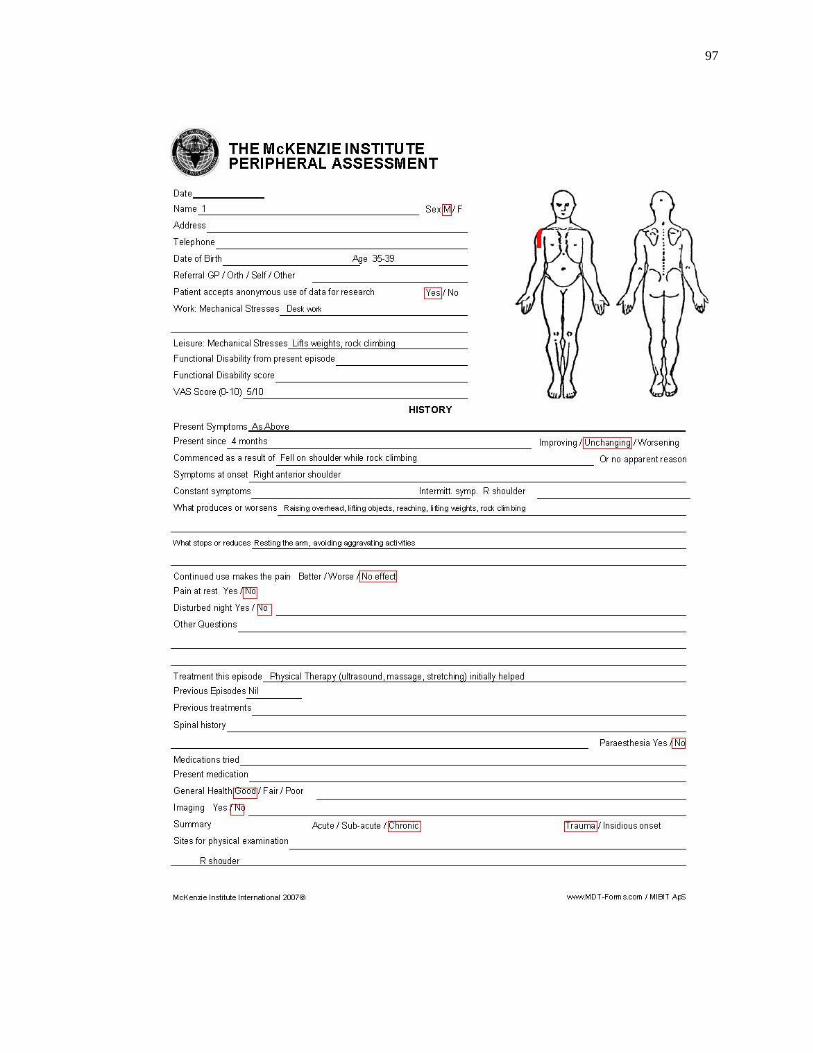
97

98

99
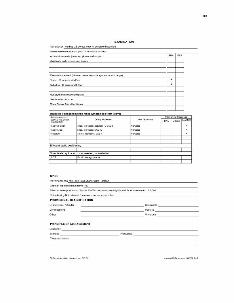
100

101

102

103

104

105

106
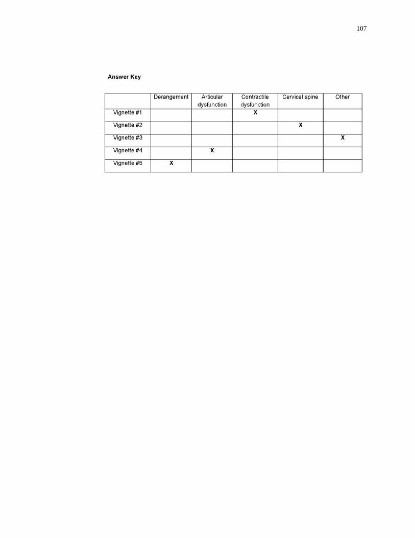
107

108
Curriculum Vitae
Name: Afshin Heidar Abady
Post-secondary Iran University of Medical Sciences
Education and Tehran, Iran
Degrees: 1993-1997, BSc (PT).
Iran University of Medical Sciences
Tehran, Iran
1997-2000, MSc (PT).
The University of Western Ontario
London, Ontario, Canada
2007-2017, PhD (PT).
Honours and Western Graduate Research Scholarships (WGRS), 2007-2012,
Awards: 2011-2012
Related Work Teaching Assistant
Experience The University of Western Ontario
2007-2012
Research Assistant
The University of Western Ontario
2007-2017
Publications: Heidar Abady A, Rosedale R, Chesworth BM, Rotondi MA, Overend TJ. Consistency of
commonly used orthopedic special tests of the shoulder when used with the McKenzie system of
mechanical diagnosis and therapy. Musculoskelet Sci Pract. DOI 10.1016/j.msksp.2017.10.001.
Heidar Abady A, Rosedale R, Chesworth B, Rotondi M, Overend T. Application of the
McKenzie system of Mechanical Diagnosis and Therapy (MDT) in patients with shoulder
pain. J Man Manip Ther. DOI 10.1080/10669817.2017.1313929.
Heidar Abady A, Rosedale R, Overend TJ, Chesworth BM, Rotondi MA. Inter-examiner
reliability of diplomats in the Mechanical Diagnosis and Therapy system in assessing
patients with shoulder pain. J Man Manip Ther. 2014;22(4):199–205.

109
Hill K, Wickerson L, Woon L, Heidar Abady A, Overend T, Goldstein R, Brooks D. The
6-min walk test: responses in healthy Canadians aged 45-85 years. Applied Physiology,
Nutrition & Metabolism, 2011; 36(5): 643-649.
Heidar Abady A, Ebrahimi E, Forough B, Maroufi N. The effects of infra-red low power
laser radiation on sensory and motor electroneurographic parameters of median nerve.
Journal of Iran University of Medical Sciences. 2001; 9(22):262-266.