applications of conventional radiology in the medical ... · 3806 a o c r i t m f f m g o a a g de...
TRANSCRIPT

3805Rev Colomb Radiol. 2013; 24(4): 3805-17
Review Articles
ApplicAtions of conventionAl RAdiology in the MedicAl foRensic fieldAplicAciones de lA RAdiologíA convencionAl en el cAMpo de lA MedicinA foRense
Guillermo Andrés Montes Loaiza1 Andrés Felipe Otálora Daza2
Guillermo Andrés Archila3
SummaryIntroduction: The field of forensics is an unexplored field of radiology in Colombia.
However, it has many applications of great importance. These applications help solve crimes and therefore, assist in the administration of justice. Radiology is applied in forensic medicine in areas such as: study of deaths from air accidents and disasters, documentation of injuries in traffic accidents, medical-legal autopsies in mechanical suffocation deaths, study of deaths associated from accidents with firearm projectiles, diagnosis of child abuse, verifying the authenticity of physical evidence, identification of corpses, examination of fetal deaths, personal injuries, age determination, examination of skeletal remains and virtual autopsy (virtuopsia), among others. Objectives: To present the reader with the most important applications of radiology in the forensic field through illustrations of medical-legal cases obtained from the National Institute of Legal Medicine and Forensic and Academic Sciences at the University Hospital San Ignacio. Materials and Methods: We performed a literature search in PubMed, ScienceDirect and MDConsult, with MESH words and free text from 1985 where 114 articles were obtained. 59 of these articles were selected due to their relevance. Conclusions: Different radiological imaging modalities can be applied in the area of forensics. The radiologist doctor plays a key role in the implementation, interpretation and reporting of radiological studies aimed at solving legal problems for the proper administration of justice.
reSumenIntroducción: La medicina forense es un campo poco explorado por la radiología en
Colombia; sin embargo, la radiología tiene aplicaciones de gran importancia para esclarecer delitos y así apoyar la administración de justicia. Entre estas aplicaciones se encuentran: estudio de muertes por accidentes aéreos y desastres, documentación de lesiones en accidentes de tránsito, necropsias médico-legales en muertes por asfixias mecánicas, estudio de muertes asociadas a heridas por proyectiles de arma de fuego, diagnóstico del maltrato infantil, verificación de la autenticidad de la evidencia física, identificación de cadáveres, examen en muertes fetales, lesiones personales, determinación de edad, examen de restos óseos y necropsia virtual (virtuopsia). Objetivos: Dar a conocer al lector las aplicaciones más importantes de la radiología en el campo forense por medio de ilustraciones obtenidas en casos médico-legales del Instituto Nacional de Medicina Legal y Ciencias Forenses y de académicos del Hospital Universitario de San Ignacio. Materiales y métodos: Se realizó una búsqueda de la literatura en PubMed, ScienceDirect y MDconsult, con palabras MeSH y texto libre a partir de 1985. Se obtuvieron 114 artículos y se seleccionaron 59 por su relevancia. Conclusiones: Distintas modalidades de imagen radiológica pueden ser aplicadas en el área
1MD, radiologist and expert in Forensic Medicine.
Department of Diagnostic Images, San Ignacio Hospital,
Bogotá, Colombia.
2MD, Radiologist. Department of Diagnostic Images, San Ignacio Hospital,
Bogotá, Colombia.
3MD, resident physician in Radiology, Javeriana
University, Bogotá, Colombia.
Key words (meSH)RadiologyForensic medicineCadaver Victims identificationForensic anthropology
Palabras clave (DeCS)RadiologíaMedicina legalCadáver Identificación de víctimasAntropología forense

3806 Applications Of Conventional Radiology In The Medical Forensic Field. Montes G., Otálora A., Archila G.
de la medicina forense. El médico radiólogo tiene un papel fundamental en la ejecución, interpretación y reporte de los estudios radiológicos encaminados a solucionar problemas legales para una adecuada administración de la justicia.
Radiology Applied to IdentificationIdentification of living persons as well as corpses is one
of the requests authorities make to medical examiners, an essential one to criminal investigation (1-3). Identification of the deceased in Colombia requires a medical-legal autopsy, an activity embedded within standard legal procedures as one of the objectives of Decree 786/1990, which regulates autopsy practice in the country.
According to the Code of Criminal Procedure, three of the available methods are considered to be reliable scientific procedu-res for identification: fingerprint analysis, dental record analysis, and genetic profiling.
When reliable identification is impossible through these methods, others may be used to gain insight into it. Such cases may involve radiology as an essential tool throughout the identi-fication process, providing support in the following manners (4):
Identification of Particular SignsIdentification is, from a broad perspective, a comparative
process. Background information is needed to perform compa-risons that lead to a successful identification. As a supporting identification method, radiology requires previous radiographic records, complete clinical records, or a description of particular signs made by people close to the subject under study; these will be contrasted with radiographic studies carried out during medical-legal examinations. Particular signs suitable for circumstantial identification through radiographic studies are: the presence of orthopedic prosthetics, or osteosynthetic material (Figure 1); bone callus formation in ancient fractures (Figure 2); and the presence of anatomical variations such as fused or supernumerary vertebrae (5,6).
Age DeterminationAge is a useful identifying characteristic in an individual. An
age estimate for a living person or a corpse can be provided at a glance; after assessing secondary sex characteristics and anthropo-metric measurements, age determination attains greater accuracy. Radiology assessment, however, is the most accurate provider of the approximate age of an individual under study.
Several age determination radiology techniques exist. The most important are radiographic assessments of carpal bone (7) and dental characteristics (3,8-14) (Figure 3). Other radiographic characteristics useful for estimating an individual’s age are the presence of ossification centers and cranial suture closure.
Other methods such as foot and kneecap radiographic as-sessments have been described as well (15); the relative simplicity of classic techniques mentioned earlier (carpal bone and dental radiography) has relegated these latter techniques to particular cases such as mutilations where wrists are unavailable for carpal bone assessment, or body fragments assessment where the cra-nium and pelvis are unavailable.
Besides being a key element in the identification process, age is an important characteristic that must be established for each individual involved in legal procedures in Colombia. For example, ascertaining whether a person who has committed a crime is of legal age or underage is required to decide if the person should be tried either as an adult or a minor. Another important age threshold in Colombian law is age 14, which indicates a mental maturity capable of decision-making on the person’s own sexual integrity; before that age the person is considered unable to do so, and therefore any sexual activity with a minor younger than 14 years is considered a felony.
Sex DeterminationSex determination through a general physical examination
is relatively simple, particularly in corpses, as an assessment of internal anatomy is possible with genital organ identification. Nonetheless, sex reassignment surgery in living persons may lead to confusions on the matter. Radiology provides insight on this regard (3), contributing to accurate sex determination using cranial and pelvic morphology radiographic assessments. Features such as foramen magnum area and pelvic ring measurements, among others, cast light on the sex of the examined individual.
Conventional radiology is not the only available approach in this field; facing limitations for a physical examination, a compu-terized axial tomography for forensic purposes may be used. This technique enables the “virtual” examination of internal genitalia for sex determination of an individual.
Disaster Victim Identification (3, 16-17) In case of a disaster, a priority objective during autopsies
is victim identification. To this end, the increased probability that examined corpses, victims of the disaster, display multiple fractures or mutilations that render impossible a circumstantial identification makes radiographic studies particularly important, also because performing definite identification techniques may become impossible due to the absence of fingerprints or dental structures. In these cases, radiology is useful in the determination of particular signs in corpses or examined body fragments.
Massive disasters in closed populations, such as a plane crash that has passenger and crew lists, particular signs identified using radiographic studies may become a reliable identification method. For example, if data collection among crew and passenger relatives reveals that a single victim has a medical history that implies the presence of osteosynthetic material, the presence of this material in a corpse’s radiographies will make its identification possible.
Similarly, certain lesions caused by such an accident may enable victim identification. For example, carpal and feet bone fractures are related to closeness to flight instruments at the mo-ment of the accident, thus being commonly found in the pilot and co-pilot. Such indication points to the corpse under study belonged to that portion of the crew.

3807Rev Colomb Radiol. 2013; 24(4): 3805-17
Review Articles
Radiology in Child Abuse (3, 19-21)Radiographic studies are a pillar for diagnosing child abuse,
and must be performed following a multidisciplinary study.Soft tissue damage is easily recorded by photography
during medical-legal examinations, but bone lesions require similar documentation as these are frequently observed in child abuse-related deaths and injuries.
The most frequent bone lesions in these cases are:
Long bonesThese are the most frequent injuries in child abuse cases,
accounting for 76% of bone lesions in such cases. Distal femoral metaphyseal fractures, proximal and distal tibia fractures, and proximal humeral fractures are characteristic of these cases and have been described as “corner fractures”, equivalent to type II Salter-Harris fractures. Also highly specific are: helical fractures of long bones, caused by applied torsion (Figure 4); costal fractures, particularly in the posterolateral region; and avulsion fractures of the clavicle or acromion. Observing several fractures at diverse healing stages is ground for suspicion of child abuse, and must lead to a multidisciplinary study for diagnosis.
CraniumCranial injuries are common in child abuse, particularly in
shaken infant syndrome patients. Intentional shaking inflicted by a caretaker causes, through an acceleration-deceleration me-chanism, lesions such as subdural or subarachnoid hematomas. Direct trauma may occur when shaking occurs against solid surfaces, leading to cranial fractures and epidural hematoma. Cranial injuries are the most frequent cause of mortality in child abuse, along with visceral lesions, and are seldom observed in accidental trauma cases (22-25).
ThoraxCostal fractures are frequent, mostly in the posterolateral
region and arising from compression exerted by the caretaker. These may cause pneumothorax and pulmonary contusions linked to increased morbility and mortality rates.
Conventional radiology is the cornerstone of child abuse injury identification. Other useful tools available are computerized axial tomography, magnetic resonance, and ultrasound, particularly when documenting cranioencephalic traumas. Bone scintigraphy is useful in long bone fractures, when the injuries’ origin and the absence of such lesions at the bone level raise suspicions about the presence of hidden fractures.
Münchhausen syndrome, a child abuse-related psychiatric disorder, may require a radiographic study depending on the modality used to provoke symptoms on the child. Foreign bodies may be found in the gastrointestinal tract, after being fed to the child by the caretaker to create symptoms.
Radiology in ballisticsApplications of radiology in ballistic studies are described below (3,17,26-25).
Determination of the Minimum Number of ProjectilesFirearm wounds with morphologies that make telling entrance
and exit wounds apart difficult, as well as estimating the number of projectiles lodged in the corpse that have to be recovered during a medical-legal examination. Notably, old lesions that underwent a healing process is a scenario frequently faced by medical exa-miners in violent deaths caused by projectiles from one or more firearms. Such cases demand support from radiographic studies to identify the number of bullets within the body (Figures 5 and 6).
Determination of Projectile TrajectoryMultiple firearm wounds with overlapping anatomical trajec-
tories may hamper individual projectile trajectory identification. These cases benefit from radiographic studies as it allows for identification of bone structure injuries as it traces a firearm projectile’s possible trajectory; its most useful feature, however, is enabling the identification of shrapnel left by the projectile along its trajectory (Figure 7) (36).
Determination of Projectile Caliber and Chain of Custody
A projectile’s caliber is a measure of its base, expressed in millimeters (i.e.: “9 mm caliber”) or inches; the latter is expressed in decimals only (i.e.: “.22 caliber”). In a firearm, the caliber reflects the barrel’s internal diameter.
Determination of projectile’s caliber is important to establish the firearms that may have fired it, which helps to associate it with a weapon as well as an alleged perpetrator.
Even if the caliber of the projectile found at the scene or victim is analyzed directly and thoroughly by a ballistics expert, this analysis may find further support in a radiographic study which also helps document evidence and preserve the chain of custody (Figure 8).
Radiological documentation of a projectile been recove-red during medical-legal procedures, before extraction and comparison with post-extraction documentation, is extre-mely useful during hearings to prove the authenticity of the evidence. (Proving that the projectile presented as evidence, and not a different one, was found in the body is possible) (Figure 9) (37).
In addition, radiographic evidence documentation is not only useful in defending its authenticity but also in main-taining the safety of those who must process it as it avoids inadequate manipulation of potentially dangerous evidence that is kept in dark packaging (Figure 10).
Determination of the Firearm TypeThere are several types of firearms: single-shot (pistols
and revolvers) and multiple-shot (shotgun or homemade guns) firearms, as well as explosives.

3808 Applications Of Conventional Radiology In The Medical Forensic Field. Montes G., Otálora A., Archila G.
Figure 1. Ultrasound of a left knee with post-surgical changes and endoprosthesis. The endoprosthesis here may become a particular signal that can help in corpse identification.
Figure 2. Ultrasound of bone remains assigned to a medical-legal study; a bone callus is evident in the femur, resulting from an old trauma -a particular sign that is very useful during the corpse identification process.
Figure 3. Radiographic study of a third molar, revealing an approximate age of 18 –evidenced in an almost complete eruption and apex closed- for an alleged robbery perpetrator who claimed to be 17 but was undocumented at the time of examination.
Figure 4. Helicoidal fracture in a 33-day-old blunt trauma victim. The father stated the minor fell off the bed. A radiographic study supported a child abuse scenario, with torsion trauma.
Figure 5. Cranial radiography revealing the presence of four projectile fragments, recovered during the autopsy.
b
a

3809Rev Colomb Radiol. 2013; 24(4): 3805-17
Review Articles
Figure 6. Ultrasound documenting a minimum number of 6 mm projectiles within the cranium. Lesions arising from these were documented during the autopsy.
Figure 7. Ultrasound documentation of the trajectory of firearm projectile shrapnel and associated lesions.
ba
ba

3810 Applications Of Conventional Radiology In The Medical Forensic Field. Montes G., Otálora A., Archila G.
In most cases, an external and thorough examination by the medical examiner leads to establishing the type of firearm used. Nonetheless, the same examination can lead to a wrong diagnosis in certain cases.
Such is the case of wounds caused by multiple-shot firearms at close range, where the power piston enters the body before releasing its contents (pellets or balls) and causes a single en-trance hole that can mislead towards a diagnosis involving a single-shot firearm.
Radiographic studies in these cases are useful when aiming to identify the type of weapon used, as during the study multiple pellets lodged in the examined body (Figure 11) may be identified, regardless of whether they come from a single-load firearm or they are projectiles from an explosion (Figure 12). Furthermore, mathematical methods can be used to calculate shooting distance based on pellet or ball dispersion within the corpse.
Applications of Radiology in the Study of Mechanical Asphyxia-Related Deaths
The study of mechanical asphyxia-related deaths is of major importance to establish whether asphyxia was caused by another person (strangulation) or self-inflicted (hanging) (3,26).
Much of the data obtained in the crime scene can help esta-blish the cause of death: homicide by strangulation or suicide by hanging. Yet many scenes do not yield enough information to identify the circumstances of the incident. Thus, proper corpse examination can lead the medical expert to accurately figure out the cause of death.
Radiology is a valuable tool in the identification of lesions in neck structures, enabling discrimination between strangula-tion and suicide. To this end, laryngeal skeleton or hyoid bone ultrasound is particularly useful; the presence of fractures at these sites is associated with strangulation, but absence of such fractures do not allow to rule out that cause of death. For this reason, all findings must be interpreted along with autopsy reports (Figure 13).
Radiology Applied to the Study of Perinatal Deaths (38,39)
Radiographic studies, besides being useful in the identi-fication and documentation of birth canal bone traumas, in perinatal deaths it’s essential to determine if it was an intra- or extra-uterine death. This study is called pulmonary docimasia;
Figure 8. Documentation of a projectile lodged within the body, during a medical-legal autopsy; determination of caliber using a radiographic study.
ba

3811Rev Colomb Radiol. 2013; 24(4): 3805-17
Review Articles
Figure 9. Radiographs taken during an autopsy, documenting the presence of a projectile in a body. After extraction, a photograph of the projectile and ultrasound is taken in order to support evidence authenticity.
Figure 10. Ultrasound of a piece of evidence, packaged in a cardboard box and labeled as a homemade weapon .To avoid the dangers of manipulation a ultrasound is taken before opening the box to check if the trigger is in its default position, minimizing the risk of accidentally shooting it.
Figure 11. Ultrasound of a corpse with multiple pellets in the thorax. Wound caused by a multiple-shot firearm projectile.
b
e
a
dc

3812 Applications Of Conventional Radiology In The Medical Forensic Field. Montes G., Otálora A., Archila G.
a forensic technique used to establish if the deceased breathed before its death (3).
Several pulmonary docimasia techniques exist, and must be interpreted collectively with other findings during the autopsy. Hydrostatic pulmonary docimasia is a test per-formed directly on the corpse’s lung to determine if there was breathing activity before death. The lung is placed in a water-filled container: if it floats it indicates some breathing occurred, while if it does not float then breathing did not occur either.
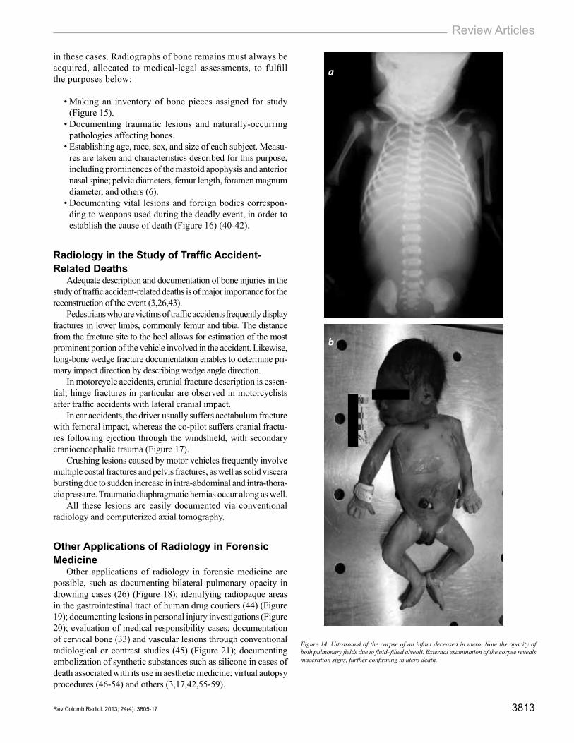
Radiological docimasia consists of a radiology-based demonstration of the absence of respiration. Since air is ra-diolucent, a corpse that once breathed will display radiolucent areas which are air-filled pulmonary alveoli. A corpse that never breathed, due to intra-uterine death, will have comple-tely opaque lungs in this test (Figure 14).
Radiology Applied to Forensic AnthropologyThe study of corpses reduced to bone remains has gai-
ned importance over the last years in Colombia, following the implementation of the Peace and Justice Law (Ley de Justicia y Paz). Autopsies of this kind are highly complex procedures due to the lack of tissues, hindering event recons-truction. Forensic radiographic studies are immensely useful
Figure 12. Body fragments found at an explosion scene; the presence of shrapnel from the explosive was documented by a radiographic study, making easier to recover it during the autopsy.
Figure 13. Ultrasound of the hyoid bone of a female, who was found in partial suspension in a motel where she walked in accompanied by her boyfriend. This is a case that requires establishing if her death was due to hanging or strangulation.
ba
c
3813Rev Colomb Radiol. 2013; 24(4): 3805-17
Review Articles
Figure 14. Ultrasound of the corpse of an infant deceased in utero. Note the opacity of both pulmonary fields due to fluid–filled alveoli. External examination of the corpse reveals maceration signs, further confirming in utero death.
in these cases. Radiographs of bone remains must always be acquired, allocated to medical-legal assessments, to fulfill the purposes below:
• Making an inventory of bone pieces assigned for study (Figure 15).
• Documenting traumatic lesions and naturally-occurring pathologies affecting bones.
• Establishing age, race, sex, and size of each subject. Measu-res are taken and characteristics described for this purpose, including prominences of the mastoid apophysis and anterior nasal spine; pelvic diameters, femur length, foramen magnum diameter, and others (6).
• Documenting vital lesions and foreign bodies correspon-ding to weapons used during the deadly event, in order to establish the cause of death (Figure 16) (40-42).
Radiology in the Study of Traffic Accident-Related Deaths
Adequate description and documentation of bone injuries in the study of traffic accident-related deaths is of major importance for the reconstruction of the event (3,26,43).
Pedestrians who are victims of traffic accidents frequently display fractures in lower limbs, commonly femur and tibia. The distance from the fracture site to the heel allows for estimation of the most prominent portion of the vehicle involved in the accident. Likewise, long-bone wedge fracture documentation enables to determine pri-mary impact direction by describing wedge angle direction.
In motorcycle accidents, cranial fracture description is essen-tial; hinge fractures in particular are observed in motorcyclists after traffic accidents with lateral cranial impact.
In car accidents, the driver usually suffers acetabulum fracture with femoral impact, whereas the co-pilot suffers cranial fractu-res following ejection through the windshield, with secondary cranioencephalic trauma (Figure 17).
Crushing lesions caused by motor vehicles frequently involve multiple costal fractures and pelvis fractures, as well as solid viscera bursting due to sudden increase in intra-abdominal and intra-thora-cic pressure. Traumatic diaphragmatic hernias occur along as well.
All these lesions are easily documented via conventional radiology and computerized axial tomography.
Other Applications of Radiology in Forensic Medicine
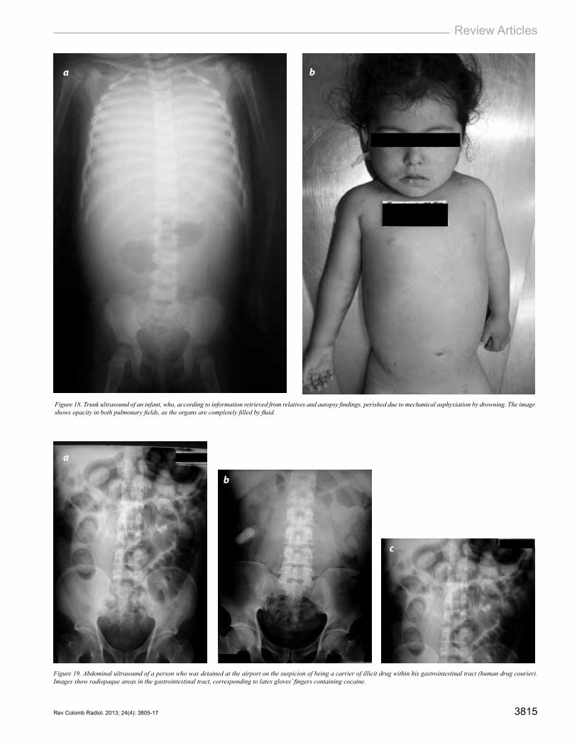
Other applications of radiology in forensic medicine are possible, such as documenting bilateral pulmonary opacity in drowning cases (26) (Figure 18); identifying radiopaque areas in the gastrointestinal tract of human drug couriers (44) (Figure 19); documenting lesions in personal injury investigations (Figure 20); evaluation of medical responsibility cases; documentation of cervical bone (33) and vascular lesions through conventional radiological or contrast studies (45) (Figure 21); documenting embolization of synthetic substances such as silicone in cases of death associated with its use in aesthetic medicine; virtual autopsy procedures (46-54) and others (3,17,42,55-59).
b
a

3814 Applications Of Conventional Radiology In The Medical Forensic Field. Montes G., Otálora A., Archila G.
Figure 15. Ultrasound of bone fragments allocated to a medical-legal assessment, related to the Law of Justice and Peace, exhumed from an armed conflict area.
Figure 16. Corpse rib, reduced to bone remnants for a medical-legal study, which displays a radiopaque area (arrow) due to shrapnel. Wound caused by a firearm projectile. Shrapnel was retrieved for analysis thanks to radiological identification.
Figure 17. Radiographs of a 6-month old infant’s corpse cranium, who was riding a bus on its mother’s arms when the vehicle crashed with another one. The infant was ejected towards the bus front and suffered a cranioencephalic trauma that led to its death. Cranial dissection of the corpse revealed a comminute fracture in the left frontoparietal area, with extensive subgaleal hematoma.
b
a
b
a
c
3815Rev Colomb Radiol. 2013; 24(4): 3805-17
Review Articles
Figure 18. Trunk ultrasound of an infant, who, according to information retrieved from relatives and autopsy findings, perished due to mechanical asphyxiation by drowning. The image shows opacity in both pulmonary fields, as the organs are completely filled by fluid.
Figure 19. Abdominal ultrasound of a person who was detained at the airport on the suspicion of being a carrier of illicit drug within his gastrointestinal tract (human drug courier). Images show radiopaque areas in the gastrointestinal tract, corresponding to latex gloves’ fingers containing cocaine.
b
b
a
a
c

3816 Applications Of Conventional Radiology In The Medical Forensic Field. Montes G., Otálora A., Archila G.
Figure 20. Male, blunt trauma victim who was taken to the medical-legal examiner’s office for personal injury inquiry and medical-legal incapacitation. A dental ultrasound revealed a hidden fracture of the tooth’s root (arrow).
Figure 21. Ultrasound of cervical (first and second) vertebrae removed from a cervical trauma corpse during autopsy. Before its death, it received medical attention and an odontoid fracture diagnosis was issued. Such fracture is absent, ruling it out as the cause of death.
References
1. Byard RW, Both K, Simpson E. The identification of submerged skeletonized rema-ins. Am J forensic Pathol. 2008;29:69-71.
2. Bilge Y, Kedici PS, Alakoç YD, et al. The identificatión of a dismembered human body: a multidisciplinary approach. Forensic Sci Int. 2003;137:141-6.
3. Di Maio VJ, Dana SE. Manual de patología forense. Madrid: Ediciones Díaz de Santos; 2003.
4. Rocha Sdos S, Ramos DL, Cavalcanti Mde G. Applicability of 3D-CT facial reconstruction for forensic individual identification. Pesqui Odontol Bras. 2003;17:24-8.
5. Silva RF, Pinto RN, Ferreira GM, et al. Importance of frontal sinus radiographs for human identification. Braz J Otorhinolaryngol. 2008;74:798.
6. Lynnerup N, Astrup JG, Sejrsen B. Thickness of the human cranial diploe in rela-tion to age, sex and general body build. Head Face Med. 2005;1:13.
7. Faruch Bilfeld M, Dedouit F, Soumah M, et al. Value of radiographic evaluation of the second metacarpal in the determination of bone age. J Radiol. 2008;89:1930-4.
8. Kirchhoff S, Fischer F, Lindemaier G, et al. Is Post-mortem CT of the dentition adequate for correct forensic identification?: comparision of dental computed tomo-grapy and visual dental record. Int J Legal Med. 2008;122:471-9.
9. Mincer HH, Chaudhry J, Blankenship JA, et al. Postmortem dental radiography. J Forensic Sci. 2008;53:405-7.
10. Minaguchi K, Maruyama S, Kasahara I, et al. Identification of unknown body using DNA analysis and dental characteristics in chest X-ray photograph. Bull Tokyo Dent Coll. 2005;46:145-53.
11. Bowers CM. Digital analysis of bite marks and human identification. Dent Clin North Am. 2001;45:327-42.
12. Wood RE. Forensic aspects of maxillofacial radiology. Forensic Sci Int. 2006;159(Suppl 1):S47-55.
13. Espina-Fereira A, Ortega AI, Barrios FA, et al. Metric and angular variables of the mandibular ramus on panoramic radiographs, as indicators for chronologic age. Invest Clin. 2007;48:403-18.
14. Vasiliadis L, Stavrianos C, Kafas P. A forensic aspect of age characteristics of denti-ne using transversal microradiography: a case report. Cases J. 2009;2:4.
15. Abdel Moneim WM, Abdel Hady RH, Abdel Maaboud RM, et al. Identification of sex depending on radiological examination of foot and patella. Am J forensic Pathol. 2008;29:136-40.
16. Blau S, Robertson S, Johnstone M. Disaster victim identification: New applications for postmortem computed tomography. J Forensic Sci. 2008;53:956-60.
17. Benjaminov O, Sklair-Levy M, Rivkind A, et al. Role of radiology in evaluation of terror attack victims. AJR Am J Roentgenol. 2006;187:609-16.
18. Campman S, Scott LA. The Sensitivity and specificity of control surface injuries in aircraft accident fatalities. Am J forensic Pathol; 2007;28:111-5.
19. Barber MA, Sibert JR. Diagnosing physical child abuse: the way forward. Postgrad Med J. 2000;76:743-9. 20. Giardino A, Randell A. Child maltreatment: A clinical guide and reference. 3rd ed.
St Louis, MO: G.W. Medical Publishing; 2005. 21. Lonergan G, Baker AM, Morey MK. From the archives of the AFIP: Child abuse:
radiologic-pathologic correlation. Radiographics. 2003;23:811-45. 22. Kemp AM. Investigating subdural haemorrhage in infants. Arch Dis Child.
2002;86:98-102.23. Demaerel P, Casteels I, Wilms G. Cranial imaging in child abuse. Eur Radiol.
2002;12:849-57. 24. Tung GA, Kumar M, Richardson RC, et al. Comparison of accidental and nonac-
cidental traumatic head injury in children on noncontrast computed tomography. Pediatrics. 2006;118:626-33.
25. Chiesa A. Abusive head trauma. Pediatr Clin North Am. 2009;56:317-31. 26. Calabuig G. Medicina legal y toxicología. 6ta ed. Barcelona: Elsevier-Masson; 2004. 27. Levy G, Goldstein L, Blachar A, et al. Postmortem computed tomography in vic-
tims of military air mishaps: radiological-pathological correlation of CT findings. Isr Med Assoc J. 2007;9:699-702.
28. Dicpinigaitis PA, Koval KJ, Tejwani NC, et al. Gunshot wounds to the extremities. Bull NYU Hosp Jt Dis. 2006;64:139-55.
29. von See C, Bormann KH, Schumann P, et al. Forensic imaging of projectiles using cone-beam computed tomography. Forensic Sci Int. 2009;190:38-41.
30. Jeffery AJ, Rutty GN, Robinson C, et al. Computed tomography of projectile inju-ries. Clin Radiol. 2008;63:1160-6.
31. JJ Hollerman, ML Fackler, DM Coldwell, et al. Gunshot wounds: 2. Radiology. Am J Roentgenol. 1990;155:691-702.
32. Kim PE. Radiographic assessment of cranial gunshot wounds. Neuroimaging Clin N Am. 2002;12:229-48.
33. Pinto A, Brunese L, Scaglione M, et al. Gunshot injuries in the neck area: ballistics elements and forensic issues. Semin Ultrasound, CT MR. 2009;30:215-20.
34. Messmer JM, Fierro MF. Radiologic forensic investigation of fatal gunshot wounds. Radiographics. 1986;6:457-73.
35. Harcke HT, Levy AD, Getz JM, et al. MDCT analysis of projectile injury in foren-sic investigation. AJR Am J Roentgenol. 2008;190:W106-11.
36. Puentes K, Taveira F, Madureira AJ, et al. Three-dimensional reconstitution of bullet trajectory in gunshot wounds: a case report. J Forensic Leg Med. 2009;16:407-10.
b
a

3817Rev Colomb Radiol. 2013; 24(4): 3805-17
Review Articles
37. Dodd GD 3rd, Budzik RF Jr. Identification of retained firearm projectiles on plain radiographs. AJR Am J Roentgenol. 1990;154:471-5.
38. Sieswerda-Hoogendoorn T, van Rijn RR. Current techniques in postmortem ima-ging with specific attention to paediatric applications. Pediatr Radiol. 2010;40:141-52.
39. McGraw E, Pless E, Pennington D, et al. Postmortem radiography after unexpected death in neonates, infants, and children: should imaging be routine? Am J Roentge-nol. 2002;178:1517-21.
40. Işcan MY, Olivera HE. Forensic anthropology in Latin America. Forensic Sci Int. 2000;109:15-30.
41. Sanabria C. Antropología forense y la investigación médico-legal de las muertes. Bogotá: Fondo Rotatorio de la Policía; 2004.
42. Lichtenstein JE, Fitzpatrick JJ, Madewell JE. The role of radiology in fatality in-vestigations. AJR Am J Roentgenol. 1988;150:751-5.
43. Alempijevic D, Jecmenica D, Pavlekic S, et al. Forensic medical examination of victims of trafficking in human beings. Torture. 2007;17:117-21.
44. Grabherr S, Ross S, Regenscheit P, et al. Detection of smuggled cocaine in cargo using MDCT. AJR Am J Roentgenol. 2008;190:1390-5.
45. Grabherr S, Djonov V, Yen K, et al. Postmortem angiography: review of former and current methods. AJR Am J Roentgenol. 2007;188:832-8.
46. Aghayev E, Staub L, Dirnhofer R, et al. Virtopsy. The concept of a centralized data-base in forensic medicine for analysis and comparision of radiological and autopsy data. J Forens Legal Med. 2008;15:135-40.
47. Manzano AC, Morillo AJ, Vallejo JM, et al. Necropsy by magnetic resonance in a case of conjoined thoracopagus twins. J Magn Reson Imaging. 2001;13:976-81.
48. Scholing M, Saltzherr TP, Fung Kon Jin PH, et al. The value of postmortem com-puted tomography as an alternative for autopsy in trauma victims: a systematic review. Eur Radiol. 2009;19:2333-41.
49. Cha JG, Kim DH, Kim DH, et al. Utility of postmortem autopsy via whole-body imaging: initial observations comparing MDCT and 3.0 T MRI findings with autop-sy findings. Korean J Radiol. 2010;11:395-406.
50. Dirnhofer R, Jackowski C, Vock P, et al. Virtopsy: minimally invasive, imaging-guided virtual autopsy. Radiographics. 2006;26:1305-33.
51. Levy AD, Abbott RM, Mallak CT, et al. Virtual autopsy: preliminary experience in high-velocity gunshot wound victims. Radiology. 2006;240:522-8.
52. Aghayev E, Christe A, Sonnenschein M, et al. Postmortem imaging of blunt chest trauma using CT and MRI: comparison with autopsy. J Thorac Imaging. 2008;23:20-7.
53. Christe A, Ross S, Oesterhelweg L, et al. Abdominal trauma—sensitivity and speci-ficity of postmortem noncontrast imaging findings compared with autopsy findings. J Trauma. 2009;66:1302-7.
54. Levy AD, Abbott RM, Mallak CT, et al. Virtual autopsy: preliminary experience in high-velocity gunshot wound victims. Radiology. 2006;240:522-8.
55. Harcke HT, Bifano JA, Koeller KK. Forensic radiology: response to the Pentagon Attack on September 11, 2001. Radiology. 2002;223:7-8.
56. Sosna J, Sella T, Shaham D, et al. Facing the new threats of terrorism: radiologists’ perspectives based on experience in Israel. Radiology. 2005;237:28-36.
57. O’Donnell C, Woodford N. Post-mortem radiology—a new sub-speciality? Clin Radiol. 2008;63:1189-94.
58. Morayati SJ, Nagle CE. The determination of death and the changing role of medi-cal imaging. Radiographics. 1988;8:967-79.
59. Castillo M. Digital forensics and the American Journal of Neuroradiology. AJNR Am J Neuroradiol. 2008;29:211-2.
Corresponding AuthorGuillermo Andrés Montes LoaizaInstituto Nacional de CancerologíaCalle 1A # 9-85Bogotá, [email protected]
Received for evaluation: July 8, 2013Accepted: August 22, 2013