applications of doppler ultrasound in fetal growth · pdf fileintroduction • the main use...
TRANSCRIPT

Applications of Doppler Ultrasound in Fetal Growth
Assessment
David Cole

Aims
• The aim of this presentation is to consider the use of Doppler ultrasound to investigate and monitor those pregnancies at risk of perinatal morbidity and mortality due to uteroplacental insufficiency

Learning outcomes• Describe the main applications of Doppler
Ultrasound in Obstetrics• Explain the principles underlying the Doppler
Technique• Describe the uteroplacental anatomy• Describe the feto-placental anatomy• Describe the Doppler ultrasound techniques for
examining the uteroplacental, feto-placental and fetal circulations
• Describe how spectral Doppler waveforms are interpreted

Introduction• The main use of Doppler Ultrasound in
Obstetrics is to identify and monitor those fetuses at risk of perinatal mortality or morbidity due to uteroplacental insufficiency
• This is achieved by investigating blood volume flow to the placenta; in the umbilical arteries and in the fetus.
Topics
• Colour flow imaging and Spectral Doppler• Interpretation of the spectral Doppler waveform• Doppler indices• Uterine artery Doppler• Umbilical artery Doppler• Middle cerebral artery Doppler• Fetal venous Doppler

Colourflow imaging and Spectral Doppler• Colour Doppler gives a map of blood flow
superimposed on the normal 2D image and is used to identify a particular blood vessel and sample the blood velocity within that vessel.
• Spectral Doppler gives a graph of blood velocity versus time – the Doppler waveform. This waveform is analysed to detect changes in resistance to blood flow

Colour flow imaging
Image courtesy of UNIVERSAL DIAGNOSTIC SOLUTIONS

Colour flow imaging - sampling
Image: Courtesy of IAME
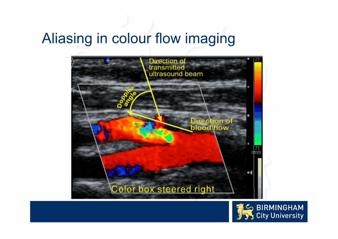
Aliasing in colour flow imaging
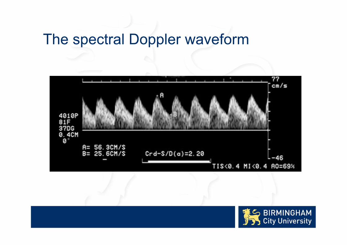
The spectral Doppler waveform
Interpretation of the spectral Doppler waveform
• The Doppler waveform represents the velocities of blood cells within the sample volume plotted against time.
Interpretation of the spectral Doppler waveform
• The Doppler waveform represents the velocities of blood cells within the sample volume plotted against time.
• The waveform can be analysed by:– Waveform pattern recognition.
– Waveform shape analysis
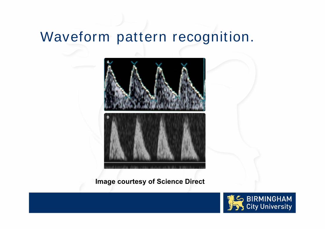
Waveform pattern recognition.
Image courtesy of Science Direct
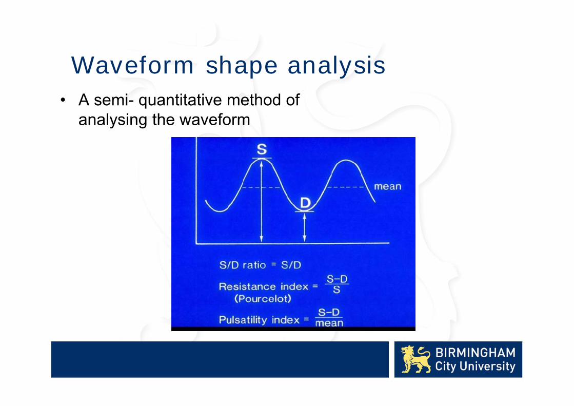
Waveform shape analysis• A semi- quantitative method of
analysing the waveform
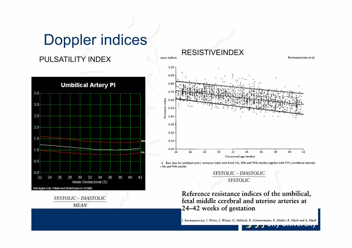
Doppler indices
MEANDIASTOLICSYSTOLIC
SYSTOLICDIASTOLICSYSTOLIC
PULSATILITY INDEXRESISTIVEINDEX

Doppler Ultrasound in Fetal Growth Assessment
• Uterine artery Doppler• Umbilical artery Doppler• Middle cerebral artery Doppler• Fetal venous Doppler

Uterine artery Doppler
• Uterine artery Doppler is usually performed as a screening test on a high risk population. It’s purpose is detect those pregnancies at risk of preeclampsia or intrauterine growth retardation (IUGR).

The Utero Placental Circulation
reproduced from Moffett-King (2002)
The spiral arteries within the wall of the uterus supply blood to the endometrium.
During the first trimester of pregnancy these spiral arteries are invaded by trophoblastic cells from the developing placenta.
This invasion of trophoblastic cells produces dilated spiral arteries and results in a low resistance blood flow to the placenta

Uterine artery Doppler
• Normally carried out at 20 -24 weeks gestation –The uterine artery is sampled to examine the spectral Doppler waveform.
• Both arteries are examined and reported on.
• In addition the position of the placenta should be recorded, especially if it is lateral.
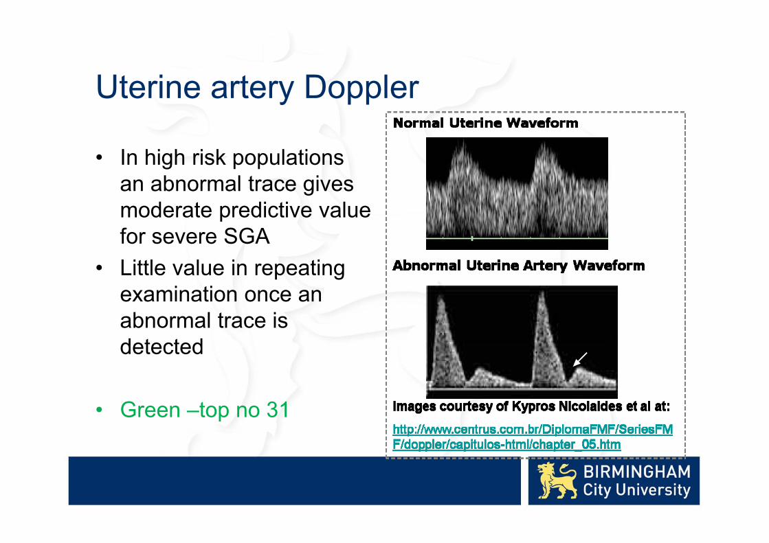
Uterine artery Doppler
• In high risk populations an abnormal trace gives moderate predictive value for severe SGA
• Little value in repeating examination once an abnormal trace is detected
• Green –top no 31

Umbilical artery Doppler
• Umbilical artery Doppler is the most widely used Doppler examination in obstetrics and abnormal results have good correlation with adverse perinatal conditions.
• Performed from 24 weeks gestation to term and it’s purpose is to detect those fetuses at risk of perinatal morbidity or mortality due to growth retardation.
• Examination Technique
• This can normally be carried out from 24 weeks gestation – a colour flow image of the cord is scanned.
• The sample should be taken from either free-floating cord or cord close to the placenta.
Image courtesy of UNIVERSAL DIAGNOSTIC SOLUTIONS

Umbilical artery normal anatomy
The Umbilical Cord
The umbilical cord normally contains two arteries and one vein. The arteries carry de-oxygenated blood from the fetus to the placenta, where it is mixed with maternal blood for oxygenation, nutrient uptake and elimination of waste products. The umbilical vein carries oxygenated blood rich in nutrients from the placenta to the fetus.
Blood Flow in the Umbilical Artery
The blood flow in the umbilical artery is pulsatile and is governed by the fetal heart rate. In the normal pregnancy the resistance to flow is low with forward flow in systole and diastole.
The Feto-placental Circulation
Schematic representation of the feto-placental circulation showing the fetus, placenta, umbilical arteries and umbilical vein.

Umbilical artery Doppler
• Primary surveillance tool in the SGA fetus
• When Doppler indices are normal it is reasonable to repeat every 14 days
• More frequent measurements if the fetus is severely SGA
• Green –top no 31
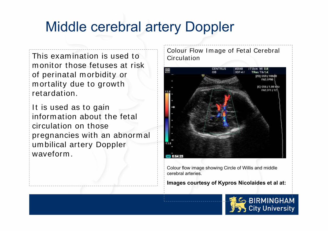
This examination is used to monitor those fetuses at risk of perinatal morbidity or mortality due to growth retardation.
It is used as to gain information about the fetal circulation on those pregnancies with an abnormal umbilical artery Doppler waveform.
Colour Flow Image of Fetal Cerebral Circulation
Colour flow image showing Circle of Willis and middle cerebral arteries.
Images courtesy of Kypros Nicolaides et al at:
Middle cerebral artery Doppler

Middle cerebral artery Doppler

Middle cerebral artery Doppler
• Examination Technique
• A transverse section of the fetal head is obtained and the colour box applied to view the Circle of Willis and middle cerebral arteries (MCAs).
• The sampling gate is placed in the proximal MCA to obtain the Doppler waveform.
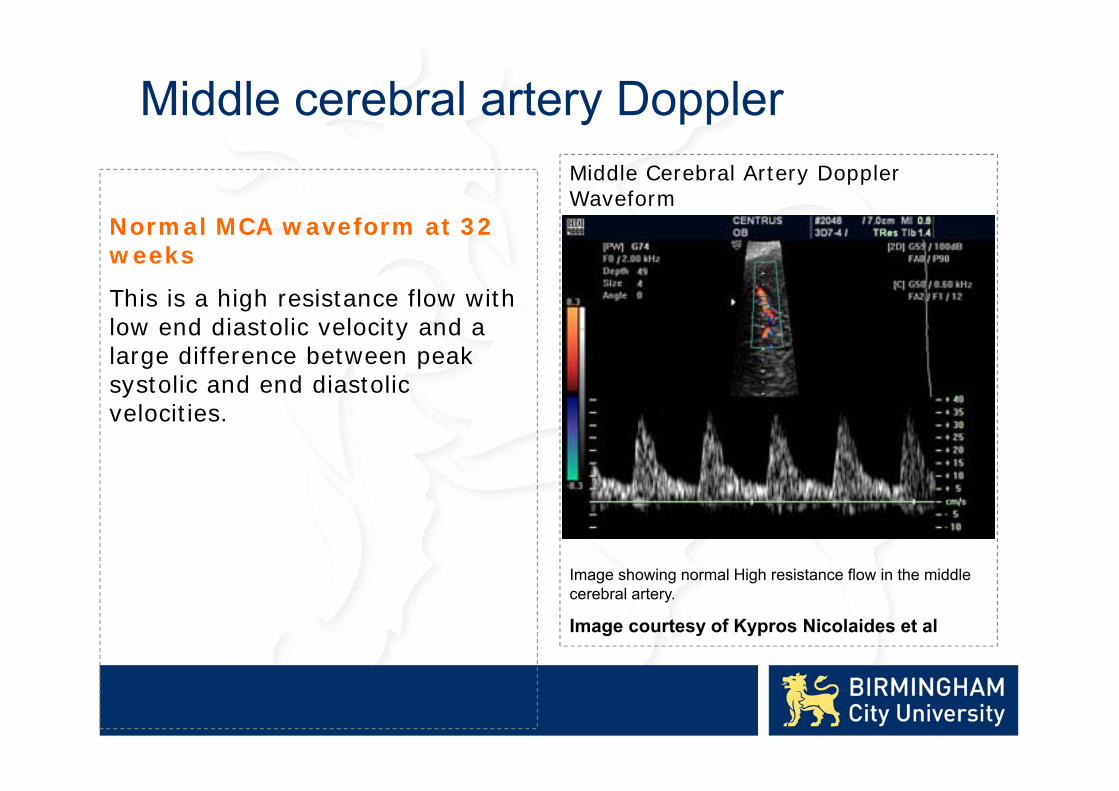
Normal MCA waveform at 32 weeks
This is a high resistance flow with low end diastolic velocity and a large difference between peak systolic and end diastolic velocities.
Middle Cerebral Artery Doppler Waveform
Image showing normal High resistance flow in the middle cerebral artery.
Image courtesy of Kypros Nicolaides et al
Middle cerebral artery Doppler

Middle Cerebral Artery Doppler Waveform
Image showing low resistance flow in the middle cerebral artery.
Image: Courtesy of Phillipe Jeanty
Middle cerebral artery Doppler
Abnormal MCA waveform at 32 weeks
This is a low resistance flow with good forward flow in diastole.
This is due to “brain sparing” in the growth retarded fetus. Blood is preferentially shunted to the life support centres in the brain heart and adrenals rather than to the abdominal organs and skeletal muscles
Middle cerebral artery Doppler
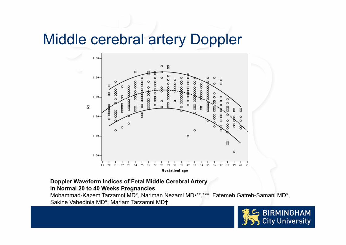
Doppler Waveform Indices of Fetal Middle Cerebral Arteryin Normal 20 to 40 Weeks PregnanciesMohammad-Kazem Tarzamni MD*, Nariman Nezami MD•**,***, Fatemeh Gatreh-Samani MD*,Sakine Vahedinia MD*, Mariam Tarzamni MD†

Middle cerebral artery Doppler
• In the preterm SGA fetus, middle cerebral artery (MCA) Doppler has limited accuracy to predict acidaemia and adverse outcome and should not be used to time delivery.
• In the term SGA fetus with normal umbilical artery Doppler, an abnormal middle cerebral artery Doppler (PI < 5th centile) has moderate predictive value for acidosis at birth and should be used to time delivery.
Green top guide 31
Arterial Doppler
• The waveforms from arterial Doppler examinations depend on both cardiac output and vascular resistance.
• Arterial Doppler alone is inadequate for examination of impaired fetal cardiac function.

Fetal venous Doppler
• Fetal venous Doppler is used to complement umbilical artery and middle cerebral artery Doppler in identifying those fetuses with IUGR at risk of death in utero.
• Includes umbilical vein, ductus venosus and IVC• Ductus venosus waveforms have been found to be the
most useful in terms of reproducibility and identifying the “at risk” fetus
• Application late second and early third trimester fetuses with IUGR.
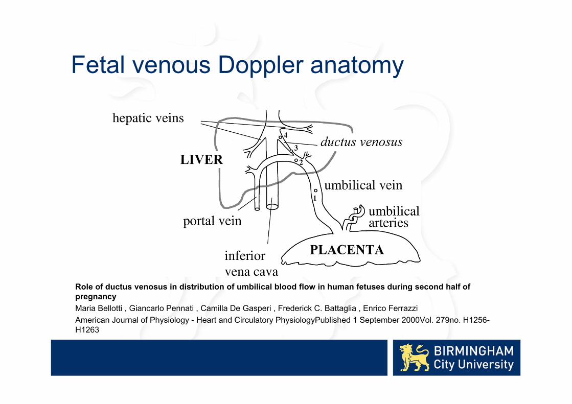
Fetal venous Doppler anatomy
Role of ductus venosus in distribution of umbilical blood flow in human fetuses during second half of pregnancyMaria Bellotti , Giancarlo Pennati , Camilla De Gasperi , Frederick C. Battaglia , Enrico FerrazziAmerican Journal of Physiology - Heart and Circulatory PhysiologyPublished 1 September 2000Vol. 279no. H1256-H1263
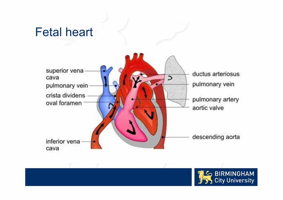
Fetal heart

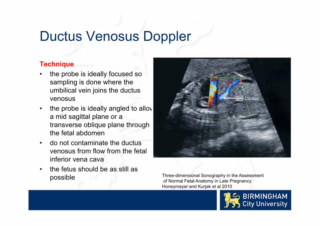
Ductus Venosus Doppler
“The deterioration of fetal condition due to severe FGR is usually accompanied by signs of cardiovascular changes that can be shown by venous Doppler studies”
“Evidence of impaired cardiac function has been documented using Doppler flow studies of the precordial veins”
Aberry and Soothill 2007

Ductus venosus waveforms
• S wave : corresponds to fetal ventricular systolic contraction
• D wave : corresponds to fetal early ventricular diastole
• A wave : corresponds to fetal atrial contraction

Ductus venosus waveforms
Ductus Venosus Doppler
Technique• the probe is ideally focused so
sampling is done where the umbilical vein joins the ductusvenosus
• the probe is ideally angled to allow a mid sagittal plane or a transverse oblique plane through the fetal abdomen
• do not contaminate the ductusvenosus from flow from the fetal inferior vena cava
• the fetus should be as still as possible
2
Three-dimensional Sonography in the Assessmentof Normal Fetal Anatomy in Late Pregnancy
Honeymayer and Kurjak et al 2010
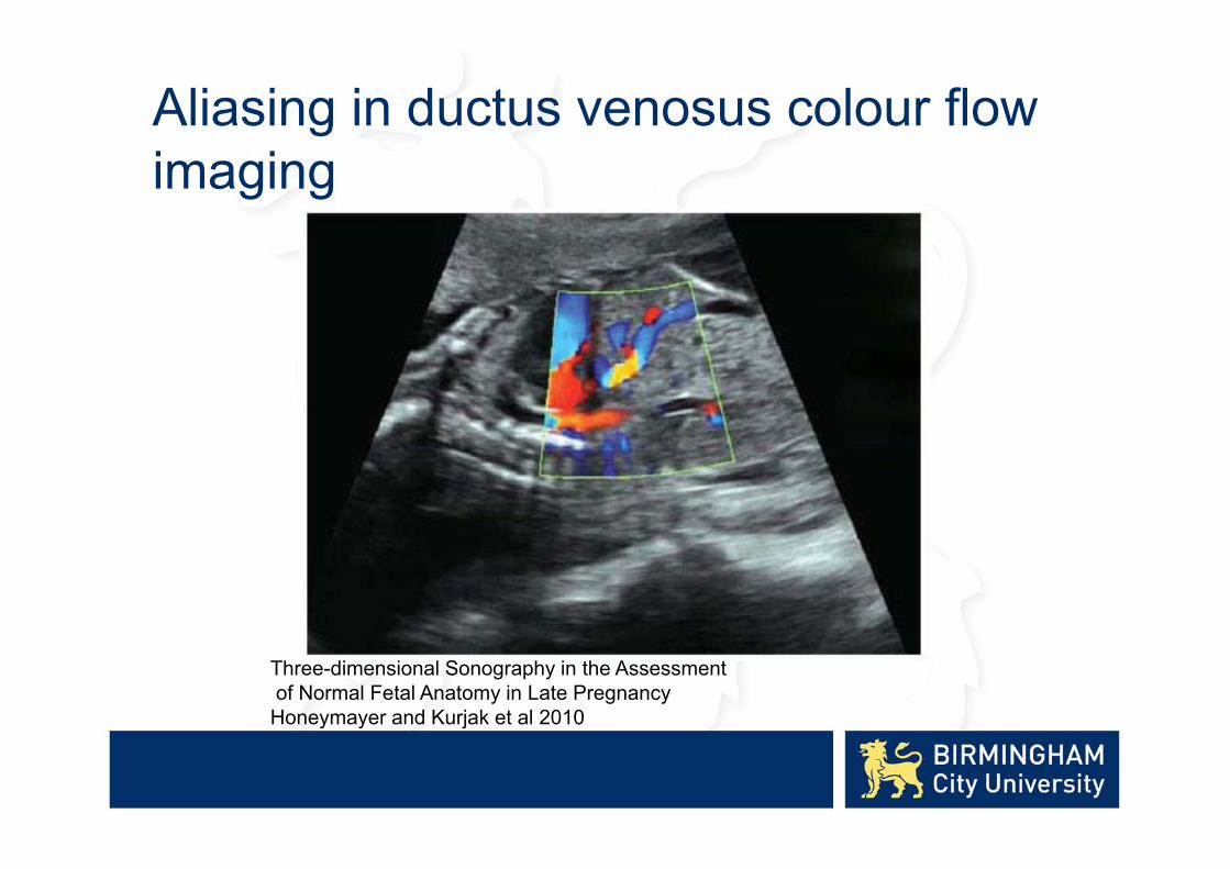
Aliasing in ductus venosus colour flow imaging
Three-dimensional Sonography in the Assessmentof Normal Fetal Anatomy in Late Pregnancy
Honeymayer and Kurjak et al 2010
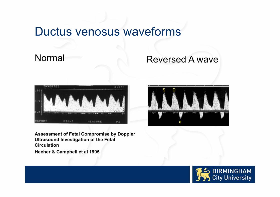
Ductus venosus waveforms
Normal
Assessment of Fetal Compromise by Doppler Ultrasound Investigation of the Fetal CirculationHecher & Campbell et al 1995
Reversed A wave
Ductus Venosus Doppler
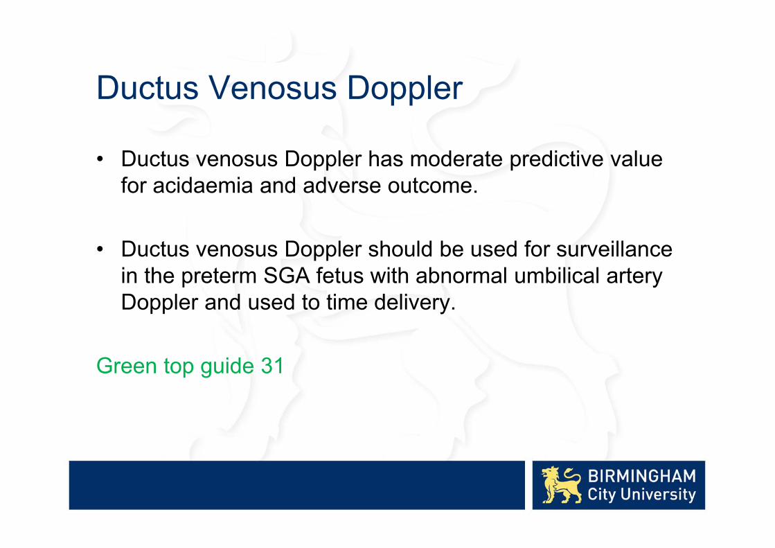
• Ductus venosus Doppler has moderate predictive value for acidaemia and adverse outcome.
• Ductus venosus Doppler should be used for surveillance in the preterm SGA fetus with abnormal umbilical artery Doppler and used to time delivery.
Green top guide 31

Applications of Doppler Ultrasound in Fetal Growth AssessmentSummary• Uterine artery Doppler – screening examination• Umbilical artery Doppler – assessment of SGA fetuses• MCA Doppler – to detect brain sparing on fetuses with
abnormal UA Dopplers• Ductus venosus Dopplers – to detect impaired cardiac
function on SGA fetuses and to time delivery
