applications of the phase-coded generalized hough
TRANSCRIPT

Copyright © 2009 Tech Science Press CMC, vol.14, no.2, pp.79-97, 2009
Applications of the Phase-Coded Generalized HoughTransform to Feature Detection, Analysis, and
Segmentation of Digital Microstructures
Stephen R. Niezgoda1 and Surya R. Kalidindi1,2
Abstract: The generalized Hough transform is a common technique for featuredetection in image processing. In this paper, we develop a size invariant Houghframework for the detection of arbitrary shapes in three dimensional digital mi-crostructure datasets. The Hough transform is efficiently implemented via kernelconvolution with complex Hough filters, where shape is captured in the magnitudeof the filter and scale in the complex phase. In this paper, we further generalize theconcept of a Hough filter by encoding other parameters of interest (e.g. orientationof plate or fiber constituents) in the complex phase, broadening the applicability ofHough transform techniques. We demonstrate the application of these techniquesto feature detection in micrographs (2-D) and three-dimensional (3-D) microstruc-ture datasets, and explore their utility to the closely related applications of featurebased image segmentation and calculation of 3-D microstructure metrics.
Keywords: microstructure, Hough transform, image processing, segmentation,feature detection
1 Introduction
The description of the internal structure (also referred to as the microstructure)of the material is at the core of all descriptions in the field of materials scienceand engineering. This internal structure is an exceptionally rich dataset that spansmultiple hierarchical length scales from the macroscopic to atomistic. Recent ad-vances in characterization techniques, such as 3-D atom probe (Blavette, Bostel,Sarrau, Deconihout and Menand 1993; Seidman 2007), x-ray micro-tomography(Flannery, Deckman, Roberge and D’amico 1987; Maire, Buffière, Salvo, Blandin,Ludwig and Létang 2001), 3-D x-ray diffraction microscopy (Schmidt, Nielsen,
1 Department of Materials Science and Engineering, Drexel University, Philadelphia, 191042 Department of Mechanical Engineering and Mechanics, Drexel University, Philadelphia, 19104.
Corresponding author.

80 Copyright © 2009 Tech Science Press CMC, vol.14, no.2, pp.79-97, 2009
Gundlach, Margulies, Huang and Jensen 2004; Chapman, Barty, Marchesini, Noy,Hau-Riege, Cui, Howells, Rosen, He, Spence, Weierstall, Beetz, Jacobsen andShapiro 2006), and automated serial sectioning, have made it possible to capture thethree-dimensional details of microstructures at multiple length scales. The datasetsgenerated from these techniques are often in the form of exceptionally large digital3-D microstructure maps. For example, the high-resolution tomography capabili-ties at the European Synchrotron Radiation Facility can capture a field of view of2048 x 2048 pixels at a 0.28µm pixel size and exceeding 8GB in the size of thedataset, with the reconstructed 3-D voxel dataset exceeding 8× 10⁹ voxels (Betz,Wegst, Weide, Heethoff, Helfen, Lee and Cloetens 2007). Visualization and anal-ysis of such large datasets is a non-trivial problem. Depending on the scale of themicrostructural features of interest, a reconstructed 3-D volume may contain up-wards of 100,000 examples. Manually scanning such a dataset for microstructuralfeatures or defects (e.g. cracks, pores) is time consuming, and often impractical.In order to rigorously study these datasets, an automated methodology to locatefeatures of interest is critically needed.
In this paper, we explore the application of a phase-coded generalized Hough trans-form to the problem of automated microstructure feature detection. We also explorethe closely related applications of feature-based image segmentation and calcula-tion of 3-D microstructure metrics. The generalized Hough transform is a namegiven to a class of algorithms for the detection of imperfect instances of a targetobject in an image or volume. The original Hough transform was conceived as amethod for extracting lines and simple curves from images (Hough 1962; Duda andHart 1972), and is probably best known to materials scientists in its application tothe automated indexing of electron backscatter diffraction (EBSD) for orientationimaging microscopy (OIM) (Adams, Wright and Kunze 1993; Schwartz, Kumarand Adams 2000). The basic Hough transform has been generalized for the de-tection of arbitrary shapes (Ballard 1981) and has been efficiently implementedvia kernel convolution (Kierkegaard 1992). Atherton and Kerbyson developed acomplex Hough filter for size invariant circle detection (Kerbyson and Atherton1995; Atherton and Kerbyson 1999). Here we further extend the general frame-work of Atherton and Kerbyson to the detection of 3-D arbitrary shapes in mi-crostructure datasets and extend the complex Hough filter to size invariant shapedetection and orientation invariant line detection. We will then demonstrate theutility of this framework in three specific examples: 1) The detection of grains witha specified morphology in a polycrystalline material, 2) The rapid calculation ofa common statistical microstructure metric referred to as the lineal path function(Lu and Torquato 1992), and 3) The segmentation of an alpha/beta titanium colonymicrostructure into individual colonies.

Applications of the Phase-Coded Generalized Hough Transform 81
2 Phase-Coded Generalized Hough Filters
The Hough transform was originally developed for the machine analysis of trajec-tories of charged sub-atomic particles in a bubble chamber (Hough 1962). TheHough transform is an elegant mapping of each pixel in a binary image to a si-nusoidal curve in an accumulator or voting space, so that curves corresponding tocollinear pixels intersect in the voting space. We quickly review the basic Houghtransform since the key concepts are carried through to the more complex Houghfilters developed in this work.
Any line in a 2-D plane can be parameterized by the length of the perpendicu-lar from the line to the origin, r, and by the angle of this perpendicular, θ , asr = xcosθ + ysinθ . Thus every possible line in the plane can be uniquely mappedto a point in the parameter space as the ordered pair (r,θ). Each pixel in an imageis mapped to a curve in the parameter space through the realization that all possi-ble lines passing through the point (x0,y0) have the form r(θ) = x0 cosθ +y0 sinθ ,which is a sinusoid through the parameter space. If the curves from two pixels inthe image are plotted in the parameter space, the intersecting point corresponds tothe line connecting those points in the image. Lines in the binary image can thenbe easily found by mapping all of the pixels from the binary image into the pa-rameter space and counting the number of intersections at each point. The pointswhere numerous curves intersect correspond to line segments in the image. Thusthe parameter space is referred to as the voting or accumulator space, as each inter-section is considered a vote for a possible line in the image. Longer or more perfectline segments will receive more votes as more curves in the accumulator space willintersect.
The circle Hough transform can be used to detect circles in an image in a verysimilar manner. The standard equation of a circle in a 2-D plane, (x− a)2 +(y−b)2 = r2 parameterizes the circle by the ordered triplet (a,b,r). Each pixel in theimage can be mapped to a right circular cone in the parameter space (see Figure1 and description below). The cones from all of the edge points on a circle willintersect at a single point in the space (Duda and Hart 1972). In parallel with theline case larger circles or more perfect circles will receive more votes. This simpleobservation will motivate the introduction of a corrective normalization in laterexamples. In general such a mapping can be constructed for any shape describedby a set of parametric equations, where the dimensionality of the accumulator spaceis determined by the number of parameters.
The conventional Hough transforms described above can be formulated as a seriesof convolutions with a series of appropriately defined Hough transform filters. Forexample, consider a binary digital image where each pixel can either take the values

82 Copyright © 2009 Tech Science Press CMC, vol.14, no.2, pp.79-97, 2009
Figure 1: (a) Hough filter for a circle of radius 10 as defined by Eq. (1). (b) Thecontribution of selected pixels in the accumulator space of the convolution of acircle (schematically shown by the dotted line) with the filter shown in (a). Theeffect of the convolution operation is to “draw” in a circle of radius 10 at eachpixel. These circles intersect at the point (a = 0,b = 0) in the accumulator space.High intensity pixels in the accumulator space indicate likely centers of circles inthe image space. (c) The (a,b,r) accumulator space resulting from the convolutionof a single pixel located at the origin with a successive series of filters from r∗ = 1to r∗ = 13. The effect of the circle Hough transform is to map each pixel in theimage to a right circular cone in the full accumulator space.
0 or 1, in which we wish to detect a circle of radius r∗ pixels. The correspondingHough filter will be of size (2r∗+1,2r∗+1) where the rows of the filter are indexedm = [−r∗,−r∗+ 1, . . . ,0, . . . ,r∗− 1,r∗], and the columns are similarly indexed byn. The filter can then be defined as
Omn =
{1
2πr∗ iff (r∗−0.5)2 < m2 +n2 < (r∗+0.5)2
0 otherwise(1)
which is simply a circle of radius r∗ normalized so that its power is 1. Convolution

Applications of the Phase-Coded Generalized Hough Transform 83
with the filter maps a pixel located at position (a0,b0) in the image to a circle withequation (x−a0)2 +(y−b0)2 = (r∗)2 in the accumulator space, with an intensity of
12πr∗ . If the image contains a perfect circle of radius r∗ centered at position (a0,b0),the set of edge pixels will each be mapped to a circle of radius r∗ in the accumu-lator space that all share the common point (a0,b0). Each circle contributes 1
2πr∗
to the intensity at the intersection point in the accumulator space (see Figure 1).The purpose of normalizing the power of the filter by 1
2πr∗ is so that the cumulativecontribution of all the edge points of a perfect circle in the image is unity. Thusthe problem of detecting circles of radius r∗ in the image is reduced to identifyingpixels in the accumulator space with intensity close to 1. By “stacking” the resultsof successive convolutions with filters ranging from r∗ = rmin to r∗ = rmax the de-sired portion of the accumulator space, (a,b,r), can be explored. The mapping ofa single pixel to a right circular cone in the accumulator space is shown in Fig-ure 1. Hough filters offer significant computational advantages as the convolutionoperations can be efficiently computed via fast Fourier transform (FFT) methods(Briggs and Henson 1995). The primary advantage of filter based techniques is theelimination of the need for an analytic parametric description of the shape beingdetected; with an appropriately defined filter the above approach is applicable tothe detection of arbitrary shapes (Ballard 1981; Samal and Edwards 1997).
The drawback of the above approach is that a different filter is needed for each r∗,i.e. the technique is not scale invariant. Atherton and Kerbyson proposed a complexHough filter for size invariant circle detection in 2-D binary images (Kerbysonand Atherton 1995; Atherton and Kerbyson 1999). Here, we present a frameworkbased on the Atherton and Kerbyson filters, with an added normalization to improveaccuracy when searching over a wide range of sizes and extended to the detectionof arbitrary shapes in 3-D microstructure datasets. The general principal behindthis approach is to reduce the dimensionality of the accumulator space by encodingscale information in the complex phase of the filter. For circles, the filter is aannulus where the phase varies linearly from rmin→ rmax, defined by
Omn =
{1
2π√
m2+n2 e2πiφmn iff (rmin)2 ≤ m2 +n2 ≤ (rmax)2
0 otherwise
φmn =
√m2 +n2− rmin
rmax− rmin
(2)
An example filter is shown in Figure 2.
When convoluted with an image containing a circle of radius r∗, the contributionfrom each edge pixel on the circle will interfere constructively for the phase cor-responding to r∗ and destructively for all other phases. Thus the intensity of the

84 Copyright © 2009 Tech Science Press CMC, vol.14, no.2, pp.79-97, 2009
Figure 2: Complex phase coded Hough filter defined by Eq. (2) with rmin = 5 andrmax = 25. The complex magnitude as a function of radius is shown in (a) and thephase angle is shown in (b).
accumulator space gives the likelihood of finding a circle centered at that locationin the image, and the phase gives the radius of the circle. The normalization by
12π√
m2+n2 in Eq. (2) is designed so that all circles contribute equal intensity to theaccumulator space regardless of size. To the best knowledge of the authors, this is anovel contribution from this work. Without such normalization, the peaks in accu-mulator space from different sized circles are proportionate to their circumference.For ideal images of perfect circles without any signal noise, this difference in peakheight is not of great consequence. However, for noisy images and images withimperfect features (such as characterized micrographs), this normalization greatlyimproves the ability to accurately detect small circles, especially when rmax� rmin.Additionally, normalizing the contribution of a perfect circle to unity, allows the in-tensity in the accumulator to be thought of as a probability on the likelihood of acircle being centered at that point. For example, an accumulator point with intensity0.9 indicates that 90% of the pixels are consistent with a perfect circle. With theadded normalization, this technique is quite robust, even for noisy images, whendetecting circles with a wide distribution of radii. The application of our normal-ized phase coded filter to circle detection in a perfect and noisy image is detailed inFigure 3.
The circular filters described above for 2-D images are an important theoreticaland intellectual exercise. However, these filters need to be extended to arbitraryshapes in 3-D in order to be of significant practical use for microstructure analysis.

Applications of the Phase-Coded Generalized Hough Transform 85
Figure 3: (a) Ideal digital image containing circles of radius 20 and 50 pixels.(b) Accumulator space plotted as surface resulting from convolution of image (a)with filter defined by Eq. (2) where rmin = 10 and rmax = 60. Height of the peakindicates complex magnitude and coloration gives complex phase. There are indeedsix main peaks in this plot, and their phase correctly reflected the correspondingcircle radii. (c) Digital image containing imperfect circles and added uniformlydistributed noise at a volume fraction of 0.15. (d) Accumulator space for image (c).The noise adds additional peaks to the space and the peaks corresponding to circleshave been diminished relative to the ideal case. (e) Reconstructed image from theaccumulator space, by thresholding the accumulator space at 0.5 to separate thedesired peaks from the spurious ones due to the noise in the image.
Note that the extension of Eq. (2) from circles to spheres is trivial, the formremains exactly the same and the appropriate normalization becomes surface arearather than circumference. Consider an arbitrary 3-D solid shape (such as a grainin a polycrystalline material) denoted ω and described by the indicator function

86 Copyright © 2009 Tech Science Press CMC, vol.14, no.2, pp.79-97, 2009
(Torquato 2002):
Imnp =
{1 if pixel m,n, p is interior to ω
0 if pixel m,n, p is exterior to ω(3)
The surface of ω can then be expressed as (Torquato 2002)
Mmnp = |∇Imnp| (4)
which is nonzero only at the surface pixels. Additionally, let A = ∑m,n,p Mmnp
indicate the number of surface pixels. A simple Hough filter to find exact instancesof ω could be defined as
Omnp =1A
Mmnp (5)
To develop a complex filter for size invariant detection we introduce the notationM s
mnp where the index indicates a scaling factor, i.e. M 2mnp are the surface pixels
when the size of is scaled by 2. Similarly, let As = ∑m,n,p M smnp ≈ A1s2 represent
the appropriate scaling for surface area. A general size invariant Hough filter canthus be defined as
Omnp =∫ smax
smin
1As
e2πiφ sM s
mnpds
φs =
s− smin
smax− smin
(6)
An example of a filter modeled on a single grain from a polycrystalline dataset isshown in Figure 4.
In effect, a complex filter allows us to reduce the dimensionality of the accumu-lator space by encoding one parameter in the phase. In the examples described inEqs. (2) and (6), the phase angle represented scale. For other feature identificationapplications, it might be more advantageous to encode a different microstructurefeature as the phase. As an example, consider the problem of rapidly identifyingthe orientation of fibers in a 2-D micrograph from a fiber-reinforced composite. Inthis case, a convolution with a simple filter of the form (in polar coordinates)
Orθ =
{1r∗ e
2πi θ
π if r ≤ r∗
0 otherwise(7)
where is approximately equal to the average fiber length, will indentify the orien-tation associated with each fiber. This example and related applications will be

Applications of the Phase-Coded Generalized Hough Transform 87
Figure 4: (a) A single grain extracted from a digital polycrystalline microstructurethat served as a template for the filter shown in (b) and (c). (b,c) Intersecting or-thogonal slices through the center of the filter corresponding to the image in (a)obtained by using Eq. (6). (b) shows the complex phase of the filter, and (c) showsthe magnitude. Only pixels that take non-zero values are visible.
explored in more detail in the Image Segmentation section below. In the imageprocessing literature, the term Hough filters is only used for feature extraction ap-plications, and would not necessarily cover Eq. (7). However, for our purposes,the implementation and interpretation are so similar that we will refer to them asgeneralized Hough filters.
3 Feature Extraction in Digital Microstructures
In order to validate the framework presented earlier, we will apply the Hough filtersdefined by Eq. (6) to extract grains of a prescribed morphology from a digitally cre-ated polycrystalline microstructure (Brahme, Alvi, Saylor, Fridy and Rollett 2006).The microstructure is 100x100x100 pixels in size and contains 997 unique grains(see Figure 5). The Hough filter techniques described above rely on the mappingof edge pixels into the accumulator space. Thus the first step in extracting grainsbased on morphology is to convert the full dataset into a grain boundary network.This was accomplished using a simple Canny edge detection algorithm on the indi-vidual 2D slices (Canny 1986), and the results are shown in Figure 5. For complexdatasets, more robust edge segmentation algorithms would be required. The datasetwas then searched for grains of 2 specific morphologies: 1) spherical grains and 2)grains similar in shape and scale to a specified target grain. The dataset shown inFigure 5 is perfectly periodic, thus it was natural to include edge grains that inter-sect the bounding box. For aperiodic digitally created or characterized datasets thedecision to include the edge grains would have to be informed by both the applica-

88 Copyright © 2009 Tech Science Press CMC, vol.14, no.2, pp.79-97, 2009
tion and the shapes being detected.
Figure 5: (a) Digitally created polycrystalline dataset 100x100x100 pixels in size.The dataset contains 997 unique grains. (b) Grain boundary network created fromedge detection on the entire dataset. The Hough filters act on the edge pixels andaccurate edge detection is critical to shape detection. (c) Equi-axed grains extractedfrom the dataset in (a) at an accumulator threshold of 0.28 (97% of the maximumintensity), the radius range of the filter was 5 to 20 pixels. (d) By lowering thethreshold to 0.26 (90% of the maximum intensity) more grains are detected, how-ever while still equi-axed, the selected grains have a larger deviation from spheroc-ity.

Applications of the Phase-Coded Generalized Hough Transform 89
The results of the spherical grain detection are shown in Figure 5. A sphericalfilter was defined based on Eq. (6), where Mmnp was a spherical shell of radius 10,smin = 0.5, and smax = 2. Thus, the working range of the filter was radii between5 and 20 pixels. After convolution, the maximum magnitude of the accumulatorspace was 0.29, indicating that for the most spherical grain, only about 30% of thepixels coincided with a perfect sphere. In this case the relatively low intensitiesin the accumulator space are not surprising; the fact that grains are space fillingstructures precludes the existence of perfectly spherical grains. Here, the higherintensity regions in the accumulator space after convolution with a spherical filtercorrespond to the locations of the most equi-axed grains in the microstructure. Theresults are shown for two threshold values in the accumulator space, 0.28 and 0.26(97% and 90% of the max value respectively). At the 0.28 level, the grains thatare extracted appear to deviate substantially from spherical. On closer inspection itis seen that these grains are largely equi-axed with local deviations; when viewedfrom another angle they appear nearly spherical. At the 0.26 level, the deviationfrom spherocity is more evident but the grains are clearly still equi-axed.
When applied to detecting near instances of natural shapes common in the dataset,the generalized Hough filters are extremely robust. A Hough filter was createdbased on an archetype grain from the dataset and was used to extract grains ofsimilar shape and scale. A pancaked archetype grain was selected to differentiatethe results from the spherical filter case, and a narrow scale range of smin = 0.8 tosmax = 1.2 was used. The archetype grain and the nearest other grains are shown inFigure 6. Since the filter was constructed from an actual grain from the sample, theintensity of the accumulator space ranges from 0 to 1, with accumulator intensity 1indicating the center of mass from the archetype grain. After the grain that servedas a template, the next highest peak in the accumulator space has an intensity of0.4. Thresholding the accumulator space at 0.30 left only the peaks correspondingto the archetype grain and the 5 closest grains.
The low values of peaks in the accumulator space for both the spherical and natu-rally shaped filter highlight the need for proper normalization of the phase codedfilter. In microstructural feature detection we are naturally seeking imperfect in-stances of the object; no two grains are the same morphology.
4 Lineal Path Function and Related Functions
The lineal path function is an important statistical descriptor of the microstructurewhich is often used in reconstructions of 3-D microstructures from 2-D sections(Yeong and Torquato 1998; Manwart, Torquato and Hilfer 2000; Zeman and Se-jnoha 2007). Additionally, the lineal path function contains linear free path infor-mation and has been used to model Knudsen diffusion and radiative transport in

90 Copyright © 2009 Tech Science Press CMC, vol.14, no.2, pp.79-97, 2009
Figure 6: Grains from the dataset shown in Figure 5 that come closest to matchingthe circled archetype grain in shape and scale. The edge pixels of the circled grainwere used to construct a Hough filter using Eq. (6). Thresholding the accumulatorspace at 30% returned the archetype grain and 5 most similar grains.
porous materials (Torquato 2002). The lineal path function, i, is defined as theprobability that a line segment of length z lies entirely in phase i when thrown ran-domly into the microstructure. The lineal path function is commonly calculated bysampling, and accurate measurement requires throwing a very large number of linesegments into the microstructure (Torquato 2002; Singh, Gokhale, Lieberman andTamirisakandala 2008). Most applications of the lineal path function are limitedto isotropic structures and the function is assumed to be independent of the orien-tation of line segment. For broad applicability to anisotropic structures, the linealpath function must be calculated as function of the length of line segment i and also

Applications of the Phase-Coded Generalized Hough Transform 91
its orientation, i.e. Li(z), where z is a 2-D or 3-D vector. This angularly resolvedlineal path function is rarely calculated, or is only calculated at a handful of angles,due to the tedious nature of the required sampling over the space of length andorientations (Singh, Gokhale, Lieberman and Tamirisakandala 2008).
Calculation of the angularly resolved lineal path function can be efficiently achievedusing generalized Hough filter approaches described earlier. For example, the filterto calculate Li(z = z∗,θ = θ ∗) in a 2-D microstructure can be expressed as
Orθ =
{1r∗ if r ≤ r∗ and θ = θ ∗
0 otherwise(8)
Convolution of a properly segmented microstructure image with the above filterwill map a value of 1 everywhere the vector z will fit in the image. Li(z = z∗,θ =θ ∗) is simply calculated as the fraction of accumulator space pixels with value 1.In a sense, the Hough filter can be thought of as sampling every possible place-ment of vector z, defined by the ordered pair (z,θ), simultaneously. Thus the entirelineal path function Li(z) can be calculated by convolution with a series of Houghfilters where the length and orientation of the vector z are systematically varied.An example calculation of the lineal path function for a 2-phase microstructure isshown in Figure 7. When calculating statistical measures in this way it is importantto consider bias due to boundary effects due to the convolution operation by ei-ther padding the image or discounting the contribution of points near the boundary(Briggs and Henson 1995)(Gokhale, Lieberman and Tamirisakandala 2008).
The use of Hough filters allows further generalization of the lineal path function.Rather than simply calculating the probability of an oriented line segment fallingcompletely in a selected phase, we can calculate the probability associated withother shapes. For example in 3-D percolating microstructures, the flux through aregion is strongly dependent on the cross-sectional area of the connected paths.In this case, a better statistical measure might be the probability that sphere ofradius r or a cylinder of radius r, length z and a orientation (θ ,φ) falls within aselected phase. As a simple example, determination of the radial path functionRi(r) (probability of a sphere of radius r lies completely in phase i when thrownrandomly into the microstructure) for a porous solid is shown in Figure 7.
5 Application to Image Segmentation
Segmentation of an experimentally characterized microstructure, especially in 3-D, based on microstructural features is a difficult task without a general solution.The approach taken is highly subjective to both the characterization technique andmicrostructure (for interesting examples, see (Chawla, Ganesh and Wunsch 2004;

92 Copyright © 2009 Tech Science Press CMC, vol.14, no.2, pp.79-97, 2009
Figure 7: (a) Digital image of a two phase microstructure showing a clearanisotropy in platelet orientation. Microstructure image is 2904x3864 pixels. (b)Angularly resolved lineal path function Li(z) calculated by convolution of the mi-crostructure shown in (a) with a series of filters defined by Eq. (8). Each meridianrepresents the lineal path function for line segments of a given angular orientation.Each radius represent a given line segment length. The radial range is from 0-240pixels with a gridline every 56 pixels. Notice that anisotropy of the microstruc-ture is clearly captured in the lineal path function. (c) Digitally created 3-D poroussolid. The white phase indicates pore and the solid is black. (d) Radial path func-tion calculated from (c). The radial path function gives the probability that a sphereof radius r lands completely in the pore space, when thrown randomly into themicrostructure.
Petushi, Katsinis, Coward, Garcia and Tozeren 2004; Stutzman 2004; Uchic, Groe-ber, Dimiduk and Simmons 2006; Simmons, Bartha, De Graef and Comer 2008)).Often, the problem of image segmentation can be simplified by the application ofa suitably defined phase-coded Hough filter. As a simple example, consider theback-scattered electron (BSE) image of an alpha/beta titanium colony microstruc-ture shown in Figure 9, obtained in a scanning electron microscope (SEM). In char-

Applications of the Phase-Coded Generalized Hough Transform 93
Figure 8: (a) Idealized lath or plate microstructure to serve as a demonstration fororientation resolution by the generalized Hough filter. (b) Complex phase of anorientation resolving Hough filter built using Eq. (7) with r∗ = 50; the polar angleis encoded as the complex phase. The magnitude of the filter is 1 for r ≤ 50 andzero otherwise. (c) Result of convolution of the image (a) and the filter (b). Theimage shows the complex phase of pixels corresponding to the laths (white) in(a). For clarity, the complex phase of the other pixels (black) are not shown. Theorientation of the lath can then be read directly from the phase information.
acterizing such a microstructure, one often desires to extract statistics from individ-ual colonies such as the mean lath separation or the volume fraction of alpha (Tiley,Searles, Lee, Kar, Banerjee, Russ and Fraser 2004; Collins, Welk, Searles, Tiley,Russ and Fraser 2009). An automated method of segmenting the microstructureinto colonies would be extremely beneficial.
The problem of colony segmentation can be effectively addressed through the ap-plication of a generalized Hough filter to resolve the lath orientation. For claritywe will demonstrate the approach on an idealized plate or lath microstructure (seeFigure 8), and subsequently show the results for the experimentally acquired mi-crograph shown in Figure 9. A line orientation encoding Hough filter was createdusing Eq. (7) with r∗ = 50 pixels. The magnitude of the filter is 1 inside the circleand zero outside, and the complex phase is shown in Figure 8. When convolvedwith the microstructure image the phase corresponding to the orientation construc-tively interferes in the accumulator space. The complex phase of each line can thenbe mapped back to a physical orientation angle. The image can be segmented inthe accumulator space by simply isolating regions of similar complex phase. No-tice that the areas near the end of the laths or where two laths are near each otherexhibit slight variance in phase relative to the center, which is largely due to theedge effects from convolution. At points near the edge of features, the filter hasless complete destructive interference of the other phases. For the same reason,
94 Copyright © 2009 Tech Science Press CMC, vol.14, no.2, pp.79-97, 2009
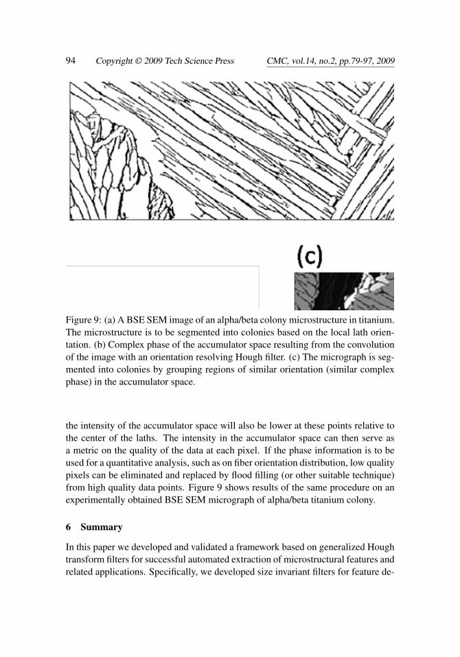
Figure 9: (a) A BSE SEM image of an alpha/beta colony microstructure in titanium.The microstructure is to be segmented into colonies based on the local lath orien-tation. (b) Complex phase of the accumulator space resulting from the convolutionof the image with an orientation resolving Hough filter. (c) The micrograph is seg-mented into colonies by grouping regions of similar orientation (similar complexphase) in the accumulator space.
the intensity of the accumulator space will also be lower at these points relative tothe center of the laths. The intensity in the accumulator space can then serve asa metric on the quality of the data at each pixel. If the phase information is to beused for a quantitative analysis, such as on fiber orientation distribution, low qualitypixels can be eliminated and replaced by flood filling (or other suitable technique)from high quality data points. Figure 9 shows results of the same procedure on anexperimentally obtained BSE SEM micrograph of alpha/beta titanium colony.
6 Summary
In this paper we developed and validated a framework based on generalized Houghtransform filters for successful automated extraction of microstructural features andrelated applications. Specifically, we developed size invariant filters for feature de-

Applications of the Phase-Coded Generalized Hough Transform 95
tection in 3-D by encoding scale information in the complex phase of the Houghfilters. The filters were formulated for the detection of arbitrary shapes eliminatingthe need for an analytical parameterization. The concept of a Hough filter was fur-ther generalized by considering complex filters that encode other parameters suchas line orientation, thus broadening the utility of the generalized Hough transformto a host of other microstructure analysis applications. Several examples were pre-sented on feature extraction in polycrystalline 3-D digital microstructure datasets aswould be obtained from serial sectioning. The application of generalized Hough fil-ters to the rapid calculation of the angularly resolved lineal path function and otherrelated statistical metrics was demonstrated in 2-D. Additionally, we demonstratedthe utility of orientation coded Hough filters to successfully address automated,feature based, microstructure segmentation.
Acknowledgement: The authors acknowledge financial support for this workfrom the Office of Naval Research, Award No. N000140510504 (Program Man-ager: Dr. Julie Christodoulou). SRN has been supported by the National ScienceFoundation Graduate Research Fellowship Program (NSF-GRFP). The authors alsowish to thank A.D. Rollett (CMU) for the polycrystalline dataset and H.L. Fraser(OSU) for the alpha/beta Ti dataset.
References
Adams, B., S. Wright, K. Kunze (1993): Orientation imaging: The emergence ofa new microscopy. Metallurgical and Materials Transactions A 24(4): 819-831.
Atherton, T. J., D. J. Kerbyson (1999): Size invariant circle detection. Image andVision Computing 17(11): 795-803.
Ballard, D. H. (1981): Generalizing the Hough transform to detect arbitrary shapes.Pattern Recognition 13(2): 111-122.
Betz, O., U. Wegst, D. Weide, M. Heethoff, L. Helfen, W.-K. Lee, P. Cloetens(2007): Imaging applications of synchrotron X-ray phase-contrast microtomogra-phy in biological morphology and biomaterials science. I. General aspects of thetechnique and its advantages in the analysis of millimetre-sized arthropod structure.Journal of Microscopy 227(1): 51-71.
Blavette, D., A. Bostel, J. M. Sarrau, B. Deconihout, A. Menand (1993): Anatom probe for three-dimensional tomography. Nature 363(6428): 432-435.
Brahme, A., M. H. Alvi, D. Saylor, J. Fridy, A. D. Rollett (2006): 3D reconstruc-tion of microstructure in a commercial purity aluminum. Scripta Materialia 55(1):75-80.
Briggs, W. L., V. E. Henson (1995): The DFT : an owner’s manual for the discrete

96 Copyright © 2009 Tech Science Press CMC, vol.14, no.2, pp.79-97, 2009
Fourier transform. Philadelphia, Society for Industrial and Applied Mathematics.
Canny, J. (1986): A Computational Approach to Edge Detection. Pattern Analysisand Machine Intelligence, IEEE Transactions on PAMI-8(6): 679-698.
Chapman, H. N., A. Barty, S. Marchesini, A. Noy, S. P. Hau-Riege, C. Cui,M. R. Howells, R. Rosen, H. He, J. C. H. Spence, U. Weierstall, T. Beetz, C.Jacobsen, D. Shapiro (2006): High-resolution ab initio three-dimensional x-raydiffraction microscopy. J. Opt. Soc. Am. A 23(5): 1179-1200.
Chawla, N., V. V. Ganesh, B. Wunsch (2004): Three-dimensional (3D) microstruc-ture visualization and finite element modeling of the mechanical behavior of SiCparticle reinforced aluminum composites. Scripta Materialia 51(2): 161-165.
Collins, P. C., B. Welk, T. Searles, J. Tiley, J. C. Russ, H. L. Fraser (2009):Development of methods for the quantification of microstructural features in [al-pha] + [beta]-processed [alpha]/[beta] titanium alloys. Materials Science and En-gineering: A 508(1-2): 174-182.
Duda, R. O., P. E. Hart (1972): Use of the Hough transformation to detect linesand curves in pictures. Commun. ACM 15(1): 11-15.
Flannery, B. P., H. W. Deckman, W. G. Roberge, K. L. D’amico (1987): Three-Dimensional X-ray Microtomography. Science 237(4821): 1439-1444.
Hough, P. V. C. (1962): Method and means for recognizing complex patterns. U.S.Patent No. 3069654.
Kerbyson, D. J., T. J. Atherton (1995): Circle detection using Hough transformfilters. Image Processing and its Applications, 1995., Fifth International Confer-ence on.
Kierkegaard, P. (1992): A method for detection of circular arcs based on theHough transform. Machine Vision and Applications 5(4): 249-263.
Lu, B., S. Torquato (1992): Lineal-path function for random heterogeneous mate-rials. Physical Review A 45(2): 922.
Maire, E., J. Y. Buffière, L. Salvo, J. J. Blandin, W. Ludwig and J. M. Létang(2001): On the Application of X-ray Microtomography in the Field of MaterialsScience. Advanced Engineering Materials 3(8): 539-546.
Manwart, C., S. Torquato, R. Hilfer (2000): Stochastic reconstruction of sand-stones. Physical Review E 62(1): 893.
Petushi, S., C. Katsinis, C. Coward, F. Garcia, A. Tozeren (2004): Automatedidentification of microstructures on histology slides. Biomedical Imaging: Nano toMacro, 2004. IEEE International Symposium on.
Samal, A., J. Edwards (1997): Generalized Hough transform for natural shapes.

Applications of the Phase-Coded Generalized Hough Transform 97
Pattern Recognition Letters 18(5): 473-480.
Schmidt, S., S. F. Nielsen, C. Gundlach, L. Margulies, X. Huang, D. J. Jensen(2004): Watching the Growth of Bulk Grains During Recrystallization of DeformedMetals. Science 305(5681): 229-232.
Schwartz, A. J., M. Kumar, B. L. Adams (2000): Electron backscatter diffractionin materials science. New York, Kluwer Academic.
Seidman, D. N. (2007): Three-Dimensional Atom-Probe Tomography: Advancesand Applications. Annual Review of Materials Research 37(1): 127-158.
Simmons, J., B. Bartha, M. De Graef, M. Comer (2008): Development of BayesianSegmentation Techniques for Automated Segmentation of Titanium Alloy Images.Microscopy and Microanalysis 14(SupplementS2): 602-603.
Singh, H., A. M. Gokhale, S. I. Lieberman, S. Tamirisakandala (2008): Im-age based computations of lineal path probability distributions for microstructurerepresentation. Materials Science and Engineering: A 474(1-2): 104-111.
Stutzman, P. (2004): Scanning electron microscopy imaging of hydraulic cementmicrostructure. Cement and Concrete Composites 26(8): 957-966.
Tiley, J., T. Searles, E. Lee, S. Kar, R. Banerjee, J. C. Russ, H. L. Fraser (2004):Quantification of microstructural features in [alpha]/[beta] titanium alloys. Materi-als Science and Engineering A 372(1-2): 191-198.
Torquato, S. (2002): Random heterogeneous materials : microstructure and macro-scopic properties. New York, NY, Springer.
Uchic, M. D., M. A. Groeber, D. M. Dimiduk, J. P. Simmons (2006): 3D mi-crostructural characterization of nickel superalloys via serial-sectioning using adual beam FIB-SEM. 55(1): 23-28.
Yeong, C. L. Y., S. Torquato (1998): Reconstructing random media. II. Three-dimensional media from two-dimensional cuts. Physical Review E 58(1): 224.
Zeman, J., M. Sejnoha (2007): From random microstructures to representativevolume elements. Modelling and Simulation in Materials Science and Engineering(4): S325.
