approaching heterogeneity of human epidermal growth factor ... · sofia asioli mda,1, francesca...
TRANSCRIPT
www.elsevier.com/locate/humpath
Human Pathology (2012) 43, 2070–2079
Original contribution
Approaching heterogeneity of human epidermalgrowth factor receptor 2 in surgical specimens ofgastric cancerSofia Asioli MDa,1, Francesca Maletta MDa,1, Ludovica Verdun di Cantogno BSc a,Maria A. Satolli MDb, Marina Schena MDb, Carla Pecchioni BSc a, Cristina Botta BSc a,Luigi Chiusa MDa, Luca Molinaro MDa, Luca Conti MDa, Giuseppe Viale MDc,Giuseppe Ingravallo MDd, Eugenio Maiorano MDd, Anna Sapino MDa,⁎
aDepartment of Biomedical Sciences and Human Oncology, University of Turin, 10126 Turin, ItalybOnco-Haematological Department, Oncology Unit, S. Giovanni Battista Hospital, 10126 Turin, ItalycDepartment of Pathology and Laboratory Medicine, European Institute of Oncology, University of Milan, 20122 Milan, ItalydDepartment of Pathological Anatomy, University of Bari, 70124 Bari, Italy
Received 2 January 2012; revised 17 February 2012; accepted 22 February 2012
es
Ere
0h
Keywords:Gastric cancer;HER2;Immunohistochemistry;Surgical specimen
Summary Gastric cancer shows intratumoral heterogeneity for human epidermal growth factor receptor2 expression. We evaluated whether the number of tissue blocks analyzed or the antibodies used mayinfluence the immunohistochemical results in gastrectomy specimens. Clinicopathologic data from 148patients receiving gastric surgery for cancer were collected. One tissue block for each of 88 primarytumors and 60 paired primary tumors and metastases was examined for human epidermal growth factorreceptor 2 status by immunohistochemistry using 3 different antibodies (HercepTest, CB11, and 4B5)and by fluorescent in situ hybridization. Two additional tissue blocks of the primary tumor were testedby immunohistochemistry if the results were negative on the first tissue block. The concordance amongthe 3 antibodies was 94.5% (testing 1 tissue block). Two cases showed a clinically significantdiscrepancy between primary tumor (score 0) and lymph nodes metastases (score 3+). Additional blockanalysis increased both the sensitivity (from 63% to 83%) and the accuracy (from 91% to 94%) ofimmunohistochemistry as compared with fluorescent in situ hybridization. The multiblock approachcould potentially identify a greater number of human epidermal growth factor receptor 2–positivegastric cancers, particularly those with higher levels of intratumor heterogeneity. In turn, human
Abbreviations: ToGA, Trastuzumab for Gastric Cancer; ESMO, European Society for Medical Oncology; IHC, immunohistochemistry; SISH, silver-nhanced in situ hybridization result; FISH, fluorescent in situ hybridization result; K, κ of Cohen-Fleiss; WK, weighted κ of Cohen; DSS, disease-specificurvival; HR, hazard ratio; CI, confidence interval; CT, chemotherapy; A, amplified; NA, not amplified; TP, true positive; TN, true negative.
This work was presented in part at the USCAP 100th Annual Meeting in San Antonio, TX, USA; February 26–March 4, 2011 (abstract no. 595).This work was supported by Project of Relevant National Interest (PRIN) - Italy 2008, Regione Piemonte Comitato Interministeriale Programmazione
conomica (CIPE) 2004 (Italy), Regione Piemonte (Italy) Ricerca Sanitaria Finalizzata 2008-2008 bis and 2009, and Rete Oncologica (Piemonte, Italy) 2010search fund grants. We also acknowledge Compagnia San Paolo and Fondazione Cassa di Risparmio di Torino (Italy) for funding supports.⁎ Corresponding author. Department of Biomedical Sciences and Human Oncology, University of Turin, 10126 Turin, Italy.E-mail address: [email protected] (A. Sapino).1 These authors contributed equally to this work.
046-8177© 2012 Elsevier Inc.ttp://dx.doi.org/10.1016/j.humpath.2012.02.017
Open access under CC BY-NC-ND license.

2071HER2 heterogeneity and gastrectomy specimens
epidermal growth factor receptor 2 positivity correlated with a worse prognosis (P = .011) and was anindependent variable in multivariate analysis (hazard ratio, 1.57). In conclusion, testing more than 1tissue block of cancer from specimens of gastric resection provides a more reliable human epidermalgrowth factor receptor 2 assessment regardless of the antibody used.© 2012 Elsevier Inc. Open access under CC BY-NC-ND license.
1. Introduction
In recent years, several studies have described the negativeprognostic role of human epidermal growth factor receptor(HER) 2 overexpression in gastric cancer [1-3]. In 2010, the“Trastuzumab for Gastric Cancer (ToGA)” study [4], arandomized, multicenter, international, phase 3, controlledtrial was designed to assess the clinical efficacy and safety ofthe anti-HER2 agent trastuzumab (Herceptin; Roche, Basel,Switzerland) as an addition to chemotherapy for first-linetreatment of advanced or metastatic gastric cancer over-expressing HER2 [4]. The results showed improved survivalfor patients treated with trastuzumab and chemotherapycompared with patients treated with chemotherapy alone. Atthe 12th European Society for Medical Oncology/WorldCongress on Gastrointestinal Cancer, it has been recom-mended that all patients with metastatic gastric adenocarci-noma who are candidates for first-line chemotherapy shouldbe tested for HER2 status, and patients with a tumoroverexpressing HER2, as defined by an immunohistochem-ical (IHC) score of 3+ or 2+ and a confirmatory silver-enhanced (SISH) or fluorescent (FISH) in situ hybridizationresult, should be treated with the cisplatin/fluoropyrimidineplus trastuzumab combination [5].
At present, the rate of HER2 positivity in gastriccarcinomas varies in the literature from 6.1% to 91%[1,4,6,7]. The HER2 status may be influenced by thehistologic type and location of the tumor or the age of thepatients. For example, Moelans et al [8] recently reported a3% rate of HER2 expression in patients with gastric canceryounger than 45 years. On the other hand, the discrepancycould be the consequence of use of anti-HER2 antibodieswith different sensitivity [9] and specificity. Another causeof the discrepancy may be the revision of the originaldefinition, as given by Hofmann et al [10], of the membranelocation of HER2 in overexpressing gastric cancer cells thatwas a “moderate to strong complete or basolateralmembranous reactivity.” This definition was slightlymodified in the ToGA trial [4], so that “intense lateralmembrane” positivity was considered valid as well. Thedifferent quantitative criteria used to define HER2 over-expression in surgical samples and biopsy specimens maybe involved as well in the aforementioned discrepancy:although the positivity of 1 cell cluster irrespective of thesize in the latter is sufficient, in a surgical specimen, at least10% of cells have to be present to be scored 3+ andclassified as overexpressing HER2 [4]. This percentage
value may be influenced by the heterogeneity [11] of HER2expression in large gastric cancer, so that the assessment ofHER2 in just 1 tissue block obtained from surgicalspecimens of gastrectomy could produce false-negativeresults. For example, Hsu et al [6] studying HER2expression in a large case series of gastric resection forcancer showed a rate of positive cases of 6.1%. This low rateof HER2 expression failed to be an independent prognosticfactor at multivariate analysis. However, it is worthmentioning that IHC tests were performed on tissuemicroarrays obtained from a single paraffin block. Recently,an up-to-date guidance on standardizing tissue processing,HER2 testing, and scoring in patients with gastric cancerremarked that HER2 results of tissue microarrays are notsuitable for clinical decision due to the heterogeneous natureof HER2 overexpression and amplification in this tumortype [12].
In the present study, taking into account the possibleheterogeneity of HER2 expression, we wanted to evaluatewhether to examine a single tissue block could impair theIHC results in surgical specimens of large gastric cancers.
2. Materials and methods
2.1. Case series
Formalin-fixed, paraffin-embedded blocks of specimens ofsurgically resected gastric cancer were retrieved from the filesof the Departments of Pathology of Ospedale S. GiovanniBattista of the University of Turin (98 cases) and of OspedalePoliclinico of the University of Bari (50 cases), Italy.
The case series was represented by 88 samples of primarytumor only and 60 samples of paired primary tumors andmetastases (58 metastatic lymph nodes and 2 livermetastases). Clinicopathologic and follow-up data (diseasepersistence, recurrence, and metastases) were collected forall cases.
Institutional review board permission was obtained, andthe study was conducted in compliance with the ethicalregulatory issues of the participating institutions for thehandling of biological specimens from tumor banks, that is,the samples exclusively available for research purposes inretrospective studies. The number of tissue blocks sampledfor each primary tumor ranged from 4 to 8. The hematoxylinand eosin slides were reviewed to select the block for HER2analysis; these blocks had to show at least 50% invasive
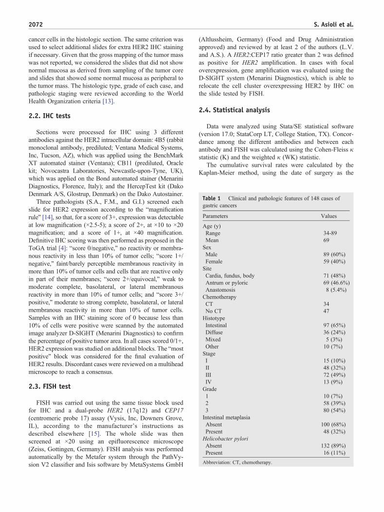
Table 1 Clinical and pathologic features of 148 cases ofgastric cancers
Parameters Values
Age (y)Range 34-89Mean 69
SexMale 89 (60%)Female 59 (40%)
SiteCardia, fundus, body 71 (48%)Antrum or pyloric 69 (46.6%)Anastomosis 8 (5.4%)
ChemotherapyCT 34No CT 47
HistotypeIntestinal 97 (65%)Diffuse 36 (24%)Mixed 5 (3%)Other 10 (7%)
StageI 15 (10%)II 48 (32%)III 72 (49%)IV 13 (9%)
Grade1 10 (7%)2 58 (39%)3 80 (54%)
Intestinal metaplasiaAbsent 100 (68%)Present 48 (32%)
Helicobacter pyloriAbsent 132 (89%)Present 16 (11%)
Abbreviation: CT, chemotherapy.
2072 S. Asioli et al.
cancer cells in the histologic section. The same criterion wasused to select additional slides for extra HER2 IHC stainingif necessary. Given that the gross mapping of the tumor masswas not reported, we considered the slides that did not shownormal mucosa as derived from sampling of the tumor coreand slides that showed some normal mucosa as peripheral tothe tumor mass. The histologic type, grade of each case, andpathologic staging were reviewed according to the WorldHealth Organization criteria [13].
2.2. IHC tests
Sections were processed for IHC using 3 differentantibodies against the HER2 intracellular domain: 4B5 (rabbitmonoclonal antibody, prediluted; Ventana Medical Systems,Inc, Tucson, AZ), which was applied using the BenchMarkXT automated stainer (Ventana); CB11 (prediluted, Oraclekit; Novocastra Laboratories, Newcastle-upon-Tyne, UK),which was applied on the Bond automated stainer (MenariniDiagnostics, Florence, Italy); and the HercepTest kit (DakoDenmark A/S, Glostrup, Denmark) on the Dako Autostainer.
Three pathologists (S.A., F.M., and G.I.) screened eachslide for HER2 expression according to the “magnificationrule” [14], so that, for a score of 3+, expression was detectableat low magnification (×2.5-5); a score of 2+, at ×10 to ×20magnification; and a score of 1+, at ×40 magnification.Definitive IHC scoring was then performed as proposed in theToGA trial [4]: “score 0/negative,” no reactivity or membra-nous reactivity in less than 10% of tumor cells; “score 1+/negative,” faint/barely perceptible membranous reactivity inmore than 10% of tumor cells and cells that are reactive onlyin part of their membranes; “score 2+/equivocal,” weak tomoderate complete, basolateral, or lateral membranousreactivity in more than 10% of tumor cells; and “score 3+/positive,” moderate to strong complete, basolateral, or lateralmembranous reactivity in more than 10% of tumor cells.Samples with an IHC staining score of 0 because less than10% of cells were positive were scanned by the automatedimage analyzer D-SIGHT (Menarini Diagnostics) to confirmthe percentage of positive tumor area. In all cases scored 0/1+,HER2 expression was studied on additional blocks. The “mostpositive” block was considered for the final evaluation ofHER2 results. Discordant cases were reviewed on a multiheadmicroscope to reach a consensus.
2.3. FISH test
FISH was carried out using the same tissue block usedfor IHC and a dual-probe HER2 (17q12) and CEP17(centromeric probe 17) assay (Vysis, Inc, Downers Grove,IL), according to the manufacturer's instructions asdescribed elsewhere [15]. The whole slide was thenscreened at ×20 using an epifluorescence microscope(Zeiss, Gottingen, Germany). FISH analysis was performedautomatically by the Metafer system through the PathVy-sion V2 classifier and Isis software by MetaSystems GmbH
(Altlussheim, Germany) (Food and Drug Administrationapproved) and reviewed by at least 2 of the authors (L.V.and A.S.). A HER2:CEP17 ratio greater than 2 was definedas positive for HER2 amplification. In cases with focaloverexpression, gene amplification was evaluated using theD-SIGHT system (Menarini Diagnostics), which is able torelocate the cell cluster overexpressing HER2 by IHC onthe slide tested by FISH.
2.4. Statistical analysis
Data were analyzed using Stata/SE statistical software(version 17.0; StataCorp LT, College Station, TX). Concor-dance among the different antibodies and between eachantibody and FISH was calculated using the Cohen-Fleiss κstatistic (K) and the weighted κ (WK) statistic.
The cumulative survival rates were calculated by theKaplan-Meier method, using the date of surgery as the
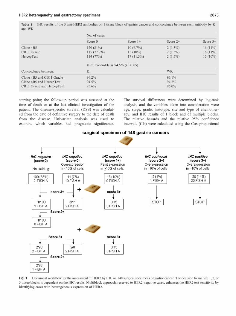
Table 2 IHC results of the 3 anti-HER2 antibodies on 1 tissue block of gastric cancer and concordance between each antibody by Kand WK
No. of cases
Score 0 Score 1+ Score 2+ Score 3+
Clone 4B5 120 (81%) 10 (6.7%) 2 (1.3%) 16 (11%)CB11 Oracle 115 (77.7%) 15 (10%) 2 (1.3%) 16 (11%)HercepTest 114 (77%) 17 (11.5%) 2 (1.5%) 15 (10%)
K of Cohen-Fleiss 94.5% (P b .05)
Concordance between: K WK
Clone 4B5 and CB11 Oracle 96.2% 96.1%Clone 4B5 and HercepTest 94.5% 94.2%CB11 Oracle and HercepTest 95.6% 96.0%
2073HER2 heterogeneity and gastrectomy specimens
starting point; the follow-up period was assessed at thetime of death or at the last clinical investigation of thepatient. The disease-specific survival (DSS) was calculat-ed from the date of definitive surgery to the date of deathfrom the disease. Univariate analysis was used toexamine which variables had prognostic significance.
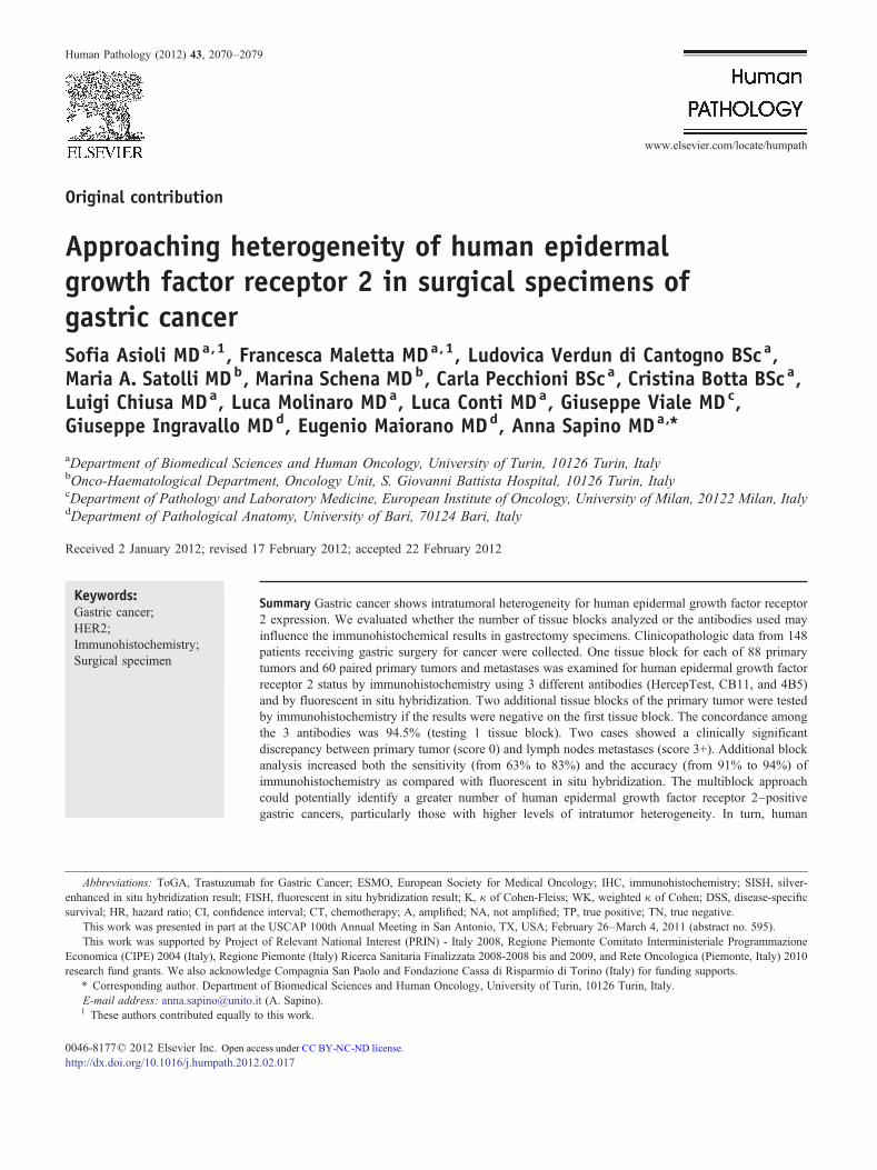
Fig. 1 Decisional workflow for the assessment of HER2 by IHC on 1483 tissue blocks is dependent on the IHC results. Multiblock approach, reseidentifying cases with heterogeneous expression of HER2.
The survival differences were determined by log-rankanalysis, and the variables taken into consideration wereage, stage, grade, histotype, site and type of chemother-apy, and IHC results of 1 block and of multiple blocks.The relative hazards and the relative 95% confidenceintervals (CIs) were calculated using the Cox proportional
surgical specimens of gastric cancer. The decision to analyze 1, 2, orrved to HER2-negative cases, enhances the HER2 test sensitivity by
Fig. 2 Example of relocation of image analysiswithD-SIGHT software (left, dark-field FISH) (right, bright-field IHC) using the 3-point alignmentmethod (points inside red circles). The software is able to exactly relocate, on the FISH-scanned slide, the same field of interest selected on the IHC-stained slides (green circles).
2074 S. Asioli et al.
hazard model. A P value less than .05 was consideredstatistically significant.
Fig. 3 Graphical correlation of HER2 expression, as detected onmultiple blocks with tumor topography in cases originally negativeon 1 block. In cases 5, 6, and 8, areas scored as 3+ (white triangle)were randomly distributed within score 2+ areas (dotted rectangle).The variance of HER2 expression was greater in tumors of groups Iand III of the classification Goseki et al.
3. Results
The clinicopathologic features of the patients arereported in Table 1. The follow-up period for the patientsranged from 1 to 64 months (mean, 29 months). Thetherapeutic protocol was known for 81 of 98 cases of theTurin series only. Twenty-eight patients received adjuvantchemotherapy (fluoropyrimidine) after surgery. Threepatients received neoadjuvant chemotherapy (platinum-fluoropyrimidine) before surgery. Two of these casesshowed HER2 overexpression/amplification both on thepreoperative biopsy and on the surgical specimen, whereas1 case was HER2 negative.
3.1. Analysis of concordances
The comparison of HER2 scores obtained using thedifferent antibodies did not show significant differences(Table 2). The 20 cases scored as 3+ were all amplifiedregardless of the antibody used, whereas only 1 of the 2cases scored 2+ was amplified. The K statistic betweenthe IHC results of the primary tumor and the metastaseswas 79% for 4B5 and CB11 and 71% for HercepTest
(P b .05). Using 4B5 and CB11, 2 cases showed aclinically relevant discrepancy because the primary tumorswere classified as negative (score 0, because HER2
2075HER2 heterogeneity and gastrectomy specimens
expression was strong but in b10% of cells), whereas thelymph node metastases were diffusely scored 3+. Inanother 2 cases, there was a minor discrepancy becausethe HER2 expression was either 3+ or 2+ in the primaryor in the metastasis, but both tumor and lymph nodemetastases were amplified. The concordance between theIHC scoring of the 3 antibodies and the FISH results wasslightly better in the metastases (82% for 4B5, 88.6% forCB11/Oracle, and 88% for HercepTest; P b .05) than inthe primary tumors (80% for 4B5, 84% for CB11/Oracle,and 82% for HercepTest; P b .05).
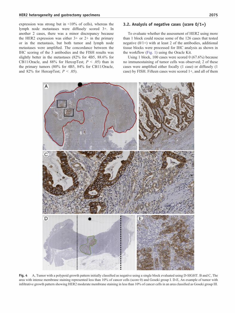
Fig. 4 A, Tumor with a polypoid growth pattern initially classified as nearea with intense membrane staining represented less than 10% of cancerinfiltrative growth pattern showing HER2moderate membrane staining in
3.2. Analysis of negative cases (score 0/1+)
To evaluate whether the assessment of HER2 using morethan 1 block could rescue some of the 126 cases that testednegative (0/1+) with at least 2 of the antibodies, additionaltissue blocks were processed for IHC analysis as shown inthe workflow (Fig. 1) using the Oracle Kit.
Using 1 block, 100 cases were scored 0 (67.6%) becauseno immunostaining of tumor cells was observed; 2 of thesecases were amplified either focally (1 case) or diffusely (1case) by FISH. Fifteen cases were scored 1+, and all of them
gative using a single block evaluated using D-SIGHT. B and C, Thecells (score 0) and Goseki group I. D-E, An example of tumor withless than 10% of cancer cells in an area classified as Goseki group III.
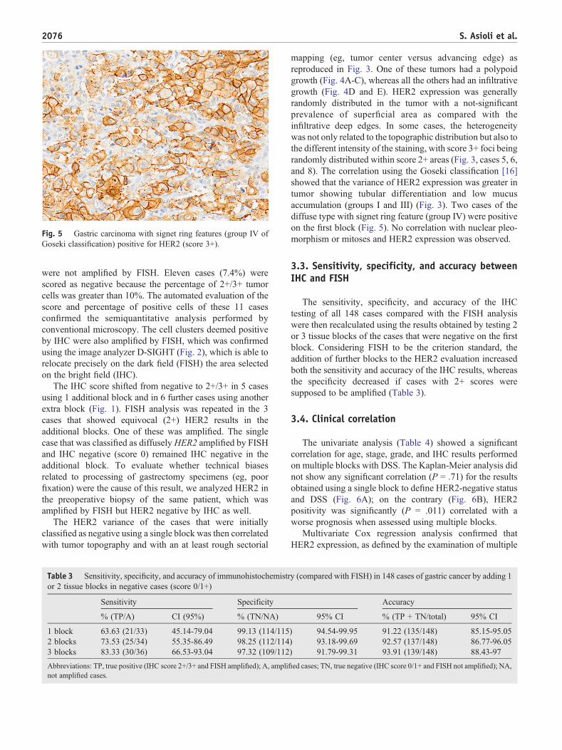
Fig. 5 Gastric carcinoma with signet ring features (group IV ofGoseki classification) positive for HER2 (score 3+).
2076 S. Asioli et al.
were not amplified by FISH. Eleven cases (7.4%) werescored as negative because the percentage of 2+/3+ tumorcells was greater than 10%. The automated evaluation of thescore and percentage of positive cells of these 11 casesconfirmed the semiquantitative analysis performed byconventional microscopy. The cell clusters deemed positiveby IHC were also amplified by FISH, which was confirmedusing the image analyzer D-SIGHT (Fig. 2), which is able torelocate precisely on the dark field (FISH) the area selectedon the bright field (IHC).
The IHC score shifted from negative to 2+/3+ in 5 casesusing 1 additional block and in 6 further cases using anotherextra block (Fig. 1). FISH analysis was repeated in the 3cases that showed equivocal (2+) HER2 results in theadditional blocks. One of these was amplified. The singlecase that was classified as diffusely HER2 amplified by FISHand IHC negative (score 0) remained IHC negative in theadditional block. To evaluate whether technical biasesrelated to processing of gastrectomy specimens (eg, poorfixation) were the cause of this result, we analyzed HER2 inthe preoperative biopsy of the same patient, which wasamplified by FISH but HER2 negative by IHC as well.
The HER2 variance of the cases that were initiallyclassified as negative using a single block was then correlatedwith tumor topography and with an at least rough sectorial
Table 3 Sensitivity, specificity, and accuracy of immunohistochemistror 2 tissue blocks in negative cases (score 0/1+)
Sensitivity Specificity
% (TP/A) CI (95%) % (TN/NA)
1 block 63.63 (21/33) 45.14-79.04 99.13 (114/1152 blocks 73.53 (25/34) 55.35-86.49 98.25 (112/1143 blocks 83.33 (30/36) 66.53-93.04 97.32 (109/112
Abbreviations: TP, true positive (IHC score 2+/3+ and FISH amplified); A, amplinot amplified cases.
mapping (eg, tumor center versus advancing edge) asreproduced in Fig. 3. One of these tumors had a polypoidgrowth (Fig. 4A-C), whereas all the others had an infiltrativegrowth (Fig. 4D and E). HER2 expression was generallyrandomly distributed in the tumor with a not-significantprevalence of superficial area as compared with theinfiltrative deep edges. In some cases, the heterogeneitywas not only related to the topographic distribution but also tothe different intensity of the staining, with score 3+ foci beingrandomly distributed within score 2+ areas (Fig. 3, cases 5, 6,and 8). The correlation using the Goseki classification [16]showed that the variance of HER2 expression was greater intumor showing tubular differentiation and low mucusaccumulation (groups I and III) (Fig. 3). Two cases of thediffuse type with signet ring feature (group IV) were positiveon the first block (Fig. 5). No correlation with nuclear pleo-morphism or mitoses and HER2 expression was observed.
3.3. Sensitivity, specificity, and accuracy betweenIHC and FISH
The sensitivity, specificity, and accuracy of the IHCtesting of all 148 cases compared with the FISH analysiswere then recalculated using the results obtained by testing 2or 3 tissue blocks of the cases that were negative on the firstblock. Considering FISH to be the criterion standard, theaddition of further blocks to the HER2 evaluation increasedboth the sensitivity and accuracy of the IHC results, whereasthe specificity decreased if cases with 2+ scores weresupposed to be amplified (Table 3).
3.4. Clinical correlation
The univariate analysis (Table 4) showed a significantcorrelation for age, stage, grade, and IHC results performedon multiple blocks with DSS. The Kaplan-Meier analysis didnot show any significant correlation (P = .71) for the resultsobtained using a single block to define HER2-negative statusand DSS (Fig. 6A); on the contrary (Fig. 6B), HER2positivity was significantly (P = .011) correlated with aworse prognosis when assessed using multiple blocks.
Multivariate Cox regression analysis confirmed thatHER2 expression, as defined by the examination of multiple
y (compared with FISH) in 148 cases of gastric cancer by adding 1
Accuracy
95% CI % (TP + TN/total) 95% CI
) 94.54-99.95 91.22 (135/148) 85.15-95.05) 93.18-99.69 92.57 (137/148) 86.77-96.05) 91.79-99.31 93.91 (139/148) 88.43-97
fied cases; TN, true negative (IHC score 0/1+ and FISH not amplified); NA,

Table 4 Univariate analysis of clinical and pathologic data correlated with DSS
n Events Mean DSS 95% CI χ2 P
Age b70 y 67 28 48.52 43.49-53.55 4.279 .039≥70 y 81 36 40.27 34.55-46.00
Stage I 15 2 54.62 45.09-64.16 11.38 .01II 48 12 48.91 41.85-55.98III 72 39 41.72 36.09-47.35IV 13 11 37.03 27.38-46.68
Grade I 10 1 63.00 63.00-63.00 6.186 .045II 58 21 42.86 36.08-49.27III 80 42 42.51 37.27-47.76
Histotype Intestinal 97 34 44.00 38.95-49.06 5.63 .13Diffuse 36 25 41.03 33.37-48.69Mixed 5 2 30.62 13.66-47.59Other 10 3 59.66 52.12-62.12
Site Cardia, fundus, body 71 28 42.82 36.99-48.65 0.722 .697Antrum, pylorus 69 34 44.62 39.05-50.20Anastomosis 8 2 46.00 18.56-73.44
Chemotherapy No 47 20 31.19 23.74-38.65 1.959 .162Yes 34 12 38.30 30.04-46.57
IHC with 1 block IHC score 0-1-2NA 127 54 44.54 40.31-48.77 0.134 .71IHC score 2A-3 21 10 42.50 32.13-52.87
IHC with new protocol IHC score 0-1-2NA 118 46 46.89 42.66-51.12 6.436 .011IHC score 2A-3 30 18 34.67 26.07-43.27
Abbreviations: NA, not amplified; A, amplified.
2077HER2 heterogeneity and gastrectomy specimens
blocks, was an independent variable for prognosis (hazardratio [HR], 1.572; 95% CI, 1.195-2.112; P = .001), togetherwith the age of patients (HR, 1.986; 95% CI, 1.187-3.324;P = .009), the disease stage (HR, 1.749; 95% CI, 1.2-2.549;P = .004), and the grade (HR, 2.797; 95% CI, 1.018-7.682;P = .046) of gastric cancer.
Fig. 6 Kaplan-Meier estimates of DSS (in months) according to HER2specimens of gastric cancer. Abbreviations: NA, not amplified; A, ampli
4. Discussion
Previous studies have described HER2 expressionheterogeneity as a frequent event in gastric cancer[7,10,17,18]. In breast cancer, the intratumor heterogeneityof the HER2 gene is defined as a tumor showing more than
status assessed using 1 block (A) and multiple blocks (B) of surgicalfied.

2078 S. Asioli et al.
5% but less than 50% of infiltrating cells, with a ratio higherthan 2.2 by FISH and the incidence ranging from 5% to 30%[19]. In gastric carcinoma, the intratumor heterogeneity hasyet to be specifically defined. For example, in a recent work,Lee et al [20] focusing on IHC 3+ or IHC 2+⁄SISH-positivetumors determined that negative or not uniform weaklystained areas were present in 50% of the surgical and in 42%of biopsy specimens, respectively. Boers et al [9] stated thatthe number of positive fragments and/or of calculated H-score did not predict amplification status in 73% of biopsyspecimens due to heterogeneity of HER2 expression ofgastric adenocarcinomas.
In the present work, we assessed heterogeneity in a seriesof 126 gastric cancers, of 148 surgically resected, that werenegative at the IHC analyses performed using differentantibodies on 1 tissue block. We can exclude a significantinfluence for the antibody's selection if the IHC proceduresare adequately performed [12] and if the scoring followsstrict rules [10,14]. On the other hand, in our case series,inadequate sample selection produced 7.1% of false-negativeresults. This suggests that the HER2 IHC analysis of a singleblock of the primary tumor could not be sufficientlyconfident to compensate for the heterogeneity of HER2expression in a large gastric cancer. For example, in thepresent series, the mean size of the tumor was around 5.5 cm.Using a single block, 14% of gastric cancers were positive,whereas the addition of further blocks increased the rate to20%, a value that was slightly higher than that obtained byHofmann et al [10] using 1 block (17%). However, theanalysis of 3 tissue blocks increased the positive rate toapproximately 50% of the cases overexpressing HER2 butscored as negative because the percentage of positive cellswas less than 10% in a single block. In our series, only 1(0.7%) of 148 case was diffusely amplified by FISH andconstantly negative by IHC, whereas 9 amplified cases wererescued from IHC negative to positive by testing at least 3blocks. The accuracy of IHC, as compared with FISH,increased from 91% to about 94% using multiple-blockanalysis. In the recently published guidelines on HER2 ingastric cancer [12], it is suggested that for surgical specimencases with strong HER2 staining in less than 10% of cells,retesting with ISH may be warranted. If such a sample isFISH-SISH positive, the tumor may be considered to beHER2 positive similar to the scoring on biopsy samples [12].
Heterogeneity may also lead to discrepancies betweenprimary tumors and metastases. For example, in the presentwork, we showed a clinically relevant difference in 2 casesthat were scored 0 (not eligible for trastuzumab therapy) inthe primary tumor because of the low percentage of HER2-positive cells and scored 3+ in the metastatic lymph node(eligible for trastuzumab).
The hypothesis that the level of HER2 protein mightinfluence the response to trastuzumab was investigated in apost hoc analysis of the ToGA trial [4]; this analysis revealeda significant increase in the response to trastuzumab in thesubgroup with high HER2 expression. However, some
benefits were also achieved by treating patients with HER2gene amplification by FISH and protein negativity by IHC.The underlying explanations for these results have not beenfully evaluated. We demonstrated that multisampling detec-tion of HER2 expression could potentially identify cases withhigher levels of intratumor heterogeneity, which may beassociated with adverse outcomes but, in turn, could have atherapeutic advantage from trastuzumab treatment. As amatter of fact, HER2 positivity correlated with a worseprognosis and was an independent variable in multivariateanalysis only when assessed using multiple blocks. Theseresults seem biologically more reliable than those reported byothers [6] on tissue microarrays obtained from a singleparaffin block, where a lower HER2 expression failed to be anindependent prognostic factor.
Thus, if the HER2 test is performed on surgical samplesof gastric carcinoma, the results may be influenced by thetissue block more than by the antibody selection. Gastrictumors at diagnosis are generally larger than breast tumors,and the analysis of a single block may not compensate for thepossible intratumor heterogeneity. Particularly, when theHER2 overexpressing cells are focally present, more than 1tissue block should be tested by IHC.
In conclusion, the workflow we propose (Fig. 1), whileproducing a relatively higher workload for pathologists,increases the accuracy of the HER2 testing in gastric cancer.
References
[1] Gravalos C, Jimeno A. HER2 in gastric cancer: a new prognostic factorand a novel therapeutic target. Ann Oncol 2008;19:1523-9.
[2] Tanner M, Hollmen M, Junttila TT, et al. Amplification of HER-2 ingastric carcinoma: association with topoisomerase IIalpha geneamplification, intestinal type, poor prognosis and sensitivity totrastuzumab. Ann Oncol 2005;16:273-8.
[3] Wagner AD, Moehler M. Development of targeted therapies inadvanced gastric cancer: promising exploratory steps in a new era.Curr Opin Oncol 2009;21:381-5.
[4] Bang YJ, Van Cutsem E, Feyereislova A, et al. Trastuzumab incombination with chemotherapy versus chemotherapy alone fortreatment of HER2-positive advanced gastric or gastro-oesophagealjunction cancer (ToGA): a phase 3, open-label, randomised controlledtrial. Lancet 2010;376:687-97.
[5] Van Cutsem E, Dicato M, Geva R, et al. The diagnosis andmanagement of gastric cancer: expert discussion and recommendationsfrom the 12th ESMO/World Congress on Gastrointestinal Cancer,Barcelona, 2010. Ann Oncol 2011;22:1-9.
[6] Hsu JT, Chen TC, Tseng JH, et al. Impact of HER-2 over-expression/amplification on the prognosis of gastric cancer patientsundergoing resection: a single-center study of 1,036 patients.Oncologist 2011;16:1706-13.
[7] Grabsch H, Sivakumar S, Gray S, Gabbert HE, Muller W. HER2expression in gastric cancer: rare, heterogeneous and of no prognosticvalue—conclusions from 924 cases of two independent series. CellOncol 2010;32:57-65.
[8] Moelans CB, Milne AN, Morsink FH, Offerhaus GJ, van Diest PJ.Low frequency of HER2 amplification and overexpression in earlyonset gastric cancer. Cell Oncol (Dordr) 2011;34:89-95.
[9] Boers JE, Meeuwissen H, Methorst N. HER2 status in gastro-oesophageal adenocarcinomas assessed by two rabbit monoclonal
2079HER2 heterogeneity and gastrectomy specimens
antibodies (SP3 and 4B5) and two in situ hybridization methods (FISHand SISH). Histopathology 2011;58:383-94.
[10] Hofmann M, Stoss O, Shi D, et al. Assessment of a HER2 scoringsystem for gastric cancer: results from a validation study. Histopa-thology 2008;52:797-805.
[11] Yang J, Luo H, Li Y, et al. Intratumoral heterogeneity determinesdiscordant results of diagnostic tests for human epidermal growth factorreceptor (HER) 2 in gastric cancer specimens. Cell Biochem Biophys2011;62:221-8.
[12] Rüschoff J, Hanna W, Bilous M, et al. HER2 testing in gastriccancer: a practical approach. Mod Pathol 2012:1-14.
[13] Lauwers GY, Carneiro F, Graham DY, et al. Gastric carcinoma. In:Bosman FT, Carneiro F, Hruban RH, Theise ND, editors. WHOclassification of tumours of the digestive system. Lyon, France: IARCPress; 2010. p. 48-58.
[14] Ruschoff J, Dietel M, Baretton G, et al. HER2 diagnostics ingastric cancer-guideline validation and development of standard-ized immunohistochemical testing. Virchows Arch 2010;457:299-307.
[15] Marchio C, Lambros MB, Gugliotta P, et al. Does chromosome 17centromere copy number predict polysomy in breast cancer? Afluorescence in situ hybridization and microarray-based CGH analysis.J Pathol 2009;219:16-24.
[16] Goseki N, Takizawa T, Koike M. Differences in the mode of theextension of gastric cancer classified by histological type: newhistological classification of gastric carcinoma. Gut 1992;33:606-12.
[17] Lee EY, Cibull ML, Strodel WE, Haley JV. Expression of HER-2/neuoncoprotein and epidermal growth factor receptor and prognosis ingastric carcinoma. Arch Pathol Lab Med 1994;118:235-9.
[18] Marx AH, Tharun L, Muth J, et al. HER-2 amplification is highlyhomogenous in gastric cancer. HUM PATHOL 2009;40:769-77.
[19] Vance GH, Barry TS, Bloom KJ. Genetic heterogeneity in HER2testing in breast cancer: panel summary and guidelines. Arch PatholLab Med 2009;133:611-2.
[20] Lee S, de Boer WB, Fermoyle S, Platten M, Kumarasinghe MP.Human epidermal growth factor receptor 2 testing in gastriccarcinoma: issues related to heterogeneity in biopsies and resections.Histopathology 2011;59:832-40.