appropriate imaging of the lower extremity · imaging modalities bony anatomy, pregnancy alignment,...
TRANSCRIPT
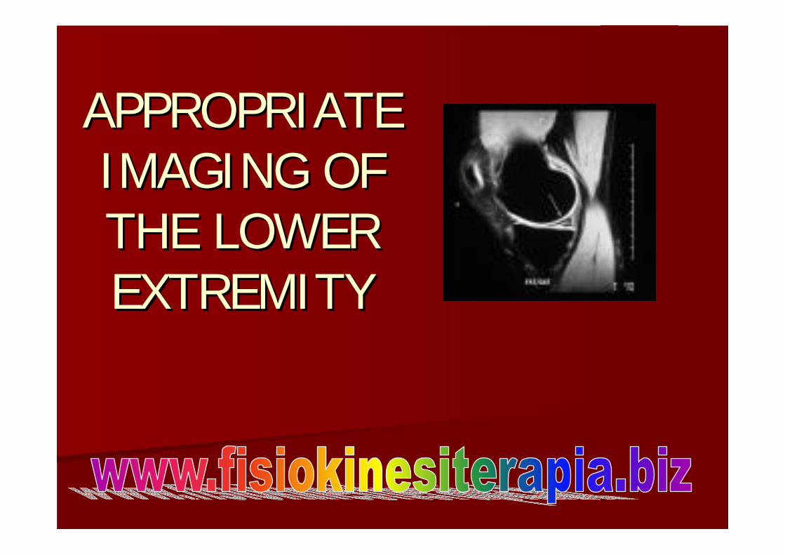
APPROPRIATE APPROPRIATE IMAGING OF IMAGING OF THE LOWER THE LOWER EXTREMITYEXTREMITY

OBJECTIVESOBJECTIVES
I. Overview of appropriate imagingI. Overview of appropriate imaging–– CostCost–– ACRACR
II. Plain xII. Plain x--ray viewsray viewsIII. Advance imagingIII. Advance imaging–– MRIMRI–– CTCT–– Bone ScanBone Scan

OBJECTIVESOBJECTIVES
IV. Hip/pelvisIV. Hip/pelvis–– AcuteAcute–– ChronicChronic
V. KneeV. Knee–– AcuteAcute–– ChronicChronic
VI. Ankle/footVI. Ankle/foot–– AcuteAcute–– ChronicChronic

OVERVIEWOVERVIEW
CostCostACRACRImaging modalitiesImaging modalities

IMAGING MODALITIESIMAGING MODALITIES
PregnancyPregnancyBony anatomy, Bony anatomy, alignment, alignment, fxfx, , periostealperiosteal rxnsrxns, , callus, noncallus, non--unionunion
55--15 15 $35$35--250250XX--RAYRAY
Pregnancy, Pregnancy, radioactive dye radioactive dye allergyallergy
Increased bone Increased bone turnover, stress turnover, stress fxfx, fractures, , fractures, tumortumor
INJECTION + INJECTION + IMAGINGIMAGING90 MINUTES90 MINUTES
$700$700BONE BONE SCANSCAN
Ferromagnetic Ferromagnetic materials, materials, pacemaker, pacemaker, defibdefib, metallic , metallic hardwarehardware
Soft tissue, bone Soft tissue, bone edema, edema, fxfx lines, lines, fluid, bursa, fluid, bursa, tumor matrixtumor matrix
6060$1500$1500MRIMRI
PregnancyPregnancyBony anatomy, Bony anatomy, clarify clarify fxfx, tumor , tumor matrixmatrix
1515--30 30 $845$845CT SCANCT SCAN
CONTRACONTRA--INDICATIONSINDICATIONS
IDEAL USESIDEAL USESTIMETIME(MINUTES)(MINUTES)
CHARGE CHARGE (APPROXIMATE)(APPROXIMATE)
MODALITYMODALITY
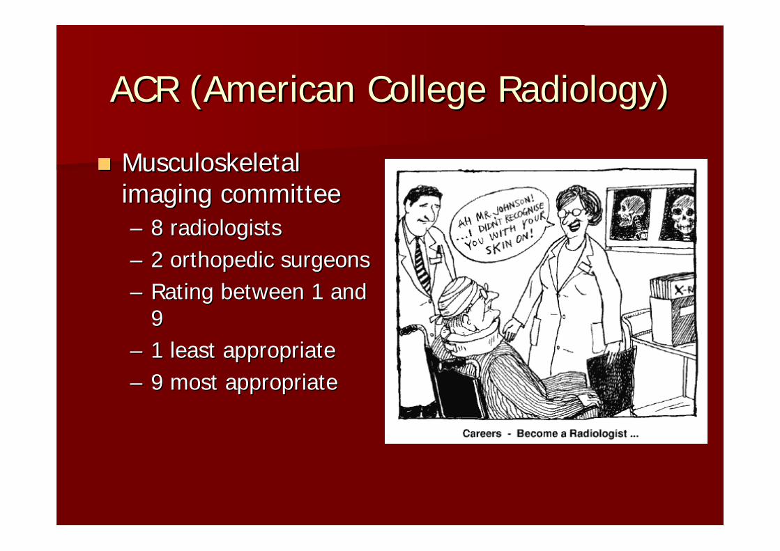
ACR (American College Radiology)ACR (American College Radiology)
Musculoskeletal Musculoskeletal imaging committeeimaging committee–– 8 radiologists8 radiologists–– 2 orthopedic surgeons2 orthopedic surgeons–– Rating between 1 and Rating between 1 and
99–– 1 least appropriate1 least appropriate–– 9 most appropriate9 most appropriate

OBJECTIVESOBJECTIVES
IV. Hip/pelvisIV. Hip/pelvis–– AcuteAcute–– ChronicChronic
V. KneeV. Knee–– AcuteAcute–– ChronicChronic
VI. Ankle/footVI. Ankle/foot–– AcuteAcute–– ChronicChronic

TIPSTIPS
TREAT THE PATIENT, TREAT THE PATIENT, NOT IMAGINGNOT IMAGINGTREAT THE TREAT THE PATHOLOGY NOT PATHOLOGY NOT PAINPAINEVALUATE FUNCTION EVALUATE FUNCTION AND CORRELATE AND CORRELATE WITH IMAGING IF WITH IMAGING IF NECESSARYNECESSARY

ADVANCED IMAGINGADVANCED IMAGING
ORDER:ORDER:–– IMAGING MODALITYIMAGING MODALITY–– WORKING DIAGNOSISWORKING DIAGNOSIS–– SPECIFICITY OF SPECIFICITY OF
LOCATIONLOCATION–– EgEg. MRI left knee. MRI left knee
Evaluate degenerative Evaluate degenerative tear posterior horn tear posterior horn medial meniscusmedial meniscus

CASE # 1CASE # 1 --Acute Hip PainAcute Hip Pain
65 65 y/oy/o female slips and female slips and falls at home. She is falls at home. She is unable to bear much unable to bear much weight, and she c/o weight, and she c/o some severe right some severe right groin pain.groin pain.

CASE # 1CASE # 1--Acute Hip PainAcute Hip Pain
SUSPECT:SUSPECT:–– Femur fracture (shaft, neck)Femur fracture (shaft, neck)–– Pelvic fracturePelvic fracture
Plain Plain PELVIS APPELVIS AP–– (NOT SINGLE HIP)(NOT SINGLE HIP)–– ACR (9)ACR (9)–– Frog leg view Frog leg view
(externally rotated) (externally rotated) view (if AP negative)view (if AP negative)

CASE #2CASE #2--Chronic Hip PainChronic Hip Pain
50 50 y/oy/o female c/o female c/o right groin pain x 6 right groin pain x 6 months. No prior months. No prior traumatic injury. Pain traumatic injury. Pain with walking and with walking and some painful loss of some painful loss of hip range of motion.hip range of motion.
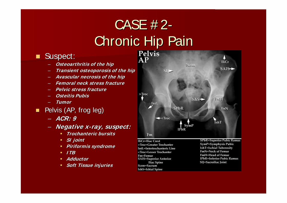
CASE #2CASE #2--Chronic Hip PainChronic Hip Pain
Suspect:Suspect:–– Osteoarthritis of the hipOsteoarthritis of the hip–– Transient osteoporosis of the hipTransient osteoporosis of the hip–– AvascularAvascular necrosis of the hipnecrosis of the hip–– Femoral neck stress fractureFemoral neck stress fracture–– Pelvic stress fracturePelvic stress fracture–– OsteitisOsteitis PubisPubis–– TumorTumor
Pelvis (AP, frog leg)Pelvis (AP, frog leg)–– ACR: 9ACR: 9–– Negative xNegative x--ray, suspect:ray, suspect:
TrochantericTrochanteric bursitsbursitsSI jointSI jointPiriformisPiriformis syndromesyndromeITBITBAdductorAdductorSoft Tissue injuriesSoft Tissue injuries

CASE #2CASE #2--Chronic Hip PainChronic Hip Pain
Consider further imaging:Consider further imaging:–– Arthritis on plain xArthritis on plain x--ray?ray?
MRI not recommendedMRI not recommendedACR: 2ACR: 2
–– No arthritisNo arthritisMRI (ACR: 9)MRI (ACR: 9)Bone Scan (no ACR rating)Bone Scan (no ACR rating)Suspect:Suspect:
–– AVN hipAVN hip–– Transient osteoporosisTransient osteoporosis–– Pelvic stress Pelvic stress fxfx–– Femoral neck stress Femoral neck stress fxfx–– LabalLabal tear (MRItear (MRI--
arthrogramarthrogram))

MRI PELVISMRI PELVIS

CASE #3CASE #3--Acute Knee PainAcute Knee Pain
35 35 y/oy/o male c/o knee male c/o knee pain after ski injury. pain after ski injury. He is unable to flex He is unable to flex his knee 90 degrees.his knee 90 degrees.

OTTAWA CRITERIAOTTAWA CRITERIA--KNEEKNEE1. Age 55 or older1. Age 55 or older2. Isolated 2. Isolated
tenderness of the tenderness of the patellapatella
3. Tenderness of the 3. Tenderness of the head of the fibulahead of the fibula
4. Inability to flex at 4. Inability to flex at 90 degrees90 degrees
5.5. Inability to bear Inability to bear weightweight
* Joint effusion * Joint effusion within 24 hourswithin 24 hours

CASE #3CASE #3--Acute Knee PainAcute Knee Pain
Walk with no limpWalk with no limpTwisting injury and no Twisting injury and no effusioneffusionSuspect:Suspect:–– Patellar instabilityPatellar instability–– Collateral ligament injuryCollateral ligament injury–– SynovialSynovial plicaplica–– Fat Pad impingementFat Pad impingement–– Stable knee injuriesStable knee injuries
–– No xNo x--raysrays–– ACR: 2ACR: 2

CASE #3CASE #3--Acute Knee PainAcute Knee Pain
Meet Ottawa Criteria:Meet Ottawa Criteria:Suspect:Suspect:–– Patellar fracturePatellar fracture–– Fibular head fractureFibular head fracture–– Loose body (OCD injury)Loose body (OCD injury)–– TibialTibial plateau fractureplateau fracture–– Femoral Femoral condylecondyle fracturefracture–– TibialTibial spine avulsionspine avulsion–– Lateral Lateral tibialtibial plateau avulsion plateau avulsion
((segundsegund’’ss fracture)fracture)
–– 22--v Knee, v Knee, wtbearingwtbearingAP or PA, lateral + AP or PA, lateral + MerchantMerchant’’s if anterior s if anterior knee painknee pain
–– ACR: 9ACR: 9

CASE #3CASE #3--Acute Knee PainAcute Knee Pain
NO ACR NO ACR recommendations for recommendations for acute twisting knee acute twisting knee injury with instability, injury with instability, recurrent swelling or recurrent swelling or mechanical symptomsmechanical symptomsSUSPECT:SUSPECT:–– CruciateCruciate ligament injuryligament injury–– MeniscalMeniscal injuryinjury–– OCD injury/loose bodyOCD injury/loose body
MRI (no ACR rating)MRI (no ACR rating)

MRI KNEEMRI KNEE

ACLACL--MeniscusMeniscus
LOCKED BUCKET LOCKED BUCKET HANDLEHANDLE--ACLACL–– *Test Passive terminal *Test Passive terminal
extension*extension*–– 22--stage arthroscopystage arthroscopy
Repair meniscusRepair meniscusDelayed ACL Delayed ACL reconstructionreconstruction

SPECIFICITY OF CONDITIONSPECIFICITY OF CONDITION
NOT ALL CRUCIATE LIGAMENT TEARS NOT ALL CRUCIATE LIGAMENT TEARS NEED SURGERYNEED SURGERY–– ACL IN MIDDLE AGE, PARTICULARLY WITH ACL IN MIDDLE AGE, PARTICULARLY WITH
ARTHRITIS MAY NOT NEED ITARTHRITIS MAY NOT NEED IT–– PCL TEARS AND SOME MENISCAL TEARS CAN PCL TEARS AND SOME MENISCAL TEARS CAN
BE TREATED CONSERVATIVELYBE TREATED CONSERVATIVELY–– MRI SHOULD BE PREOPERATIVE TOOL.MRI SHOULD BE PREOPERATIVE TOOL.
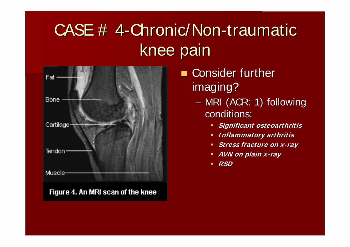
CASE #4CASE #4--Chronic/NonChronic/Non--traumatic traumatic knee painknee pain
53 53 y/oy/o male with male with medial compartment medial compartment pain and mild swelling pain and mild swelling x 2 months. Stable x 2 months. Stable knee exam, knee exam, ttpttp of of medial compartment.medial compartment.

CASE #4CASE #4--Chronic/NonChronic/Non--traumatic traumatic knee painknee pain
Suspect:Suspect:–– Arthritis (medial, lateral, Arthritis (medial, lateral,
patellofemoralpatellofemoral))–– Patellar Patellar malalignmentmalalignment–– AVN femoral AVN femoral condyecondye–– Loose bodiesLoose bodies–– OsteochondralOsteochondral lesionslesions–– Stress fracturesStress fractures–– TumorTumor–– PellegriniPellegrini--StiedaStieda
22--v Knee, v Knee, wtbearingwtbearing PA PA or AP, lateral + or AP, lateral + MerchantMerchant’’s if anterior s if anterior knee painknee painACR: 9ACR: 9

WEIGHTBEARING KNEE XWEIGHTBEARING KNEE X--RAYSRAYS
CASE # 4CASE # 4--Chronic/NonChronic/Non--traumatic traumatic knee painknee pain
Consider further Consider further imaging?imaging?–– MRI (ACR: 1) following MRI (ACR: 1) following
conditions:conditions:Significant osteoarthritisSignificant osteoarthritisInflammatory arthritisInflammatory arthritisStress fracture on xStress fracture on x--rayrayAVN on plain xAVN on plain x--rayrayRSDRSD
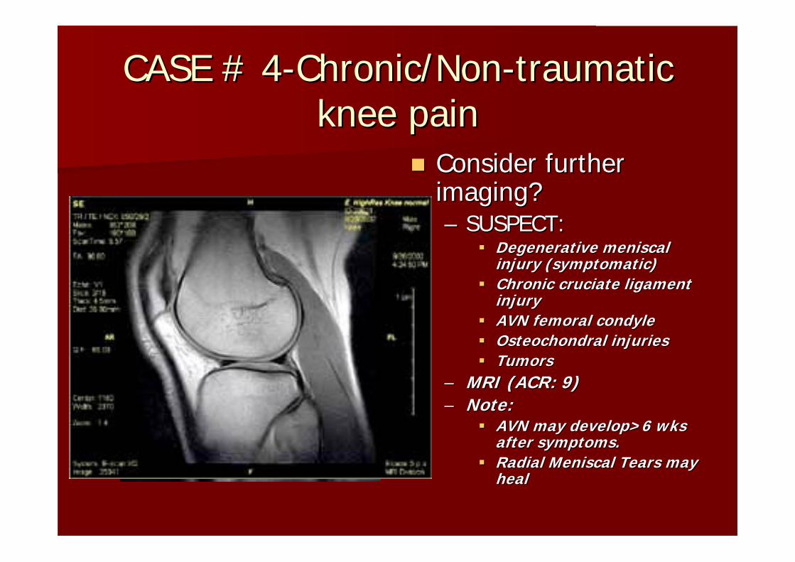
CASE # 4CASE # 4--Chronic/NonChronic/Non--traumatic traumatic knee painknee pain
Consider further Consider further imaging?imaging?–– SUSPECT:SUSPECT:
Degenerative Degenerative meniscalmeniscalinjury (symptomatic)injury (symptomatic)Chronic Chronic cruciatecruciate ligament ligament injuryinjuryAVN femoral AVN femoral condylecondyleOsteochondralOsteochondral injuriesinjuriesTumorsTumors
–– MRI (ACR: 9)MRI (ACR: 9)–– Note: Note:
AVN may develop>6 wks AVN may develop>6 wks after symptoms.after symptoms.Radial Radial MeniscalMeniscal Tears may Tears may healheal

MRIMRI--OCD LESIONSOCD LESIONS

CASE # 4CASE # 4--Chronic/NonChronic/Non--traumatic traumatic knee painknee pain
Consider further Consider further imaging?imaging?–– SUSPECT:SUSPECT:
PatellofemoralPatellofemoral syndromesyndromeOsteoarthritisOsteoarthritisTendonitis Tendonitis (Hamstring/Patellar)(Hamstring/Patellar)ITB syndromeITB syndromeBursitis (PreBursitis (Pre--Patellar/ITB)Patellar/ITB)SynovialSynovial PlicaPlicaSynovitisSynovitisMeniscalMeniscal TearTear
–– NO MRI NO MRI

ANKLE VS. FOOTANKLE VS. FOOT
ANKLE ANKLE –– TibiotalarTibiotalar jointjoint
FOOTFOOT–– HindfootHindfoot–– MidfootMidfoot–– ForefootForefoot
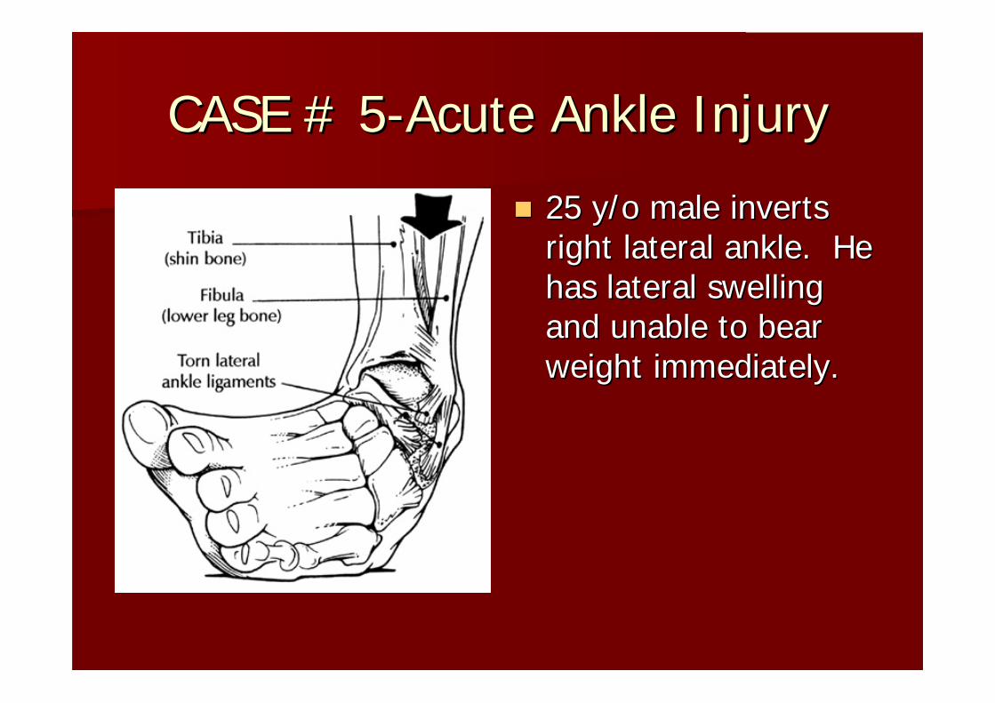
CASE # 5CASE # 5--Acute Ankle InjuryAcute Ankle Injury
25 25 y/oy/o male inverts male inverts right lateral ankle. He right lateral ankle. He has lateral swelling has lateral swelling and unable to bear and unable to bear weight immediately.weight immediately.

Case #5Case #5--Acute Ankle InjuryAcute Ankle Injury
OTTAWA CRITERIA:OTTAWA CRITERIA:–– NonNon--weightbearingweightbearing
after injury or in after injury or in emergency emergency dept/clinicdept/clinic
–– Tenderness over Tenderness over malleolimalleoli (posterior (posterior ½½ lateral lateral malleolusmalleolus), talus, ), talus, calcaneuscalcaneus
–– Inability to Inability to ambulate 4 stepsambulate 4 steps

Case #5Case #5--Acute Ankle InjuryAcute Ankle Injury
SUSPECT:SUSPECT:–– Fibular Fibular fxfx/lateral /lateral malleolimalleoli–– Distal tibia Distal tibia fxfx/medial /medial malleolusmalleolus–– Talus Talus fxfx (lateral process/dome, (lateral process/dome,
neck)neck)–– Calcaneus(anteriorCalcaneus(anterior process)process)–– SyndesmoticSyndesmotic injuryinjury
Ankle 3Ankle 3--v (AP, lateral, v (AP, lateral, mortise)mortise)–– ACR: 9ACR: 9
Continued Continued sxsx, repeat 3v, repeat 3v–– Suspect:Suspect:
Missed/occult Missed/occult fxfxTalarTalar dome OCDdome OCD

CASE #6CASE #6--CHRONIC ANKLE PAINCHRONIC ANKLE PAIN
33 33 y/oy/o male with male with recurrent ankle recurrent ankle injuries and injuries and anterolateralanterolateral ankle ankle pain with mild pain with mild swelling x 6 months.swelling x 6 months.

CASE #6CASE #6--CHRONIC ANKLE PAINCHRONIC ANKLE PAIN
SUSPECT:SUSPECT:–– TalarTalar dome OCDdome OCD–– Loose bodiesLoose bodies–– Ankle/Ankle/subtalarsubtalar arthritisarthritis–– TumorTumor
Ankle 3Ankle 3--vv–– ACR: 9ACR: 9

CASE #6CASE #6--CHRONIC ANKLE PAINCHRONIC ANKLE PAIN
Improved with Improved with rehabrehabSUSPECT:SUSPECT:–– DeconditionedDeconditioned ankleankle–– Chronic ankle Chronic ankle ligamentousligamentous
instabilityinstability–– TendinopathyTendinopathy–– Other soft tissue injuriesOther soft tissue injuries
No further imagingNo further imaging

CASE #6CASE #6--CHRONIC ANKLE PAINCHRONIC ANKLE PAIN
Continued Continued sxsx and and negative xnegative x--raysraysSUSPECT:SUSPECT:
–– Posterior Posterior tibialistibialis tendonitis/teartendonitis/tear–– PeronealPeroneal tendonitis/teartendonitis/tear–– TalarTalar Dome OCDDome OCD–– Tarsal CoalitionTarsal Coalition–– Stress Stress fxfx (distal fibula/tibia)(distal fibula/tibia)
MRI (ACR: 9)MRI (ACR: 9)SUSPECT:SUSPECT:
–– TalarTalar Dome OCDDome OCD–– Tarsal CoalitionTarsal Coalition
CT SCAN (ACR: 2)CT SCAN (ACR: 2)

MRI ANKLEMRI ANKLE--TALAR DOME OCDTALAR DOME OCD
CASE#7CASE#7--Acute foot injuryAcute foot injury
37 37 y/oy/o female twists female twists foot, has swelling on foot, has swelling on dorsum of foot. dorsum of foot.

CASE#7CASE#7--Acute foot injuryAcute foot injury
MIDFOOT/FOREFOOTMIDFOOT/FOREFOOTSUSPECT:SUSPECT:–– Metatarsal Metatarsal fxfx–– Jones Jones fxfx–– PhalynxPhalynx fxfx–– LisFrancLisFranc injuryinjury–– Tarsal coalitionTarsal coalition–– Accessory Accessory navicularnavicular–– Anterior process of Anterior process of calcaneuscalcaneus fxfx–– Lateral process of talus Lateral process of talus fxfx–– Turf toe (MTP sprain)Turf toe (MTP sprain)
Foot 3Foot 3--v v (AP/lat/oblique)(AP/lat/oblique)–– ACR: 9ACR: 9

CASE#7CASE#7--Acute foot injuryAcute foot injury
NEGATIVE XNEGATIVE X--RAYRAYConsider further Consider further imaging?imaging?–– Uncommon injuries:Uncommon injuries:
SUSPECT:SUSPECT:–– Posterior Posterior tibialistibialis tendon teartendon tear–– PeronealPeroneal tendon teartendon tear–– LisFrancLisFranc injury (should have had injury (should have had
weigthbearingweigthbearing feet with feet with comparison views)comparison views)
MRI foot (ACR: 9)MRI foot (ACR: 9)

ACUTEACUTE--HINDFOOT INJURYHINDFOOT INJURY
Direct fall on Direct fall on hindfoothindfootSUSPECT SUSPECT –– CalcaneusCalcaneus fxfx
CalcaneusCalcaneus 22--vv–– Lateral, HarrisLateral, Harris--BeathBeath–– ACR: 9ACR: 9

CASE #8CASE #8--CHRONIC FOOT PAINCHRONIC FOOT PAIN
54 54 y/oy/o female with female with lateral midlateral mid--foot pain x foot pain x 6 months with mild 6 months with mild swelling and limp.swelling and limp.

CASE #8CASE #8--CHRONIC FOOT PAINCHRONIC FOOT PAIN
MIDFOOT/FOREFOOTMIDFOOT/FOREFOOTSUSPECT:SUSPECT:–– Metatarsal stress Metatarsal stress fxfx–– Tarsal Tarsal navicularnavicular stress stress fxfx–– CuboidCuboid stress stress fxfx–– MidfootMidfoot arthritisarthritis–– Accessory Accessory navicularnavicular–– Os Os cuboidiscuboidis–– FreibergFreiberg’’s infractions infraction–– SesamoiditisSesamoiditis ((sesamoidsesamoid/axial /axial
view helpful)view helpful)–– HalluxHallux valgusvalgus–– TumorTumor–– Jones stress Jones stress fxfx
Foot 3Foot 3--v v –– ACR: 9ACR: 9
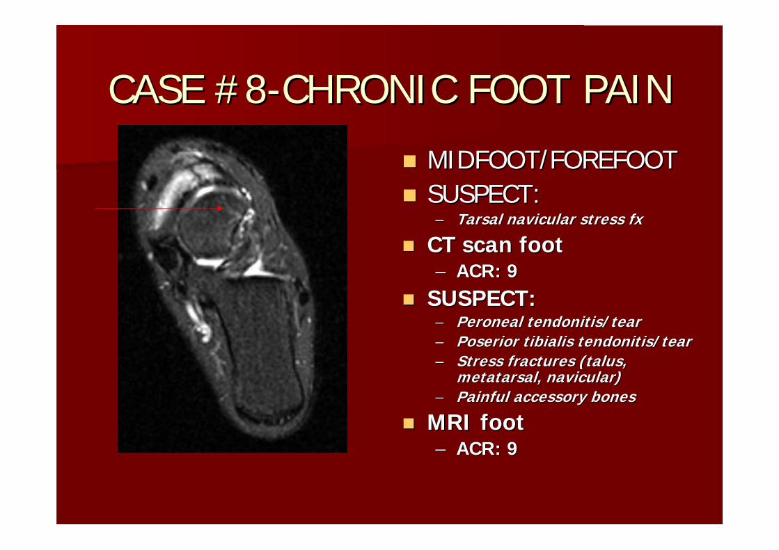
CASE #8CASE #8--CHRONIC FOOT PAINCHRONIC FOOT PAIN
MIDFOOT/FOREFOOTMIDFOOT/FOREFOOTSUSPECT:SUSPECT:–– Tarsal Tarsal navicularnavicular stress stress fxfx
CT scan foot CT scan foot –– ACR: 9ACR: 9
SUSPECT:SUSPECT:–– PeronealPeroneal tendonitis/teartendonitis/tear–– PoseriorPoserior tibialistibialis tendonitis/teartendonitis/tear–– Stress fractures (talus, Stress fractures (talus,
metatarsal, metatarsal, navicularnavicular))–– Painful accessory bonesPainful accessory bones
MRI footMRI foot–– ACR: 9ACR: 9

CASE #8CASE #8--CHRONIC FOOT PAINCHRONIC FOOT PAIN
MIDFOOT/FOREFOOTMIDFOOT/FOREFOOTSUSPECT STRESS FX:SUSPECT STRESS FX:–– Tarsal Tarsal navicularnavicular–– MetatarsalMetatarsal–– TalusTalus–– CuboidCuboid–– CalcaneusCalcaneus ((hindfoothindfoot))
Bone scanBone scan–– ACR: 6ACR: 6–– + scan for + scan for NavicularNavicular or or
Talus stress Talus stress fxfx–– CT scan or MRI/referCT scan or MRI/refer–– All others and negative All others and negative
study follow clinicallystudy follow clinically

CASE #8CASE #8--CHRONIC FOOT PAINCHRONIC FOOT PAIN
SUSPECT:SUSPECT:–– Plantar Plantar fascitisfascitis–– NeuromaNeuroma–– MetarsalgiaMetarsalgia–– Painful Painful pespes planusplanus–– Achilles tendonitisAchilles tendonitis–– Fat pad insufficiencyFat pad insufficiency
No further imaging No further imaging necessarynecessary

CASE#9CASE#9--CHRONIC HINDFOOT CHRONIC HINDFOOT PAINPAIN
SUSPECT:SUSPECT:–– CalcaneusCalcaneus stress stress fxfx–– TalarTalar neck stress neck stress fxfx–– SubtalarSubtalar arthritisarthritis–– Painful Painful osos trigonumtrigonum–– HaglundHaglund’’ss deformitydeformity–– Tarsal coalition Tarsal coalition
((CalcaneonavicularCalcaneonavicular coalition coalition seen on foot oblique), Obtain seen on foot oblique), Obtain foot 3foot 3--v as wellv as well
CalcaneusCalcaneus 22--vv–– Lateral, HarrisLateral, Harris--BeathBeath–– ACR: 9ACR: 9

SUMMARYSUMMARY
CLASSIFY MUSCULOSKELETAL CONDITIONS AS CLASSIFY MUSCULOSKELETAL CONDITIONS AS ACUTE OR CHRONIC/NONACUTE OR CHRONIC/NON--TRAUMATICTRAUMATICHAVE SPECIFICITY OF LOCATION OF HAVE SPECIFICITY OF LOCATION OF SYMPTOMS/EXAM FINDINGSSYMPTOMS/EXAM FINDINGSHAVE LINEAR THOUGHT PROCESS FOR HAVE LINEAR THOUGHT PROCESS FOR DIFFERENTIAL DIAGNOSES AND SUBSEQUENT DIFFERENTIAL DIAGNOSES AND SUBSEQUENT IMAGINGIMAGINGCONSERVATIVE TREATMENT AND IMAGING IS CONSERVATIVE TREATMENT AND IMAGING IS OFTEN WARRANTEDOFTEN WARRANTED
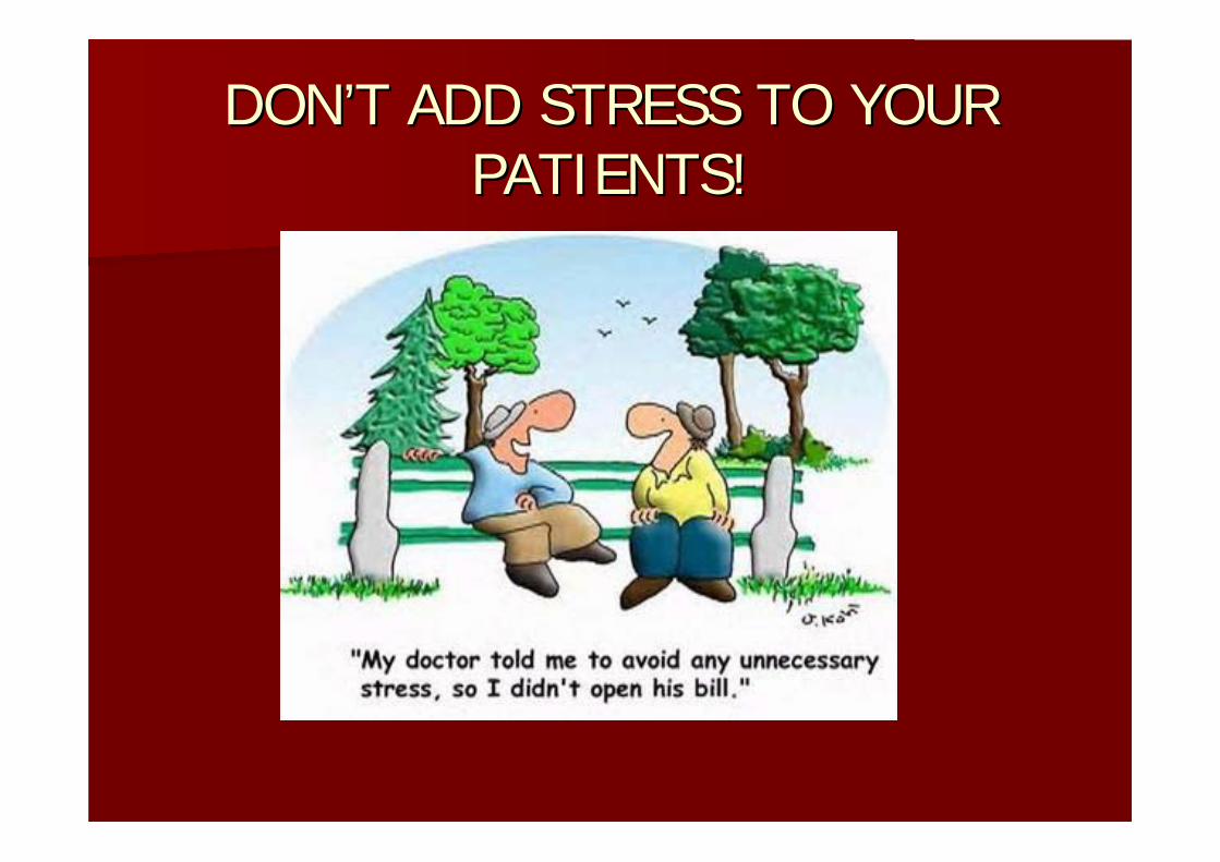
DONDON’’T ADD STRESS TO YOUR T ADD STRESS TO YOUR PATIENTS!PATIENTS!