appropriate utilization microbiology laboratorywickup.weebly.com/uploads/1/0/3/6/10368008/... ·...
TRANSCRIPT

Appropriate utilization of the microbiology laboratory
11 April 2013

Lecture Plan
• Revision of infectious disease• Triad of infectious disease• Interaction between host and infectious agent• Pathogenesis • Phases of laboratory testing• Conclusion

Introduction
• Infectious disease predominant cause of illness and death in many countries
• Improved sanitation • Improved medical standards• Poor socioeconomic conditions of majority –quagmire of infectious disease
• Introduction of antimicrobials early 20thcentury – erroneously thought all infectious disease could be controlled / eradicated

Introduction (2)
• Negative consequences of “progress”– Prolong human life– Success premature survival– Correct and incorrect use of antimicrobials
• Human incursion into environments not well adapted to
• Free movements of animals and plants across the globe
• Need to utilize the laboratory correctly –understanding the inter‐connections between humans and infectious agents

Triad of infectious diseases
infectious agent
affected host environment

Interactions between hosts and infectious agents
• Commensalistic– Infectious agent and host do not affect each other
• Mutualistic / Symbiotic– Both host and infectious agent benefit from each other
• Saprophytic– Infectious agent benefits from host but does no harm
• Parasitic – Infectious agent benefits from host and causes harm
• Colonizing bacteria– Exists on surface as commensal, saprophyte or parasite
• Opportunistic agents– Potential pathogen causing disease in immuno‐compromised host

Relationships to hostsRelationship Infectious agent Example
Free living most bacteriafungi parasites
S. aureus, E coli, K. pneumoniaeCandida sp, Cryptococcus sp.Taenia sp, Trypanosoma sp.
Facultatively intra‐cellular
some bacteriasome fungimycobacteria
Legionella sp, Brucella spHistoplasma spMycobacterium tuberculosis
Obligatory intracellular
some bacteria some parasitesviruses
Rickettsia, Chlamydia spToxoplasma sp, Plasmodium spInfluenza virus, measles virus

Pathogenesis of infection
• Attachment to the surface epithelium –fimbria, adhesins
• Multiply on the surface (respiratory, gastrointestinal tract, endocardium )
• Express virulence factors – exotoxins, endotoxins
• Invade the surface• Dissemination to other organs

Classes of infectious agentsBacteria
• Gram positive bacilli– Clostridium difficile– Listeria monoytogenes
• Gram positive cocci– Staphylococcus aureus– Streptococcus pneumoniae
• Gram negative bacilli– Klebsiella pneumoniae– Escherichia coli
• Gram negative cocci– Neisseria meningitidis

Classes of infectious agentsFungi
• Yeasts– Candida albicans– Cryptococcus neoformans
• Moulds– Mucorales– Aspergillus fumigatus
• Dermatophytes– Microsporum canus– Trychophyton rubrum– Epidermophyton floccosum
• Dimorphic fungi– Histoplasma capsulatum– Sporothrix schenkii
• Pneumocystis jiroveci
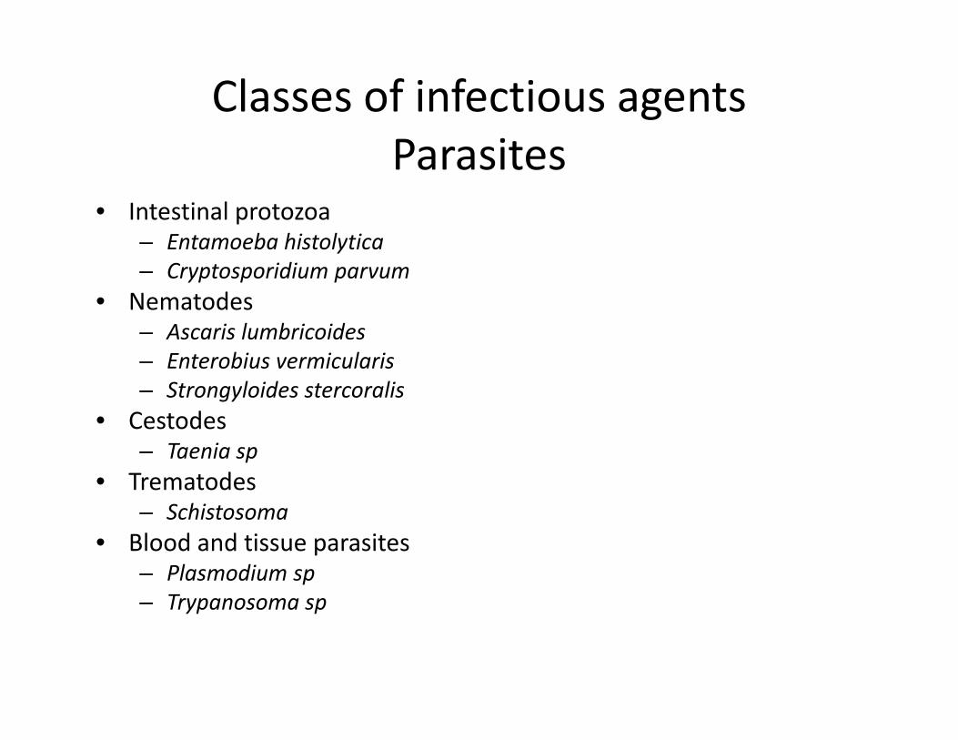
Classes of infectious agentsParasites
• Intestinal protozoa– Entamoeba histolytica – Cryptosporidium parvum
• Nematodes– Ascaris lumbricoides– Enterobius vermicularis– Strongyloides stercoralis
• Cestodes– Taenia sp
• Trematodes– Schistosoma
• Blood and tissue parasites– Plasmodium sp– Trypanosoma sp

Classes of infectious agentsViruses
• DNA viruses– Herpes viruses – herpes, cytomegalovirus, varicella zoster, Epstein‐Barr virus
– Parvoviruses– Adenoviruses– Hepadnaviruses –hepatitis B
• RNA viruses– Orthomyxoviruses – influenza– Paramyxoviruses – measles, mumps, respiratory syncytial– Retroviruses ‐ human immunodeficiency– Reoviruses – rotavirus– Piconaviruses ‐ polio

Phases of laboratory testing
• Before analysis (Pre‐analytical)• During analysis (Analytical)• After analysis ,before receipt of results (Post analytical )

Pre‐analytical
• Specimen collection• Specimen transport• Specimen reception• Specimen rejection
• Specimen analysis
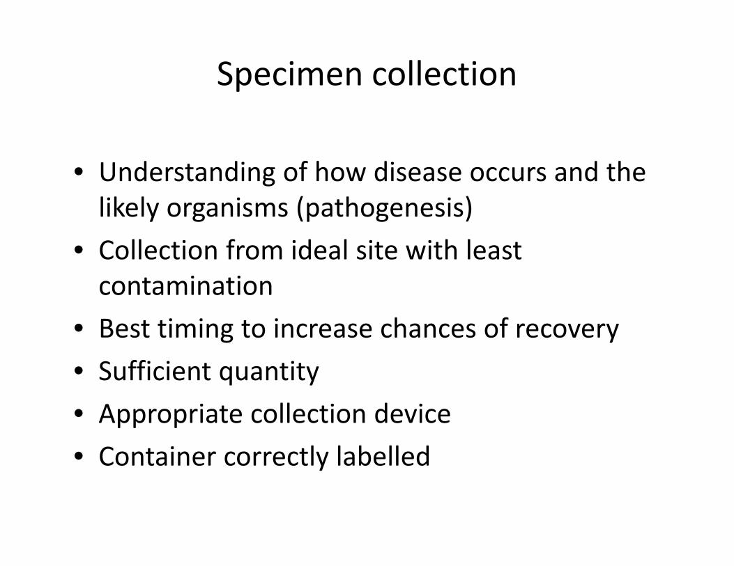
Specimen collection
• Understanding of how disease occurs and the likely organisms (pathogenesis)
• Collection from ideal site with least contamination
• Best timing to increase chances of recovery• Sufficient quantity• Appropriate collection device• Container correctly labelled

Specimen transport
• Aim to maintain specimen to as near original as possible
• Appropriate collection device, sterile• Transported in a safe way (Occupational Health and Safety Act)
• Transport as rapidly as possible• Appropriate temperature, refrigeration• Transport medium e.g. Stewart’s transport medium

Specimen reception
• Safe environment, protective clothing• Discard leaking containers • Preliminary observation (sputum vs urine)

Rejection of specimens
• Criteria must be established and communicated
• Reason for rejection must be given– Specimens received in formalin for culture– Old (24hr) sputum specimen– Specimen in improper container
• If specimen too little, incorrect, incorrectly transported communicate with clinician

Specimen analysis
• Visual observation• Microscopic specimen analysis• Culture and sensitivity• Serological analysis• Molecular diagnosis

Specimen analysis
• Microscopic examination (differential staining)– Gram stain (bacteria, scoring of sputum Bartlett’s)– India ink (capsule e.g. cryptococcosis)– Ziehl Neelson stain (tuberculosis, leprosy)– Auramine stain(tuberculosis, leprosy)– Geimsa stain (malaria, sleeping sickness)– Fluorescent microscopy (parasites)– Dark field microscopy (syphilis)– Electron microscopy(viruses)

Specimen analysis
• Culture and susceptibility testing / sensitivity (utilization of media and inhibition by antibiotics susceptible to)– Select primary culture media –enriched/selective– Isolate microorganism– Identification and antimicrobial susceptibility testing (manual or automated)
– Interpretation of results

Specimen analysis
• Serological diagnoses (antigen and antibody reaction)– Detection of antigen (organism) or antibody– Visualise reaction – charcoal, fluorescent and non fluorescent linked reagents (ELISA)
– Measure titres– Prevalence of disease important for interpretation – Increase sensitivity and specificity– Rapid onsite, automated immunoassays, incubation

Specimen analysis
• Molecular diagnosis– Identification of sequences in genetic material– Amplification by PCR detects small numbers– Identification of organism– Identification of resistant mutations– Rapid – Sensitive and specific

Post analytical
• Review of results• Quality confirmed• Pre‐determine whether routine, urgent or panic values?
• Reflex testing• Interpretation will be given• Report sent to clinicians, hard copy or electronic• Correct demographic data essential• Ensure turn‐around‐time (TAT) achieved
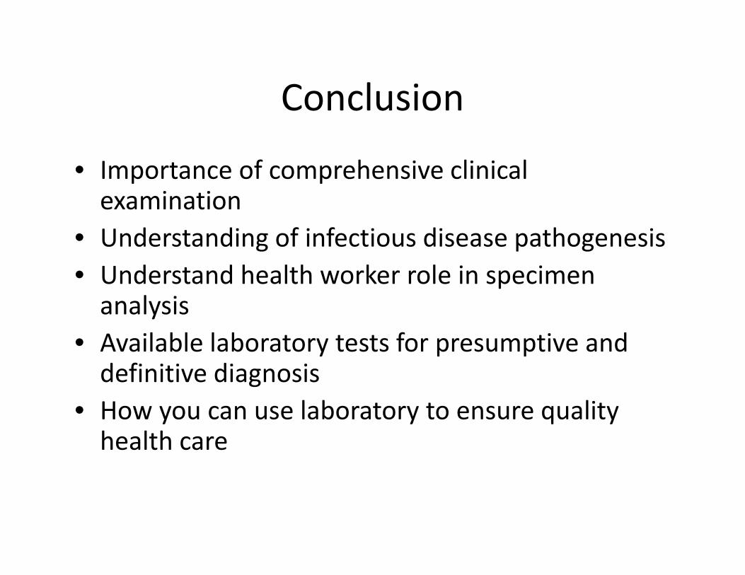
Conclusion
• Importance of comprehensive clinical examination
• Understanding of infectious disease pathogenesis• Understand health worker role in specimen analysis
• Available laboratory tests for presumptive and definitive diagnosis
• How you can use laboratory to ensure quality health care