apr2014
DESCRIPTION
Pashubandha Monthly eBulletinTRANSCRIPT

Pashubandha 2014 Volume No : 3 Issue : 04
Prof. H. A. Upendra# and Dr. Madhukar*
#Director, *Assistant Professor, Institute of Wildlife Veterinary Research, KVAFSU,
Doddaluvara, Kodagu – 571232. ([email protected])
Veterinary medicine is a branch of science that deals with prevention, diagnosis and treatment of
diseases and disorders in all animal species, both domesticated and wild. Further, veterinary profession
also aids in production of food as well as other items of animal origin.
Veterinary services are provided by a veterinary physician (also known as a vet, veterinary surgeon
or veterinarian), with assistance from para veterinary workers such as livestock veterinary inspectors and
assistants.
In addition to protecting the animal wealth, ensuring food security and generating livelihood,
veterinary professionals protect human health through the monitoring and control of zoonotic disease
(infectious disease transmitted from non-human animals to humans) in coordination with epidemiologists.
Development of Veterinary Profession
All the successful civilizations had a wealth of food, draft and security
animals, which necessitated use of herbs and surgical procedures in management
of the ailments in these animals. The Egyptian Papyrus of Kahun (1900 BCE)
and Vedic Indian Literatures are one of the ancient accounts of Veterinary
medicine. Infact, Nakula from the mythological pages of Mahabharata is
considered as father of veterinary medicine in India. Most of the ancient
literature focused on horse as they were economically very important.
The first modern veterinary college was established by
Claude Bourgelat in Lyon in 1761-62 to prevent future occurrences of
devastations due to cattle plague (Rinderspest) in French herds. This was indeed
achieved in France which continued successfully with the whole world declared
free of Rinderpest in 2011. Formal veterinary education in India began in 1862
with the establishment of an army veterinary school in Pune. The first civil
veterinary school was started in Babugarh (Hapur), in Uttar Pradesh, in 1877.
Newsletter Date : 30th April 2014 Volume No: 3 Issue : 04
Veterinary College, Bengaluru Monthly e-Bullletin

These schools had the limited objective of training Indians to serve as assistants in remount depots
and on military farms. This followed establishment of the first veterinary college at Lahore, now in
Pakistan, in 1882; veterinary research laboratory at Pune in 1889 and Bombay Veterinary College in
1886.
Subsequently, the importance of veterinary education was widely realized and numerous veterinary
colleges were established. To regulate the education and maintain the standards, Veterinary Council of
India was established in 1984. The veterinary council of India has listed 34 colleges in its website which
are managed by 25 Universities across the nation.
Veterinary education in Karnataka is provided by Karnataka Veterinary Animal and Fisheries
Science University, located at Bidar. Four veterinary colleges located at Bangalore, Bidar, Shimoga and
Hassan are involved in teaching, whereas diagnostic services are provided by Institute of Animal Health &
Veterinary Biologicals, and research and extension activities are conducted at numerous research and
information centers distributed across the state.
Pashubandha 2014 Volume No : 3 Issue : 01 Pashubandha 2014 Volume No : 3 Issue : 04
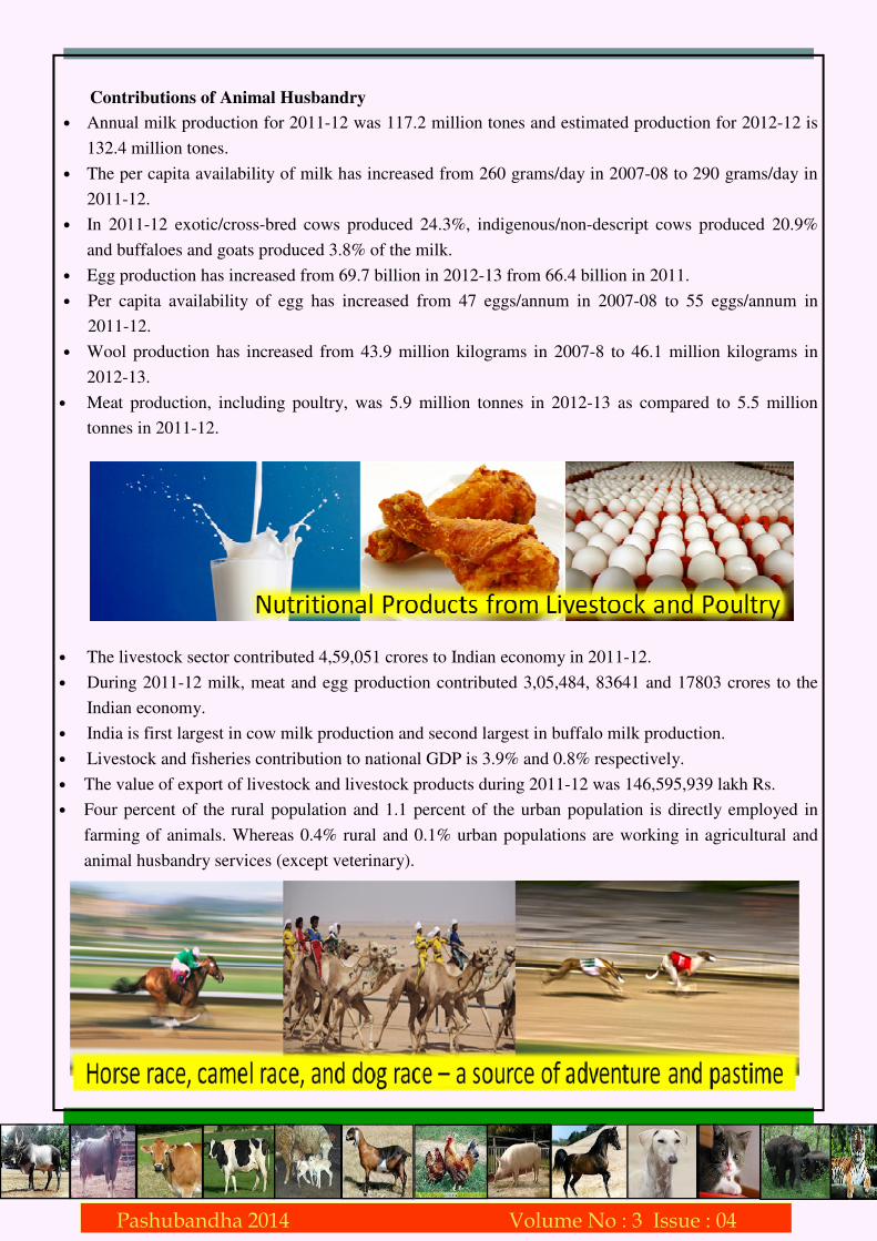
Contributions of Animal Husbandry
• Annual milk production for 2011-12 was 117.2 million tones and estimated production for 2012-12 is
132.4 million tones.
• The per capita availability of milk has increased from 260 grams/day in 2007-08 to 290 grams/day in
2011-12.
• In 2011-12 exotic/cross-bred cows produced 24.3%, indigenous/non-descript cows produced 20.9%
and buffaloes and goats produced 3.8% of the milk.
• Egg production has increased from 69.7 billion in 2012-13 from 66.4 billion in 2011.
• Per capita availability of egg has increased from 47 eggs/annum in 2007-08 to 55 eggs/annum in
2011-12.
• Wool production has increased from 43.9 million kilograms in 2007-8 to 46.1 million kilograms in
2012-13.
• Meat production, including poultry, was 5.9 million tonnes in 2012-13 as compared to 5.5 million
tonnes in 2011-12.
• The livestock sector contributed 4,59,051 crores to Indian economy in 2011-12.
• During 2011-12 milk, meat and egg production contributed 3,05,484, 83641 and 17803 crores to the
Indian economy.
• India is first largest in cow milk production and second largest in buffalo milk production.
• Livestock and fisheries contribution to national GDP is 3.9% and 0.8% respectively.
• The value of export of livestock and livestock products during 2011-12 was 146,595,939 lakh Rs.
• Four percent of the rural population and 1.1 percent of the urban population is directly employed in
farming of animals. Whereas 0.4% rural and 0.1% urban populations are working in agricultural and
animal husbandry services (except veterinary).
Pashubandha 2014 Volume No : 3 Issue : 01 Pashubandha 2014 Volume No : 3 Issue : 04

• India has 84243 centrally registered and 36306 state registered dairy plants.
• During 2011-12, India produced 8666450 tonnes of fish.
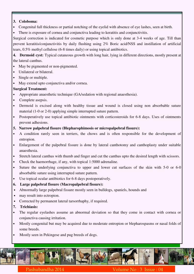
• Apart from these contributions, animals are extensively used in military, sports, laboratory research
and as pets, all of which are supported by veterinarians.
• Wild animals are also a big strength of Indian biodiversity and generate millions of dollars through
tourism annually.
The contributions of veterinary services have been always essential and forms backbone of the
livelihood and food security of India. On this World Veterinary Day, let’s salute this noble profession and
hope for even higher achievements in both quality and quantity of contributions to human betterment.
B. N. Nagaraja, Sangeeta Jadhav, A. S. Patil, Ramesh Rathod and L. Ranganath Department of Veterinary Surgery & Radiology,Veterinary College, Hebbal, Bangalore-24
(email: [email protected] )
Anatomy and physiology:
• Consists of movable folds of skin, loose connective tissue, muscular tissue, tarsus and conjunctiva.
• Dorsal and ventral folds form palpebral fissure.
• Sensory supply is by ophthalmic to upper eyelid and maxillary to the lower eyelid.
• The upper eyelid is more movable.
• Have protective eyelashes (cilia) at the margins
• Upper and lower eyelids unite to form lateral and median canthi.
• There is a well developed orbicularis oculi muscle. Sphincter muscle responsible for blinking.
Innervated by palpebral branch of facial nerve.
• Posterior to muscle is dense connective tissue layer called tarsus. A poorly defined fibrous sheet (well
developed in upper eyelid) which continues with septum orbitale.
Pashubandha 2014 Volume No : 3 Issue : 01 Pashubandha 2014 Volume No : 3 Issue : 04

• Dorsal to tarsus is levator palpebrae superiosis which helps to raise the upper eyelid. Innervated by
oculomotor nerve.
• Deep to levator is Miller’s muscle which is innervated by sympathetics. Paralysis of this muscle causes
a disease called Ptosis i.e. drooping of upper eyelid (Horner’s syndrome).
• Contains meibomian glands or tarsal glands having outlets as tiny apertures along the lid margins.
• The innermost layer of eyelid is palpebral conjunctiva containing lymphoid follicles, accessory
lacrimal glands of Krause and Wolfring.
Blinking:
• 90% bilateral in human beings.
• 80% in dogs.
• 60% in cattle and other animals.
• Voluntary due to trauma.
• Involuntary, normally 25/5min in all. May go as high as 100/5min.
• Helps in continuous spreading of precorneal tear film over cornea.
• Helps in continuous removal of precorneal tear film towards median canthus.
• Helps in the removal of foreign bodies, if any.
Precorneal Tear Film: Constantly baths the cornea and conjunctiva and anterior of eyelid. Gives nutrition
to avascular cornea. Consists of three layers;
1. Outer oily layer: Secretions come from meibomian glands.
2. Middle aqueous layer: True tear film and the secretions come from lacrimal gland, accessory lacrimal
glands of 3rd eyelid and accessory glands of conjunctiva.
3. Inner mucin layer: Secretions come from accessory glands and helps in uniform spread of precorneal
tear film.
Congenital Affections:
1. Ablepharon: Rarest condition in which there is congenital absence of eyelids.
2. Ankyloblepharon: Mostly seen in dogs.
Fusion of upper and lower eyelids.
• Normal physiological for first 10-15 days of life in dogs.
May be pathological due to Staph. Infection usually associated with purulent discharge.
Treated by surgical separation under suitable anaesthesia after removal of the discharge.
Topical antibiotic ointments are used qid for 7-8 days.
Pashubandha 2014 Volume No : 3 Issue : 01 Pashubandha 2014 Volume No : 3 Issue : 04
3. Coloboma:
• Congenital full thickness or partial notching of the eyelid with absence of eye lashes, seen at birth.
• There is exposure of cornea and conjunctiva leading to keratitis and conjunctivitis.
Surgical correction is indicated for cosmetic purpose which is only done at 3-4 weeks of age. Till than
prevent keratitis/conjunctivitis by daily flushing using 2% Boric acid/NSS and instillation of artificial
tears, 0.5% methyl cellulose (6-8 times daily) or using topical antibiotics.
4. Dermoid cyst: Typical cutaneous growth with long hair, lying in different directions, mostly present at
the lateral canthus.
• May be pigmented or non-pigmented.
• Unilateral or bilateral.
• Single or multiple.
• May extend upto conjunctiva and/or cornea.
Surgical Treatment:
• Appropriate anaesthetic technique (GA/sedation with regional anaesthesia).
• Complete asepsis.
• Dermoid is excised along with healthy tissue and wound is closed using non absorbable suture
material (1-0 or 2-0) applying simple interrupted suture pattern.
• Postoperatively use topical antibiotic ointments with corticosteroids for 6-8 days. Uses of ointments
prevent adhesions.
5. Narrow palpebral fissure (Blepharophimosis or micropalpebral fissure):
• A condition rarely seen in terriers, the chows and is often responsible for the development of
entropion.
• Enlargement of the palpebral fissure is done by lateral canthotomy and canthoplasty under suitable
anaesthesia.
• Stretch lateral canthus with thumb and finger and cut the canthus upto the desired length with scissors.
• Check the haemorrhage, if any, with topical 1:5000 adrenaline.
• Suture the underlying conjunctiva to upper and lower cut surfaces of the skin with 5-0 or 6-0
absorbable suture using interrupted suture pattern.
• Use topical ocular antibiotics for 6-8 days postoperatively.
6. Large palpebral fissure (Macropalpebral fissure):
• Abnormally large palpebral fissure mostly seen in bulldogs, spaniels, hounds and
• may result into ectropion.
• Corrected by permanent lateral tarsorrhaphy, if required.
7. Trichiasis:
• The regular eyelashes assume an abnormal deviation so that they come in contact with cornea or
conjunctiva causing irritation.
• Mostly congenital but may be acquired due to moderate entropion or blepharospasms or nasal folds of
some breeds.
• Mostly seen in Pekingese and pug breeds of dogs.
Pashubandha 2014 Volume No : 3 Issue : 01 Pashubandha 2014 Volume No : 3 Issue : 04

Symptoms:
• Epiphora, may be corneal damage and keratitis or conjunctivitis.
• Mucoid discharge is normally seen.
Treatment: Mostly electro-epilation is done. Treat entropion if present. Remove nasal folds as per need.
Adopt appropriate anaesthetic technique before adopting any treatment. Postoperatively, topical eye
antibiotics for 6-8 days.
8. Distichiasis:
• Congenital second abnormal row of eyelashes is present.
• Mostly seen in American Cocker Spaniel, Pekingese, Poodle and terrier breeds
• The row is usually incomplete with one to many eyelashes arising from the orifices of meibomian
glands.
• Usually upper eyelid is involved.
• Such eyelashes may occur spontaneously at any age and any breed.
• Rarely cause any clinical symptom except when causing irritation of cornea or conjunctiva.
Symptoms: Epiphora, may be keratitis or conjunctivitis.
Treatment: Removal of unwanted cilia by plucking, electro-epilation or by lid-splitting. Adopt
appropriate anaesthetic technique before adopting any treatment. Postoperatively, topical eye antibiotics
for 6-8 days.
9. Districhiasis: A rare condition where multiple cilia arise from one follicle.
10. Ectopic Cilia: These are eyelashes or cluster of lashes that grow through the conjunctiva.
• Mostly seen on the upper palpebral surface. Highly irritating to the cornea and conjunctiva.
Symptoms: Unilateral blepharospasms, Epiphora or Mucoid discharge, conjunctivitis, keratitis are
common clinical symptoms.
Treatment: Electro-epilation. Adopt appropriate anaesthetic technique before adopting any treatment.
Postoperatively, topical eye antibiotics for 6-8 days.
11. Entropion: Inversion of lid margins of the eyelid seen in almost all the animals.
Etiology:
• Congenital mostly involving the lower eyelid primarily because
of the weakness of the tarsal plate. Mostly seen in Chow-chow,
Blood hound, Labrador, Doberman, St. Bernard, Irish setter
breed of dogs.
• Acquired as a result of cicatricial contraction following injuries/
chronic inflammation.
• Acquired due to blepharospasms associated with the painful eye disease also known as spastic en-
tropion.
Symptoms:
• Typical in rolling of the lid margin.
• Epiphora.
Pashubandha 2014 Volume No : 3 Issue : 01 Pashubandha 2014 Volume No : 3 Issue : 04

• Blepharospasms.
• May be conjunctivitis or keratitis.
• May be vascularization (Pannus) or ulceration of the cornea.
Treatment: Modified Hotz Celsus blepharoplasty is done under suitable anaesthetic technique.
Postoperatively, topical eye antibiotics with corticosteroids for 6-8 days. Local antiseptic dressing of the
suture line for 8-10 days or till the sutures are removed. If corneal ulcers treat accordingly.
12. Ectropion: Typical eversion of the lid margin exposing the palpebral and bulbar conjunctiva.
Etiology:
• Congenital due to the inadequacy of lateral retractor muscle of the eye.
• Acquired due to decreased tone of the orbicularis oculi muscle, secondary to trauma, surgery, thermal
or chemical injury or chronic inflammation.
Symptoms: Typical turning out of the lid margin.
• Epiphora.
• Blepharospasms.
• May be conjunctivitis or keratitis.
Treatment: V-Y blepharoplasty is done under suitable anaesthetic technique. Postoperatively, topical eye
antibiotics with corticosteroid for 6-8 days. Local antiseptic dressing of the suture line for 8-10 days or till
the sutures are removed.
13. Blepharitis: Inflammation of the eyelids may be superficial/deep, acute/sub acute/chronic.
Etiology: Traumatic.
• Bacterial – Staph.
• Fungal – Microsporum, Trichophyton.
• Parasitic – Demodex, strongyloids.
• Allergic – pollens.
• Neoplastic.
Symptoms: Pain, mostly photophobia, Blepharospasms, Hyperaemia,
Oedema., Serous to purulent discharge, Alopecia, Epiphora, May be associated with keratitis or
conjunctivitis.
Treatment: Adopted as per the etiological factor (both local as well as systemic). Includes topical
antibiotic or antifungal (Nystatin BID daily for 3-4 weeks or amphotericin B BID daily for 3-4 weeks).
Topical corticosteroids are beneficial in allergic blepharitis.
14. Hordeolum: Localized supporative inflammation of lid margin usually due to Staph.
Types:
• External Hordeolum: Also known as ‘Stye’ involves glands of
Zies and Moll. Mostly seen in
young dogs characterized by solitary or multiple small abscesses on
the lid margin.
• Internal Hordeolum: Also known as ‘Chalazion’ and involves
meibomian glands. The swelling is mostly seen on the palpebral
conjunctiva. The condition is mostly seen in middle aged dogs.
Treatment: Hot compression of the swelling followed by manual
Pashubandha 2014 Volume No : 3 Issue : 01 Pashubandha 2014 Volume No : 3 Issue : 04

compression with cotton plug and then flushing the affected eye using 2% Boric acid or NSS. Topical
antibiotics with corticosteroids qid daily for 6-8 days.
15. Traumatic eyelid injuries: Mechanical involving skin or full thickness of the eyelid mostly leading
to lacerations. Plastic surgery is done with minimum debridement under suitable anaesthesia along with
suitable postoperative management.
16. Neoplasms: Adenomas, adenocarcinomas, Melanoma or Papillomas. Mostly the tumors of the
eyelids are benign. Treated as per method adopted for the management of any neoplastic condition
following radical surgery and appropriate postoperative management.
17. Symblepharon: Adhesions between palpebral conjunctiva
and cornea or between palpebral and bulbar conjunctivae following
conjunctival injuries. Manual severing of such adhesions is done
under suitable anaesthetic technique to separate the palpebral
conjunctiva from either cornea or bulbar conjunctiva.
Postoperatively always use ocular antibiotic ointments for 6-7 days
to prevent reoccurrence.
Dr. Madhukar* and Prof. H. A. Upendra#
*Assistant Professor, #The Director, Institute of Wildlife Veterinary Research, KVAFSU,
Doddaluvara, Kodagu – 571232, (E-mail: [email protected] )
As we have seen in first part of this article, dart syringe and needles have major differences
compared to regular ones. This fact, coupled with handling of potent anesthetics under pressurized
conditions necessitates understanding of proper loading and unloading of dart syringes.
In this pictorial article, we outline the procedure for loading and unloading of the dart syringes.
The filling procedure should be first practiced thoroughly with water before handling the anesthetics.
DART LOADING
1. Remove the hypodermic needle from the protective case.
Slide the silicone sleeve onto the needle so that the injection
holes are located at the centre of the sleeve. This can be
achieved by rotating the needle whilst exerting pressure on
the sleeve. The silicone sleeve must only be used once.
Pashubandha 2014 Volume No : 3 Issue : 01 Pashubandha 2014 Volume No : 3 Issue : 04

Pashubandha 2014 Volume No : 3 Issue : 01 Pashubandha 2014 Volume No : 3 Issue : 04
2. Remove the stabilizer from the dart if necessary using a
venting pin, release any retained air from the dart syringe
air chamber.
3. Hold the dart with the air chamber uppermost. Using an
air filler syringe fitted with a coupling adapter connected to
the drug chamber, position the black plunger at the rear of
the chamber.
4. Reverse the dart so that the drug chamber is now
uppermost. Using a suitable syringe filled with the required
tranquillizing/medicating liquid slowly inject the drug into
the chamber. Ensure that you use a sufficiently small gauge
needle to allow air from the drug chamber to be expelled
without displacing the drug.
5. Mount the hypodermic needle onto the dart syringe boss
using pliers and locate it firmly by rotating it slightly
whilst applying pressure.
6. Apply a safety cap over the needle and seat it firmly on
the dart.
7. Pressurizing the dart: Hold the dart vertically with the
needle/protection cap uppermost. Mount the coupling
adapter onto a dry standard 12 – 20 ml syringe.
Introduce 12 ml of air into the syringe, (correct air pressure
for a 3 ml dart). Connect the syringe securely to the air
chamber of the dart and, with a smooth continuous action,
inject the 12 ml of air into the dart air chamber. Now 12 ml
of air is let in to the syringe though the coupling. The
plunger will act as a non-return valve and retain the air
within the dart. As the dart is loaded and under
pressure, extreme care is warranted.
8. Place the red stabilizer firmly on the rear of the dart. If
the stabilizer becomes soiled, it can be washed at 30 °C.
The stabilizer must not be wet before use as the syringe
may be unstable in flight.

DART UNLOADING
Similar to the precautions during loading, unloading of a loaded pressurized dart also warrants
extreme precautions.
When an undischarged or unused dart is recovered, the first step must always be to render the dart
into a safe state by fitting a protection cover and releasing any remaining air pressure. The following
illustrated steps should be adapted to the situation encoun-
tered. Darts can be removed from rifle/pistol barrels using
tweezers.
1. Remove the red stabilizer from the dart.
2. Holding the dart vertically, depress the red plunger to
release air pressure using the venting pin.
3. Remove the protection cap from the dart.
4. If using proprietary medication/drug bottles with
rubberised seals, insert a plain hypodermic needle, directed
away from the operator, into the seal before proceeding to
the next step. This will allow excess air pressure to vent
from the bottle.
5. Holding the dart with the needle pointing down, insert
the needle into a suitable receptacle as far as possible. The
silicon sleeve will slide along the needle shaft exposing the
injection ports.
6. Hold the dart with the attached bottle. Using a coupling
adapter on an air filler syringe, steadily and smoothly insert
12ml of air into the dart air chamber. The air pressure will
slowly force the black plunger forwards and inject the drug
into the bottle. Do not store drugs or water in the dart
syringes for prolonged periods. The extreme pH of most
drug formulations will adversely affect the plunger and dart
barrel after 3-4 weeks. Always clean the dart and needle after use.
Next article will cover the details on maintenance of dart set. The authors have used images and
illustrations for educational purpose only and neither claims their ownership nor endorses these brands.
Pashubandha 2014 Volume No : 3 Issue : 01 Pashubandha 2014 Volume No : 3 Issue : 04
Dr.M.A.Kshama & Dr.A.Muralidhara Dept of TVCC, Veterinary College, Bangalore, KVAFSU
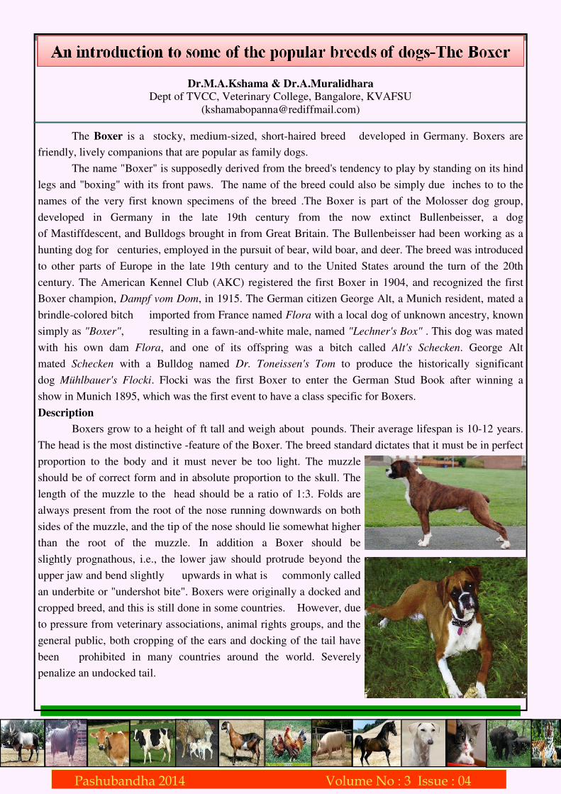
The Boxer is a stocky, medium-sized, short-haired breed developed in Germany. Boxers are
friendly, lively companions that are popular as family dogs.
The name "Boxer" is supposedly derived from the breed's tendency to play by standing on its hind
legs and "boxing" with its front paws. The name of the breed could also be simply due inches to to the
names of the very first known specimens of the breed .The Boxer is part of the Molosser dog group,
developed in Germany in the late 19th century from the now extinct Bullenbeisser, a dog
of Mastiffdescent, and Bulldogs brought in from Great Britain. The Bullenbeisser had been working as a
hunting dog for centuries, employed in the pursuit of bear, wild boar, and deer. The breed was introduced
to other parts of Europe in the late 19th century and to the United States around the turn of the 20th
century. The American Kennel Club (AKC) registered the first Boxer in 1904, and recognized the first
Boxer champion, Dampf vom Dom, in 1915. The German citizen George Alt, a Munich resident, mated a
brindle-colored bitch imported from France named Flora with a local dog of unknown ancestry, known
simply as "Boxer", resulting in a fawn-and-white male, named "Lechner's Box" . This dog was mated
with his own dam Flora, and one of its offspring was a bitch called Alt's Schecken. George Alt
mated Schecken with a Bulldog named Dr. Toneissen's Tom to produce the historically significant
dog Mühlbauer's Flocki. Flocki was the first Boxer to enter the German Stud Book after winning a
show in Munich 1895, which was the first event to have a class specific for Boxers.
Description
Boxers grow to a height of ft tall and weigh about pounds. Their average lifespan is 10-12 years.
The head is the most distinctive -feature of the Boxer. The breed standard dictates that it must be in perfect
proportion to the body and it must never be too light. The muzzle
should be of correct form and in absolute proportion to the skull. The
length of the muzzle to the head should be a ratio of 1:3. Folds are
always present from the root of the nose running downwards on both
sides of the muzzle, and the tip of the nose should lie somewhat higher
than the root of the muzzle. In addition a Boxer should be
slightly prognathous, i.e., the lower jaw should protrude beyond the
upper jaw and bend slightly upwards in what is commonly called
an underbite or "undershot bite". Boxers were originally a docked and
cropped breed, and this is still done in some countries. However, due
to pressure from veterinary associations, animal rights groups, and the
general public, both cropping of the ears and docking of the tail have
been prohibited in many countries around the world. Severely
penalize an undocked tail.
Pashubandha 2014 Volume No : 3 Issue : 01 Pashubandha 2014 Volume No : 3 Issue : 04

The Boxer is a short-haired breed, with a shiny, smooth coat that lies tight to the body. The
recognized colors are fawn and brindle, frequently with a white underbelly and white feet. These
white markings, called flash, often extend onto the neck or face, and dogs that have these markings are
known as "flashy". "Fawn" denotes a range of color, the tones of which may be described variously as
light tan or yellow, reddish tan, mahogany or stag/deer red, and dark honey-blonde. The Boxer does not
carry the gene for a solid black coat color and therefore purebred black Boxers do not exist.
Boxers with white markings covering more than one-third of their coat – conventionally called
"white" Boxers – are neither albino nor rare and approximately 20–25% of all Boxers born are white.
Genetically, these dogs are either fawn or brindle, with excessive white markings overlying the base coat
color. White Boxers have a higher risk of sunburn and associated skin cancers than colored Boxers and it
is found that about 18% of white Boxers are deaf in one or both ears.
Temperament
Boxers are large, muscular, square-headed dogs who look
imposing — that is, until you look into their eyes and see the
mischief and joy of life reflected there. Because of their playful
nature and boundless energy, they are sometimes called the
"Peter Pan" of the dog breeds. Boxers aren't considered fully
mature until they are three years old, meaning they have one of
the longest puppyhoods in the world of dogs.
The typical Boxer is intelligent, alert, and fearless, yet
friendly. He's loyal to his family and loves to play with them, but he's also headstrong, especially if you
try to use harsh training methods with him.
With minimal grooming needs ,legendary patience and gentleness with children, Boxers are great
family companions, as long as you provide them with the physical exercise and mental stimulation they
need. If you're willing and able to provide them with adequate exercise in the form of walks or runs, they
can even adapt to apartment living, so long as they are able to be close to their beloved people.
Their suspicion of strangers, alertness, agility, and strength make them formidable guard dogs. As
puppies, Boxers demonstrate a fascinating combination of mood-mirroring expressions, energetic
curiosity, flexible attention spans and charming characteristics. These strong and intelligent animals have
been used as service dogs, guide dogs for the blind, therapy dogs, police dogs and occasionally for herding
cattle or sheep. The versatility of Boxers was recognized early on by the military, which has used them as
valuable messenger dogs, pack carriers, and attack and guard dogs in times of war.
Issues regarding health
Boxers are generally healthy, but like all breeds, they're prone to certain health conditions. Not all
boxers will get any or all of these diseases, but it's important to be aware of them if you're considering
this breed.
• Aortic stenosis/ sub-aortic stenosis (AS/SAS)-This is one of the most common heart defects found in
Boxers. This condition can cause fainting and even sudden death. It's an inherited condition, but its
mode of transmission isn't known at this time. Dogs with this condition should not be bred.
Pashubandha 2014 Volume No : 3 Issue : 01 Pashubandha 2014 Volume No : 3 Issue : 04

• Boxer cardiomyopathy (BCM)-Also called Boxer Arrythmic Cardiomyopathy (BAC), Familial
Ventricular Arrhythmia (FVA) and Arrhythmogenic Right Ventricular Cardiomyopathy (ARVC).
BCM is an inherited condition. occuring due to an electrical conduction disorder. This can cause
weakness, collapse, or sudden death. Because it is difficult to detect this condition, it can cause an
unexpected death. Boxers who show signs of this condition should not be bred.
• Hip Dysplasia-Some dogs show pain and lameness on one or both rear legs while others don’t. As the
dog ages, arthritis can develop. Dogs with hip dysplasia should not be bred Hip dysplasia is hereditary,
but it can also be triggered by environmental factors, such as rapid growth from a high-calorie diet or
injuries incurred from jumping or falling on slick floors. Treatment ranges from supplements that
support joint function to total hip replacement.
• Hypothyroidism- Hypothyroidism is caused by a deficiency of thyroid hormone and may produce
signs that include infertility, obesity, mental dullness, and lack of energy. The dog's fur may become
coarse and brittle and begin to fall out, while the skin becomes tough and dark. Hypothyroidism can be
managed very well with a thyroid replacement therapy. Medication must continue throughout the dog's
life.
• Demodectic Mange-Generalized demodectic mange covers the entire body and affects older puppies
and young adult dogs. The dog develops patchy skin, bald spots, and skin infections all over the body.
The American Academy of Veterinary Dermatology recommends neutering or spaying all dogs that
develop generalized demodectic mange because there is said to be a genetic link.
• Gastric dilatation-volvulus (GDV)-This is a life-threatening condition that can affect large,
deep-chested dogs like Boxers, especially if they are fed one large meal a day, eat rapidly, drink large
volumes of water after eating, and exercise vigorously after eating.
• Allergies- Boxers are prone to allergies, both environmental allergies and food-related allergies. If you
notice that your Boxer has itchy, scaly skin, have him checked out by your vet.
• Deafness- White Boxers are especially susceptible to deafness. About 20 percent of white Boxers are
deaf, and white Boxers should not be bred because the genes that cause deafness in white Boxers can
be inherited.
Additionally, Boxers that carry the extreme white spotting gene can increase the incidence of deafness
in the breed.
Some points to remember about the breed
• Boxers are high-energy dogs and need a lot of exercise.
• Boxers are exuberant and will greet you ecstatically.
• Early, consistent training is critical — before your Boxer gets too big to handle!
• Although they are large, Boxers are not "outdoor dogs." Their short noses and short hair make them
uncomfortable in hot and cold weather, and they need to be kept as housedogs.
• Boxers mature slowly and act like rambunctious puppies for several years.
• Boxers don't just like to be around their family they need to be around them! If left alone for too long
or kept in the backyard away from people, they can become ill-tempered and destructive.
• Boxers drool, a lot. Boxers also snore, loudly.
Pashubandha 2014 Volume No : 3 Issue : 01 Pashubandha 2014 Volume No : 3 Issue : 04

Dr. K. Chandrapal Singh and Dr. Y. Madhura Veterinary College, Hebbal, Bangalore 560 024
Heat Stress in dairy cattle can be defined as an ill effect of high environmental temperature,
influencing the physiological and productive performance of a cow. The heat stress will last for a period
of 2 to 5 months in a year during summer season, when the cow is having difficulty in maintaining its
normal temperature. The normal temperature of a dairy cow is 390C and the cow is said to be in a
“comfort zone” of environmental temperature between 5 to 250C. Cows will be able to tolerate ambient
beyond 250C with the homeostatic mechanism, but with increasing temperature and humidity, the health
and productivity of the animal suffers.
The heat tolerance limit, that is capacity of animal to maintain normal temperature, above certain
environmental temperature (upper critical temperature) varies with the breed. For example, in Holstein
and Jersey cattle it is 280C and in case of indigenous cattle (Indian breeds) and buffaloes it is 380C. The
response of cows due to heat stress will be in the form of 1. Decreased feed intake, 2. lowered milk
production, 3. decreased milk fat percentage, 4. decreased fertility and 5. depressed immune system. The
magnitude of such lowered performance could be to the extent of 20 to 40 percent decreased feed intake
and milk production. For example, every one degree increase in the environmental temperature above
390C, would lead to a decrease in milk production by one litre.
Having understood the principle of heat stress and its impact on the production, let us briefly
consider the feeding and managemental practices to mitigate heat stress in dairy cows.
Decreased feed intake: Most livestock species including dairy cows, drastically decrease their feed
intake and consequently suffer from energy deficiency. This could be overcome by increasing energy
concentration of the diet or supplementing energy in the diet. Practically speaking, encourage cows to eat
more, by offering more number of times per day. For example, against normal feeding of two times per
day, feed 4 or 5 times, but with lesser quantities of feed offered per meal. Supplement cattle feed with 5 to
10 per cent jaggery (some time sugar can be cheaper than jaggery and sugar can be used instead of
jaggery). This would add energy and increase the palatability of the feed. Animals should be offered feed
during early hours of morning or late evening hours, that is cooler times of the day. If available, feed
adequate green fodder or silage.
In order to dissipate heat, the dairy cows lose water from the body in the form of respiration and
evaporative loss through skin, and obviously the water intake increases. Provide plenty of cool and clean
water. Provide automatic taps or Bucket filled with water, always kept in front of the animal.
Along with water, the cows also lose electrolytes, specially sodium and potassium. Cows tend to
lose nearly five times the electrolyte through urine or perspiration during the heat stress. Supplement cattle
feed with mineral mixture containing salt. Sodium bicarbonate (50-100gms) can also be fed per day per
cow.
Pashubandha 2014 Volume No : 3 Issue : 01 Pashubandha 2014 Volume No : 3 Issue : 04

It is also important to keep the animal cool by bathing 2 times per day, allowing animals for wallowing,
or sprinkling water on the animal and in the sheds. Provide sprinklers and fans in the ceiling of cow sheds.
Provide plenty of shade. Animals should not be tied in closed sheds during summer. Let the
animals lose and roam freely in open shade, like under the shade of tree etc.
Summary: Heat stress could lead to the decreased milk production in dairy cows and therefore result in
economic losses to the farmers. In most situations, the loss of productivity due to summer stress in dairy
cows go unnoticed by the dairy farmers. Alleviating dairy cows from heat stress and providing comfort
would make them more productive and in turn increase economic returns. Suggestions to mitigate heat
stress during hot weather include
1. Providing open shades and protecting cows from direct sunlight
2. Provide additional cooling using fan and sprinklers in cow sheds
3. Provide additional energy, minerals and vitamins
4. Supplement ration with green good quality forages
5. Feeding smaller meals several times daily to promote intake
6. Feeding during cooler periods of the day
7. Cleaning feed mangers daily to prevent feed spoilage
8. Provide unlimited clean, cool water.
Dr. Anant Rao Desai*, Dr. Harisha M and and Dr. Mahesh Savanur
*Assistant Professor, Veterinary College, Bidar
(email: [email protected])
Project can be defined as; a scientifically evolved work plan, devised to achieve specific objectives,
within specified time limit, consuming planned resources.
To formulate and analyze effective projects, those responsible must consider many aspects that
determine how remunerative a proposed investment will be. All aspects must be considered and
reconsidered at every stage in the project planning and implementation cycle.
Project report is a means to convince a banker/ financing institution to finance your project. Type
and contents of the report varies with the amount asked for funding. Guidelines to formulate a detailed
bankable project report are discussed below.
The cover page of the project report should have the following:
1. Project Subject: Mention the title of the project in bold letters and large fonts. It should be mentioned
as either Dairy/ Poultry/ Processing unit etc.,
2. Unit Size: Size of the unit should be mentioned. No of animals/ birds/ capacity of processing unit is
declared.
3. Indicate whether it is a layer/ broiler unit in case of poultry, cow or buffalo unit in case of dairy and
goat or sheep unit in case of small ruminant project.
4. Breed of the animals selected for the project should be mentioned.
5. Project cost, Loan amount, Margin money, BCR, IRR should also be mentioned.
Pashubandha 2014 Volume No : 3 Issue : 01 Pashubandha 2014 Volume No : 3 Issue : 04

Particulars/Content of the Report
1. Introduction: General information about subject (e.g. dairy/poultry etc) is stated. The present status of
the sector, position of the country in production (e.g. dairy/ poultry etc), per capita consumption in
India, scope for increase consumption, scope for future expansion should be highlighted. Strengths and
opportunities should be described.
2. Scope & objectives of the project should be mentioned
3. Development initiatives of Government Departments, Corporations, Board, Co-operatives, NGOs,
State Agricultural /Veterinary University, KVKs etc., in the form of technical, financial support.
4. Status of beneficiary: Individual/ Partnership/ Company/ Corporation/ Co-operative Society/ Public
Private Partnership/ NGO/ Board/ Others.
5. Details of borrowers’ profile/Background of company: Name, Age, Address, Other income generating
activities if any,etc.,
6. Technical & financial background of the borrower: Land holding, present income, education,
experience & training- related to project subject.
7. Site/Location of project: Survey No., Village, Dist, etc. land area in acre/hectare, cropping pattern,
land development, fencing approach to site, Roads/ railways and other transportation connectivity/
Proximity, etc.
8. Suitability of climate (Meteorological data): season wise avg. temperature, rainfall & humidity etc.
9. Infrastructure available on site:
a . Power supply: Electricity/LPG/ DG set connection availability. Alternative arrangement made in case
of power failure.
b. Irrigation facilities: well, water tank, motor, pump & pipeline etc
c. Housing: sheds, quarters, godown etc. with their No. & size/area available on the farm suitable for
project.
d. Equipments & machinery with their No. & size/capacity related to project already available on site
10. Livestock & poultry farming system: Give details of housing pattern/system etc. Size, type and
number of buildings required, equipment and machinery required. Strategy followed to purchase or
construct the same
11. Backward Linkages (related to project subject)
a. Availability of livestock & poultry birds: cow/buffalo/sheep/goat/chicks etc. List of livestock markets/
fairs, supplying agencies, Government/University Breeding farms etc.
b. Availability of Feed & fodder: dry, green & concentrates etc. cropping pattern & fodder cultivation etc.
List the markets/ authorized suppliers of feed & fodder etc
c. Veterinary aid & breeding facility: Hospital/ dispensary, AI/Natural breeding, vaccination, availability
suppliers of medicine, vaccines etc. etc., Place &distance from project site. Name, address and contact
number of consultant veterinarian if any. Contract made with the Veterinary consultant/ dealers
d. Manpower requirement: Availability of trained & experienced, Technical / Non technical staff, labours
etc. Contracts made if any
e. Financing Bank & branch location: Name, address & Contact No. etc.
Pashubandha 2014 Volume No : 3 Issue : 01 Pashubandha 2014 Volume No : 3 Issue : 04

12. Forward Linkages
a. Marketing of end products: name & place of market for milk, Eggs, broilers, culled animals, young
one, manure etc. price to be realized.
b. Processing or value addition of produce
c. Specific marketing strategy, if any.
d. Arrangement for farm waste processing and value addition and sale/ disposal.
13. Schedule of project implementation: Time frame required for the project to be fully functional and
financial details of work within that period are discussed.
14. Project Outlay: Total Financial Outlay (TFO) = Fixed cost &working capital at a glance (Item wise)
is given.
15. Project Economics: Cash flow statement, Financial Analysis (Benefit Cost Ratio (BCR), Net Present
Worth (NPW), Internal Rate of Return (IRR), etc.
Net Present Worth (NPW) or Net Present Value (NPV)
The Net Present Worth is the difference between the present worth of all cash inflows and outflows
of a project.
The investments made in the projects are generally called costs or cash outflows. The receipts that
accrued during different time periods are called as cash inflows or gross returns. The cash flows
discounted with an appropriate discount rate will give the net present worth of the project.
(Bt is cash flows in tth year, Ct is cash outflows in tth year,
t is 1 to 10 years that is life span of the project.)
The choice criterion using NPW is that the project with positive NPW is accepted for
implementation and the project with negative NPW is rejected. If he is to choose among different projects,
the project with highest NPW has to be chosen.
Benefit Cost Ratio (BCR): An indicator used in cost-benefit analysis that attempts to summarize the
overall “value for money” of a project. It is the ratio of the benefits of a project relative to its costs, being
expressed in discounted present values.
BCR is worked out by dividing the present value of cash inflows by the present value of cash outflows. If
the BCR is more than one, that project is accepted and if BCR is less than one the project is
rejected. Among the different projects, the project with highest BCR is to be selected.
Net Present Value of Benefits
BCR = ---------------------------------------------------
Net Present Value of Costs
Pashubandha 2014 Volume No : 3 Issue : 01 Pashubandha 2014 Volume No : 3 Issue : 04

Internal Rate of Returns (IRR): It is the rate of return per rupee invested in a project over its life
span. For example if the IRR is 30 per cent in a livestock project, it means that this project gets an average
annual return of Rs. 30/ per Rs. 100/ invested in the project over its life span.
For a project to be viable it should have a BCR of one or greater than one at the opportunity cost of capital
and a NPW of zero or greater than zero at the opportunity cost of capital and the discount rate for IRR
should be greater than the opportunity cost.
16. Repayment of loan: amount & No. of installments per month/quarter/ annum, moratorium/grace period
if any etc.
17. Lending terms: Term loan, Cash Credit (CC Limit), Margin money & its sources, Rate of Interest,
Security, Hypothecation, Insurance, working capital, repayment holiday etc.
18. Other Financial Assistance: Refinance/ Subsidy (NABARD/ Govt.) etc. Under any government or
co-operative schemes.
19. Records & registers to be maintained.
20. Conclusion.
21. List of Statements/ Annexure
a. Project Cost in details with items of Investment, bank loan, margin etc.
b. Techno- financial norms/assumptions.
c. Herd projection charts: Lactation/Kidding/lambing/flock/poultry housing schedule/kindling/ farrowing
etc.
d. Working capital requirement
e. Income/ Expenditure Statement
f. Cash flow analysis: BCR, NPW, IRR and DER, DSCR etc.
Repayment Schedule & adjustment of back ended subsidy if any.
22. List of attachments/ enclosures
a. RTC (pahani of the farmland), registration certificate of the land, lease agreement copy etc.,
b. Site/layout plan (Maps)
c. Detail cost estimates of civil work from Regd. Civil Engineering
d. Quotations for equipments & machinery from reputed suppliers/dealers
e. Well water test report (potable water)
f. A copy of training/experience certificate, if any.
g. NOC from different agencies (e.g. pollution control, Municipal Corporation, gram panchayat.) if
applicable
h. State Electricity Board approval for connection.
i. A copy of contract farming agreement if any.
J. Quotation for supply of livestock & chicks from reputed supplier/hatchery.
k. Quotation for supply of feed & fodder, concentrate if any.
l. Prevention of Food Adulteration Act License. (In case of food processing-milk & Meat) and other
licenses as applicable.
m. Any other documents as & when asked by the financing agency/bank.
Pashubandha 2014 Volume No : 3 Issue : 01 Pashubandha 2014 Volume No : 3 Issue : 04

Contact :
Dept of Veterinary and Animal Husbandry Extension Education
Veterinary College, Hebbal Bangalore
email: [email protected]
Blog: pashubandhavcheb.in
monthly e-Bulletin
Published and circulated by Veterinary College, Hebbal Bengaluru
Editor: Associate Editior:
Dean, Veterinary College, Hebbal, Bengaluru Head, Dept of Vety & Animal Husbandry Extension Education
Dr.S.Yathiraj (Ex-Officio) Dr.K.Satyanarayana (Ex-Officio)
• PELVIC
n. Subsidy application form duly filled in, if any (to be finalized & signed by the bank officials)
o. Veterinary Certificate
23. Additional points to be considered while preparing a project report for large projects
a. Executive Summary of the Project (After cover page): Summary stating the vision, mission of the
project should be given. The background of why this project was selected should also be discussed in
detail.
b. STEP- Social, Technical, Economic, Environmental & Political analysis Social, Economic and Political
implications of the project are clearly discussed. What would be the employment generation,
livelihoods created, any issues against the local community feelings and how are they handled etc.,
c. SWOT–Strengths, Weaknesses, Opportunities & Threats analysis: Each of them is discussed in detail
with explanation on how weakness is converted into strengths and threats into opportunities.
d. Stake Holder – Primary, Secondary & Key stake holder analysis: Identify all the stakeholders involved
in the project, their role etc.,
e. Careful project preparation is the best available means to ensure efficient, economic use of capital funds
and to increase the chances of implementation of project on schedule. Unless projects are carefully
prepared in substantial detail, inefficient or even wasteful expenditure is almost sure to result in loss of
capital.
Information on use of Microsoft Excel in preparing project report will be published in next issue.
Pashubandha 2014 Volume No : 3 Issue : 01 Pashubandha 2014 Volume No : 3 Issue : 04