aq4n, extra cellular matrix, hypoxia and...
TRANSCRIPT

Citation: Kazmi, Nabila, Hossain, Alamgir and Phillips, Roger (2011) Modelling of Tirapazamine effects on solid tumour morphology. In: 5th International Conference on Practical Applications of Computational Biology & Bioinformatics (PACBB 2011). Advances in Intelligent and Soft Computing, 93 (2011). Springer, pp. 125-132. ISBN 978-3-642-19913-4
Published by: Springer
URL: http://dx.doi.org/10.1007/978-3-642-19914-1_18 <http://dx.doi.org/10.1007/978-3-642-19914-1_18>
This version was downloaded from Northumbria Research Link: http://nrl.northumbria.ac.uk/3463/
Northumbria University has developed Northumbria Research Link (NRL) to enable users to access the University’s research output. Copyright © and moral rights for items on NRL are retained by the individual author(s) and/or other copyright owners. Single copies of full items can be reproduced, displayed or performed, and given to third parties in any format or medium for personal research or study, educational, or not-for-profit purposes without prior permission or charge, provided the authors, title and full bibliographic details are given, as well as a hyperlink and/or URL to the original metadata page. The content must not be changed in any way. Full items must not be sold commercially in any format or medium without formal permission of the copyright holder. The full policy is available online: http://nrl.northumbria.ac.uk/policies.html
This document may differ from the final, published version of the research and has been made available online in accordance with publisher policies. To read and/or cite from the published version of the research, please visit the publisher’s website (a subscription may be required.)

Abstract Bioreductive drugs are in clinical practice to exploit the resistance from
tumour microenvironments especially in the hypoxic region of tumour. We pre-
sented a tumour treatment model to capture the pharmacology of one of the most
prominent bioreductive drugs, Tirapazamine (TPZ) which is in clinical trials I and
II. We calculated solid tumour mass in our previous work and then integrated that
model with TPZ infusion. We calculated TPZ cytotoxicity, concentration, penetra-
tion with increasing distance from blood vessel and offered resistance from micro-
environments for drug penetration inside the tumour while considering each cell
as an individual entity. The impact of these factors on tumour morphology is also
showed to see the drug behaviour inside animals/humans tumours. We maintained
the heterogeneity factors in presented model as observed in real tumour mass es-
pecially in terms of cells proliferation, cell movement, extracellular matrix (ECM)
interaction, and the gradients of partial oxygen pressure (pO2) inside tumour cells
during the whole growth and treatment activity. The results suggest that TPZ high
concentration in combination with chemotherapy should be given to get maximum
abnormal cell killing. This model can be a good choice for oncologists and re-
searchers to explore more about TPZ action inside solid tumour.
Index Terms— AQ4N, Extra Cellular Matrix, Hypoxia and Tirapazamine.
INTRODUCTION
Most common cancer treatments like chemotherapy and radiotherapy are facing a
strong resistance from hypoxic regions inside the tumour. When a tumour reaches
to a critical size approximately 106 cells the nutrients diffusion is insufficient to
supply required amount of oxygen to the inner parts of the tumour initiating a situ-
ation called hypoxia (Gerlee and Anderson 2007).
N. Kazmi, A. Hossain is in Department of Computing, University of Bradford, Bradford, BD7 1DP,
UK. Email: {N.Kazmi5, M.A.Hossain1}@Bradford.ac.uk
Roger Phillips is in Institute of Cancer Therapeutics, University of Bradford, Bradford, BD7 1DP, UK.
Email: [email protected]
Modelling of Tirapazamine Effects on Solid Tumour
Morphology
1Kazmi, N.,
1Hossain, M. A. and
2Phillips, R.M.
1School of Computing, Informatics and Media, 2Institute of Cancer Therapeutics,
University of Bradford, Bradford, BD7 1DP, UK
{N.Kazmi5; M.A.Hossain1; R.M.Phillips}@Bradford.ac.uk
2 N Kazmi, M A Hossain and R Phillips
Hypoxia is recognized as a factor that helps the tumour cells survival by giving
them more aggressive phenotypes. Majority of tumours with size greater than
1mm3 have got hypoxic regions because of irregular blood vessel structure and in-
creased distance from blood vessels. The high rate of glycolysis has been shown in
hypoxic regions of most tumours. (Shannon et al. 2003). Hypoxic cells were
thought to be present at about 100-150 µm from functional blood vessels but now
recent studies showed that hypoxia can be found at about 20-25 µm from blood
vessels (Marcu and Olver 2006). Deformed capillary vessels of tumour and in-
creased distance of tumour cells and blood vessels result in poor drug penetration
(Marcu and Olver 2006). The hypoxia is a major challenge in the control of tu-
mour while either using radiation or chemotherapy. Chemotherapy’s major aim is
to decrease down the number of tumour cells with a number of treatment cycles
(Algoul et al. 2010). Tumour cells that are at distal locations from blood vessels
are difficult to treat with chemotherapy. With increasing distance drug penetration
slows down making it less effective. Cells with good distance from blood vessels
are deficient in oxygen supply and slow in proliferation rate, show resistance to-
wards effective chemotherapy treatment. Chemotherapy is not an effective way to
treat hypoxic regions, as it is developed to kill cells having rapid division cycles
(Brown 1999). Radiation, another cancer treatment works against tumour cells by
damaging their DNA. This DNA damage remains permanent under the presence
of oxygen molecules and results in cell death. So this cancer treatment is also
most effective to those cells having sufficient oxygen (Bronwyn et al. 2003).
Some bioreductive drugs are under high consideration to exploit these hypoxic re-
gions with no or less harm to normal cells. Tirapazamine (TPZ) and AQ4N are
under experiments and are in clinical practice as bioreductive drugs (Patterson and
McKeown 2000). TPZ is in clinical phase II and III trials with radiotherapy and
anti cancer drug cisplatin respectively. Its effectiveness can be determined by two
factors one is the action of reductive enzymes and secondly the extent of hypoxia.
Potential cell killing has been measured for both radiation and TPZ combination in
three murine tumours. In SCCVII, DNA damage decreased with increasing oxy-
gen concentration and this damage was half of the best possible value at 0.2%
pO2. These in-vitro experiments showed the oxygen dependence of DNA damage
when treated with TPZ. In well oxygenated cells back oxidation of radical con-
verts it to parent compound with no toxicity (Shannon et al. 2003). Results
showed that less TPZ concentration is highly toxic at low pO2 regions (0.2% O2)
and more concentration is required if oxygen pressure increases. Current studies
still show no confirmed answer for its actions, toxicity and optimal administration.
Mathematical and computational modelling has introduced a new horizon
for biologists, scientists and doctors in making hypothesis and experimentations
about complex biological phenomenons and in curing diseases. Several attempts
have been made to model tumour growth process but mostly considered it as a
whole entity at tissue level. An in-silico model was developed to investigate early
tumour growth under the influence of Extracellular Matrix (ECM), Cell-Cell and
Cell-Matrix adhesion and cell movement as growth constraints while used a po-

Modelling of Tirapazamine Effects on Solid Tumour Morphology 3
werful artificial intelligence decision making tool; the neural network considering
each cell as an individual and independent entity (Kazmi et al. 2010). Now we ex-
tended our previous computational modeling technique to capture TPZ preferen-
tial action towards tumour at cellular level. The initial TPZ concentrations 10 µM,
50 µM and 100 µM were infused to tumour mass from surrounding blood vessel
and model calculated drug cytotoxicity, its penetration, metabolism inside each
cell. The results section showed the impact of all these factors on tumour mor-
phology. This model with some parameter modifications can be served as a tool in
assumptions and experiments for bioreductive drugs in laboratories and in clinical
trials by oncologists, researchers and pharmacologists.
The model
We did not consider tumour as a whole entity, our model explored the beha-
vioral characteristics of the tumour at basic cell level. The model is developed us-
ing a 2 dimensional plot of size 400 that can simulate tumour of radius 200. Each
element shows the availability or absence of a tumour cell. A well known decision
making artificial intelligence technique; neural network is used to calculate the re-
sponse or phenotype of each abnormal cell. The model calculates tumor microen-
vironments values and passes them to input layer and used one hidden layer of
neurons to calculate the middle values and then pass them to the output layer as
final phenotype of that specific cell using standard transfer function at each layer.
Partial differential equation set PDE (1) was used to calculate nu-
trients/microenvironments i.e. the consumption of oxygen, glucose and production
of hydrogen ions for each cell at specific location x and at specific time instance t,
during tumour growth process (Gerlee and Anderson 2007). ),(),(/),( txftxcDttxc cc
),(),(/),( txftxgDttxg gg
),(),(/),( txftxhDttxh hh
(1)
Dc, Dg and Dh are the diffusion constants for oxygen, glucose and hydrogen. They
are given values Dc=1.8×10⁻⁵
cm2s
-1 (Grote et al. 1997), Dg =9.1×10
-5cm
2s
-1 and
Dh=1.1×10⁻⁵
cm2s
-1 Crone and Levitt (1984). The actual target was to explore the
TPZ effects on tumour morphology and cell killing during the treatment. The
whole tumour mass was divided into five hypoxic regions based upon available
pO2 for each cell. These five identified hypoxic regions were hypoxia I, II, III, IV
and V based upon pO2 values; 20.9%, 10%, 2%, 0.2% and 0.02% respectively.
Using experimentally measured and published data in the literature, the values of
drug concentrations required to kill cells at various oxygen tensions was entered
into the model: Above 500 µM TPZ was cytotoxic towards all cells at O2 tensions

4 N Kazmi, M A Hossain and R Phillips
of 20.9% and 10% 50 µM was toxic towards cells at 2% O2 while 10 µM was
enough to kill cells at 0.2% and 0.02% O2 (Lartigau and Guichard 1994). We in-
troduced 10 µM, 50 µM and 100 µM as initial concentrations of TPZ in our simu-
lations. In the model, blood vessel has surrounded the tumour using boundary
condition and the cell that is residing at outer most edge closest to the blood vessel
and first to be infused with TPZ. Now the tumour is surrounded by blood vessel,
an infused drug penetrates downward passing through the cells at outer boundary
of tumour i.e. the proliferating rim moving towards the inner part and reaches to
the inner most area i.e. the severe hypoxic region. With increasing distance from
blood vessel drug penetration decreases and becomes less effective at distal areas.
As shown from literature TPZ is less toxic to rapidly dividing cells, so we as-
sumed it as a nontoxic agent to proliferating rim. We calculated the drug concen-
tration for each cell following (2) (Kevin et al. 2003).
tMdvTpzDtTpz MCL /// 22
TpzKTpzVTpzktM mmet // max (2)
Where DMCL is the diffusion coefficient for TPZ Tpz gives the individual cell
captured tirapazamine concentration at the time instance t and at position x. Now
the drug diffusion is function of time and the distance from blood vessel dv. The
initial drug concentration was considered as an initial condition for Tpz. The me-
tabolism factor of the available drug concentration for the specific position at spe-
cific time step has been calculated using (3). The description of the other used pa-
rameters is given in table 1.
RESULTS AND DISCUSSION
Main aim was to explore TPZ pharmacology, especially inside hypoxic regions
of tumour. The model was executed to calculate tumour mass for continuous 6
days and divided the tumour mass into five different hypoxic regions based upon
their pO2 criteria. Fig. 1 differentiated these five regions using five different co-
lours: cells that fall in hypoxic I region have been shown in blue, hypoxia 2 with
yellow, hypoxia 3 with red, hypoxia 4 with black and the most severe and oxygen
deprived one i.e. the hypoxia 5 in magenta colour. During the growth of tumours,
the oxygen level decreases in gradients as shown in fig 1. The cell closest to the
blood vessel was considered to be first infused with TPZ. The drug infusion was
modelled in layers, from top to bottom one. First drug penetrates to cells of proli-
ferating region when infused through surrounding blood vessel. Then it penetrates
to hypoxia I region underlying the dividing cells and further penetrates to down-
ward areas. We selected 10 µM, 50 µM and 100 µM as three TPZ initial concen-
trations in separate simulations and continued the treatment for 25 continuous
cycles. The model must meets the criteria of supplying drug first to hypoxia layer
I completely. Then it comes to hypoxia II, III and so on. Fig. 2 shows the tumour

Modelling of Tirapazamine Effects on Solid Tumour Morphology 5
morphology after 5 days with TPZ 100 µM as an initial concentration. It shows
that no cell is alive from hypoxia IV and hypoxia V regions. Because in simula-
tions it was assumed that if the available penetrated drug concentration is greater
than 10 µM then it is enough to kill cells of these two regions. This was the reason
that on day 5 no cell of this area was alive. Then drug was infused and results
were collected on 10th
day of treatment in fig. 3. On 10th
continuous TPZ cycle all
the cells from hypoxia III region were dead. As the cell killing threshold of this
region was set at 50 µM and when the cells from this region experienced enough
damage equivalent to 50 µM drug exposure, they were got killed. On 16th
day of
treatment no hypoxic cell was observed, only cells with good amount of oxygen
were alive. Only the cells with good amount of oxygen were present because TPZ
was nontoxic to these cells (fig. 4). Results showing TPZ toxicity and number of
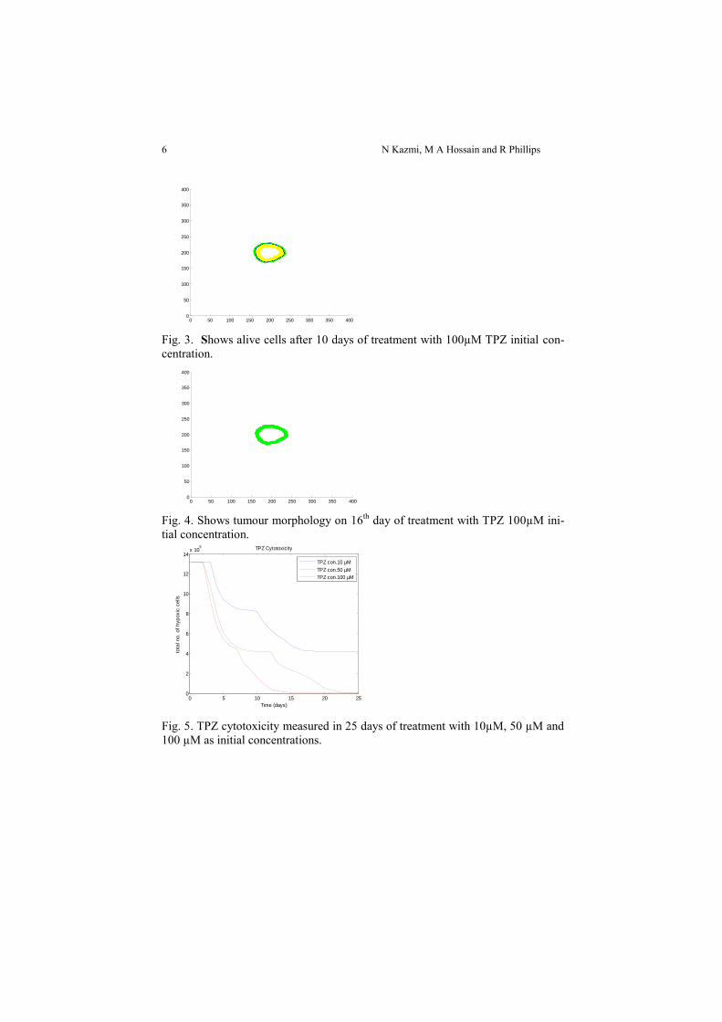
survived hypoxic cells using initial drug concentrations of 10 µM, 50 µM and 100
µM are compared in fig. 5. Total number of hypoxic cells was plotted against the
number of days (TPZ cycles). This comparison showed highest toxicity i.e. high-
est cell death rate against 100 µM concentration. Cell death was observed with
first few treatment cycles and on 16th
day hypoxic cell survival rate approached
zero. Cytotoxicity level was also high at 50 µM concentrations but was bit less
than that observed using100 µM. The cell survival approached to zero on the 23rd
day of treatment. Cytotoxicity at 10 µM concentration was the lowest and failed to
kill all hypoxic cells on 25th
day of treatment.
Fig. 1. Tumour mass is divided into 5 different hypoxic regions.
Fig. 2. On 5
th day of treatment with TPZ 100 µM as initial concentration.
0 50 100 150 200 250 300 350 4000
50
100
150
200
250
300
350
400
0 50 100 150 200 250 300 350 4000
50
100
150
200
250
300
350
400
6 N Kazmi, M A Hossain and R Phillips
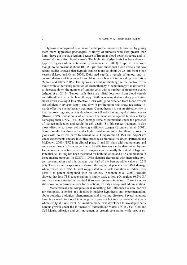
Fig. 3. Shows alive cells after 10 days of treatment with 100µM TPZ initial con-
centration.
Fig. 4. Shows tumour morphology on 16
th day of treatment with TPZ 100µM ini-
tial concentration.
Fig. 5. TPZ cytotoxicity measured in 25 days of treatment with 10µM, 50 µM and
100 µM as initial concentrations.
0 50 100 150 200 250 300 350 4000
50
100
150
200
250
300
350
400
0 50 100 150 200 250 300 350 4000
50
100
150
200
250
300
350
400
0 5 10 15 20 250
2
4
6
8
10
12
14x 10
5 TPZ Cytotoxicity
Time (days)
tota
l no.
of
hypoxic
cells
TPZ con.10 µM
TPZ con.50 µM
TPZ con.100 µM
Modelling of Tirapazamine Effects on Solid Tumour Morphology 7
CONCLUSIONS AND FUTURE WORK
This paper presented an in-silico model to observe the pharmacology of biore-
ductive drug tirapazamine inside solid tumour. The model calculated the amount
of TPZ and its efficient cell killing on each day of continuous drug infusion during
the whole treatment cycle. Drug metabolism and drug concentration inside each
cell was calculated using PDEs and solved in one dimension inside each cell. Drug
resistance and cytotoxicity effects on tumour morphology were also calculated us-
ing 10 µM, 50 µM and 100 µM as initial TPZ concentrations. Highest toxicity was
measures at 100 µM and lowest at 10 µM while we proposed that its highest
amount should be given to solid tumour to exploit hypoxia fully. As TPZ have no
or less toxic effects on normal cells. This model with integration of laboratory data
and parameters modifications can be used by oncologists and pharmacologist to
explore TPZ dynamic behaviour inside multicellular spheroids, animal and human
tumours. A quite strange behavior of bioreductive drugs has observed in clinical
practice that its considerable amount disappears inside the blood vessel before
reaching to the targeted locations. Our next aim is to capture the effects of Tirapa-
zamine inside the blood vessel and then its combination with chemotherapy treat-
ment.
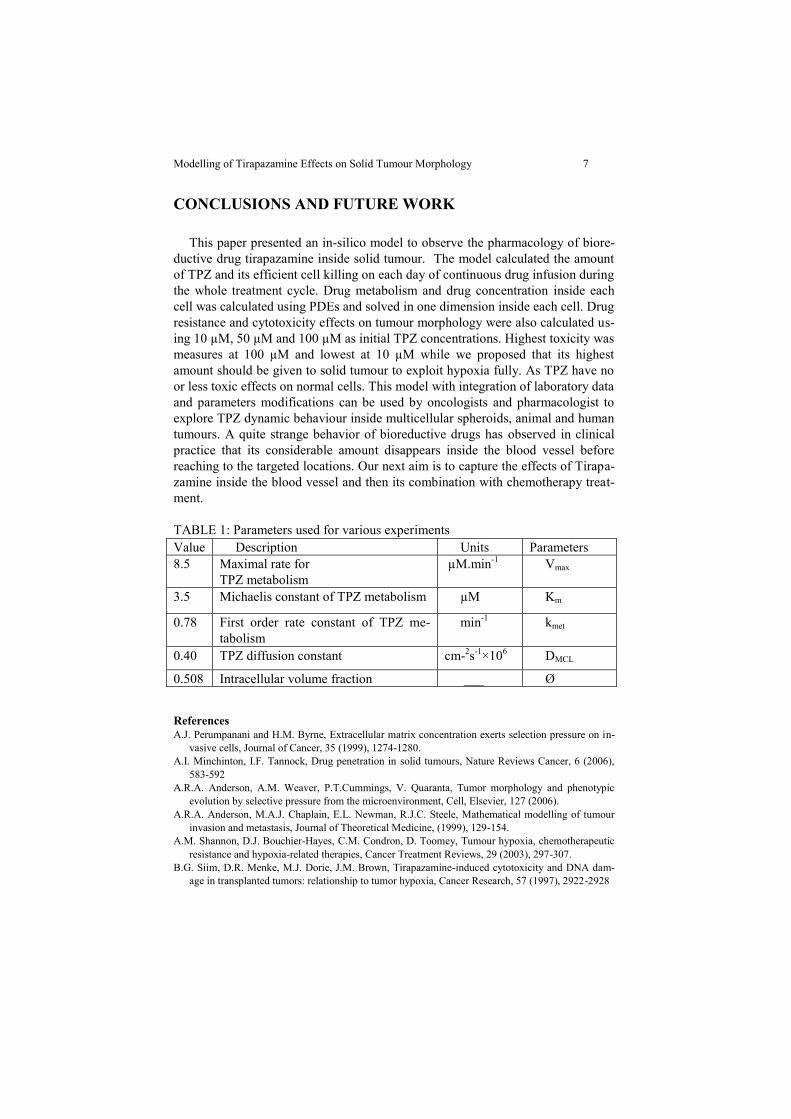
TABLE 1: Parameters used for various experiments
References
A.J. Perumpanani and H.M. Byrne, Extracellular matrix concentration exerts selection pressure on in-
vasive cells, Journal of Cancer, 35 (1999), 1274-1280.
A.I. Minchinton, I.F. Tannock, Drug penetration in solid tumours, Nature Reviews Cancer, 6 (2006),
583-592
A.R.A. Anderson, A.M. Weaver, P.T.Cummings, V. Quaranta, Tumor morphology and phenotypic
evolution by selective pressure from the microenvironment, Cell, Elsevier, 127 (2006).
A.R.A. Anderson, M.A.J. Chaplain, E.L. Newman, R.J.C. Steele, Mathematical modelling of tumour
invasion and metastasis, Journal of Theoretical Medicine, (1999), 129-154.
A.M. Shannon, D.J. Bouchier-Hayes, C.M. Condron, D. Toomey, Tumour hypoxia, chemotherapeutic
resistance and hypoxia-related therapies, Cancer Treatment Reviews, 29 (2003), 297-307.
B.G. Siim, D.R. Menke, M.J. Dorie, J.M. Brown, Tirapazamine-induced cytotoxicity and DNA dam-
age in transplanted tumors: relationship to tumor hypoxia, Cancer Research, 57 (1997), 2922-2928
Value Description Units Parameters
8.5 Maximal rate for
TPZ metabolism
µM.min-1
Vmax
3.5 Michaelis constant of TPZ metabolism µM Km
0.78 First order rate constant of TPZ me-
tabolism
min-1
kmet
0.40 TPZ diffusion constant cm-2s
-1×10
6 DMCL
0.508 Intracellular volume fraction ___ Ø

8 N Kazmi, M A Hossain and R Phillips
D. Hanahan, R.A. Weinberg, The hallmarks of cancer, Department of Biochemistry and Biophysics,
Cell, Elsevier, (2000) 57-70.
E. Lartigau, M. Guichard, Does tirapazamine (SR-4233) have any cytotoxic or sensitizing effect on
three human tumour cell lines at clinically relevant partial oxygen pressure?, International Journal
of Radiation Biology, 67 (1995), 211-216.
F. Kozusko, Z. Bajzer, Combining Gompertzian Growth and Cell Population Dynamics, Mathemati-
cal Biosciences, Elsevier, 185 (2003), 153-167.
H. Enderling, M.A.J. Chaplain, A.R.A. Anderson, J.S. Vaidya, A mathematical model of breast cancer
development, local treatment and recurrence, Journal of Theoretical Biology, Elsevier, 246 (2007),
245-259.
H. Enderling, A.R.A. Anderson, M.A.J. Chaplain, A.J. Munro, S.V. Vaidya, Mathematical modelling
of radiotherapy strategies for early breast cancer, Journal of Theoretical Biology, Elsevier, 241
(2006), 158-171.
H.M. Byrne , using mathematics to study solid tumour growth, In the preecedings of the 9 th general
meetings of euorpean women in mathematics, (1999), 81-107.
H.M. Byrne and M.A.J. Chaplain, Growth of nonnecrotic tumours in the presence and absence of inhi-
bitors, Journal of Mathematical Biosciences, 130 (1995), 151-181.
I.R. Conde, M.A.J. Chaplain, A.R.A. Anderson, Mathematical modeling of cancer cell invasion of tis-
sue, Mathematical and Computer Modelling, Elsevier, (2007).
J.J. Kim, L.F. Tannock, Repopulation of Cancer Cells during Therapy: an Important Cause of Treat-
ment Failure, Nature Cancer Review, 5, (2005), 516.
J.M. Brown, Exploiting the hypoxic cancer cell: mechanisms and therapeutic strategies, Molecular
Medicine Today, 6 (2000), 157-162.
J.M. Brown, SR 4233 (Tirapazamine): a new anticancer drug exploiting hypoxia in solid tumours, Brit-
ish Journal of Cancer, 67 (1993), 1163-1170.
J.M. Lee, S. Dedhar, R. Kalluri, and E. W. Thompson, The epithelial–mesenchymal transition: new in-
sights in signaling, development, and disease, Journal of Cell Biology, 172 (2006) 973-981.
J. Folkman, Tumor angiogenesis, Advance Cancer Research, 19 (1974), 331-358.
J. Vohardsky, Neural model of the Genetic Network, Journal of Biological Chemistry, 276 (2001),
36168-36173.
L.H. Patterson and S.R. Mckeown, AQ4N: a new approach to hypoxia-activated cancer chemotherapy,
British Journal of Cancer, 83(2000), 1589-1593.
L. Marcu, I. Olver, Tirapazamine: From Bench to Clinical Trials, Current Clinical Pharmacology, 1
(2006), 71-79.
L. Preziosi and A. Farina, On decay‘s law for growing porous media, Journal of non-linear mathemat-
ic, 37 (2002), 485-491.
N. Ferrara, R.S. Kerbel, Angiogenesis as a Therapeutic Target, Nature, 438 (2005), 967-974.
N.Kazmi, M.A.Hossain, R.M. Phillips, Intelligent modeling of benign tumour growth with cell-cell and
cell-matrix adhesion, 10th IEEE International Conference on Computer and Information Technol-
ogy, (2010).
P. Gerlee, A.R.A. Anderson, A hybrid cellular automaton model of clonal evolution in cancer: the
emergence of the glycolytic phenotype, Journal of Theoretical Biology, Elsevier, (2007), 705-722.
P. Gerlee, A.R.A. Anderson, An evolutionary hybrid cellular automaton model of solid tumour growth,
Journal of Theoretical Biology, Elsevier, (2007).
R.E. Durand, P.L. Olive, Physiologic and cytotoxic effects of tirapazamine in tumor-bearing mice,
Radiation Oncology Investigations, 5 (1997), 213-219.
S.M Tse, Y.Liang, K.S.Leung, K.H. Lee and T.S. Mok, A mimetic algorithm for multiple-drug
cancer chemotherapy schedule optimization, IEEE transaction on systems man, cybernatics,
37(2007), 84-91.
S.Algoul, M.A.Hossain, M.A.A. Majumderand M.S. Alam, Multi-objective optimal chemothera-
py model for cancer treatment, Medical Biology Engineering and Computing, 2010.