aqa biology unit 1.1 disease and pathogens
TRANSCRIPT
AQA BIOLOGY UNIT 1.1-DISEASE AND PATHOGENS
Pathogens- Gain entry, Colonise tissues, Resist defences, Cause damage Pathogens (organisms that cause disease) include-
Bacteria
Fungi
Viruses’
Pathogens can enter the body via penetration of an organisms interface mainly via-
The gas exchange system (breathe in air containing pathogen)
The digestive system (eat or drink food containing the pathogen)
The skin
Pathogens cause disease by-
Releasing toxins in to the body
Direct damage to the cell (rupturing the cell to release its nutrients, breaking down nutrients
in the cell or replicating inside the cell causing it to burst)
Preventing pathogen entry-
Mucus lining in the lung epithelium
Blood clots at the area of skin damage
Acidic conditions of the stomach
Enzymes which break down pathogens
Lifestyle A risk factor is something that increases your chance of developing a disease.
A disease is a malfunction of part or whole of the body with a characteristic set of symptoms.
CHD (Coronary heart disease) risk factors-
HIGH SATURATED FAT, HIGH SATURATED SALT-poor diet
HIGH BLOOD PRESSURE- due to smoking, lack of exercise or excessive alcohol intake causing
damage to the heart and blood vessels.
Cancer (uncontrolled cell division) risk factors-
Smoking
Carcinogenic chemicals
Ionising radiation
Excessive alcohol intake
A change in lifestyle would result in reduced risk of contracting these conditions.

Data analysis Shows a positive/negative correlation
Correlation does not mean causation
Other named factor
Sample size
Data analysed

AQA BIOLOGY UNIT 1.2- THE DIGESTIVE SYSTEM
Digestion When large insoluble molecules are broken down into smaller more soluble molecules by hydrolysis
from digestive enzymes so they can be absorbed and assimilated.
Oesophagus- takes food from the mouth to the stomach, mouth contains salivary glands
secreting salivary amylase which breaks starch into maltose, mucus on its walls to lubricate
food passage
Stomach- gastric juice to break down food containing HCl, pepsin and mucus, pepsin is an
enzyme which hydrolyses proteins
Small intestine- alkaline bile and pancreatic juices from the pancreas neutralise the acidity of
the HCl from the stomach, small soluble molecules are absorbed in the gut wall by villi
Large intestine- absorbs water, salt and minerals, bacteria that decompose some undigested
nutrients
Rectum- faeces stored here until defecation
Proteins Proteins have a variety of functions such as enzymes, antibodies, transport and structural function.
Primary Structure- sequence of amino acids in a polypeptide chain
Secondary Structure- hydrogen bonds form between amino acids, the chain coils into an
alpha helix or folds into beta pleated sheets
Tertiary Structure- the secondary structure is coiled or folded further, more bonds are
formed, final 3D structure for proteins made from one polypeptide chain
Quaternary Structure- final 3D structure for proteins made of more than one polypeptide
chain
Polypeptides are more than two amino acids are joined.
A dipeptide is when two amino acids join.
A peptide bond is the bond formed between two amino acids.
A condensation reaction, removal of a water molecule, joins two amino acids together, forms a
peptide bond.
A hydrolysis reaction, addition of a water molecule, separates two amino acids, breaks a peptide
bond.
Biuret test
Add NaOH to make the solution alkaline
Add copper(II) sulphate
If protein is present a purple layer forms
If is no protein the solution will remain blue
Enzymes Enzymes are proteins which speed up rate of reaction by acting as catalysts and lowering the
activation energy required for a reaction.
‘Lock and Key’ Model-
Substrate will fit into the enzymes active site
This will form an enzyme substrate complex and break up the substrate
‘Induced Fit’ Model-
Any substrate binds to the active site of the enzyme causing the active site to change shape
The substrate can now form a complimentary enzyme substrate complex and break up the
substrate
Induced Fit model is better as shows how the active site can be specific and only bind to one
substrate.
Factors affecting enzyme activity Temperature
Increase causes an increase in kinetic energy, faster moving molecules, enzymes more likely
to collide with substrates, higher energy collisions which are more likely to result in a
reaction
If it is too high, the enzyme molecule vibrates more, some bonds are broken, the tertiary
structure is altered, active site changes shape, enzyme and substrate no longer fit and the
enzyme is denatured
pH
All enzymes have an optimum pH, most work best at pH 7,
If the pH of an enzyme is not optimum the ionic bonds and hydrogen bonds break, this
changes the shape of the active site and denatures the enzyme
Concentration
Increase causes faster reaction, more substrate molecules means a collision is more likely so
more active sites are filled, levels off when all the active sites are filled so no more enzyme
substrate complexes can be formed
Inhibition of enzymes Competitive Inhibition-
Similar shape to the substrate molecule
Bind to the active site of the enzyme in place of the substrate but no reaction takes place
They block enzyme-substrate complexes forming
Non-competitive Inhibition-
Bind to the enzyme on a place away from the active site
Cause the active site to change shape so the substrate can no longer bind to it
Enzyme-substrate complexes can no longer form
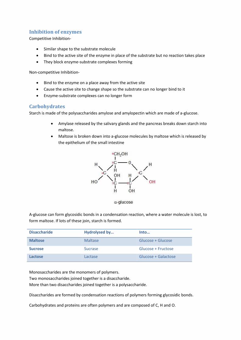
Carbohydrates Starch is made of the polysaccharides amylose and amylopectin which are made of a-glucose.
Amylase released by the salivary glands and the pancreas breaks down starch into
maltose.
Maltose is broken down into a-glucose molecules by maltose which is released by
the epithelium of the small intestine
A-glucose can form glycosidic bonds in a condensation reaction, where a water molecule is lost, to
form maltose. If lots of these join, starch is formed.
Monosaccharides are the monomers of polymers.
Two monosaccharides joined together is a disaccharide.
More than two disaccharides joined together is a polysaccharide.
Disaccharides are formed by condensation reactions of polymers forming glycosidic bonds.
Carbohydrates and proteins are often polymers and are composed of C, H and O.
Disaccharide Hydrolysed by… Into…
Maltose Maltase Glucose + Glucose
Sucrose Sucrase Glucose + Fructose
Lactose Lactase Glucose + Galactose

Tests Reducing sugars
All monosaccharides and some disaccharides
Add Benedict’s reagent to a sample and heat it
If it contains reducing sugars, the sample will turn from blue to brick red
Non-Reducing sugars
Sucrose
Boil the test solution with dilute HCl
Neutralise the solution with sodium hydrogen carbonate
Add Benedict’s reagent to the sample and heat it
If it turns brick red it contains either a reducing sugar or a non-reducing sugar.
Iodine test for starch
Add iodine dissolved in potassium iodide solution
If starch is present, colour change from orange to a dark blue/black
Lactose Intolerance Lactose is a sugar found in milk
Lactase, an enzyme released the epithelium of the intestine, breaks down lactose
Some people don’t have enough lactase so can’t break down the lactose
Can cause stomach cramps, diarrhoea and flatulence
Milk can be purified with lactase to make it suitable for lactose intolerant people
AQA BIOLOGY UNIT 1.3- ORGANISMS AND SURFACE INTERCHANGES
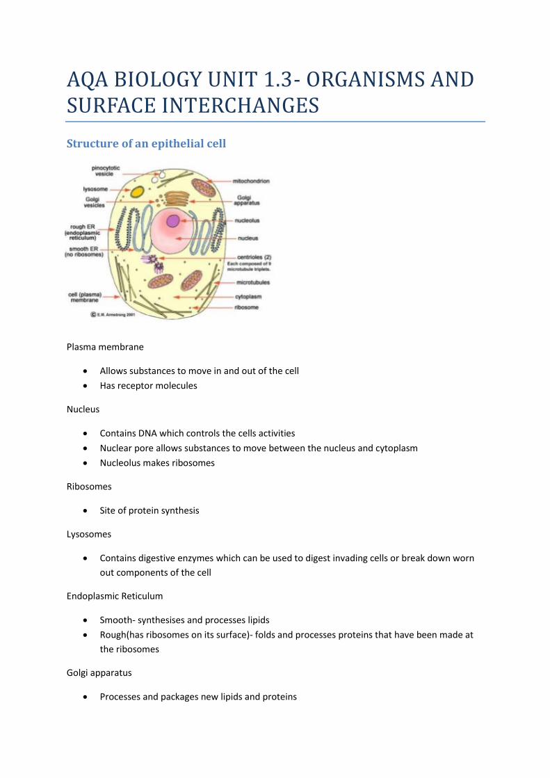
Structure of an epithelial cell
Plasma membrane
Allows substances to move in and out of the cell
Has receptor molecules
Nucleus
Contains DNA which controls the cells activities
Nuclear pore allows substances to move between the nucleus and cytoplasm
Nucleolus makes ribosomes
Ribosomes
Site of protein synthesis
Lysosomes
Contains digestive enzymes which can be used to digest invading cells or break down worn
out components of the cell
Endoplasmic Reticulum
Smooth- synthesises and processes lipids
Rough(has ribosomes on its surface)- folds and processes proteins that have been made at
the ribosomes
Golgi apparatus
Processes and packages new lipids and proteins

Makes lysosomes
Microvilli
Increase the surface area of the plasma membrane for absorption
Mitochondrion
Site of aerobic respiration
Produce ATP for energy
Have a double membrane, the inner one is folded to form cristae, inside is the matrix which
contain enzymes
Microscopes Magnification- How much bigger a sample appears to be under a microscope than it is in real life
Resolution- ability to distinguish between two points on an image
Electron microscopes are better than light microscopes as they have higher resolutions, detailed
images and 3D images. A vacuum is needed however which requires the specimen to be dead, it’s
expensive and skills and training are needed. Light has a longer wavelength than electrons. Electron
microscopes both produce black and white images compared to the colour image produced by light
microscopes. Staining process, thin sample and vacuum needed for electron microscopes. TEM and
SEM may contain artefacts because of the complex staining process.
TEM (Transmission Electron Microscope)
2D image
Highest magnification
Best resolution
Electron beam passes through a thin sample
Magnification Size of object
Size of Image
SEM (Scanning Electron Microscope)
3D image
High resolution
Good resolution
Electron beam bounces off the sample
Cell fractionation and ultracentrifugation 1. Homogenisation- Breaking up the cells
Break open the plasma membrane to release organelles into the solution
2. Filtration- Getting rid of the big bits
Homogenised solution filtered through a gauze to remove any large cell or tissue
debris.
3. Ultracentrifugation- Separating the organelles
Put the solution in a tube un a centrifuge and spin at low speeds
The heaviest organelles will go to the bottom of the centrifuge, the pellet
The supernatant is the suspended fluid above the pellet with the remaining
organelles
The pellet can be removed
The process is repeated at higher speeds to separate all the organelles out until you
are finally left with the lightest organelle.
Plasma membranes The plasma membrane contains proteins, carbohydrates (usually attached to proteins or lipids) and
lipids(mainly phospholipids)
Triglycerides are formed by condensation reactions between glycerol and a fatty acid.
Triglycerides have a glycerol head and three fatty acid tails.
Phospholipids have a glycerol head, two fatty acid tails and a phosphate group attached to
the head.
The phosphate group is hydrophilic and the fatty acid tail is hydrophobic.
A fatty acid may be unsaturated or saturated.
Emulsion test for lipids- Shake the substance with ethanol then pour in water. A lipid will show a
milky emulsion.
Diffusion Diffusion is the passive movement of substances down a concentration gradient from an area with
high concentration to an area with lower concentration.
Factors affecting diffusion-
Surface area
Difference in concentration
Thickness of exchange surface
Particles can diffuse across the plasma membrane as they can move freely through the membrane(if
they are small enough).

Facilitated diffusion-
Still the diffusion of particles down a concentration gradient
Both the carrier proteins and the protein channels are specific to the molecule they are transporting
Carrier proteins- move large molecules in or out of the cell, the molecule attaches to the
carrier protein then the protein changes shape allowing the release of the molecule on the
other side of the membrane
Protein channels- pores in the membrane for charged particles to diffuse through.
Osmosis Diffusion of water molecules across a partially permeable membrane from an area of a high water
potential to an area with low water potential
Water potential is the potential of water molecules to diffuse into or out of a solution, water has the
highest water potential
Isotonic is when two solutions have the same osmotic pressure
Active transport Active transport uses energy to move molecules and ions across plasma membranes, against a
concentration gradient.
Carrier proteins- a molecule attaches to the protein, it changes shape, the molecule is
released on the other side of the membrane, energy is used from ATP to move the solute
against its concentration gradient
Co-transporters- a type of carrier protein, bind two molecules at a time, sodium ions and
glucose, concentration gradient of one molecule used to move the other molecule against its
concentration gradient
Absorption The products of carbohydrate digestion are absorbed in different ways-
Some glucose diffuses across the intestinal epithelium to the blood- there’s a higher
concentration of glucose in the small intestine than the blood when carbohydrates are first
digested so glucose diffuses out.
Some glucose enters the intestinal epithelium by active transport with sodium ions-
1. Sodium ions are actively transported out of the small intestine epithelial cells, into the
blood, by the sodium potassium pump. This causes a concentration gradient of more
sodium ions in the small intestine lumen than in the cell.
2. Sodium ions diffuse into the cell from the small intestine lumen. They do this via sodium-
glucose co-transporter proteins.
3. The co-transporter protein carries glucose into the cell with sodium, glucose
concentration inside the cell increases.
4. Glucose diffuses out of the cell into the blood down its concentration gradient, via a
protein channel by facilitated diffusion.

Cholera Cholera is an example of a prokaryotic organism. Prokaryotic organisms have some different
features than eukaryotic organisms; here is what they consist of-
Flagellum- Long hair like structure that helps the bacterium move
DNA- Free circular DNA in the cytoplasm
Plasma membrane- made mainly of lipids and proteins, controls movement of substances
into and out of the cell
Cell wall- supports the cell
Plasmids- small loops of DNA which contain genes and can be passed between bacteria
Capsule- made of secreted slime, protects the bacteria from attack
Cholera produces a toxin when it infects the body
1. The toxin causes chloride ion protein channels in the plasma membranes of the small
intestine epithelial cells to open
2. Chloride ions move into the small intestine lumen and lower the water potential of the
lumen
3. Water moves out of the blood and into the lumen by osmosis
4. The increase in water in the lumen leads to diarrhoea and dehydration
Oral rehydration solutions are used to treat diarrhoeal disease-
Water for rehydration
Sodium ions to replace those lost from epithelium
Glucose to provide energy and stimulate uptake of sodium ions by co-transport
Potassium ions to replace those lost and stimulate appetite
Other electrolytes to prevent an electrolyte imbalance
AQA BIOLOGY UNIT 1.4- THE LUNGS
The lungs of a mammal act as an interface with the environment
Lung function may be affected by pathogens and lifestyle
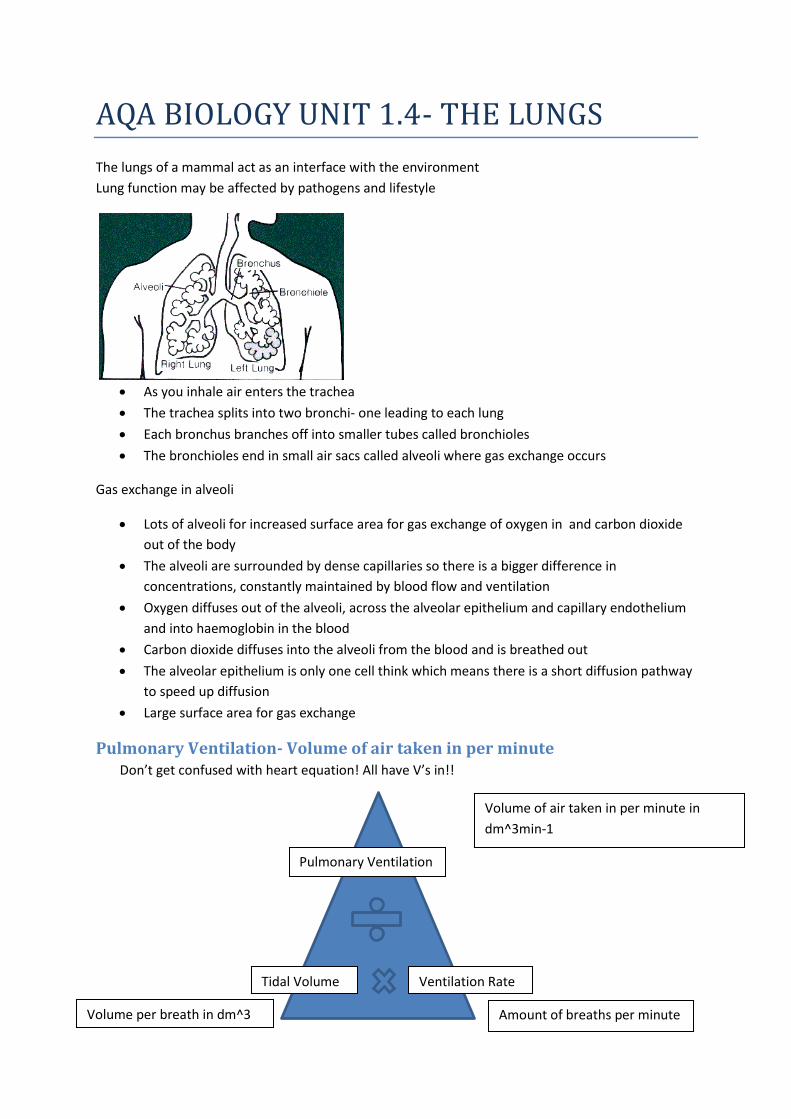
As you inhale air enters the trachea
The trachea splits into two bronchi- one leading to each lung
Each bronchus branches off into smaller tubes called bronchioles
The bronchioles end in small air sacs called alveoli where gas exchange occurs
Gas exchange in alveoli
Lots of alveoli for increased surface area for gas exchange of oxygen in and carbon dioxide
out of the body
The alveoli are surrounded by dense capillaries so there is a bigger difference in
concentrations, constantly maintained by blood flow and ventilation
Oxygen diffuses out of the alveoli, across the alveolar epithelium and capillary endothelium
and into haemoglobin in the blood
Carbon dioxide diffuses into the alveoli from the blood and is breathed out
The alveolar epithelium is only one cell think which means there is a short diffusion pathway
to speed up diffusion
Large surface area for gas exchange
Pulmonary Ventilation- Volume of air taken in per minute Don’t get confused with heart equation! All have V’s in!!
Ventilation Rate Tidal Volume
Pulmonary Ventilation
Volume per breath in dm^3 Amount of breaths per minute
Volume of air taken in per minute in
dm^3min-1
Inspiration VS Exhalation Inspiration
Intercostal muscles and diaphragm contract
Ribcage moves upwards and outwards
Volume increases
Lung pressure decreases
Air moves in the lungs
Exhalation
Intercostal and diaphragm muscles relax
Ribcage moves down and inwards
Volume decreases
Lung pressure increases
Air moves out of the lungs
Pulmonary Tuberculosis (TB) Caused by bacteria
Transmitted by droplets in air being breathed in- from mucus/saliva in coughing or sneezing
Phagocytes build a wall around bacteria in the lungs called tubercles
Bacteria destroy tissue of lungs resulting in cavities and scar tissue arises, causing damage to
the gas exchange surface
Bacteria can spread once in blood
The inactive form may stay in the body until your immune system is weakened and then the
active form is stimulated
Coughing mucus or blood, chest pains, fatigue, shortness of breath, fever, weight loss
Emphysema- Smoking/Air pollution Foreign particles are trapped in the alveoli causing inflammation that attracts phagocytes
Phagocytes break down alveoli elastin so the alveoli can’t recoil to expel air
The alveoli walls have a smaller surface area and a lower gas exchange rate
Fibrosis- Infection/Dust/Asbestos Formation of scar tissue
Thicker, less elastic lung tissue, less expansion so lower tidal volume
Slower gas exchange
Faster breathing rate
Asthma- Allergic reaction to pollen/Dust Inflamed and irritated airways
Muscle lining bronchioles contracts and produces mucus
Airways are constricted so tidal volume lower, faster breathing rate
Air flow reduced
Less oxygen enters the alveoli and therefore the blood, slower gas exchange
AQA BIOLOGY UNIT 1.5- THE HEART
The functioning of the heart plays a central role in the circulation of blood and relates to the
level of activity of an individual.
Heart structure
Cardiac cycle 1. Relaxation of the heart- Diastole
Blood enters the atria and ventricles via the pulmonary veins and vena cava
Atria are relaxed and fill with blood
Ventricles are relaxed
Semi lunar valves closed
Left and right Atrioventricular valves open
Relaxed ventricles draws blood from atria
2. Contraction of the atria- Atrial systole
Atria contract to push remaining blood into the ventricles
Semi lunar valves closed
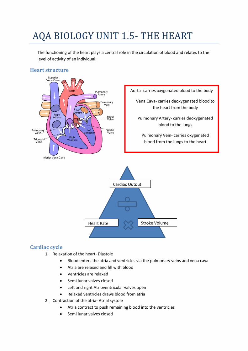
Aorta- carries oxygenated blood to the body
Vena Cava- carries deoxygenated blood to
the heart from the body
Pulmonary Artery- carries deoxygenated
blood to the lungs
Pulmonary Vein- carries oxygenated
blood from the lungs to the heart
Cardiac Output
Heart Rate Stroke Volume
Left and right Atrioventricular valves open
Blood pumped from atria to ventricles
Ventricles remain relaxed
3. Contraction of the ventricles- Ventricular systole
Blood pumped into pulmonary arteries and the aorta
Atria relax
Semi lunar valves open
Left and right atrioventricular valves closed
Ventricles contract
Blood is pushed away from the heart
Myogenic Heart Muscle The atrioventricular node and the sinoatrial node pass electrical activity to each other in order to
make the heart contract.
A wave of electrical activity passes from the SAN causing the atria to contract
Non-conductive tissue stops this electrical activity passing to the ventricles
The electrical activity passes to the AVN which causes a delay so the atria can fully empty
and the atrioventricular valves can fully close so this prevents backflow of blood to the atria
The AVN sends a wave of electrical activity to the bundle of his
It travels down the bundle of his to the purkinje fibres were it makes the ventricles contract
thus forcing blood out of the heart
Heart Problems An atheroma is a build-up of fatty deposits under the epithelium of the artery wall
Causes the lumen to narrow restricting blood flow
Increases chances of aneurysm and thrombosis
Myocardial infarction-
When the lumen is so narrowed that not enough blood can get to the heart muscle
The heart is not supplied with enough oxygen so dies
Thrombosis-
When an atheroma breaks through the lining of the artery
Forms a rough surface
As the body repairs it, a clot may be formed (a thrombus)
Will block the blood vessel further and supply of blood to tissue
Tissue dies due to lack of oxygen and nutrients (glucose)
Clot could detach and block another artery blocking blood flow and thus oxygen flow to the
heart muscle
Aneurysm-
Caused by thrombosis weakening the artery walls
Weakened parts swell to form balloon like structures called aneurysms
These can burst causing haemorrhaging and blood loss- in the brain this is a stroke
Risk factors of CHD A diet high in saturated fats or salts
High blood cholesterol
Smoking
Lack of exercise and excess of alcohol causing high blood pressure
AQA BIOLOGY UNIT 1.6- IMMUNOLOGY
Phagocytosis (Cellular Response) When a pathogen with a foreign antigen enters the body this stimulates the phagocyte
The phagocyte engulfs the pathogen into a vesicle in its cytoplasm
Lysosomes bind to the vesicle and release digestive enzymes to break down the pathogen
The pathogens antigen is then presented on the phagocyte to aid the T cells in the immune
response
T- Cells (Cellular Response) Bind to the antigen presented by the phagocyte
T-Cells bind to the antigen with the complimentary receptor, it is activated and clones
Helper T-Cells stimulate B-Cells
Killer T-Cells locate and destroy the body cells infected by the pathogen/with the antigen
B- Cells (Humoral Response) Helper T-Cells stimulate B-Cells to clone into two types
Plasma cells produce specific antibodies to form antibody antigen complexes with the
pathogen
Memory cells ( of T-Cells and B-Cells ) remain in the body in case of re- infection
An antigen is a molecule which stimulates an immune response and results in producing specific
antibodies
An antibody is a protein made in response to a foreign antigen with binding sites which bind
specifically to the antigen to form an antigen-antibody complex
Antibody shape is complimentary to the antigen
Each antibody will only bind to a specific antigen
Secondary Response Body recognises the foreign antigen
Memory T cells and B cells are stimulated
T cells recognise and bind to the foreign antigen
B cells produce antibodies specific to the antigen and form antigen- antibody complexes to
kill the pathogen
This response is much faster, no symptoms shown
Antigenic variability (Influenza virus) Some pathogens have different strains with different surface antigens
Antigens mutate and change shape
Can’t use secondary response
If your body has suffered from one strain but is re-attacked with another strain the memory
cells do not recognise the antigen

Must go through the cellular and humoral response again
Hard to produce vaccines against
Vaccines (active immunity) Injection of a dead or weakened version of a pathogen
Initiate primary response
No symptoms
Allows for fast secondary response if pathogen invades the body in future
Booster doses increase efficiency
Herd Immunity- When most of the population is vaccinated so it is less likely an individual
will develop a disease even if they are not vaccinated
Monoclonal antibodies- genetically identical B cells/plasma cells Antibodies bind to specific antigens
Can inject mouse with any pathogen to stimulate it to produce an antibody against this
Monoclonal antibodies can be made to bind to tumour markers on cancer cells and we can
attach an anti-cancer drug to the antibody so they bind and target only the cancer cell