architectures of mammalian and fungal fatty acid synthases presentation based on: t. maier, s....
TRANSCRIPT
Architectures of Mammalian and Fungal Fatty Acid Synthases
Presentation based on:
T. Maier, S. Jenni, N. Ban, Science 311, 1258 (2006). -- Mammalian fatty acid at 4.5 Å resolution
S. Jenni, M. Leibundgut, T. Maier, N. Ban, Science 311, 1263 (2006). -- Fungal fatty acid at 5 Å resolution

Agenda
1. fatty acid quick peak
2. catalytic cycle of fatty acid synthesis
3. mammalian fatty acid synthase structure
4. fungal fatty acid synthase structure

QuickTime™ and aTIFF (Uncompressed) decompressor
are needed to see this picture.
1. Fatty acid quick peak
Common fatty acids are carboxylic acids with long hydrocarbon tails:
comes with a COOH head and a tail of many CH2.

2. Fatty acid catalytic cycle
2.1 Common elongation scheme
starter substrates Acetyl coenzyme A (Acetyl-CoA) and Malonyl-CoA transfer the active functionals to acyl carrierprotein (ACP).
ACP transports substrate to different reaction sites, catalyzed by different enzyme. A complete cycle gives the acyl group an additional two carbon units.
This step-wise elongation repeats until a substrate length of C16 to C18 is achieved.
Another enzyme then release the substrate from ACP, completing the synthesis process.
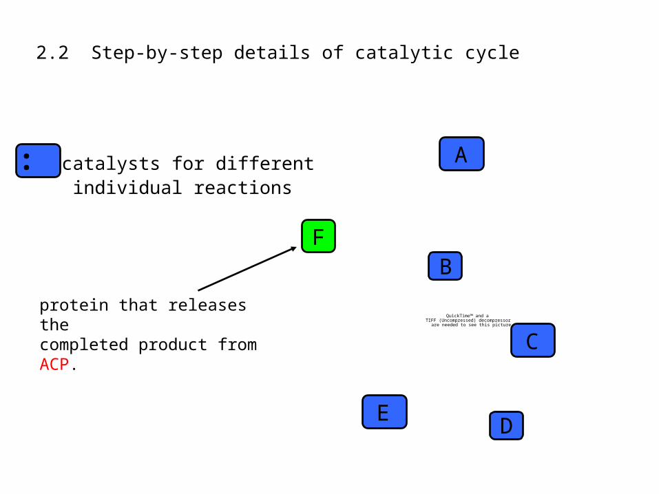
2.2 Step-by-step details of catalytic cycle
QuickTime™ and aTIFF (Uncompressed) decompressor
are needed to see this picture.
: catalysts for different individual reactions
protein that releases the completed product from ACP.
A
B
C
DE
F
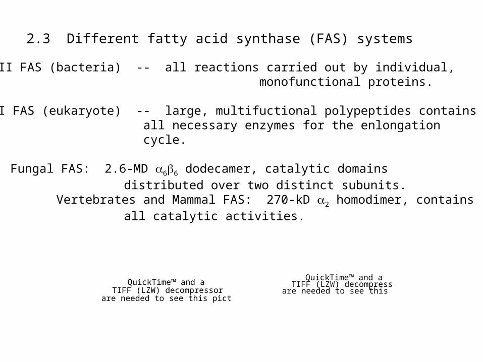
2.3 Different fatty acid synthase (FAS) systems
Type II FAS (bacteria) -- all reactions carried out by individual, monofunctional proteins.
Type I FAS (eukaryote) -- large, multifuctional polypeptides contains all necessary enzymes for the enlongation cycle.
Fungal FAS: 2.6-MD 66 dodecamer, catalytic domains distributed over two distinct subunits.
Vertebrates and Mammal FAS: 270-kD 2 homodimer, contains all catalytic activities.
QuickTime™ and aTIFF (LZW) decompressor
are needed to see this picture.
QuickTime™ and aTIFF (LZW) decompressor
are needed to see this picture.
3. Mammalian fatty acid synthase
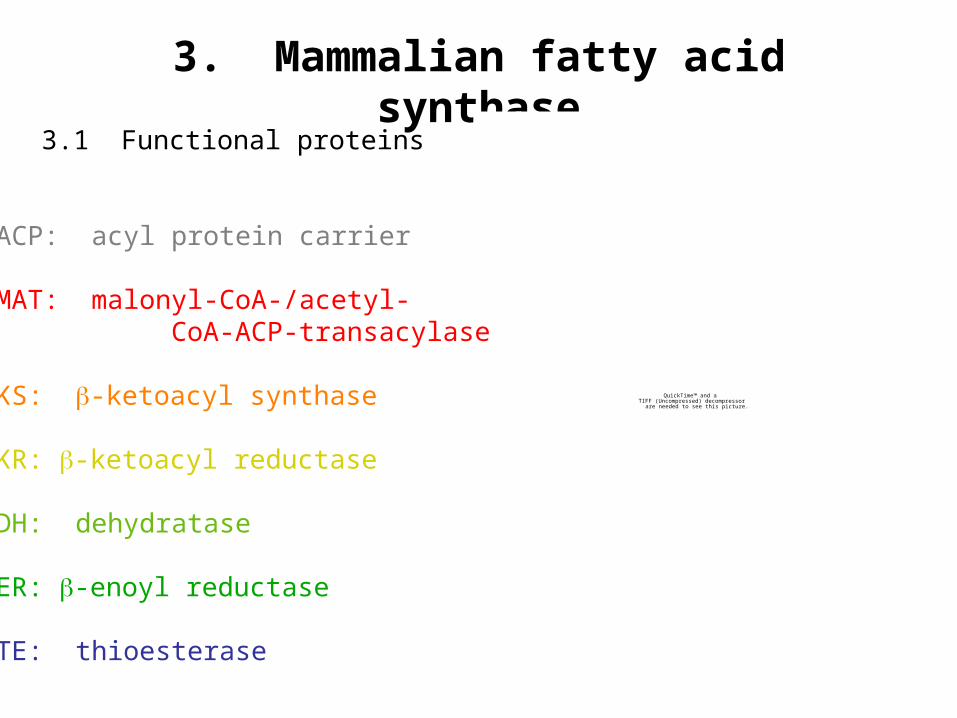
3.1 Functional proteins
QuickTime™ and aTIFF (Uncompressed) decompressor
are needed to see this picture.
ACP: acyl protein carrier
MAT: malonyl-CoA-/acetyl- CoA-ACP-transacylase
KS: -ketoacyl synthase
KR: -ketoacyl reductase
DH: dehydratase
ER: -enoyl reductase
TE: thioesterase
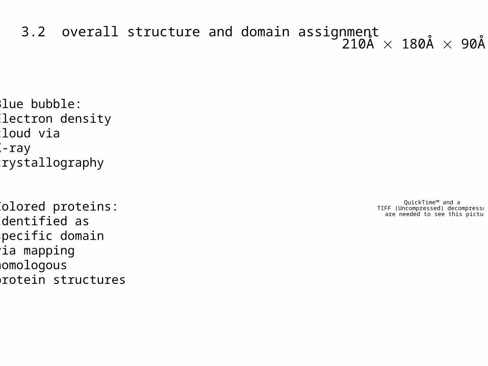
3.2 overall structure and domain assignment
QuickTime™ and aTIFF (Uncompressed) decompressor
are needed to see this picture.
210Å 180Å 90Å
Blue bubble:Electron density cloud viaX-ray crystallography
Colored proteins:Identified as specific domainvia mapping homologous protein structures
QuickTime™ and aTIFF (Uncompressed) decompressor
are needed to see this picture.
QuickTime™ and aTIFF (Uncompressed) decompressor
are needed to see this picture.

3.2.1 KS domain
QuickTime™ and aTIFF (Uncompressed) decompressor
are needed to see this picture.
Mammalian KS closely resembles the Escherichia coli KS I (FabB).So KS domain was fitted with E. coli FabB.
QuickTime™ and aTIFF (LZW) decompressor
are needed to see this picture.
QuickTime™ and aTIFF (Uncompressed) decompressor
are needed to see this picture.
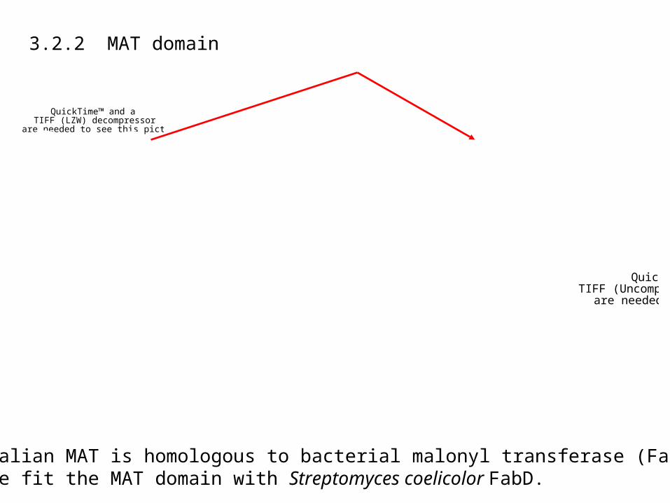
3.2.2 MAT domain
Mammalian MAT is homologous to bacterial malonyl transferase (FabD).So we fit the MAT domain with Streptomyces coelicolor FabD.
QuickTime™ and aTIFF (LZW) decompressor
are needed to see this picture.
QuickTime™ and aTIFF (Uncompressed) decompressor
are needed to see this picture.
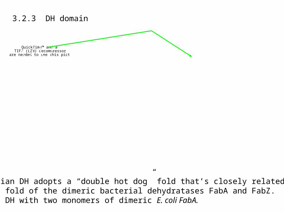
3.2.3 DH domain
Mammalian DH adopts a “double hot dog” fold that’s closely related to the fold of the dimeric bacterial dehydratases FabA and FabZ. So we fit DH with two monomers of dimeric E. coli FabA.
QuickTime™ and aTIFF (LZW) decompressor
are needed to see this picture.

QuickTime™ and aTIFF (Uncompressed) decompressor
are needed to see this picture.
3.2.4 ER domain
The best structural match for ER was obtained with a zinc-free bacterial quinone reductase. Here the particular model is quinone reductase of T. thermophilus.
QuickTime™ and aTIFF (LZW) decompressor
are needed to see this picture.
QuickTime™ and aTIFF (Uncompressed) decompressor
are needed to see this picture.
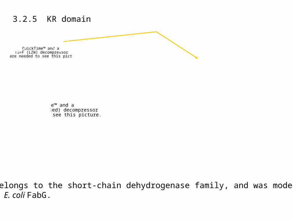
3.2.5 KR domain
KR belongs to the short-chain dehydrogenase family, and was modeled with E. coli FabG.
QuickTime™ and aTIFF (LZW) decompressor
are needed to see this picture.
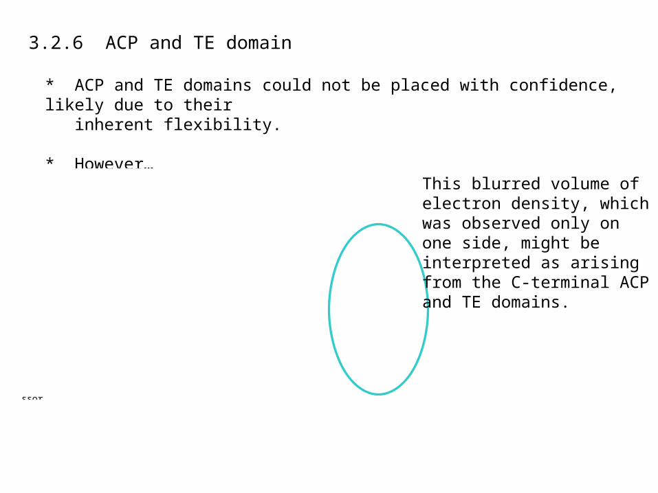
3.2.6 ACP and TE domain
* ACP and TE domains could not be placed with confidence, likely due to their inherent flexibility.
* However…
QuickTime™ and aTIFF (Uncompressed) decompressor
are needed to see this picture.
This blurred volume of electron density, which was observed only onone side, might be interpreted as arising from the C-terminal ACPand TE domains.

QuickTime™ and aTIFF (LZW) decompressor
are needed to see this picture.
3.2.7 Table of structural and functional analogs
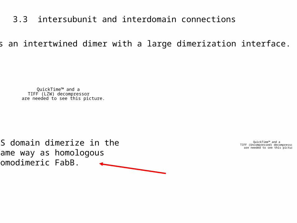
3.3 intersubunit and interdomain connections
FAS is an intertwined dimer with a large dimerization interface.
QuickTime™ and aTIFF (Uncompressed) decompressor
are needed to see this picture.KS domain dimerize in the same way as homologous homodimeric FabB.
QuickTime™ and aTIFF (LZW) decompressor
are needed to see this picture.
QuickTime™ and aTIFF (Uncompressed) decompressor
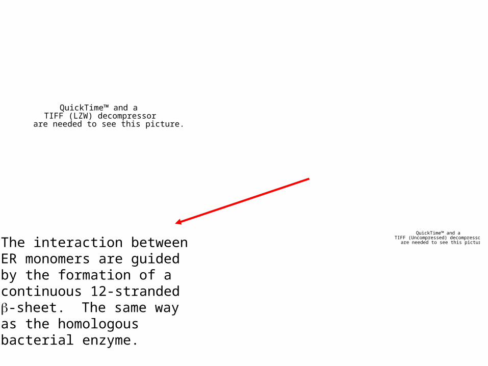
are needed to see this picture.The interaction betweenER monomers are guidedby the formation of a continuous 12-stranded-sheet. The same way as the homologousbacterial enzyme.
QuickTime™ and aTIFF (LZW) decompressor
are needed to see this picture.

QuickTime™ and aTIFF (LZW) decompressor
are needed to see this picture.
There are other substantialintersubunit contacts in the unassigned region of electrondensity map.
COLORED: identified domains
GREY: unassigned region.
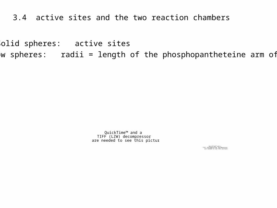
3.4 active sites and the two reaction chambers
QuickTime™ and aTIFF (LZW) decompressor
are needed to see this picture.QuickTime™ and a
TIFF (Uncompressed) decompressorare needed to see this picture.
Solid spheres: active sites
Hollow spheres: radii = length of the phosphopantheteine arm of ACP
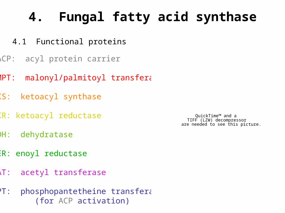
4. Fungal fatty acid synthase
4.1 Functional proteins
ACP: acyl protein carrier
MPT: malonyl/palmitoyl transferase
KS: ketoacyl synthase
KR: ketoacyl reductase
DH: dehydratase
ER: enoyl reductase
AT: acetyl transferase
PT: phosphopantetheine transferase (for ACP activation)
QuickTime™ and aTIFF (LZW) decompressor
are needed to see this picture.

4.2 overall structure and domain assignment 230Å 230Å 260Å
QuickTime™ and aTIFF (LZW) decompressor
are needed to see this picture.
MPT: malonyl/palmitoyl transferase
KS: ketoacyl synthase
KR: ketoacyl reductase
DH: dehydratase
ER: enoyl reductase
AT: acetyl transferase
ACP and PT structures could not be identified.
White regions denote unidentified electron density.

QuickTime™ and aTIFF (LZW) decompressor
are needed to see this picture.
QuickTime™ and aTIFF (LZW) decompressor
are needed to see this picture.
QuickTime™ and aTIFF (LZW) decompressor
are needed to see this picture.
QuickTime™ and aTIFF (LZW) decompressor
are needed to see this picture.

4.2.1 KS domain
QuickTime™ and aTIFF (LZW) decompressor
are needed to see this picture.
QuickTime™ and aTIFF (LZW) decompressor
are needed to see this picture.
KS dimer domain was identified by finding the thiolase fold in the FAS electron density map (bacterial KS is known to adopt a thiolase fold and form homodimers).
Bacterial KS homolog fits almost perfectly into the density map.
QuickTime™ and aTIFF (LZW) decompressor
are needed to see this picture.

4.2.2 KR domain
QuickTime™ and aTIFF (LZW) decompressor
are needed to see this picture.
QuickTime™ and aTIFF (LZW) decompressor
are needed to see this picture.
QuickTime™ and aTIFF (LZW) decompressor
are needed to see this picture.
The 4-helix bundle is a characteristic trait of one of the dimerization interface in type-II tetrameric KR homolog of Brassica napus.It also contains a Rossmann fold.
The Brassica napus KR homolog fits FASelectron density remarkably well.
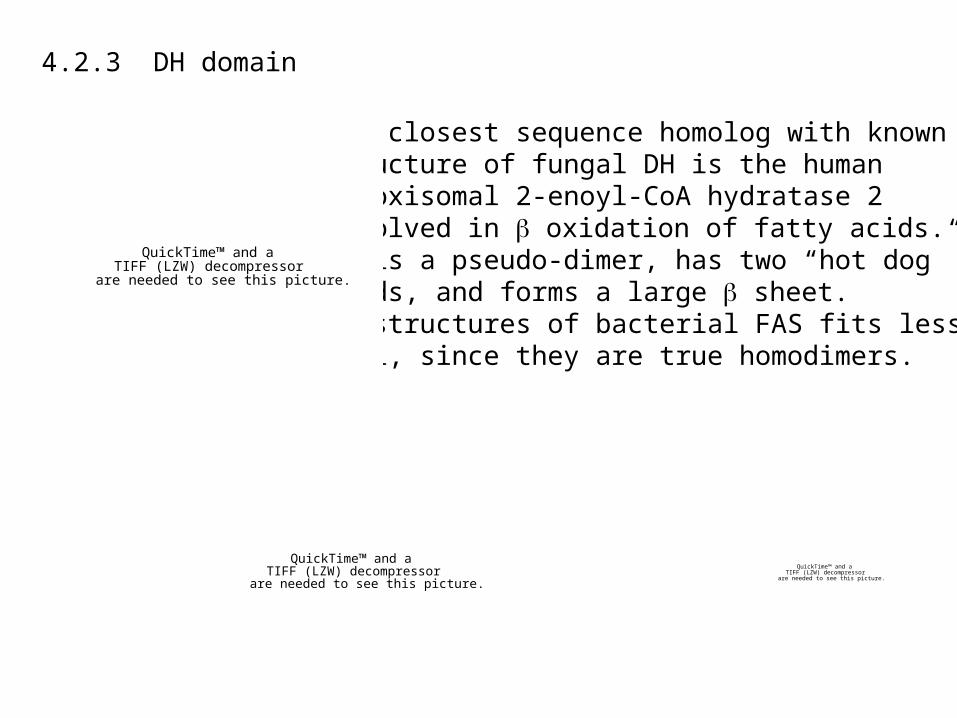
4.2.3 DH domain
The closest sequence homolog with known structure of fungal DH is the human Peroxisomal 2-enoyl-CoA hydratase 2 involved in oxidation of fatty acids. It is a pseudo-dimer, has two “hot dog”folds, and forms a large sheet.DH structures of bacterial FAS fits less well, since they are true homodimers.
QuickTime™ and aTIFF (LZW) decompressor
are needed to see this picture.
QuickTime™ and aTIFF (LZW) decompressor
are needed to see this picture.
QuickTime™ and aTIFF (LZW) decompressor
are needed to see this picture.
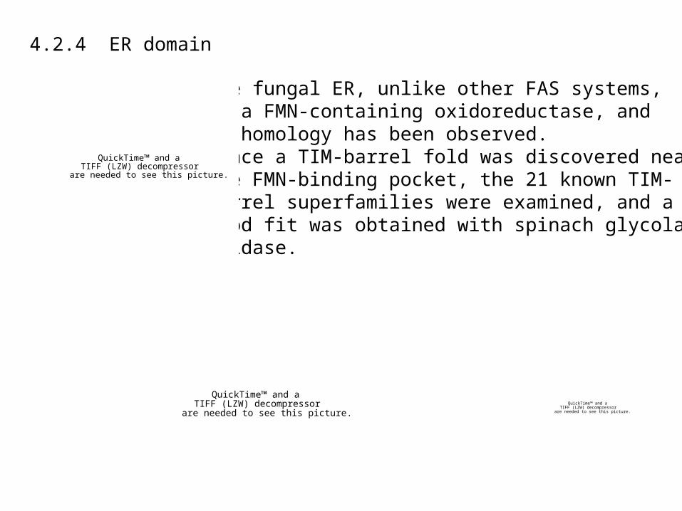
4.2.4 ER domain
The fungal ER, unlike other FAS systems,is a FMN-containing oxidoreductase, andno homology has been observed.Since a TIM-barrel fold was discovered nearthe FMN-binding pocket, the 21 known TIM-barrel superfamilies were examined, and a good fit was obtained with spinach glycolate oxidase.
QuickTime™ and aTIFF (LZW) decompressor
are needed to see this picture.
QuickTime™ and aTIFF (LZW) decompressor
are needed to see this picture.
QuickTime™ and aTIFF (LZW) decompressor
are needed to see this picture.

4.2.5 AT and MPT
QuickTime™ and aTIFF (LZW) decompressor
are needed to see this picture.
AT
MPT
QuickTime™ and aTIFF (LZW) decompressor
are needed to see this picture.
QuickTime™ and aTIFF (LZW) decompressor
are needed to see this picture.
AT and MPT are homologous in sequence, catalyze similar reactions, and have same protein fold. Therefore they are both fittedwith malonyl transferase from Streptomycescoelicolor.
QuickTime™ and aTIFF (LZW) decompressor
are needed to see this picture.
In order to unambiguously assign AT andMPT domain, the locations of their N termini relative to the C terminus of DHwere observed.
QuickTime™ and aTIFF (LZW) decompressor
are needed to see this picture.
QuickTime™ and aTIFF (LZW) decompressor
are needed to see this picture.
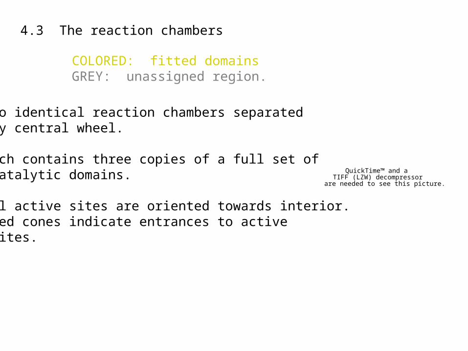
4.3 The reaction chambers
QuickTime™ and aTIFF (LZW) decompressor
are needed to see this picture.
QuickTime™ and aTIFF (LZW) decompressor
are needed to see this picture.
** Two identical reaction chambers separated by central wheel.
** Each contains three copies of a full set of catalytic domains.
** All active sites are oriented towards interior. Red cones indicate entrances to active sites.
COLORED: fitted domainsGREY: unassigned region.

4.3 The reaction chambers
QuickTime™ and aTIFF (LZW) decompressor
are needed to see this picture.
QuickTime™ and aTIFF (LZW) decompressor
are needed to see this picture.
A set of active sites in the reaction chamber with allenzymatic activities required for the synthesis cycle.
Green sphere: reaction chamber center.

4.3 The reaction chambers
QuickTime™ and aTIFF (LZW) decompressor
are needed to see this picture.
Schematic path of ACP, shuttlingsubstrate between the active sites.
QuickTime™ and aTIFF (LZW) decompressor
are needed to see this picture.
QuickTime™ and aTIFF (LZW) decompressor
are needed to see this picture.