arginine dependence of acute myeloid leukaemia blast
TRANSCRIPT
MUSSAI et al. BCT-100 ARGINASE THERAPY FOR AML
1
Arginine dependence of Acute Myeloid Leukaemia blast proliferation: a novel
therapeutic target.
Francis Mussai,1 Sharon Egan,2 Joseph Higginbotham-Jones,1 Tracey Perry,1 Andrew Beggs,1
Elena Odintsova,1 Justin Loke,1 Guy Pratt,1 Kin Pong U,3 Anthony Lo,4 Margaret Ng,4 Pamela
Kearns,1 Paul Cheng,3 Carmela De Santo.1
1School of Cancer Sciences, University of Birmingham, Birmingham, United Kingdom
2School of Veterinary Medicine and Science, University of Nottingham, Nottingham, United
Kingdom
3Bio-cancer Treatment International Ltd, Hong Kong
4Department of Anatomic Pathology, The Chinese University of Hong Kong, Hong Kong
4Bio-cancer Treatment International Ltd, Hong Kong
Corresponding author: Francis Mussai, School of Cancer Sciences, University of
Birmingham, Birmingham, United Kingdom. Tel: 0121 333 8234
Email: [email protected]
Scientific Category: Myeloid Neoplasia
Short title: BCT-100 Arginase therapy for AML
Key Points: Arginase depletion with BCT-100 pegylated recombinant human arginase is
cytotoxic to AML blasts.
MUSSAI et al. BCT-100 ARGINASE THERAPY FOR AML
2
Abstract
Acute Myeloid Leukaemia (AML) is one of the most common acute leukaemias in adults and
children, yet significant numbers of patients relapse and die of disease. In this study we identify
the dependence of AML blasts on arginine for proliferation. We show AML blasts
constitutively express the arginine transporters CAT-1 and CAT-2B, and that the majority of
newly diagnosed patients’ blasts have deficiencies in the arginine recycling pathway enzymes
arginosuccinate synthase (ASS) and ornithine transcarbamylase (OTC), making them arginine
auxotrophic. BCT-100, a pegylated human recombinant arginase, leads to a rapid depletion in
extracellular and intracellular arginine concentrations, resulting in arrest of AML blast
proliferation and a reduction in AML engraftment in vivo. BCT-100 as a single agent causes
significant death of AML blasts from adults and children, and acts synergistically in
combination with cytarabine. Using RNA-sequencing 20 further candidate genes which
correlated with resistance have been identified. Thus AML blasts are dependent on arginine for
survival and proliferation, and depletion of arginine with BCT-100 of clinical value in the
treatment of AML.
MUSSAI et al. BCT-100 ARGINASE THERAPY FOR AML
3
Introduction
Treatment for Acute Myeloid Leukaemia (AML) has seen significant progress, but overall
survival rates have plateaued and significant numbers of patients continue to die of the
disease.1,2 New therapeutic approaches are needed that complement standard chemotherapy
without increasing the burden of toxicity. Arginine is an amino acid metabolized by cells to
provide precursors for cell cycle activity, protein synthesis, and a number of other cell
functions. In certain circumstances there can be a high demand for arginine, including rapid
growth periods during development, inflammation, organ dysfunction and tumour growth.
Arginine requirements may not be met by synthesis from citrulline alone, thus requiring
arginine from the diet, leading to its classification as a semi-essential amino acid.3-5 Cancers
also pose a unique demand on nutrient requirements, including dependency on arginine
supplementation to sustain growth, i.e. arginine auxotrophism.6 Thus control over arginine
availability and metabolism represents a potential therapeutic approach that can be exploited
(see below).
BCT-100 is a clinical grade pegylated (PEG) recombinant human arginase that catalyses the
conversion of arginine to ornithine and urea, leading to arginine depletion.7-10 BCT-100 has
shown significant benefit against solid tumours in preclinical studies and early phase clinical
trials.8 Here we characterise the mechanisms of dependence of AML blasts on arginine and the
potential for arginine depletion with BCT-100 as a therapeutic approach.
MUSSAI et al. BCT-100 ARGINASE THERAPY FOR AML
4
Methods
AML patient samples
Blood samples were obtained from 20 patients with newly diagnosed or newly relapsed AML,
before the start of treatment, at the Birmingham Children’s Hospital, University Hospitals
Birmingham, or Heartlands Hospital Birmingham (Table 1). The cells were separated from
fresh samples as previously described.14 AML samples were investigated within 12 hours of
blood sampling from patients and only samples with >98% viability by trypan blue staining
were used. Bone marrow samples from 39 newly diagnosed AML patients were obtained from
the Chinese Hospital, Hong Kong.
Cytotoxicity assay
Cell lines or sorted AML blasts from patients were re-suspended in complete media and 2x10⁵
AML blasts or 0.5x10⁵ cell lines were added to each well of 96 well plates. On day 1, BCT-
100 was added at final concentrations of 0, 200, 400, 600, 800, 1000, 1500, 2000 or 4000
ng/mL to triplicate wells. The cytotoxicity of cytarabine (500 ng/mL) was also tested in
combination with BCT-100. Cells were incubated for a further 72 hours. The effect of arginine
deprivation was similarly tested by culturing AML cell lines and patients’ blasts in SILAC
arginine free RPMI-1640 (Fisher Scientific), 10% heat-inactivated arginine free fetal bovine
serum (Fisher Scientific), glutamine (1x) (Sigma) and sodium pyruvate (1x) (Sigma) .
Flow cytometric analysis
Cells from cell lines and patient samples were collected and labelled with propidium iodide
(PI) to assess viability by flow cytometry. The relative percentage of viable cells at the end of
the assay (72hours) was calculated using the following formula: (mean no. of viable blasts
recovered in treatment wells/mean no. of viable blasts in untreated wells x 100). Apoptosis
MUSSAI et al. BCT-100 ARGINASE THERAPY FOR AML
5
was estimated by cells being re-suspended in 1x Annexin Binding Buffer and labelled with 7-
AAD and Annexin conjugated to phycocyanin (FITC) (FITC Annexin V Apoptosis Detection
Kit I, BD Pharmingen, Cat. No. 556547). The cells were analyzed with a Cyan flow analyser
(Beckman Coulter) using FlowJo software (Tree Star Inc.). The 50% inhibitory concentration
(IC50) is defined as the concentration of BCT-100 that killed 50% of the viable cells at the
termination of the assay.
Cell cycle analysis was performed using propidium iodide (PI) staining and flow cytometry.
1x10⁶ cells/well incubated with RPMI 10% with or without BCT-100 (600 ng/mL) in 24 well
plates for 72 hours were harvested, washed twice in PBS and fixed in cold ethanol for 1h at
4oC. Following washing with PBS, cells were stained with PI solution and 50µl of RNase A
stock solution (10g/mL, Invitrogen) at 4oC for 3 hours before analysis with a Cyan flow
analyser in combination with ModFit software.
To investigate effects of AML blast proliferation, Carboxyfluorescein succinimidyl ester
(CFSE) labelled AML blasts, were cultured in the presence or absence of BCT-100 (600ng/ml)
for 72 h. Propidium iodide added to allow viable cells to be gated on flow cytometry. Cell
proliferation was determined according to CFSE dilution.
AML murine xenografts
NOD/Shi-scid/IL-2R SCIDnull (NOG) mice aged 10-14 weeks were irradiated with 1.25 Gy.
One day later 5x10⁶ HL60 leukaemia cells were injected into the tail vein. 5mg/kg BCT-100
was injected intraperitoneally (i.p.) twice weekly. A second group of mice were treated with
25mg/kg cytarabine (i.p. once weekly). Bone marrow was harvested from the leg bones of
mice sacrificed after 5 weeks of treatment. AML engraftment was defined by the detection of
human CD45+ cells using flow cytometry.
MUSSAI et al. BCT-100 ARGINASE THERAPY FOR AML
6
Immunoblotting
Following cell lysis (20nM Tris-Hcl pH7.5, 150nM NaCl, 2mM EDTA, 1.0% triton X-100 and
protease and phosphatase inhibitors Roche Applied Science, Indianapolis, IL) equal amounts
of protein were loaded onto 12% Tris-Glycine SDS-PAGE (BioRad) gels and transferred to
PVDF membranes. Hybridisation was carried out using antibodies to PARP, caspases -3, and
-9, LC3 (Cell Signalling), and actin (Sigma). HRP-conjugated secondary antibodies, goat anti-
rabbit (Cell Signalling), and sheep anti-mouse (GE Healthcare) were used for blots, which were
developed with ECL substrate (BioRad) and exposed on Kodak film.
Transmission Electron microscopy
AML blasts were treated with BCT-100 (600ng/ml) in culture for 72hours. Following
harvesting they were fixed in 2.5% glutaraldehyde followed by 1% osmium tetroxide. The
samples were dehydrated through ethanol and embedded in propylene oxide/resin mixture at
60°C for 16h prior to sectioning at 80 nm in thickness and placement on 300 mesh copper slot
grids for examination by transmission electron microscopy.
Immunohistochemistry
Paraffin-embedded tissue sections of bone marrow trephines from AML patients at diagnosis
were deparaffinised and rehydrated. Antigen were demasked was performed in 50 mM Tris/2
mM EDTA pH 9.0 using a Philips Whirlpool Sixth Sense microwave on a steaming program.
Staining with anti-human argininosuccinate synthase (ASS; Abcam) and anti-human ornithine
transcarbamylase (OTC; Abcam) using the Novolink Polymer Detection System (RE7280-K,
Leica). Primary antibody incubation was performed overnight in a cold room. Sections were
counterstained with Gill Nr 3 haematoxylin (Sigma Aldrich) and mounted in Aquatex (Merck).
RNA sequencing

MUSSAI et al. BCT-100 ARGINASE THERAPY FOR AML
7
RNA was derived from 6 sensitive (P1, P3, P8, P9, P10, P12) and 6 (P11, P6, P4, P5, P7, P13)
resistant AML patients’ blasts, as identified by IC50. Samples were prepared with the Illumina
TruSeq RNA Sample Preparation Kit v2 by Oxford Gene Technologies (Oxford, UK). They
were sequenced on the Illumina HiSeq2000 platform using TruSeq v3 chemistry, over 100
cycles. Read files (Fastq) were generated via the manufacturer’s proprietary software. Reads
were mapped by their location to the appropriate Illumina iGenomes built using Bowtie version
2.02. Splice junctions were identified using Tophat v2.0.9. Cufflinks (version 2.1.1) was used
to perform transcript assembly. Visualisation of differential expression results used
Cummerbund. RNA-Seq alignment metrics were generated using Picard. A table of arginine
related genes, concerned with arginine recycling and transport and associated pathways were
constructed based on current knowledge of arginine metabolism. Genes were compared to
demonstrate differences in Fragments Per Kilobase of transcript per Million mapped reads
(FPKM) between resistant and sensitive cells and subtracted to demonstrate change and
direction in FPKM. Genes of interest were highlighted if the different in FPKM was +/- 1.96
(2SD from mean).
Statistical analysis
A Wilcoxon-rank-sum test was used to determine the statistical significance of the difference
in unpaired observations between 2 groups (GraphpPad Prism, USA). Correlations between
parameters were evaluated using Spearman rank correlation analyses. p values are 2-tailed and
where values were <0.05,they were considered statistically significant. For combination studies
of BCT-100 with cytarabine, the interaction effect of the two drugs was tested in a two-way
analysis of variance (ANOVA).12 Analysis of synergism was assessed according to the Chou
& Talalay method, using CompuSyn software (ComboSyn Inc, NJ, USA).13 AML blasts from
patients were cultured with BCT-100 alone (0, 200, 400, 600, 800, 1000ng/mL), cytarabine (0,
MUSSAI et al. BCT-100 ARGINASE THERAPY FOR AML
8
200, 400, 600,800,1000ng/mL) or both for 72hours. The percentage of viable cells relative to
control after 72hours was measured by flow cytometry.Using this method a Combination Index
(CI) at IC50 for individual patient samples is calculated, synergism is defined as CI < 1, while
antagonism is CI > 1, and an additive effect is considered as CI = 1.
Study approval
In accordance with the Declaration of Helsinki, patient samples were obtained after written,
informed consent prior to inclusion in the study. Regional Ethics Committee (REC Number
10/H0501/39) and local hospital trust research approval for the study was granted for United
Kingdom hospitals and at the Chinese University Hospital, Hong Kong. The Birmingham
Biomedical Ethics Review Subcommittee (BERSC) approved all animal protocols in this
study. Procedures were carried out in accordance with UK Home Office Guidelines.
Results
AML proliferation is dependent on arginine
MUSSAI et al. BCT-100 ARGINASE THERAPY FOR AML
9
AML blasts create an immunosuppressive microenvironment through arginase activity and
release, contributing to AML growth and pathogenesis.14 However, the dependence of AML
blasts on arginine for survival has not been reported. Arginine deprivation resulted in a
profound decrease in the number of viable AML blasts, providing proof of principle that this
amino acid plays a key role in blast viability (Figure 1a).
Blood arginine levels are maintained by dietary consumption, protein turnover and endogenous
synthesis from citrulline through an intestinal-renal arginine cycle.15-18 In mammalian cells
arginine is imported from the microenvironment predominantly by a Na+-independent (System
y+) family of transmembrane cationic amino acid transporters (CAT1, CAT2A, CAT2B,
CAT3) with tissue-specific expression patterns.19 We identified that AML blasts express CAT-
1 and CAT2B, regardless of blast subtype, thus allowing AML blasts to utilise extracellular
arginine (Figure 1b).
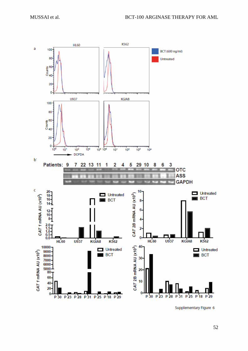
To understand if AML blasts express the key arginine recycling enzymes ASS and OTC we
examined 39 diagnostic patient samples by immunohistochemistry (Figure 1c, Figure 1d). Of
29 adult AML samples 10% had no staining, 45% showed low, 24% showed moderate, and
21% showed high ASS expression; whilst 0% had no staining 35% showed low, 55% showed
moderate, and 10% showed high OTC expression. Of 10 paediatric AML samples 20% had no
staining, 60% had low, and 20% had moderate ASS expression; whilst 20% had no staining,
0% had low, 50% had moderate, and 30% had high OTC expression. One paediatric sample
received a 0 score for both ASS and OTC.
AML blasts can therefore lack one or both, of the important enzymes for endogenous arginine
synthesis. As such their dependence on extracellular arginine levels should become rate-
MUSSAI et al. BCT-100 ARGINASE THERAPY FOR AML
10
limiting on many metabolic reactions and protein synthesis thereby preventing proliferation.
Proliferation of cultured AML blasts was arrested when arginine fell below 10Consistent
with these findings in newly diagnosed patients, where AML disease burden is highest, plasma
arginine concentrations is significantly lower than in healthy controls (mean: 131 M healthy
vs 9.0 M AML patients; p=<0.0001) (Figure 1e). Together these findings highlight the
dependency of AML blasts on extracellular arginine - arginine auxotrophism.
BCT-100 reduces the number of AML blasts in vitro and in vivo
BCT-100 is a pegylated recombinant human arginase being investigated for the treatment of
solid tumours.8 We first demonstrated BCT-100 catalyses a dose-dependent reduction in media
arginine concentrations in vitro (Sup. Figure 1a). Arginine was reduced to <2M within 8 h at
concentrations of 600ng/mL BCT-100 or higher.
AML cell lines are sensitive to BCT-100 arginine depletion: IC50s ranged between 50 and 180
ng/mL (Sup. Figure 1b). No further decrease in cell number was seen by increasing
concentrations of BCT-100 above 600ng/mL, consistent with the depletion of arginine at this
drug concentration and the specificity of drug action. In vivo experiments previously
demonstrated that a single BCT-100 dose results in a sustained depletion of plasma arginine
levels persisting for 6 days.7 We confirmed that BCT-100 led to a significant decrease in
plasma arginine (mean: 20 M healthy vs 8.5 M AML mice; p=0.0244) (Figure 2a) and
treated mice had a significant reduction in AML engraftment (human CD45+ cells: median
21% untreated vs 5% BCT treated, p=0.029) (Figure 2b), which was approximately equivalent
to 25mg/kg cytarabine (median 6%). No evidence of toxicity or weight loss was observed
(Figure 2c).
MUSSAI et al. BCT-100 ARGINASE THERAPY FOR AML
11
BCT-100 demonstrates activity against primary AML blasts from patients
The activity of BCT-100 was tested against sorted, fresh blasts from 20 AML patients. Samples
varied in response, ranging from >95% cell death to completely resistant (Figure 3a). IC50s
ranged from 100ng/mL to 2000ng/mL (Figure 3b). In 5 samples < 50% death occurred, and no
increase in activity was seen even with doses up to 16,000ng/mL, confirming that BCT-100
does not act through off-target toxicity. Sensitivity to BCT-100 did not correlate with clinical
characteristics. BCT-100 was also cytotoxic to 3 of the 4 relapse samples (P7, P14, P19, P20).
This is the first report of the efficacy of arginine depletion against human AML blasts.
BCT-100 synergises with cytarabine against AML blasts from patients
Cytarabine is a key agent in AML chemotherapy protocols for adults and children20,21 However
patient AML blasts can develop cytarabine resistance. Therefore a new drug which re-sensitises
blasts to cytarabine may play a key role in future therapy.22,23 When BCT-100 was combined
with cytarabine, cytotoxicity that is greater than the sum of the 2 individual compounds alone
was seen in AML samples (F(1,57) = 6.405, p<0.0001) (Figure 3c). Analysis of individual patient
samples, showed that BCT-100 synergised with cytarabine (Combination Index <1) for almost
all samples (Table 2). BCT-100 sensitivity correlated moderately with sensitivity to cytarabine
(r=0.5182, p=0.0280), suggesting complementary mechanisms of activity of these two drugs.
(Figure 3d).
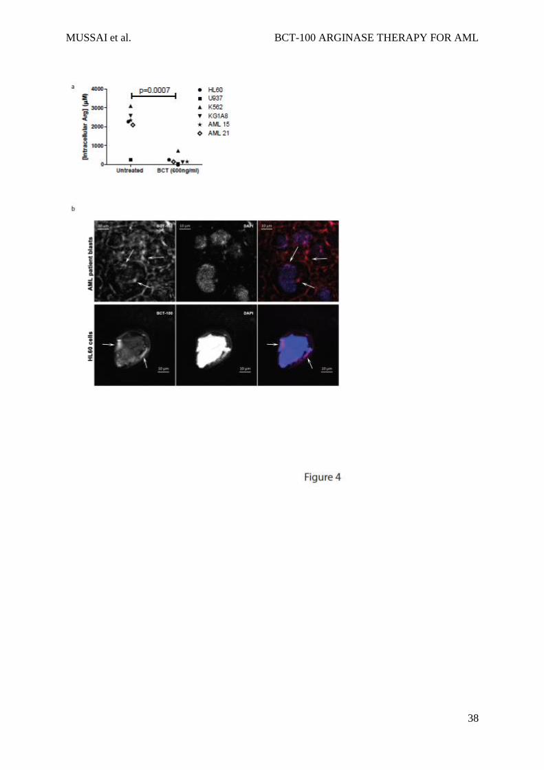
BCT-100 reduces intracellular arginine concentrations
Having demonstrated above that BCT-100 reduces local arginine concentrations, we also
showed that BCT-100 led to a significant decrease in AML intracellular arginine (p<0.0007)
(Figure 4a). Although BCT-100 is a relatively large molecule and acts extracellularly, some

MUSSAI et al. BCT-100 ARGINASE THERAPY FOR AML
12
BCT-100 molecules could also be internalised. Using fluorescently labelled BCT-100, this
drug conjugate bound to the cell surface of AML blasts and was internalised (Figure 4b, Sup.
Fig 2). The percentage of surface bound BCT-100 increased moderately over time (Sup Figure
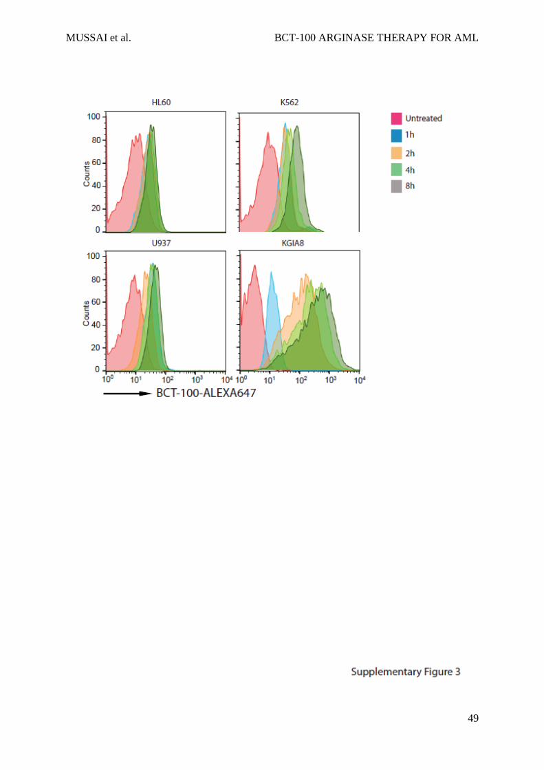
3). No correlation was identified between the sensitivity of AML blasts and the percentage of
internalised BCT-100.
BCT-100 induces cell cycle arrest and death by necrosis in AML blasts
Conventional chemotherapy agents lead to a reduction in leukaemia cell numbers through
either cell cycle arrest or cell death. To investigate BCT-100 cytotoxicity and its mechanism,
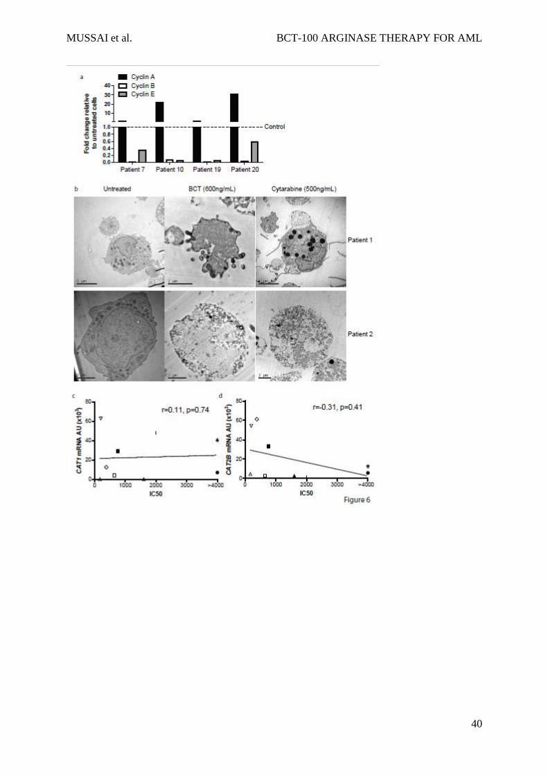
we show that arginine depletion leads to a significant arrest of AML proliferation (Figure 5a,b).
Cell cycle analysis showed an increase in the percentage of cells in G0/G1, with decreases of
those in S phase (Figure 5c and Figure 5d). G0/G1 arrest was confirmed in the blasts of AML
patients treated with BCT-100, by the relative increase in Cyclin A expression, and decreases
in cyclin B and E, compared to untreated cells. (Figure 6a).
Cell cycle arrest may result in either a steady-state of viable cells or in cell death. Examining
cell morphology by TEM, BCT-100 caused cell death of AML blasts, with features more
consistent with necrosis – including cell membrane permeabilisation and organelle
enlargement (Figure 6b, and Supp Figure 4 top panel).24 In contrast cytarabine treated AML
blasts showed nuclear fragmentation bodies characteristic of apoptosis. There was no evidence
of cell death in treated normal T cells or monocytes, confirming the low toxicity against non-
malignant haematopoietic cells seen in adult early phase trials of BCT-100 (Supplementary
Figure 4).

MUSSAI et al. BCT-100 ARGINASE THERAPY FOR AML
13
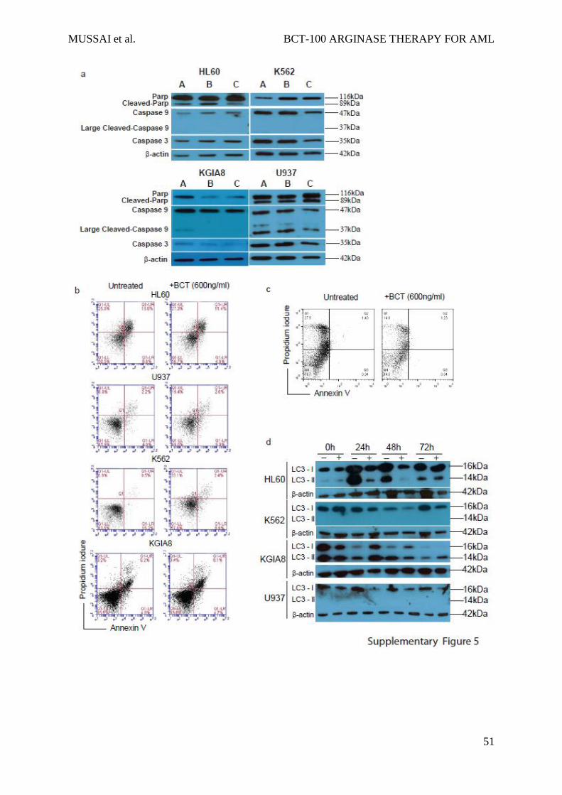
Non-specific growth factor withdrawal has been associated with induction of the intrinsic
pathway of apoptosis in AML.25 BCT-100 did not induce any significant activation of the
requisite effector caspases -9 and -3 or PARP cleavage (Supplementary Figure 5a).26 In
addition no significant increase in PI+/Annexin+ cells were seen following BCT-100 treatment
(Sup Figure 5b and 5c). Amino acid deprivation has also been associated with the induction of
death by autophagy in AML, identified by the conversion of cytoplasmic LC3-I to
autophagosomic LC3-II.27-30 No increase in LC3-II was seen following BCT-100 treatment,
confirming the absence of autophagy seen in EM (Supplementary Figure 5d). Amino acid
deprivation can induce the rapid production of reactive oxygen species which induce cell death,
but no evidence for this was found following BCT treatment (Sup Fig 6a, data not shown).31
Biomarkers of sensitivity to BCT
Understanding characteristics of AML blasts which correlate with drug sensitivity is important
for patient risk stratification. Of the 20 AML samples tested for sensitivity to BCT-100 in
vitro, ASS and OTC expression did not correlate with response to BCT-100, consistent with
our previous reports in adult solid tumours (Sup Fig 6b). CAT 1 and CAT 2B expression did
not correlate strongly with BCT sensitivity (CAT1 r=0.11, p=0.74; CAT2B r=-0.31, p=0.41)
(Figure 6c, 6d, Sup Fig 6c).
As multiple pathways, other than arginine recycling may be important in AML pathogenesis,
we used RNA-sequencing to identify genes predictive of BCT-100 response. RNA was isolated
from 6 sensitive and 6 resistant patient samples, based on their IC50s post treatment with BCT-
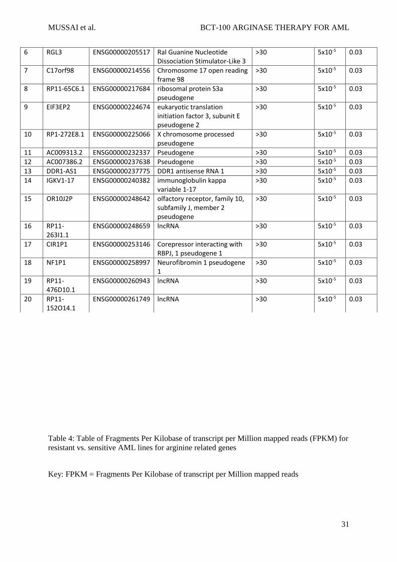
100. (Figure 3b). We identified 20 genes which were differentially expressed between resistant
and sensitive samples (Table 3) The top differentially expressed gene was EREG (epiregulin),
which codes for a ligand of the epidermal growth factor family (EGF) capable of binding to
MUSSAI et al. BCT-100 ARGINASE THERAPY FOR AML
14
the EGF receptor and the ERBB family of tyrosine kinase receptors. Other genes of interest
included ZIC5, which have a zinc finger protein known to be an upstream regulator of the Wnt
pathway; EN2, a homeobox gene that regulates the forkhead box transcription factor FOXA2;
HSPA6, a heat shock associated protein that seems to be associated with drug sensitivity in
AML; and RGL3, a paralog of RASGRF2.
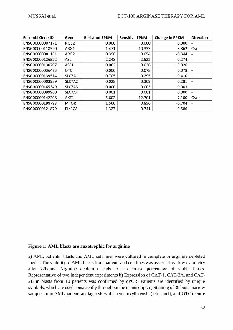
In comparing FPKM between resistant and sensitive cells (Table 4), there were 2 differentially
expressed arginine pathway associated genes. Over-expression of the AKT1 gene (FPKM =
7.1) was observed in sensitive cells, although there was no change in MTOR gene expression
(FPKM=-0.704). Type I arginase (ARG1,FPKM=8.87) was also overexpressed in sensitive
cells as compared to resistant cells. No changes in arginine transporter genes (SLC7A1-4) or
ASS and OTC were identified. These findings identify pathways that might be predictive of
sensitivity to therapeutic arginine depletion in AML, and shed light on new aspects of AML
disease biology.
Discussion
Normal myeloid cells differ in their requirement for arginine, ranging from maintenance of
neutrophil activity, through to the consumption of arginine by Myeloid Derived Suppressor
Cells and inflammatory macrophages.32-34 We previously showed that AML blasts have a high
MUSSAI et al. BCT-100 ARGINASE THERAPY FOR AML
15
arginase activity, creating an immunosuppressive microenvironment.13 However the
mechanism by which arginine is imported from the microenvironment by AML blasts had not
been described. We identify that human AML blasts predominantly express the CAT-1 and
CAT-2B isoforms. Interrogation of the 'R2: microarray analysis and visualization platform’
(http://r2.amc.nl) confirms the expression of these transporters across all FAB subtypes of
AML. The role of the CAT family of proteins in haematopoiesis has only received limited
study, identifying CAT-1 and CAT-2B as the main transporters responsible for arginine uptake
in in vitro models of non-malignant myeloid and erythroid cells.35-37
Arginine can be produced within healthy cells, by the ornithine-citrulline-arginine cycle.38,39
We identified that the majority of patients’ blasts are deficient in either ASS or OTC enzymes,
strongly suggesting that AML blasts are reliant on extracellular arginine availability. The
contribution of bone marrow stroma in producing arginine to support AML expansion is
unknown, but analogous mechanisms exist. For example CNS neurons are reliant on astrocyte
derived arginine, and asparaginine is produced by bone marrow stromal cells in cases of
ALL.40,41 Additionally it has been shown that ASS has a tumour suppressor function in
sarcoma,42,43 perhaps suggesting ASS may play a more fundamental role in the malignant
transformation of blasts.
These findings are consistent with the auxotrophic requirement of arginine by AML blasts. We
identified that the need for arginine by AML blasts lead to a significant decrease in arginine
concentrations both locally, but also in the plasma of patients at diagnosis. To our knowledge
this is the first report of a fall in plasma arginine due to a haematological malignancy and
plasma arginine may correlate with AML disease burden. Decreases in plasma arginine
concentrations occur in cervical cancer and renal cell carcinoma patients.44,45 Physiological
compensation through protein breakdown, contributing to cancer induced cachexia, and
MUSSAI et al. BCT-100 ARGINASE THERAPY FOR AML
16
arginine recycling by the intestinal-renal axis may try to compensate for lowered arginine
levels.46
Since arginine is essential for proliferation and the maintenance of AML viability, arginine
starvation by the pegylated recombinant arginase BCT-100 should be cytotoxic to AML, as in
other tumours types. Normal human arginase has limited clinical value because of a short
plasma-half life.7 The addition of a 5000 MW polyethylene glycol molecule (PEG)
significantly increases the plasma half-life of arginase 1 from 10-20 min to up to 3.5 days in
man with minimal loss of enzyme activity.8
We report for the first time that the majority of AML patients’ blasts from children and adults,
including those at relapse, are sensitive to arginine depletion, leading to necrotic cell death.
BCT-100 may be internalised by AML blasts, consistent with other pegylated molecules in
myeloid cells, which would further contribute to low intracellular arginine concentrations.47
BCT-100 can work in combination with cytarabine. Cytarabine in its triphosphorylated form
is a substrate for DNA polymerases and is incorporated into phosphodiester linkages in the
DNA strand. The addition of subsequent deoxynucleotides are inhibited, resulting in S-phase
arrest and cell death.20,21 As BCT-100 may induce G0/G1 arrest, the synergistic effect of the
two agents may be due to their targeting of different phases of the cell cycle. Similar findings
have been described against T-ALL.48 Non-malignant cells show little cytotoxicity to BCT-100
because the cells tend to become quiescent, a state they can survive in for prolonged periods,
due to intact restriction (R) checkpoints.49,50 However tumour cells are less tolerant of this
condition, and metabolic stressors may induce G1 cell cycle arrest and ultimately death by
necrosis in tumour cells.51 Tumour-specific arginine requirements and the concurrent use of
drugs to drive cell death along a particular mechanistic pathway may explain tumour-specific
effects of arginine depletion.52-54
MUSSAI et al. BCT-100 ARGINASE THERAPY FOR AML
17
RNA sequencing of sensitive and resistant samples identified 20 genes which predict response
to arginine depletion. Pathway analysis confirmed that expression of arginine recycling or
transport molecules did not correlate with sensitivity to arginine depletion. Most intriguing was
the finding that epiregulin (EREG), was differentially expressed, with arginase sensitive AML
overexpressing EREG. Overexpressed EREG has been linked to dysregulation of MAPK
signalling via the EGF pathway as well as the ERBB pathway, and this overexpression in the
sensitive cells may reflect an underlying drive towards the MAPK/ERBB pathways.55,56
Conversely, in the arginase resistant cells, over-expression of the heat shock protein HSPA6
was demonstrated, suggesting that this may be a mechanism by which these cells attain
resistance, especially in light of the finding that HSP inhibition is cytotoxic to AML.57 EREG
and EGFR signalling, as well as the heat shock protein family, play key roles in solid tumour
cell proliferation. Small molecule inhibitors are currently under development in AML to target
these pathways, providing the potential for rationale combinations of other drugs with BCT-
100 for maximal anti-leukaemia effect.58,59
The findings could important translational consequences for a disease which is in desperate
need of new therapies. Although small molecule arginase pathway inhibitors are available for
laboratory use (NG-hydroxy-L-arginine: NOHA and L-NG-monomethyl arginine: L-
NMMA),14 their clinical application has been limited by the requirement for the molecules to
be given together and by off-target toxicity in vivo. An alternative approach is through the
depletion of arginine from the microenvironment, thus starving AML blasts of this key amino
acid.
Pre-clinically BCT-100 has demonstrable activity against hepatocellular, melanoma and
prostate carcinoma. A Phase I and II clinical trial has been completed in adults with refractory
hepatocellular carcinoma, in which 1600U/kg BCT-100 (OBD) resulted in plasma arginine
falling below 8µM (Adequate Arginine Depletion - ADD) and can be maintained for up to 5
MUSSAI et al. BCT-100 ARGINASE THERAPY FOR AML
18
or more days after a single treatment (the maintenance level of arginine in human blood is
~40µM). Interim analysis of patients enrolled on a Phase II trial in hepatocellular carcinoma
suggest that patients experience significant improvements in overall survival, comparable to
the standard of care (personal communication Dr P. Cheng, BCTI).8 For both paediatric and
elderly AML patients in particular, concerns over treatment side-effects limits the type of
therapies that can be given safely.60,61 In this study we show that arginine depletion is not
cytotoxic to T cells and monocytes and is well tolerated in vivo. Clinically this is supported by
the excellent toxicity profile of BCT-100 in trial with no evidence of increased patient
infections.
Two alternative arginine depleting enzymes are also undergoing early preclinical and clinical
evaluation, but have been subject to a number of limiting toxicities. The first is ADI-PEG, a
pegylated form of mycoplasma derived arginine deiminase.62 ADI converts arginine into
citrulline and ammonia, potentially leading to toxic hyperammonemia, and ensuing
neutropenia.62 The bacterial origin of the molecule leads to neutralising antibody formation and
intramuscular injection site hypersensitivity reactions, limiting continued drug administration
and a failure to sustain adequately low plasma arginine.63,65 An alternative pegylated human
arginase has also been described, in which the enzyme co-factor has been replaced with cobalt
to increase arginase activity,53 but unfortunately seems from in early preclinical studies to be
significantly more toxic. Thus the natural enzyme seems to be the best option.
A similar paradigm of bacterial versus recombinant protein therapy occurred in paediatric ALL,
with PEG-asparaginase. The bacterial derivative (Erwinia) was eventually superseded by
recombinant asparaginase, due to side-effects such as immunogenicity. Recombinant
asparaginase has resulted in significant improvements in overall survival for children and its
incorporation into upfront treatment protocols.66

MUSSAI et al. BCT-100 ARGINASE THERAPY FOR AML
19
The findings of our study highlight a role for arginine in AML pathogenesis and support the
ongoing development of BCT-100 and similar arginase preparations in early Phase trials for
patients with AML.
Acknowledgements
The authors thank the patients and parents who contributed samples to the study. Thank you to
Jane Cooper and Cay Shakespeare for consent and collection of patient samples. We thank
Chen-Li for provision of BCT-100. Thank you to Paul Stanley and Theresa Morris for technical
assistance with electron microscopy. Thank you to Manoj Raghavan for identifying patients.
MUSSAI et al. BCT-100 ARGINASE THERAPY FOR AML
20
Thank you to Denys Wheatley for review of the manuscript. This work was supported by the
Amber Phillpott Trust, Children with Cancer, the Birmingham Children’s Hospital Research
Fund, and Cancer Research UK.
Authorship
Contribution: F.M. and C.D.S designed the study, performed research, analysed data and wrote
the manuscript. F.M. additionally secured ethical approval and was chief investigator of the
study. S.E designed and performed research, J.H-J performed research, T.P. and P.K. provided
access to murine xenografts, A.B performed RNA-Sequencing analysis, E.O. performed
confocal microscopy, J.L. provided patient samples, G.P. provided patient samples, A.L. and
M.N. provided patient samples and performed immunohistochemistry, P.C. and K.P.U.
provided BCT-100.
Conflict of Interest Disclosure
The authors declare no competing financial interests.
References
1. Marcucci G, Haferlach T, Dohner H. Molecular genetics of adult acute myeloid
leukemia:prognostic and therapeutic implications. J Clin Oncol. 2011;29(15):475-486.
MUSSAI et al. BCT-100 ARGINASE THERAPY FOR AML
21
2. Gibson B, Wheatley K, Hann I, et al. Treatment strategy and long-term results in
paediatric patients treated in consecutive UK AML trials. Leukemia.
2005;19(12):2130-2138
3. De Santo C, Serafini P, Marigo I, Dolcetti L, Bolla M, Del Soladato P. Nitroaspirin
corrects immune dysfunction in tumor-bearing hosts and promotes tumor eradication
by cancer vaccination. PNAS. 2005;102(11):4185-4190
4. Brenner T, Fleming TH, Rosenhagen C, Krauser U, Mieth M, Bruckner T, Martin E,
Nawroth PP, Weigand MA, Bierhaus A, Hofer S. L-arginine and asymmetric
dimethylarginine are early predictors for survival in septic patients with acute liver
failure. Mediators Inflamm. 2012; 210454
5. De Santo C, Salio M, Masri SH, Lee LY, Dong T, Speak AO, Porubsky S, Booth S,
Veerapen N, Besra GS, Gröne HJ, Platt FM, Zambon M, Cerundolo V. Invariant NKT
cells reduce the immunosuppressive activity of influenza A virus-induced myeloid-
derived suppressor cells in mice and humans. J Clin Invest. 2008;118(12):4036-4048.
6. Wheatley DN. Arginine deprivation and metabolomics: important aspects of
intermediary metabolism in relation to the differential sensitivity of normal and tumour
cells. Semin Cancer Biol. 2005; 15(5): 247-253
7. Cheng PN, Lam TL, Lam WM, Tsui SM, Cheng AW, Lo WH et al. Pegylated
recombinant human arginase (rhArg-peg5,000mw) inhibits the in vitro and in vivo
proliferation of human Hepatocellular carcinoma through arginine depletion. Cancer
Res. 2007;67(1):309-17
8. Yau T, Cheng PN, Chan P, Chan W, Chen L, Yuen J et al. A phase 1 dose-escalating
study of pegylated recombinant human arginase 1 in patients with advanced
hepatocellular carcinoma. Invest New Drugs. 2013; 31(1):99-107
MUSSAI et al. BCT-100 ARGINASE THERAPY FOR AML
22
9. Lam TL, Wong GK, Chow HY, Cong HC, Chow TL, Kwok SY et al. Recombinant
human arginase inhibits the in vitro and in vivo proliferation of human melanoma by
inducing cell cycle arrest and apoptosis. Pigment Cell Melanoma Res. 2011; 24(2):366-
376
10. Hsueh EC, Knebel SM, Lo WH, Leung YC, Cheng PN, Sueh CT. Deprivation of
arginine by recombinant human arginase in prostate cancer cells. J Hematol Oncol
2012; 5:17
11. Nenutil R, Smardova J, Pavlova S, Hanzelkova Z, Muller P, Fabian P et al.
Discriminating functional and non-functional p53 in human tumours by p53 and
MDM2 immunohistochemistry. J Pathol. 2005; 207(3):251-259
12. Slinker BK. The statistics of synergism. J Mol Cell Cardiol. 1998; 30(4):723-731
13. Chou TC and Talalay P. Quantitative analysis of dose-effect relationships: the
combined effects of multiple drugs or enzyme inhibitors. Adv Enzyme Regul. 1984;
22:27-55
14. Mussai FJ, De Santo C, Abu-Dayyeh I, Booth S, Quek L, McEwen-Smith R et al. Acute
myeloid leukaemia creates an arginase-dependent immunosuppressive
microenvironment. Blood. 2013; 122(5):749-758
15. Morris Jr SM. Arginine Metabolism: Boundaries of Our Knowledge. J Nutr. 2007: 137;
1602S-1609S.
16. Windmueller HG, Spaeth AE. Source and fate of circulating citrulline. Am J
Physiol.1981;241(6):E473-E80
17. Ryall J, Nguyen M, Bendayan M, Shore GC. Expression of nuclear genes encoding
the urea cycle enzymes, carbamoyl-phosphate synthetase I and ornithine carbomyl
transferase in rat liver and intestinal mucosa. Eur J Biochem. 1985:152:287-292
MUSSAI et al. BCT-100 ARGINASE THERAPY FOR AML
23
18. Levillain O, Hus-Citharel A, Morel F, Bankir L. Localization of arginine synthesis
along rat nephron. Am J Physiol. 1990;259:F916-F923
19. Closs EL, Simon A, Vekony N, Rotmann A. Plasma membrane transporters for
arginine. J Nutr. 2004; 134(10 Suppl): 2752S-2759S
20. Major PP, Egan EM, Beardsley GP, Minden MD, Kufe DW. Lethality of human
myeloblasts correlates with the incorporation of arabinofuanosylcytosine into DNA.
Proc Natl Acad Sci USA. 1981; 78(5): 3235-3239
21. Major PP, Egan EM, Herrick DJ, Kufe DW. Effect of ARA-C incorporation on
deoxyribonucleic acid synthesis in cells. Biochem Pharmacol. 1982; 31:2937-2940
22. Jabbour E, Daver N, Champlom R, Mathisen M, Oran B, Ciurea S, et al. Allogeneic
stem cell transplantation as initial salvage for patients with acute myeloid leukemia
refractory to high-dose-cytarabine-based induction chemotherapy. Am J Hematol.
2014; 89(4):395-398
23. Leonard SM, Perry T, Woodman CB, Kearns P. Sequential treatment with cytarabine
and decitabine has an increased anti-leukemia effect compared to cytarabine alone in
xenograft models of childhood acute myeloid leukaemia. Plos One. 2014; 9(1):e87475
24. Ziegler U, Groscurth P. Morphological features of cell death. Physiology.
2004;19:124-128
25. Pallis M, Russell N. P-glycoprotein plays a drug-efflux-independent role in
augmenting cell survival in acute myeloblastic leukemia and is associated with
modulation of a sphingomyelin-ceramide apoptotic pathway. Blood. 2000; 95(9):2897-
2904
26. Brentnall M, Rodriguez-Monocal L, De Guevara RL, Cepero E, Boise LH. Caspase-9,
caspase-3 and caspase-7 have distinct roles during intrinsic apoptosis. BMC Cell Biol.
2013;14:32
MUSSAI et al. BCT-100 ARGINASE THERAPY FOR AML
24
27. Willems L, Jacque N, Jacquel A, Neveux N, Maciel TT, Lambert M et al. Inhibiting
glutamine uptake represents an attractive new strategy for treating acute myeloid
leukemia. Blood. 2013;122(20):3521-3522
28. Cheong H, Lu C, Lindstein T, Thompson CB. Therapeutic targets in cancer cell
metabolism and autophagy. Nat Biotechnol. 2012; 30(7):671-678
29. Taatjes DJ, Sobel BE, Budd RC. Morphological and cytochemical determination of
cell death by apoptosis. Histochem Cell Biol. 2008; 129:33-43
30. Eskelinen EL, Reggiori F, Baba M, Kovacs AL, Seglen PO. Seeing is believing: the
impact of electron microscopy on autophagy research. Autophagy. 2011; 7(9):935-956
31. Brown RD, Burke, GA, Brown GC. Dependence of leukemic cell proliferation and
survival on H2O2 and L-arginine. Free Radic Biol Med. 2009; 46(8):1211-1220
32. Kapp K, Prufer S, Michel CS, Habermeier A, Luckner-Minden C, Gieste T et al.
Granulocyte functions are independent of arginine availability. J Leukoc Biol. 2014;
96(6):1047-1053
33. Mussai F, De Santo C, Cerundolo V. Interaction between invariant NKT cells and
myeloid-derived suppressor cells in cancer patients: evidence and therapeutic
opportunities. J Immunother. 2012; 35(6);449-459
34. Chang CI, Liao JC, Kuo L. Macrophage arginase promotes tumor cell growth and
suppresses nitric oxide-mediated tumor cytotoxicity. Cancer Res. 2001; 61(3):1100-
1106
35. Fotiadis D, Kanai Y, Palacin M. The SLC3 and SLC7 families of amino acid
transporters. Mol Aspects Med. 2013; 34(2-3):139-158
36. Barilli A, Rotoli BM, Visigalli R, Bussolati O, Gazzola GC, Dall'Asta V. Arginine
transport in human monocytic leukemia THP-1 cells during macrophage
differentiation. J Leukoc Biol 2011.;90(2):293-303.
MUSSAI et al. BCT-100 ARGINASE THERAPY FOR AML
25
37. Shima Y, Maeda T, Aizawa S, Tsuboi I, Kobayashi D, Kato R, Tamai I. L-arginine
import via cationic amino acid transporter CAT1 is essential for both differentiation
and proliferation of erythrocytes. Blood. 2006;107(4):1352-1356.
38. Evans RW, Fernstrom JD, Thompson J, Morris SM Jr, Kuller LH. Biochemical
responses of healthy subjects during dietary supplementation with L-arginine. J Nutr
Biochem. 2004;15(9):534-539.
39. Wu G, Morris SM Jr. Arginine metabolism: nitric oxide and beyond. Biochem J
1998;336:1-17.
40. Wiesinger H. Arginine metabolism and the synthesis of nitric oxide in the nervous
system. Prog Neurobiol. 2001; 64(4):365-391
41. Iwamoto S, Mihara K, Downing JR, Pui C-H, Campana D. Mesenchymal cells regulate
the response of acute lymphoblastic leukemia cells to asparaginase. J Clin Invest. 2007;
117:1049-1057
42. Huang HY, Wu WR, Wang YH, Wang JW, Fang Fm, Tsai JW. ASS1 as a novel tumor
suppressor gene in myxofibrosarcomas: aberrant loss via epigenetic DNA methylation
confers aggressive phenotypes, negative prognostic impact and therapeutic relevance.
Clin Cancer Res. 2013; 19(11):2861-2872
43. Lan J, Tai, HC, Lee SW, Chen TJ, Huang HY, Li CF. Deficiency in expression and
epigenetic DNA methylation of ASS1 gene in nasopharyngeal carcinoma: negative
prognostic impact and therapeutic relevance. Tumour Biol. 2014; 35(1):161-169
44. Hasim A, Aili A, Maimaiti A, Mamtimin B, Abudula A, Upur H. Plasma-free amino
acid profiling of cervical cancer and cervical intraepithelial neoplasia patients and its
application for early detection. Mol Biol Rep. 2014; 40(10):5853-5859
MUSSAI et al. BCT-100 ARGINASE THERAPY FOR AML
26
45. Rodriguez PC, Ernstoff MS, Hernandez C, Atkins, Zabaltea J, Sierra R et al. Arginase
I-producing myeloid-derived suppressor cells in renal cell carcinoma are a
subpopulation of activated granulocytes. Cancer Res. 2009; 69(4):1553-1560
46. Buijs N, Luttikhold J, Houdijk AP, can Leeuwen PA. The role of a disturbed
arginine/NO metabolism in the onset of cancer cachexia: a working hypothesis. Curr
Med Chem. 2012; 19(31)5278-5286
47. Zhan X, Tran KK, Shen H. Effect of the poly(ethylene glycol) (PEG) density on access
and uptake of particles by antigen-presenting cells (APCs) after subcutaneous
administration. Mol Pharm. 2012; 9(12):3442-3451
48. Hernandez CP, Morrow K, Lopez-Barcons LA, Zabaleta J, Sierra R, Velasco C et al.
Pegylated arginase I: a potential therapeutic approach in T-ALL. Blood. 2011;
115(25):5214-21
49. Philip R, Campbell E, Wheatley DN. Arginine deprivation, growth inhibition and
tumour cell death: 2. Enzymatic degradation of arginine in normal and malignant cell
cultures. Br J Cancer. 2003; 88(4):613-62
50. Scott, L, Lamb J, Smith S, Wheatley DN. Single amino acid (Arginine) deprivation:
rapid and selective death of cultured transformed and malignant cells. Br J Cancer.
2000; 83(6):800-810
51. Degenhardt K, Mathew R, Beaudoin B, Bray K, Anderson D, Chen G et al. Autophagy
promotes tumor cell survival and restricts necrosis, inflammation, and tumorigenesis.
Cancer Cell. 2006; 10(1): 51-64
52. Lam TL, Wong GK, Chong HC, Cheng PN, Choi SC, Chow TL et al. Recombinant
human arginase inhibits proliferation of human hepatocellular carcinoma by inducing
cell cycle arrest. Cancer Lett. 2009; 277(1):91-100
MUSSAI et al. BCT-100 ARGINASE THERAPY FOR AML
27
53. Morrow K, Hernandex CP, Raber P, Del Valle, L, Wilk AM, Majumdar S et al. Anti-
leukemic mechanisms of pegylated arginase I in acute lymphoblastic T-cell leukemia.
Leukemia. 2013; 27(3):569-577
54. Zeng X, Li Y, Fan J, Zhao H, Sun Y, Wang Z et al. Recombinant human arginase
induced caspase-dependent apoptosis and autophagy in non-Hodgkin’s lymphoma
cells. Cell Death Dis. 2013;4:e840
55. Ufkin ML, Peterson S, Yang X, Driscoll H, Duarte C et al. miR-125a regulates cell
cycle, proliferation and apoptosis by targeting the ErbB pathway in acute myeloid
leukemia. Leuk Res. 2014; 38(3):402-410
56. Riese DJ, Cullum RL. Epiregulin: roles in normal physiology and cancer. Semin Cell
Dev Biol. 2014; 49-56
57. Al Shaer L, Walsby E, Gilkes A, Tonks A, Walsh V et al. Heat shock protein 90
inhibition is cytotoxic to primary AML cells expressing mutant FLT3 and results in
altered downstream signalling. Br J Haematol. 2008; 141(4):483-493
58. Deangelo DJ, Neuberg D, Amrein PC, Berchuck J, Wadleigh M, Sirlnkik LA, et al. A
Phase II study of EGFR inhibitor gefitinib in patients with acute myeloid leukemia.
Leuk Res. 2014; 38(4):430-434
59. Lancet JE, Gojo I, Burton M, Quinn , Tighe SM, Kersey K et al. Phase I study of th
heat shock protein 90 inhibitor alvespimycin (KOS-1022, 17-DMAG) administered
intravenously twice weekly to patients with acute myeloid leukemia. Leukemia. 2010;
24(4):699-705
60. Pollyea DA, Kohrt HE, Medeiros BC. Acute myeloid leukaemia in the elderly: a
review. Br J Haematol. 2011; 152(5):524-542
61. Creutzig U, Aimmermann M, Lehrnbecher T, Graf N, Hermann J, Niemeyer CM. Less
toxicity by optimizing chemotherapy, but not by addition of granulocyte colony
MUSSAI et al. BCT-100 ARGINASE THERAPY FOR AML
28
stimulating factor in children and adolescents with acute myeloid leukemia: results of
AML-BFM 98. J Clin Oncol. 2006; 24(27):499-4506
62. Glazer ES, Piccirillo M, Albino V, Di Giacomo R, Palaia R, Mastro AA et al. Phase II
study of pegylated arginine deiminase for nonresectable and metastatic hepatocellular
carcinoma J Clin Oncol. 2009; 28:2220–2226
63. Shawcross DL, Wright GA, Stadbauer V, Hodges SJ, Davies NA, Wheeler-Jones C.
Ammonia impairs neutrophil phagocytic function in liver disease. Hepatology. 2008;
48(4):1202-1212
64. Glazer ES, Piccirillo M, Albino V, Di Giacomo R, Palasia R, Mastro AA et al. Phase
II study of pegylated arginine deiminase for nonresectable and metastatic
hepatocellular carcinoma. J Clin Oncol. 2010; 28(13):2220-2226
65. Synakiewicz A, Stachowicz-Stencel T, Adamkiewicz-Drozynska E. The role of
arginine and the modified arginine deiminase enzyme DI-PEG20 in cancer therapy
with special emphasis on Phase I/II clinical trials. Expert Opin Investig Drugs. 2014;
1-13
66. Dinndorf PA, Gootenberg J, Cohen M, Keegan P, Pazdur R. FDA drug approval
summary: pegasparginase for the first-line treatment of children with acute
lymphoblastic leukemia. Oncologist. 2007; 12(8):991-998
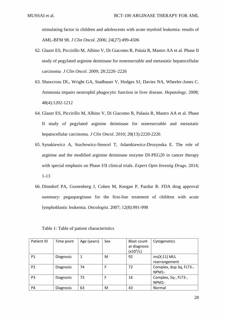
Table 1: Table of patient characteristics
Patient ID Time point Age (years) Sex Blast count at diagnosis (x109/L)
Cytogenetics
P1 Diagnosis 1 M 92 ins(X;11) MLL rearrangement
P2 Diagnosis 74 F 72 Complex, dup 3q, FLT3-, NPM1-
P3 Diagnosis 73 F 16 Complex, 5q-, FLT3-, NPM1-
P4 Diagnosis 63 M 43 Normal
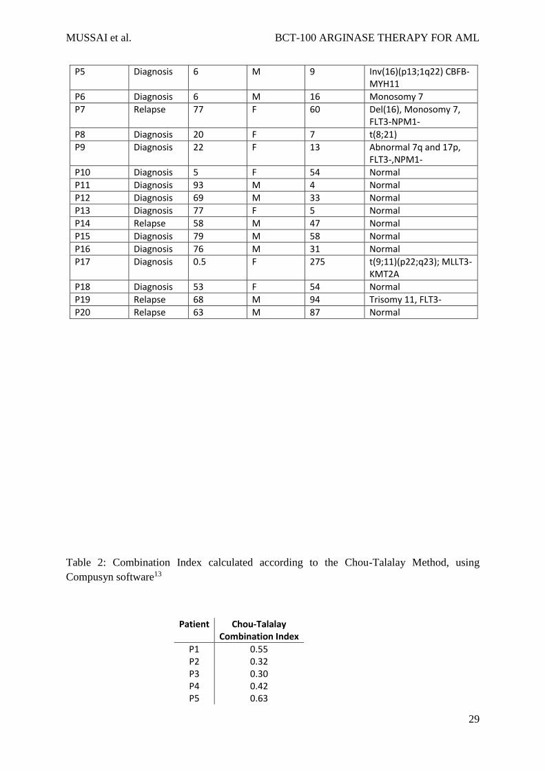
MUSSAI et al. BCT-100 ARGINASE THERAPY FOR AML
29
P5 Diagnosis 6 M 9 Inv(16)(p13;1q22) CBFB-MYH11
P6 Diagnosis 6 M 16 Monosomy 7
P7 Relapse 77 F 60 Del(16), Monosomy 7, FLT3-NPM1-
P8 Diagnosis 20 F 7 t(8;21)
P9 Diagnosis 22 F 13 Abnormal 7q and 17p, FLT3-,NPM1-
P10 Diagnosis 5 F 54 Normal
P11 Diagnosis 93 M 4 Normal
P12 Diagnosis 69 M 33 Normal
P13 Diagnosis 77 F 5 Normal
P14 Relapse 58 M 47 Normal
P15 Diagnosis 79 M 58 Normal
P16 Diagnosis 76 M 31 Normal
P17 Diagnosis 0.5 F 275 t(9;11)(p22;q23); MLLT3-KMT2A
P18 Diagnosis 53 F 54 Normal
P19 Relapse 68 M 94 Trisomy 11, FLT3-
P20 Relapse 63 M 87 Normal
Table 2: Combination Index calculated according to the Chou-Talalay Method, using
Compusyn software13
Patient Chou-Talalay Combination Index
P1 0.55 P2 0.32 P3 0.30 P4 0.42 P5 0.63
MUSSAI et al. BCT-100 ARGINASE THERAPY FOR AML
30
P6 0.13 P7 0.81 P8 0.27 P9 0.43
P10 0.43 P11 0.02 P12 0.48 P13 0.37 P14 0.27 P15 0.04 P16 0.81 P17 0.21 P18 0.99 P19 0.36 P20 0.35
Table 3: Table of ranked gene expression in arginase sensitive vs. resistant cells as
determined via RNA-seq
Rank Gene Ensembl ID Name log2(fold_change) p_value q_value
1 EREG ENSG00000124882 Epiregulin 4.61947 5x10⁻⁵ 0.03
2 CCL4 ENSG00000129277 Chemokine (C-C motif) ligand 4
-3.42043 5x10⁻⁵ 0.03
3 ZIC5 ENSG00000139800 Zic Family Member 5 >30 5x10⁻⁵ 0.03
4 EN2 ENSG00000164778 Engrailed homeobox 2 >30 5x10⁻⁵ 0.03
5 HSPA6 ENSG00000173110 Heat Shock 70kDa Protein 6 -4.11747 5x10⁻⁵ 0.03
MUSSAI et al. BCT-100 ARGINASE THERAPY FOR AML
31
Table 4: Table of Fragments Per Kilobase of transcript per Million mapped reads (FPKM) for
resistant vs. sensitive AML lines for arginine related genes
Key: FPKM = Fragments Per Kilobase of transcript per Million mapped reads
6 RGL3 ENSG00000205517 Ral Guanine Nucleotide Dissociation Stimulator-Like 3
>30 5x10⁻⁵ 0.03
7 C17orf98 ENSG00000214556 Chromosome 17 open reading frame 98
>30 5x10⁻⁵ 0.03
8 RP11-65C6.1 ENSG00000217684 ribosomal protein S3a pseudogene
>30 5x10⁻⁵ 0.03
9 EIF3EP2 ENSG00000224674 eukaryotic translation initiation factor 3, subunit E pseudogene 2
>30 5x10⁻⁵ 0.03
10 RP1-272E8.1 ENSG00000225066 X chromosome processed pseudogene
>30 5x10⁻⁵ 0.03
11 AC009313.2 ENSG00000232337 Pseudogene >30 5x10⁻⁵ 0.03
12 AC007386.2 ENSG00000237638 Pseudogene >30 5x10⁻⁵ 0.03
13 DDR1-AS1 ENSG00000237775 DDR1 antisense RNA 1 >30 5x10⁻⁵ 0.03
14 IGKV1-17 ENSG00000240382 immunoglobulin kappa variable 1-17
>30 5x10⁻⁵ 0.03
15 OR10J2P ENSG00000248642 olfactory receptor, family 10, subfamily J, member 2 pseudogene
>30 5x10⁻⁵ 0.03
16 RP11-263I1.1
ENSG00000248659 lncRNA >30 5x10⁻⁵ 0.03
17 CIR1P1 ENSG00000253146 Corepressor interacting with RBPJ, 1 pseudogene 1
>30 5x10⁻⁵ 0.03
18 NF1P1 ENSG00000258997 Neurofibromin 1 pseudogene 1
>30 5x10⁻⁵ 0.03
19 RP11-476D10.1
ENSG00000260943 lncRNA >30 5x10⁻⁵ 0.03
20 RP11-152O14.1
ENSG00000261749 lncRNA >30 5x10⁻⁵ 0.03
MUSSAI et al. BCT-100 ARGINASE THERAPY FOR AML
32
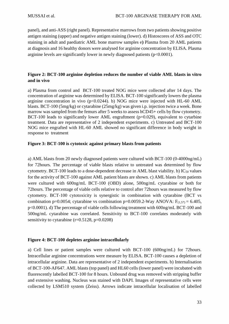
Figure 1: AML blasts are auxotrophic for arginine
a) AML patients’ blasts and AML cell lines were cultured in complete or arginine depleted
media. The viability of AML blasts from patients and cell lines was assessed by flow cytometry
after 72hours. Arginine depletion leads to a decrease percentage of viable blasts.
Representative of two independent experiments b) Expression of CAT-1, CAT-2A, and CAT-
2B in blasts from 10 patients was confirmed by qPCR. Patients are identified by unique
symbols, which are used consistently throughout the manuscript. c) Staining of 39 bone marrow
samples from AML patients at diagnosis with haematoxylin eosin (left panel), anti-OTC (centre
Ensembl Gene ID Gene Resistant FPKM Sensitive FPKM Change in FPKM Direction
ENSG00000007171 NOS2 0.000 0.000 0.000 -
ENSG00000118520 ARG1 1.471 10.333 8.862 Over
ENSG00000081181 ARG2 0.398 0.054 -0.344 -
ENSG00000126522 ASL 2.248 2.522 0.274 -
ENSG00000130707 ASS1 0.062 0.036 -0.026 -
ENSG00000036473 OTC 0.000 0.078 0.078 -
ENSG00000139514 SLC7A1 0.705 0.295 -0.410 -
ENSG00000003989 SLC7A2 0.028 0.309 0.281 -
ENSG00000165349 SLC7A3 0.000 0.003 0.003 -
ENSG00000099960 SLC7A4 0.001 0.001 0.000 -
ENSG00000142208 AKT1 5.602 12.701 7.100 Over
ENSG00000198793 MTOR 1.560 0.856 -0.704 -
ENSG00000121879 PIK3CA 1.327 0.741 -0.586 -
MUSSAI et al. BCT-100 ARGINASE THERAPY FOR AML
33
panel), and anti-ASS (right panel). Representative marrows from two patients showing positive
antigen staining (upper) and negative antigen staining (lower). d) Histoscores of ASS and OTC
staining in adult and paediatric AML bone marrow samples e) Plasma from 20 AML patients
at diagnosis and 16 healthy donors were analysed for arginine concentration by ELISA. Plasma
arginine levels are significantly lower in newly diagnosed patients (p<0.0001).
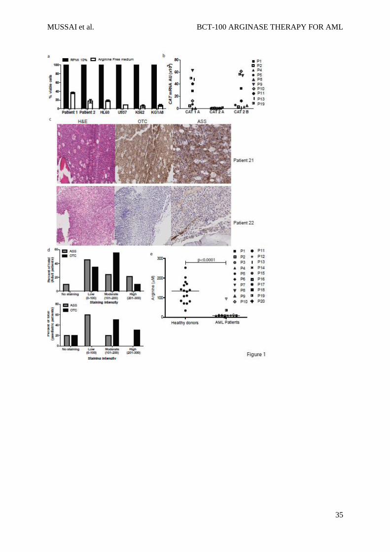
Figure 2: BCT-100 arginine depletion reduces the number of viable AML blasts in vitro
and in vivo
a) Plasma from control and BCT-100 treated NOG mice were collected after 14 days. The
concentration of arginine was determined by ELISA. BCT-100 significantly lowers the plasma
arginine concentration in vivo (p<0.0244). b) NOG mice were injected with HL-60 AML
blasts. BCT-100 (5mg/kg) or cytarabine (25mg/kg) was given i.p. injection twice a week. Bone
marrow was sampled from the femurs after 5 weeks to assess hCD45+ cells by flow cytometry.
BCT-100 leads to significantly lower AML engraftment (p=0.029), equivalent to cytarbine
treatment. Data are representative of 2 independent experiments. c) Untreated and BCT-100
NOG mice engrafted with HL-60 AML showed no significant difference in body weight in
response to treatment
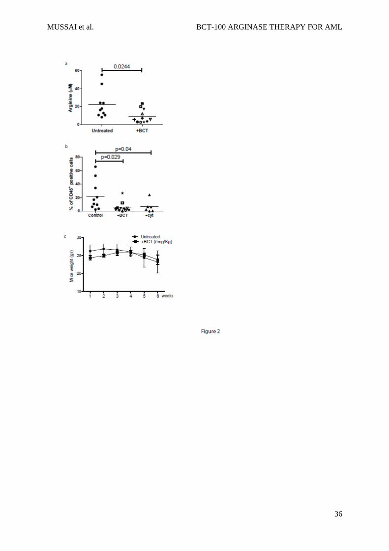
Figure 3: BCT-100 is cytotoxic against primary blasts from patients
a) AML blasts from 20 newly diagnosed patients were cultured with BCT-100 (0-4000ng/mL)
for 72hours. The percentage of viable blasts relative to untreated was determined by flow
cytometry. BCT-100 leads to a dose-dependent decrease in AML blast viability. b) IC50 values
for the activity of BCT-100 against AML patient blasts are shown. c) AML blasts from patients
were cultured with 600ng/mL BCT-100 (OBD) alone, 500ng/mL cytarabine or both for
72hours. The percentage of viable cells relative to control after 72hours was measured by flow
cytometry. BCT-100 cytotoxicity is synergistic in combination with cytarabine (BCT vs
combination p=0.0054; cytarabine vs combination p=0.0059.2-Way ANOVA: F(1,57) = 6.405,
p<0.0001). d) The percentage of viable cells following treatment with 600ng/mL BCT-100 and
500ng/mL cytarabine was correlated. Sensitivity to BCT-100 correlates moderately with
sensitivity to cytarabine (r=0.5128, p=0.0208)
Figure 4: BCT-100 depletes arginine intracellularly
a) Cell lines or patient samples were cultured with BCT-100 (600ng/mL) for 72hours.
Intracellular arginine concentrations were measure by ELISA. BCT-100 causes a depletion of
intracellular arginine. Data are representative of 2 independent experiments. b) Internalisation
of BCT-100-AF647. AML blasts (top panel) and HL60 cells (lower panel) were incubated with
fluorescently labelled BCT-100 for 8 hours. Unbound drug was removed with stripping buffer
and extensive washing. Nucleus was stained with DAPI. Images of representative cells were
collected by LSM510 system (Zeiss). Arrows indicate intracellular localisation of labelled

MUSSAI et al. BCT-100 ARGINASE THERAPY FOR AML
34
BCT-100; arrowheads indicate surface-bound drug. Scale 10um.Representative patient sample
of 3 different patient samples.
Figure 5: BCT-100 halts proliferation and cell cycle arrest
a) BCT-100 halts AML cell division. CFSE labelled cell lines were cultured in the presence of
600ng/mL BCT-100. Representative histogram plots shown. Independent experiments were
performed on two separate occasions. b) Cell lines were cultured with BCT-100 (0-
2000ng/mL) for 72hours. AML proliferation was measured by 3H-thymidine incorporation
after 72hours. Data are representative of 2 independent experiments. BCT-100 causes a dose-
dependent decrease in AML proliferation. c) AML cell lines were cultured with 600ng/ml
BCT-100. Cell cycle analysis was performed after 72 hours. BCT-100 increases the percentage
of cells in G0/G1 arrest. Representative histogram plots for untreated and treated HL-60 shown.
Independent experiments were performed on four separate occasions. d) Table showing the
relative percentages of cells in G0/G1, S, G2/M based on flow cytometry cell cycle analysis.
Figure 6: BCT-100 induced cell cycle arrest leads to necrotic cell death
a) Relative expression of cyclins A, B, E in BCT-100 treated AML patient blasts compared to
untreated controls (hashed line) were investigated by qPCR. Representative data of 4 patients
shown. b) AML blasts from patients were treated with BCT-100 (600ng/mL) or cytarabine
(500ng/mL) for 72 hours. Analysis of cell death was performed by transmission electron
microscopy. Representative micrographs of 2 out of 5 patients shown. Left panel – untreated
cells. Middle panels – post treatment with 600ng/mL BCT-100. Features consistent with
organelle enlargement and cell membrane permeablisation. Right panels – post treatment with
500ng/mL cytarabine. Features consistent with nuclear fragmentation bodies and preserved
membrane integrity. Experiments performed on 3 separate occasions. c) Sensitivity to BCT-
100 does not correlate with CAT1 expression (d) and only mildly with CAT-2B expression
(r=-0.31, p=0.41)
MUSSAI et al. BCT-100 ARGINASE THERAPY FOR AML
35
MUSSAI et al. BCT-100 ARGINASE THERAPY FOR AML
36
MUSSAI et al. BCT-100 ARGINASE THERAPY FOR AML
37
MUSSAI et al. BCT-100 ARGINASE THERAPY FOR AML
38

MUSSAI et al. BCT-100 ARGINASE THERAPY FOR AML
39
MUSSAI et al. BCT-100 ARGINASE THERAPY FOR AML
40
MUSSAI et al. BCT-100 ARGINASE THERAPY FOR AML
41
Supplemental Methods
Cell lines
Cell lines were cultured in RPMI-1640 (Invitrogen, CA, USA) with 10% heat-inactivated fetal
bovine serum, glutamine (1x), sodium pyruvate (1x) and Penicillin-Streptomycin (RPMI 10%)
using T-75 flasks kept in a humidified air atmosphere with 5% CO2 at 37oC.
Arginine ELISA
Complete media was treated with BCT-100 as described above. Aliquots were collected after
8, 24, 48, and 72h. The concentration of arginine within the media was quantified using a
competitive enzyme linked immunoassay (Immunodiagnostik K7733) according to the
manufacturers’ instructions. In brief this assay uses a competitive enzyme immunoassay in
which L-arginine is derivatized from samples, and competes with an L-arginine-tracer for
binding of polyclonal antibodies, in the microtiter wells. The concentration of the tracer-bound
antibody is inversely proportional to the L-arginine concentration in the samples. Plasma
collected from the blood of NOG AML murine xenografts was similarly tested. For
intracellular concentrations of arginine, AML blasts were treated with BCT-100 (600ng/ml)
for 72hours. Cells were collected, washed twice in PBS and counted. Equal numbers of treated
and untreated cells were lysed using lysis buffer (20nM Tris-Hcl pH7.5, 150nM NaCl, 2mM
EDTA, 1.0% triton X-100) and lysates tested for arginine concentration as above.

MUSSAI et al. BCT-100 ARGINASE THERAPY FOR AML
42
3H-thymidine incorporation
The effect of BCT-100 on AML cell line proliferation was tested by 3H-thymidine
incorporation. Cells were plated as above with BCT-100 for 50h and then 1Ci/well 3H-
thymidine (Perkin Elmer Life Sciences, Beaconsfield, UK) was added for 12-16 hours. 3H-
thymidine incorporation was measured using a Wallac Microbeta Jet 1450 reader (Perkin
Elmer).
PCR analysis
RT-PCR was used to detect ASS and OTC in patient-derived AML blasts. RNA was extracted
using an RNeasy Mini kit (Qiagen). cDNA was prepared using SuperScriptTM III Reverse
Transcriptase (Invitrogen) following the manufacturer’s instructions. The PCR products
separated by electrophoresis on a 2% agarose gel were visualised by staining with ethidium
bromide. Primers sequence (Eurofin) were: OTC forward 5'-tcccaattatcaatgggctg-
3' and reverse 5'-catgcttatccaaagtgtctg-3', ASS: forward 5'-
GGGGTCCCTGTGAAGGTGACC-3' and reverse 5'-CGTTCATGCTCACCAGCTC-3'.
mRNA levels following treatment of blasts with BCT-100, were measured using
Q-PCR. cDNA was generated from RNA of untreated and treated AML blasts from
patients using the protocol described above. RT-Q-PCR was done in duplicate
using FAST SYBR Green Master Mix (Applied Biosystems) and the Applied
Biosystems 7500 Fast Real-Time PCR system. Analysis of gene expression was
MUSSAI et al. BCT-100 ARGINASE THERAPY FOR AML
43
calculated according to 2-ΔT method described by Livak et al. plotted as arbitrary units
of mRNA relative to GAPDH. Primer sequences (Eurofins) were:
Cyclin A: forward 5'-AATGGGCAGTACAGGAGGAC-3'
reverse 5'-CCACAGTCAGGGAGTGCTTT-3',
Cyclin B1: forward 5'-CATGGTGCACTTTCCTCCTT-3'
Reverse 5'-AGGTAATGTTGTAGAGTTGGTGTCC-3',
Cyclin E: forward 5'-GGCCAAAATCGACAGGAC-3'
reverse 5'-GGGTCTGCACAGACTGCAT-3',
CAT1: forward 5'-ATGGGTGGAAACGCTGATGATAC-3'
reverse 5'-ACCTTGCCTGTTAAGTCTGGGTG-3',
CAT2A: forward 5'-TTAACACTTATGATGCCGTACTACCT-3'
reverse 5'-GCAACTGGTGACTGCCTCTTACT-3',
CAT 2B: forward 5'-ATGCCTCGTGTAATCTATGCTATG-3
reverse 5'-ACTGCACCCGATGATAAAGTAGC-3';
GAPDH: forward 5'-CCAGCCGAGCCACATCGCTC-3'
reverse 5'-ATGAGCCCCAGCCTTCTC-3'
Immunohistochemistry scoring
Antigen expression in immunohistochemistry sections were assigned independently by 2
experienced pathologists, as described by Nenutil et al.11 Briefly, to evaluate the
immunostaining intensity each slide was examined on an Olympus BX51 microscope.
Representative 400x magnification fields of at least 100 tumor cells were selected and
photographed with an Olympus DP70 camera and accompanying image software. Fields were
assigned an antigen staining intensity score of 0 = negative, 1= weak, 2 = moderate, 3 = strong.
MUSSAI et al. BCT-100 ARGINASE THERAPY FOR AML
44
The product of the percent positive cells and staining intensity was then derived to create a
histoscore of 0-300 for each high power field. A final histoscore was then given to each
specimen for each antigen.
BCT-100 Internalisation and Confocal Microscopy
BCT-100 was labeled with Alexa 647 using the Alexa-Fluro 647 Protein Labelling Kit
according to manufacturer’s instructions (Molecular Probes A20173, Invitrogen, Carlsbard,
CA). To measure the internalisation BCT-100, 2x10⁵ AML cells were suspended in 200L of
RPMI-10% with 1g/ml BCT-Alexa-647 on ice for 30 minutes to saturate the cell surface. The
cells were placed on a 37o C heat block and incubated for 0, 1, 2, 4, and 8h. They were washed
with FACS buffer and stripped with 0.2M glycine-HCl (pH 2.2, with 1mg/ml BSA) on ice for
15 minutes, to remove unbound BCT-Alexa-647. After resuspension in 200mL of FACS buffer
they were analysed by flow cytometry. To confirm internalization of BCT-Alexa-647, stripped
AML cells were cytospun (1000 rpm) for 5 minutes, fixed for 30 minutes in 2%
paraformaldeyde (Sigma), washed and mounted onto glass slides using DAPI-Fluromount G
(Southern Biotech). Z-stacks of 0.7m sections were collected at 0.5 m intervals using
LSM510-META confocal system with 63x/NA1.4 oil objective. 2.5D projections were
produced using ZEN2009 software from Zeiss. XZ and YZ sections through Z-stacks were
produced using ImageJ software. In instances where images of single cells are presented, these
images are representative of the population of cells studied.
MUSSAI et al. BCT-100 ARGINASE THERAPY FOR AML
45
Reactive Oxygen Species (ROS) detection
To measure ROS production cells were cultured in the presence of 2.5 μM DCFDA (Molecular
Probes/Invitrogen) for 30 min prior to analysis by flow cytometry as described above. The
Greiss Reaction (Cayman) was used to estimate reactive nitrate/nitric species.
Supplementary Figures:
S1: BCT-100 leads to arginine depletion and reduction in AML blast viability
a) Complete media was treated with BCT-100 (0-4000ng/mL). A dose- and time-dependent
decrease in arginine concentration was determined after 72hours by ELISA. The OBD was
determined as 600ng/mL in vitro. Representative of 2 independent experiments. b) Cell lines
were cultured with BCT-100 (0-2000ng/mL) for 72hours. The percentage of viable cells
relative to untreated controls was determined by flow cytometry. BCT-100 leads to a dose-
dependent decrease in AML cell line viability.
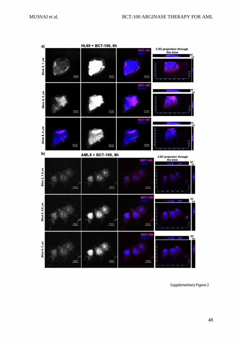
S2: Internalisation of BCT-100-ALEXA647 into AML cells
Internalisation of BCT-100-AF647 was confirmed by confocal microscopy. Z-stacks of 0.7m
sections through individual cells were collected: HL60 (a) and AML5 (b). Three sections at
various distances from the cell surface indicate intracellular localisation of BCT-100. 2.5D
projections and XZ and YZ sections through the slices confirm intracellular distribution of the
drug.
S3: BCT-100 is internalised into AML blasts over time
Internalisation of BCT-100-ALEXA647 into AML cell lines was confirmed by flow
cytometery. Cell lines were incubated with labelled BCT-100 for 0,1,2,4, 8hours. Unbound
drug was removed with stripping buffer and washing. Intracellular accumulation of labelled
BCT was measured by flow cytometry.
S4: Arginine depletion by BCT-100 is not cytotoxic to non-malignant haematopoietic
cells.
a) Cell lines, and patient-derived monocytes and T cells were treated with BCT-100
(600ng/mL) for 72 hours. Analysis of cell death was performed by electron microscopy.
Representative micrographs. Left panel – untreated cells. Right panels – post treatment with
600ng/mL BCT-100. HL60 post-treatment undergoing necrotic cell death. Monocytes and T
cells display no features of cell death. Experiments performed on 3 separate occasions.
MUSSAI et al. BCT-100 ARGINASE THERAPY FOR AML
46
S5: BCT-100 does not activate apoptotic or autophagy pathways
a) Cell lines were cultured alone (A), PEG (B), or BCT-100 (C). Expression of PARP, Caspase
9, and Caspase 3 in whole cell lystaes was determined by immunoblotting. Actin was used as
the housekeeping gene to ensure equal loading. No significant cleavage of PARP, Caspase 9
or 3 is seen, indicating the apoptosis pathway was not activated. b) AML cell lines were treated
with BCT-100 for 72hours and stained with Annexin-PI. Percentages of Annexin and PI
positive cells, compared to untreated controls, is shown, as tested by flow cytometry. No
significant increase in Annexin+PI+ cells is seen, confirming the lack of apoptosis.
Representative of 3 independent experiments c) AML patient blasts were treated with BCT-
100 for 72hours and stained with Annexin-PI. Percentages of Annexin and PI positive cells,
compared to untreated controls, is shown, as tested by flow cytometry. No significant increase
in Annexin+PI+ cells is seen, confirming the lack of apoptosis. One representative patient
sample of 5. d) Cell lines were treated with BCT-100 in the presence or absence of bafilomycin.
Whole cell lysates were tested for LC3-I and LC3-II turnover by immunoblotting at
0H,24H,48H, 72H of culture. No increase in conversion of LC3-I to LC3-II is seen, confirming
the lack of autophagy induction above baseline
S6: BCT-100 activity is not related to reactive oxygen species or arginine recycling
enzyme expression
a) AML patient blasts were treated with BCT-100 for 72hours and stained with DCFDA. No
increase ROS species evidence by DCFDA staining is seen. Representative of 2 independent
experiments b) ASS and OTC expression was determined by RT-PCR. Representative data
from eleven patients are shown. GAPDH was used as the housekeeping gene to ensure equal
loading. c) Relative expression of CAT 1 and CAT2B in BCT-100 treated AML cell lines or
patient blasts compared to untreated controls were investigated by qPCR. Representative data
of 4 cell lines and 7 patients shown.

MUSSAI et al. BCT-100 ARGINASE THERAPY FOR AML
47
MUSSAI et al. BCT-100 ARGINASE THERAPY FOR AML
48
MUSSAI et al. BCT-100 ARGINASE THERAPY FOR AML
49

MUSSAI et al. BCT-100 ARGINASE THERAPY FOR AML
50
MUSSAI et al. BCT-100 ARGINASE THERAPY FOR AML
51
MUSSAI et al. BCT-100 ARGINASE THERAPY FOR AML
52