arq. bras. med. vet. zootec., v.66, n.3, p.688-696, 2014arq. bras. med. vet. zootec., v.66, n.3,...
TRANSCRIPT
Arq. Bras. Med. Vet. Zootec., v.66, n.3, p.688-696, 2014
Investigations in associated protozoa-bacterial infections of cyprinids from
a fish farm situated on the Jijia river in N-E of Romania
[Investigações sobre infecções protozoárias-bacterianas associadas a ciprinídeos da
exploração piscícola no rio Jijia, situado no NE da Romênia]
M. Lazăr
1, L. Miron
1, I. Gostin
2, C. Rîmbu
1, R. Lazăr
3, E. Guguianu
1
1UASVM of Iasi, Faculty of Veterinary Medicine, 8 Mihail Sadoveanu Alley, Iasi, Romania 2Al. I. Cuza University of Iasi, Faculty of Biology, 11 Carol I Boulevard, Iasi, Romania
3UASVM of Iasi, Faculty of Animal Science Iasi, 3 Mihail Sadoveanu Alley, Iasi, Romania
ABSTRACT
In autumn 2011 in cyprinid farms located in Iasi on the Jijia river, several infections with bacterial strains
and macroscopical external cysts on the skin were diagnosed, which developed as a result of the stress
induced by biotic and abiotic factors. On the examination of the cyst contents the presence of numerous
spores was observed, mostly of the Dermocystidium sp genus. The samples were taken from the common
carp (Cyprinus carpio) and crucian carp (Carassius auratus gibelio) species from the fish farm as well as
from the Jijia River. 35 fish were examined, all of them showing cysts, fragmentation of their dorsal fin
and congestion of the gills. Histological examination of the skin showed a field of multiple dermal cysts
with round light eosinophilic formations (14-16µm) containing a central refractable body similar to that
reported for Dermocystidium sp. Gills samples were taken from the affected areas for the SEM
examination with the purpose of evaluating not only aspects of normal morphology, but also aspects of
some modifications of the affected areas as well as the presence of the etiologically incriminated bacteria
Pseudomonas fluorescens. The isolates were identified through phenotypic methods. All the strains that
showed mobility and oxidase-positivity were tested using API 20 NE strip. Consequently, they were
taxonomically grouped into the species Pseudomonas fluorescens. The scanning electron microscope
(SEM) was used for the first time in the characterization of the bacterial lesions produced by
Pseudomonas strains on Cyprinus carpio and Carassius auratus gibelio gills. The diagnosis of septicemia
with conditional pathogen species of Pseudomonas fluorescens was correlated with the results of the
physico-chemical investigations of water and the data concerning the breeding conditions of the
investigated livestock.
Keywords: cyprinids, Pseudomonas, Dermocystidium, SEM (Scanning Electron Microscopy),
histopathology
RESUMO
No outono de 2011, em fazendas de ciprinídeos localizadas em Iasi, no rio Jijia, diversas infecções
bacterianas e cistos externos macroscópicos na pele se desenvolveram como resultado do estresse
induzido por fatores bióticos e abióticos. No exame do conteúdo dos cistos, a presença de diversos
esporos foi observada, a maioria do gênero Dermocystidium sp. As amostras foram colhidas das
seguintes espécies: carpa comum (Cyprinus carpio) e carpa cruciana (Carassius auratus gibelio) de
fazenda piscícola, além do rio Jijia. Assim sendo, 35 peixes foram examinados, todos demonstrando
cistos, fragmentação da barbatana dorsal e congestão das guelras. O exame histológico da pele mostrou
um campo de múltiplos cistos dérmicos com formações circulares claras eosinofílicas (14-16µm)
contendo corpo central refratado similar ao relatado para Dermocystidium sp. Amostras de guelras
foram retiradas das áreas afetadas para exame MEV, com o propósito de se avaliar não apenas os
aspectos da morfologia normal, mas também os aspectos de algumas modificações das áreas afetadas,
Recebido em 12 de julho de 2012
Aceito para publicação em 21 de outubro de 2013 E-mail: [email protected]
Investigations in associated…
Arq. Bras. Med. Vet. Zootec., v.66, n.3, p.688-696, 2014 689
além da presença da bactéria etiologicamente incriminada: Pseudomonas fluorescens. Os isolados foram
identificados por meio de métodos fenotípicos. Todas as amostras que mostraram mobilidade e
positividade-oxidase foram testadas usando-se fita API 20 NE. Consequentemente, estas foram
taxonomicamente agrupadas na espécie Pseudomonas fluorescens. O microscópio eletrônico de
varredura (MEV) foi usado pela primeira vez na caracterização de lesões bacterianas produzidas por
Pseudomonas nas guelras de Cyprinus carpio e Carassius auratus gibelio. O diagnóstico de septicemia
com espécies condicionais de patogênico de Pseudomonas fluorescens foi correlacionado com os
resultados das investigações físico-químicas da água e de dados sobre as condições de reprodução dos
animais investigados.
Palavras-chave: ciprinídeos, Pseudomonas, Dermocystidium, MEV (microscópio eletrônico de
varredura), histopatologia
INTRODUCTION
Dermocystidium sp. – the studied group of
organisms – is causing white, macroscopic
nodules on the skin or gills of many fish species
(Hatai, 1989). Dermocystidium is probably
related to the protozoa or to the fungi, but its
classification is unclear (Dyková and Lom, 1992;
Hoffman, 1999).
Members of the Dermocystidium genus infect a
variety of fish hosts. Several species of
Dermocystidium infecting skin, fins and gills
have been reported. Apart from a few pathogenic
species, most infections are considered to be
relatively innocuous, although many are still
poorly registered in scientific literature (Feist,
2004).
The systemic infection with parasitic organisms
such as Dermocystidium sp. caused visceral
granuloma and mortality in farmed gold fish
Carassius auratus in commercial fishfarms in
Israel (Landsberg, 1992).
The fish bacterial infections may develop into
bacteremia, which leads to the occurrence of
bacterial organisms in the bloodstream without
clinical signs. In other cases septicemia may
occur, which indicates the presence of bacteria
and toxins in the circulatory system, accelerating
the disease as well as the clinical signs. Gram-
negative bacteria are responsible for the
production of either exotoxins or endotoxins,
which are composed of proteolytic enzymes that
destroy host cells and may produce necrosis or
cause blood vessel porosity or hemorrhaging
(Inglis et al., 2001; Kirjusina et al., 2007).
The Pseudomonas species are uniformly
distributed and can be isolated from a large
variety of environmental and water samples. The
aeromonads and pseudomonads are frequently
considered the most widespread secondary
invaders of the damaged tissues isolated from
diseased carp and responsible for clinical disease
and mortality.
As a general rule, the conditionally pathogenic
bacteria inherent to the natural aquatic
environment and fauna microbiota are
manifesting their pathogeny especially in the
breach of the natural anti-infectious barriers (the
skin and the mucosa), as well as in
immunosuppressant conditions generated by
various stress factors. The stress factors
triggering these diseases include low water
temperature, pH (<6 and >9), nitrites (>0.03 mg/l
NO2), or ammonia (>0.2mg/l NH3) (Austin,
2007; Hoole, 2001).
The surface of the lamellar epithelium of the gills
is rough and the epidermal surface has
microvillus. These prolongations help cuticular
mucus to reduce infections, abrasions, and they
also play an important role in gas, water and ion
exchange processes. Each lamella is considered
to be a thin cell coating with two surfaces joined
by supporting cells (Kenneth, 1996; Eiras-
Stofella et al. 1997; Ferguson 2006).
In the following report we describe the
microscopic and ultramicroscopic features of the
infection induced by Dermocystidium sp. and
Pseudomonas fluorescens in farmed cyprinids
from Iasi.
MATERIALS AND METHODS
The investigations were carried out on 35
cyprinids: 7 common carps (Cyprinus carpio)
and 25 crucian carps (Carassius auratus gibelio)
Lazăr et al.
690 Arq. Bras. Med. Vet. Zootec., v.66, n.3, p.688-696, 2014
patients from Piscicola – Larga Jijia fish farm
and 3 crucian carps (Carassius auratus gibelio)
free from the watercourse of the Jijia River,
ranging in size from 20 to 60cm. All 35 fish
presented visible cysts breaking through the skin.
All the investigations included bacteriological,
histological and scanning electron microscopy
(SEM) analyses.
The skin samples were fixed in 10% formalin,
then were embedded in paraffin and cut at 5 µm
thickness and stained with Haematoxilin and
Eosin for general histomorphology. Additional
sections were stained using Grocott and Periodic-
acid Schiff (PAS) in an attempt to identify the
possible presence of the hyphal elements in the
tissues.
Photographs and digital images were taken using
a LEICA ICC50 HD photomicroscope.
The bacteriological exam consisted of
microscopic investigation of skin and gill lesions
on Gram stained smears and of inoculated
samples prelevated from kidney and blood by
heart puncture, following the necropsy in strict
aseptic conditions.
The used media were blood agares, which, after
being inoculated, were incubated at 25°C, for 24-
48 hours.
The identification of isolates was based on
culture aspects, on the ability to grow at
refrigerated temperature (40
C), on the bacterial
cell morphology in Gram stained smears as well
as metabolic properties. The metabolic properties
have been tested using classic tests and API 20
NE strip (bioMérieux), following the instructions
recommended by the manufacturers.
The oxidase test is a preliminary differentiation
exam between Enterobacteriaceae (oxidase
negative) and other Gram negative bacteria.
The isolates antibiotic susceptibility was carried
out using the standardized CLSI diffusimetric
method on MÜLLER Hinton agar.
Subsequently, several physicochemical exams of
water from tanks with sick fish were conducted:
temperature, pH, nitrite concentration, nitrates,
ammonium, chloride and dissolved oxygen.
Scanning electron microscopy (SEM)
investigations: the investigated material consists
of small gill pieces. The material was fixed in
glutaraldehyde (2%) for 2 hours, osmium
tetraoxide (1%) for 4 hours and washed with
phosphate buffer. After dehydration in a graded
ethanol series (40%, 70%, 80%, 90% and 100%)
and acetone, the material was critical-point dried
with CO2 (using an EMS 850 Critical Point
Dryer), sputter-coated with a thin layer of gold
(30 nm) (using an EMS 550X Sputter Coater)
and, finally, examined by scanning electron
microscopy (Tescan Vega II SBH) at an
acceleration voltage of 27.88 V.
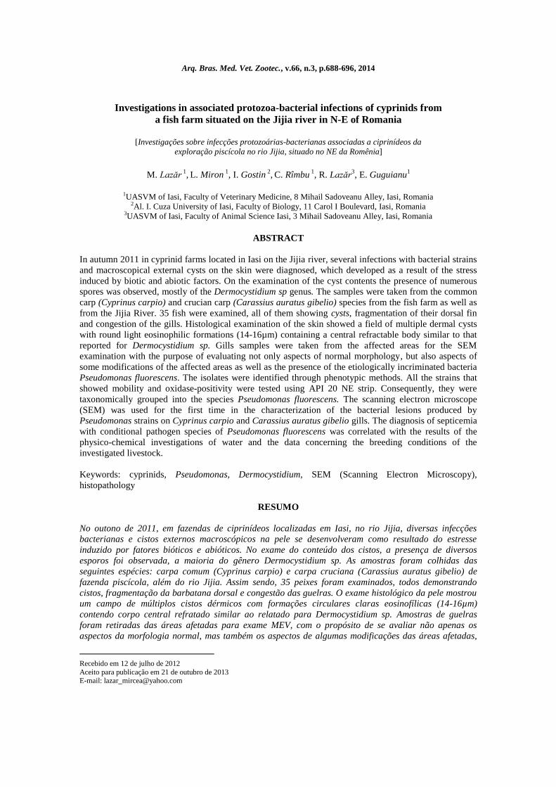
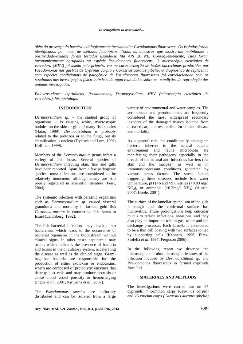
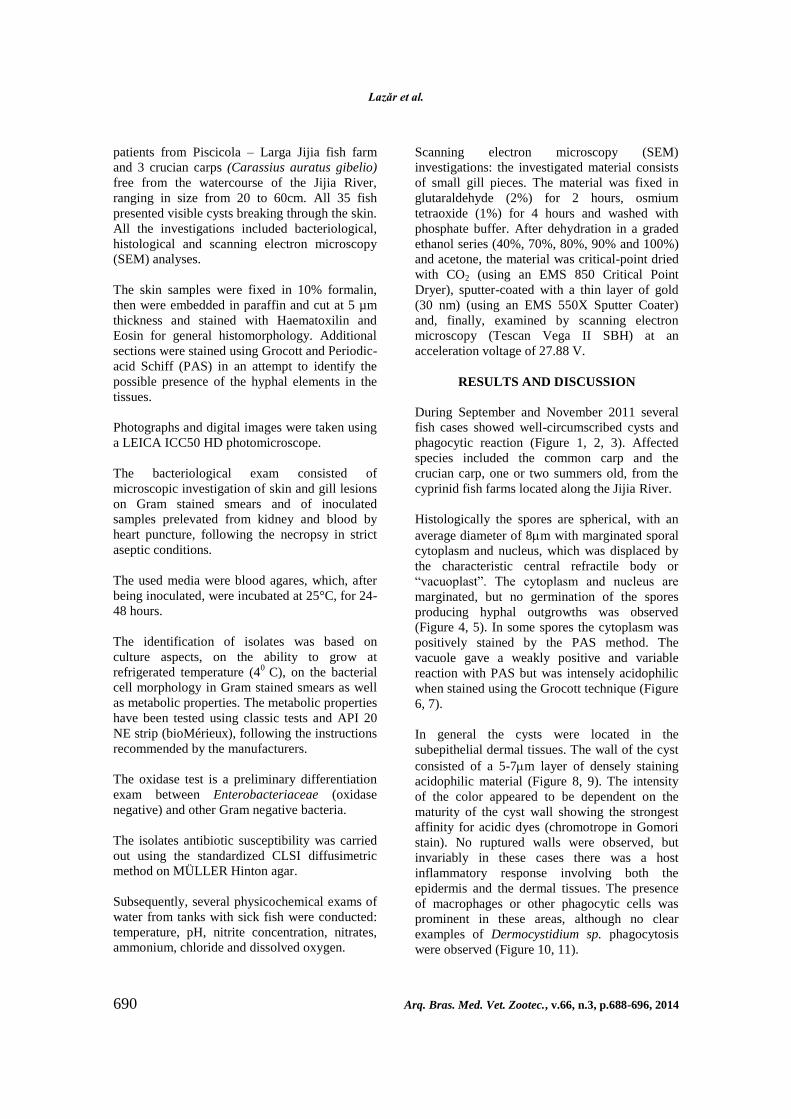
RESULTS AND DISCUSSION
During September and November 2011 several
fish cases showed well-circumscribed cysts and
phagocytic reaction (Figure 1, 2, 3). Affected
species included the common carp and the
crucian carp, one or two summers old, from the
cyprinid fish farms located along the Jijia River.
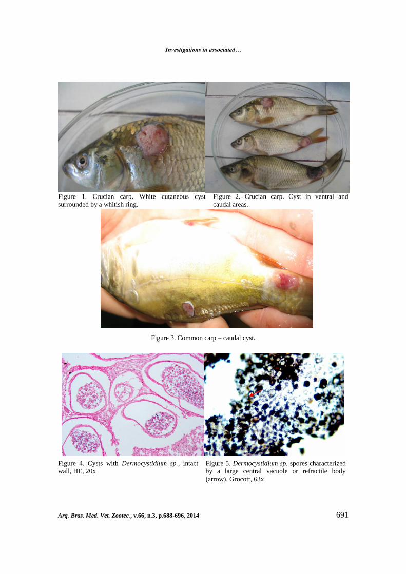
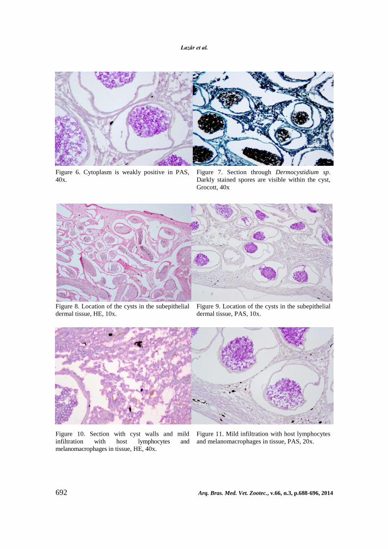
Histologically the spores are spherical, with an
average diameter of 8m with marginated sporal
cytoplasm and nucleus, which was displaced by
the characteristic central refractile body or
“vacuoplast”. The cytoplasm and nucleus are
marginated, but no germination of the spores
producing hyphal outgrowths was observed
(Figure 4, 5). In some spores the cytoplasm was
positively stained by the PAS method. The
vacuole gave a weakly positive and variable
reaction with PAS but was intensely acidophilic
when stained using the Grocott technique (Figure
6, 7).
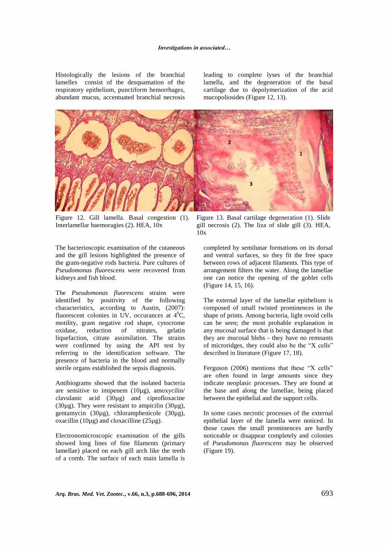
In general the cysts were located in the
subepithelial dermal tissues. The wall of the cyst
consisted of a 5-7m layer of densely staining
acidophilic material (Figure 8, 9). The intensity
of the color appeared to be dependent on the
maturity of the cyst wall showing the strongest
affinity for acidic dyes (chromotrope in Gomori
stain). No ruptured walls were observed, but
invariably in these cases there was a host
inflammatory response involving both the
epidermis and the dermal tissues. The presence
of macrophages or other phagocytic cells was
prominent in these areas, although no clear
examples of Dermocystidium sp. phagocytosis
were observed (Figure 10, 11).
Investigations in associated…
Arq. Bras. Med. Vet. Zootec., v.66, n.3, p.688-696, 2014 691
Figure 1. Crucian carp. White cutaneous cyst
surrounded by a whitish ring.
Figure 2. Crucian carp. Cyst in ventral and
caudal areas.
Figure 3. Common carp – caudal cyst.
Figure 4. Cysts with Dermocystidium sp., intact
wall, HE, 20x
Figure 5. Dermocystidium sp. spores characterized
by a large central vacuole or refractile body
(arrow), Grocott, 63x
Lazăr et al.
692 Arq. Bras. Med. Vet. Zootec., v.66, n.3, p.688-696, 2014
Figure 6. Cytoplasm is weakly positive in PAS,
40x.
Figure 7. Section through Dermocystidium sp.
Darkly stained spores are visible within the cyst,
Grocott, 40x
Figure 8. Location of the cysts in the subepithelial
dermal tissue, HE, 10x.
Figure 9. Location of the cysts in the subepithelial
dermal tissue, PAS, 10x.
Figure 10. Section with cyst walls and mild
infiltration with host lymphocytes and
melanomacrophages in tissue, HE, 40x.
Figure 11. Mild infiltration with host lymphocytes
and melanomacrophages in tissue, PAS, 20x.
Investigations in associated…
Arq. Bras. Med. Vet. Zootec., v.66, n.3, p.688-696, 2014 693
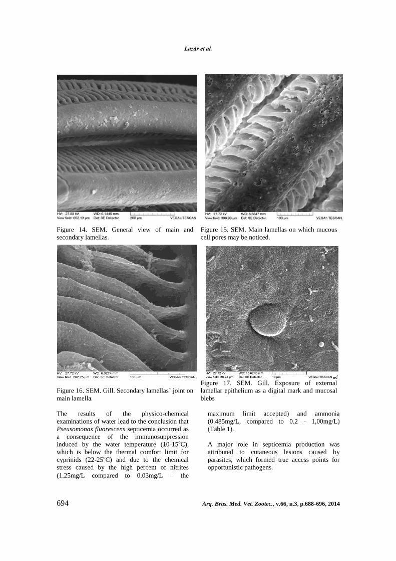
Histologically the lesions of the branchial
lamelles consist of the desquamation of the
respiratory epithelium, punctiform hemorrhages,
abundant mucus, accentuated branchial necrosis
leading to complete lyses of the branchial
lamella, and the degeneration of the basal
cartilage due to depolymerization of the acid
mucopoliosides (Figure 12, 13).
Figure 12. Gill lamella. Basal congestion (1).
Interlamellar haemoragies (2). HEA, 10x
Figure 13. Basal cartilage degeneration (1). Slide
gill necrosis (2). The liza of slide gill (3). HEA,
10x
The bacterioscopic examination of the cutaneous
and the gill lesions highlighted the presence of
the gram-negative rods bacteria. Pure cultures of
Pseudomonas fluorescens were recovered from
kidneys and fish blood.
The Pseudomonas fluorescens strains were
identified by positivity of the following
characteristics, according to Austin, (2007):
fluorescent colonies in UV, occurances at 40C,
motility, gram negative rod shape, cytocrome
oxidase, reduction of nitrates, gelatin
liquefaction, citrate assimilation. The strains
were confirmed by using the API test by
referring to the identification software. The
presence of bacteria in the blood and normally
sterile organs established the sepsis diagnosis.
Antibiograms showed that the isolated bacteria
are sensitive to imipenem (10µg), amoxycilin/
clavulanic acid (30µg) and ciprofloxacine
(30µg). They were resistant to ampicilin (30µg),
gentamycin (30µg), chloramphenicole (30µg),
oxacillin (10µg) and cloxacilline (25µg).
Electronomicroscopic examination of the gills
showed long lines of fine filaments (primary
lamellae) placed on each gill arch like the teeth
of a comb. The surface of each main lamella is
completed by semilunar formations on its dorsal
and ventral surfaces, so they fit the free space
between rows of adjacent filaments. This type of
arrangement filters the water. Along the lamellae
one can notice the opening of the goblet cells
(Figure 14, 15, 16).
The external layer of the lamellar epithelium is
composed of small twisted prominences in the
shape of prints. Among bacteria, light ovoid cells
can be seen; the most probable explanation in
any mucosal surface that is being damaged is that
they are mucosal blebs - they have no remnants
of microridges, they could also be the “X cells”
described in literature (Figure 17, 18).
Ferguson (2006) mentions that these “X cells”
are often found in large amounts since they
indicate neoplasic processes. They are found at
the base and along the lamellae, being placed
between the epithelial and the support cells.
In some cases necrotic processes of the external
epithelial layer of the lamella were noticed. In
those cases the small prominences are hardly
noticeable or disappear completely and colonies
of Pseudomonas fluorescens may be observed
(Figure 19).
1
2
3
Lazăr et al.
694 Arq. Bras. Med. Vet. Zootec., v.66, n.3, p.688-696, 2014
Figure 14. SEM. General view of main and
secondary lamellas.
Figure 15. SEM. Main lamellas on which mucous
cell pores may be noticed.
Figure 16. SEM. Gill. Secondary lamellas’ joint on
main lamella.
Figure 17. SEM. Gill. Exposure of external
lamellar epithelium as a digital mark and mucosal
blebs
The results of the physico-chemical
examinations of water lead to the conclusion that
Pseusomonas fluorescens septicemia occurred as
a consequence of the immunosuppression
induced by the water temperature (10-15oC),
which is below the thermal comfort limit for
cyprinids (22-25oC) and due to the chemical
stress caused by the high percent of nitrites
(1.25mg/L compared to 0.03mg/L the
maximum limit accepted) and ammonia
(0.485mg/L, compared to 0.2 - 1,00mg/L)
(Table 1).
A major role in septicemia production was
attributed to cutaneous lesions caused by
parasites, which formed true access points for
opportunistic pathogens.
Investigations in associated…
Arq. Bras. Med. Vet. Zootec., v.66, n.3, p.688-696, 2014 695
Figure 18. Gill. Mucosal blebs with ovoid
appearance and light color, placed between 2
secondary lamellas.
Figure 19. Gill. Necrosis zones and colonies of
Pseudomonas fluorescens.
Table 1. Chemical parameters of river water fish
growth
Parameters Recorded value
pH 7.02
Nitrites (mg/L) 1.25mg/L
Nitrates (mg/L) Absent
Ammonia (mg/L) 0.485mg/L
Chlorures (mg/L) 21.5mg/L
CONCLUSIONS
The Dermocystidium spores are spherical, with
marginated sporal cytoplasm and a nucleus
which is displaced by the characteristic
central refractile body. The presence of
melanomacrophages and other phagocytic cells
is proeminent in the cyst area, but there
are no clear examples of phagocytosis of
Dermocystidium sp. The cytoplasm of the spores
is positively stained by the PAS method and
intensely acidophilic when it is stained using
the Grocott technique. The Pseudomonas
fluorescens septicemia in cyprinids from farms in
the county of Iasi was favored by low water
temperature (10-15oC), by Dermocystidium sp.
infestations and by improper water quality,
considering the nitrites and ammonia content. In
the areas where the gills were injured, the
electronomicroscopic examinations indicated the
presence of the Pseudomonas fluorescens
bacteria as a result of some necrotic lesions, with
denudation of the lamellae due to bacteria
infestations.
REFERENCES
AUSTIN, B.; AUSTIN, D. Bacterial Fish
Pathogens. Diseases of Farmed and Wild Fish. 4.
ed. Chichester: Praxis Publishing, p. 20-553,
2007.
DYKOVA, I.; LOM, J. New evidence of fungal
nature of Dermocystidium koi Hoshina and
Saraha. J. Appl. Ichthyol., v.8, p.180-185, 1992.
FEIST, S.W.; LONGSHAW, M.; HURRELL,
R.H.; MANDER, B. Observations of
Dermocystidium sp. infections in bullheads,
Cottus gobio L., from a river in southern
England. J. Fish Diss, v.27, p.225-231, 2004.
HATAI, K. Fungal pathogens/parasites of
aquatic animals. In: AUSTIN, B.; AUSTIN,
D.A. editors: Methods for microbiological
examination of fish and shellfish. John Wiley and
Sons, New York, p. 240-272, 1989.
HOFFMAN, L.; GLENN. Parasites of North
American Freshwater Fishes. 2.ed. Correll
University Press, SUA, p. 9-92, 1999.

Lazăr et al.
696 Arq. Bras. Med. Vet. Zootec., v.66, n.3, p.688-696, 2014
HOOLE, D.; BUCKE, D.; BURGESS, P.;
WELLBY, I. Diseases of Carp and Other
Cyprinid Fishes. London: Fishing News Books,
p.53, 2001.
INGLIS, V.; ROBERTS, R.J.; BROMAGE, N.R.
Bacterial Diseases of Fish. London: Blackwell
Scientific Publications, p.1-59 e p.122 -156,
1993.
KENNETH, R.O. Scanning Electron Microscopy
Of the Fish Gill. In. DATTA MUNSHI, J.S.;
DUTTA, M. Fish morphology, horizon of new
research: New Hampshire: Science Publishers
Inc., p.31-45, 1996.
KIRJUSINA, M.; BRIEDE, I.; BONDAD-
REANTASO, M.G. Extension Manual on Some
Important Viruses, Parasites and Bacteria of
Aquatic Animals in Latvia: NDC/LZRA/FAO,
Riga, p.69, 2007.
EIRAS-STOFELLA, D.R.; CHARVET-ALMEIDA,
P. Gills of the freshwater fish Hypostomus
commersonii Val., 1840 (Loricariidae) analysed
through electron microscopy techniques. Arq.
Biol. Tecnol., v.40, p.785-792, 1997.
FERGUSON, H.W. Systemic Pathology of Fish:
a text and atlas of normal tissues in teleosts and
their responses in disease. Second Edition,
Scotian Press, London, p.5-263, 2006.
LANDSBERG, J.H.; PAPERNA, I. Systemic
granuloma in goldfish caused by
Dermocystidium like aetiological agent. Dis.
Aquat. Org., v.13, p.75-78, 1992.