ars.els-cdn.com · web viewadd 100 µl of diluted secondary antibody (horseradish...
TRANSCRIPT

Supporting Information
The detailed indirect competition ELISA for BPA detection as follows:
1. Coating each well of 96-well plate with 100 µL of BPA antigen dilution (diluted by the coating
solution).
2. Incubate the 96-well plate for 2h at room temperature.
3. Remove the coating solution and wash three times with 200 µL of PBS-Tween 20 and pat dry of the
plate on a paper towel.
4. Blocking the remaining protein-binding sites in each well with 100 µL of blocking solution(0.5%
ovalbumin in PBS) and incubate the plate at 37 ℃ for 2 h.
5. Remove the blocking solution and wash three times with 200 µL of PBS-Tween 20 solution.
6. Add 100 µL of BPA at various concentration or samples with 100 µL anti-BPA monoclonal
antibody, and then incubate the plate at 37 ℃ for 0.5 h.
7. Remove the excess antibody and BPA in each well and wash three times with PBS-Tween 20 and
pat dry of the plate on a paper towel.
8. Add 100 µL of diluted secondary antibody (horseradish peroxidase-conjugated goat anti-rabbit
antibody, 1: 3000 with PBS-Tween 20) and incubate the plate at 37 ℃ for 0.5 h.
9. Remove the excess secondary antibody of each well and wash three times with PBS-Tween 20 and
pat dry of the plate on a paper towel.
10. Add 100 µL of the substrate solution per well (3,3',5,5'-Tetramethylbenzidine (TMB) and
hydrogen peroxide) and incubate the plate at room temperature for 15 min in dark.
11. Add 100 µL of the stop solution per well (H2SO4, 2 mol/L) and record the absorption of each well
with a plate reader at 450 nm.
1
1
2
3
4
5
6
7
8
9
10
11
12
13
14
15
16
17
18
19
20
21
22
12

Fig. S1. Representative TEM images of (A) Au nanoroads and (B) Au nanoparticles.
Fig. S2. The corresponding UV-vis of Au NRs, Au NPs, and the assemblies.
2
1
23
456
12
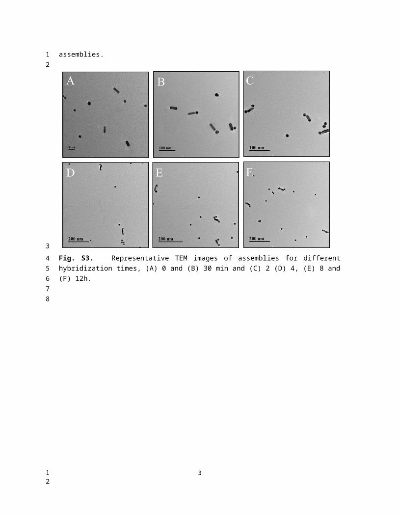
Fig. S3. Representative TEM images of assemblies for different hybridization times, (A) 0 and (B) 30 min and (C) 2 (D) 4, (E) 8 and (F) 12h.
3
1
2345
6
12

Fig. S4. Representative SERS spectra of different targets. The concentration of different targets: BPA (0.5 ng/mL), BPC (50 ng/mL), DPA (50 ng/mL), DES (50 ng/mL). Control was without any targets spiked in.
Fig. S5. UV-Vis spectra for BPA detection with various concentrations.
4
123
4
56
12