arthroscopic anterior stabilization - fowler...
TRANSCRIPT
1
ARTHROSCOPIC ANTERIOR STABILIZATION
The intent of this protocol is to provide the clinician with instruction, direction, rehabilitative guidelines and functional goals for all stabilization procedures. It is not intended to be a substitute for clinical decision making regarding the progression of a patient’s post-operative course based on physical exam/findings and individual progress. The physiotherapist must exercise their best professional judgment to determine how to integrate this protocol into an appropriate treatment plan. The general treatment for a variety of shoulder procedures involves protection of the repair, stretching/mobilizing tight or restricted structures, strengthening the rotator cuff and strengthening and retraining the scapular musculature. Progression of treatment from one phase to the next is based on achieving the appropriate level of soft tissue healing and physical performance criteria. As an individual’s progress is variable and each will possess various pre-operative deficiencies, this protocol must be individualized for optimal return to activity. Some exercises may be adapted depending on the equipment availability at each facility. There may be slight variations in this protocol or additional restrictions placed by the surgeon post-operatively depending on findings at the time of the surgery. If a clinician requires assistance in treatment progression please contact the referring physician or the physiotherapy department. DEFINITIONS Bankart: detachment of the anteroinferior glenohumeral ligament complex from the glenoid Hill-Sachs: cortical depression on the posterior lateral aspect of the humeral head from impaction against the
anteroinferior glenoid rim with an anterior shoulder dislocation. This lesion has been reported in as many as 80% of traumatic anterior dislocations and 88% in recurrent dislocations
1.
SLAP: Superior Labrum lesion from Anterior to Posterior in the shoulder. The 4 types are surgically managed in different ways and post surgical rehabilitation is strongly dependent on the stability of the biceps origin:
Type I: debridement Type II: sutured/tacked Type III: excision of bucket handle tear Type IV: excision of bucket handle tear and the attached bicep if < 30-40% of tendon
2-4
HEALING TIMELINES After the initial inflammatory phase (1-3 days post surgery), tissue repair begins by laying down collagen/scar tissue along the surgical sites and repaired areas (days 3-20) and only minimal stress is tolerated. In the first 3 weeks post surgery, the rehabilitation program is designed to relieve pain, minimize inflammation and normalize scapulothoracic movement. From 3-12 weeks, the scar tissue is progressively stronger and more responsive to remodelling. At this point gradual stress can be placed on the surgical repair areas and glenohumeral joint range of motion (ROM) can be progressed
5.
STRUCTURES WHICH REQUIRE PROTECTION DURING REHABILITATION With the arthroscopic nature of this surgery, the rotator cuff is not significantly disturbed. As a result, active range of motion (AROM), dynamic stability activities, and strengthening does not need to be delayed to protect the rotator cuff. However, sutures, anchors, capsule, ligament and labrum need significant protection for undue stress for a period of time (usually 6 weeks) to facilitate appropriate tissue healing
6. As a result, specific restrictions will
be outlined by the surgeon depending on the associated injuries found at the time of surgery. GLENOHUMERAL LIGAMENTS The glenohumeral joint is stabilized by the capsuloligamentous complex. The 3 anterior stabilizing structures are the superior, middle and inferior glenohumeral ligament. The inferior glenohumeral ligament consists of an anterior and posterior band and an axillary pouch. With an anterior dislocation, it is typical to have a disruption of the inferior glenohumeral ligament which consists of an anterior band, an axillary pouch and a posterior band. At 90° of abduction with external rotation (ER), the anterior band is the main restraint that consequently gets damaged
7.
2
ROM GUIDELINES Generally, 2-4 weeks of immobilization is common after arthroscopic instability repair
8, 9. There is evidence that
immediate staged ROM is safe and may provide earlier return to functional activity and ROM, however; long term results are not significantly different
9.
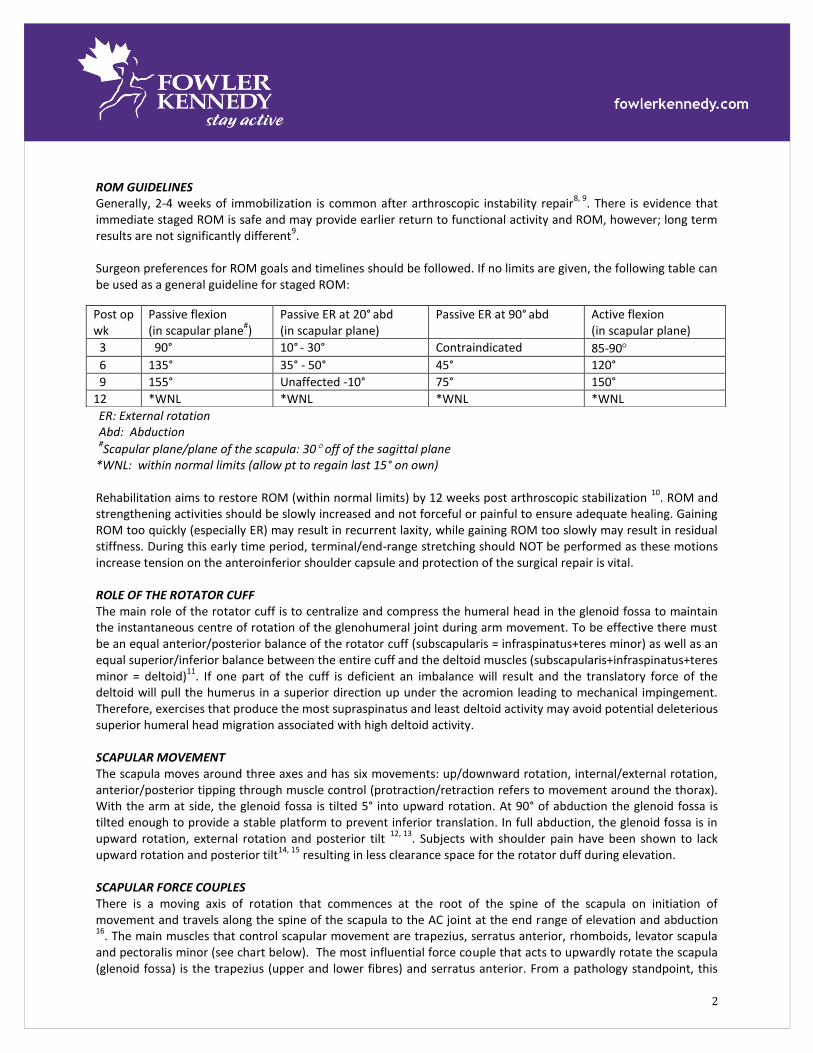
Surgeon preferences for ROM goals and timelines should be followed. If no limits are given, the following table can be used as a general guideline for staged ROM:
ER: External rotation Abd: Abduction
#Scapular plane/plane of the scapula: 30 off of the sagittal plane
*WNL: within normal limits (allow pt to regain last 15° on own) Rehabilitation aims to restore ROM (within normal limits) by 12 weeks post arthroscopic stabilization
10. ROM and
strengthening activities should be slowly increased and not forceful or painful to ensure adequate healing. Gaining ROM too quickly (especially ER) may result in recurrent laxity, while gaining ROM too slowly may result in residual stiffness. During this early time period, terminal/end-range stretching should NOT be performed as these motions increase tension on the anteroinferior shoulder capsule and protection of the surgical repair is vital. ROLE OF THE ROTATOR CUFF The main role of the rotator cuff is to centralize and compress the humeral head in the glenoid fossa to maintain the instantaneous centre of rotation of the glenohumeral joint during arm movement. To be effective there must be an equal anterior/posterior balance of the rotator cuff (subscapularis = infraspinatus+teres minor) as well as an equal superior/inferior balance between the entire cuff and the deltoid muscles (subscapularis+infraspinatus+teres minor = deltoid)
11. If one part of the cuff is deficient an imbalance will result and the translatory force of the
deltoid will pull the humerus in a superior direction up under the acromion leading to mechanical impingement. Therefore, exercises that produce the most supraspinatus and least deltoid activity may avoid potential deleterious superior humeral head migration associated with high deltoid activity. SCAPULAR MOVEMENT The scapula moves around three axes and has six movements: up/downward rotation, internal/external rotation, anterior/posterior tipping through muscle control (protraction/retraction refers to movement around the thorax). With the arm at side, the glenoid fossa is tilted 5° into upward rotation. At 90° of abduction the glenoid fossa is tilted enough to provide a stable platform to prevent inferior translation. In full abduction, the glenoid fossa is in upward rotation, external rotation and posterior tilt
12, 13. Subjects with shoulder pain have been shown to lack
upward rotation and posterior tilt14, 15
resulting in less clearance space for the rotator duff during elevation. SCAPULAR FORCE COUPLES There is a moving axis of rotation that commences at the root of the spine of the scapula on initiation of movement and travels along the spine of the scapula to the AC joint at the end range of elevation and abduction 16
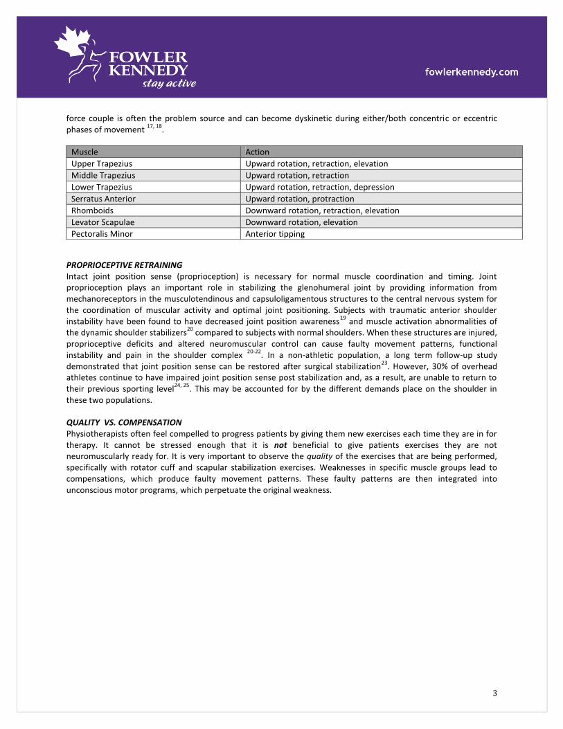
. The main muscles that control scapular movement are trapezius, serratus anterior, rhomboids, levator scapula and pectoralis minor (see chart below). The most influential force couple that acts to upwardly rotate the scapula (glenoid fossa) is the trapezius (upper and lower fibres) and serratus anterior. From a pathology standpoint, this
Post op wk
Passive flexion (in scapular plane
#)
Passive ER at 20° abd
(in scapular plane) Passive ER at 90°
abd Active flexion
(in scapular plane)
3 90° 10° - 30° Contraindicated 85-90
6 135° 35° - 50° 45° 120°
9 155° Unaffected -10° 75° 150°
12 *WNL *WNL *WNL *WNL
3
force couple is often the problem source and can become dyskinetic during either/both concentric or eccentric phases of movement
17, 18.
PROPRIOCEPTIVE RETRAINING Intact joint position sense (proprioception) is necessary for normal muscle coordination and timing. Joint proprioception plays an important role in stabilizing the glenohumeral joint by providing information from mechanoreceptors in the musculotendinous and capsuloligamentous structures to the central nervous system for the coordination of muscular activity and optimal joint positioning. Subjects with traumatic anterior shoulder instability have been found to have decreased joint position awareness
19 and muscle activation abnormalities of
the dynamic shoulder stabilizers20
compared to subjects with normal shoulders. When these structures are injured, proprioceptive deficits and altered neuromuscular control can cause faulty movement patterns, functional instability and pain in the shoulder complex
20-22. In a non-athletic population, a long term follow-up study
demonstrated that joint position sense can be restored after surgical stabilization23
. However, 30% of overhead athletes continue to have impaired joint position sense post stabilization and, as a result, are unable to return to their previous sporting level
24, 25. This may be accounted for by the different demands place on the shoulder in
these two populations. QUALITY VS. COMPENSATION Physiotherapists often feel compelled to progress patients by giving them new exercises each time they are in for therapy. It cannot be stressed enough that it is not beneficial to give patients exercises they are not neuromuscularly ready for. It is very important to observe the quality of the exercises that are being performed, specifically with rotator cuff and scapular stabilization exercises. Weaknesses in specific muscle groups lead to compensations, which produce faulty movement patterns. These faulty patterns are then integrated into unconscious motor programs, which perpetuate the original weakness.
Muscle Action
Upper Trapezius Upward rotation, retraction, elevation
Middle Trapezius Upward rotation, retraction
Lower Trapezius Upward rotation, retraction, depression
Serratus Anterior Upward rotation, protraction
Rhomboids Downward rotation, retraction, elevation
Levator Scapulae Downward rotation, elevation
Pectoralis Minor Anterior tipping
4
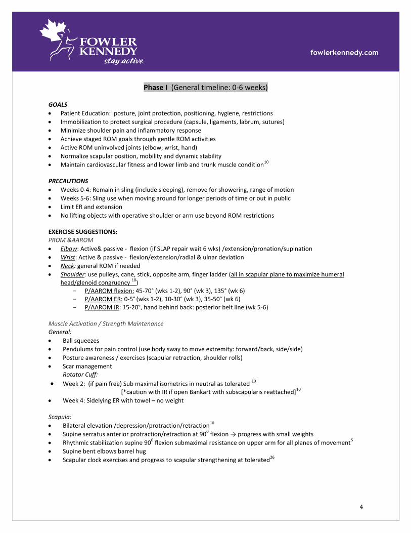
Phase I (General timeline: 0-6 weeks) GOALS
Patient Education: posture, joint protection, positioning, hygiene, restrictions
Immobilization to protect surgical procedure (capsule, ligaments, labrum, sutures)
Minimize shoulder pain and inflammatory response
Achieve staged ROM goals through gentle ROM activities
Active ROM uninvolved joints (elbow, wrist, hand)
Normalize scapular position, mobility and dynamic stability
Maintain cardiovascular fitness and lower limb and trunk muscle condition10
PRECAUTIONS
Weeks 0-4: Remain in sling (include sleeping), remove for showering, range of motion
Weeks 5-6: Sling use when moving around for longer periods of time or out in public
Limit ER and extension
No lifting objects with operative shoulder or arm use beyond ROM restrictions EXERCISE SUGGESTIONS: PROM &AAROM
Elbow: Active& passive - flexion (if SLAP repair wait 6 wks) /extension/pronation/supination
Wrist: Active & passive - flexion/extension/radial & ulnar deviation
Neck: general ROM if needed
Shoulder: use pulleys, cane, stick, opposite arm, finger ladder (all in scapular plane to maximize humeral head/glenoid congruency
10)
P/AAROM flexion: 45-70° (wks 1-2), 90° (wk 3), 135° (wk 6) P/AAROM ER: 0-5°
(wks 1-2), 10-30° (wk 3), 35-50° (wk 6)
P/AAROM IR: 15-20°, hand behind back: posterior belt line (wk 5-6) Muscle Activation / Strength Maintenance General:
Ball squeezes
Pendulums for pain control (use body sway to move extremity: forward/back, side/side)
Posture awareness / exercises (scapular retraction, shoulder rolls)
Scar management Rotator Cuff:
Week 2: (if pain free) Sub maximal isometrics in neutral as tolerated 10
[*caution with IR if open Bankart with subscapularis reattached]
10
Week 4: Sidelying ER with towel – no weight Scapula:
Bilateral elevation /depression/protraction/retraction10
Supine serratus anterior protraction/retraction at 900 flexion → progress with small weights
Rhythmic stabilization supine 900 flexion submaximal resistance on upper arm for all planes of movement
5
Supine bent elbows barrel hug
Scapular clock exercises and progress to scapular strengthening at tolerated26
5
Proprioceptive Retraining
Week 3: Upper extremity weight-bearing exercises for scapular movements at GH angles below 60 degrees elevation
10
i.e. Standing with swiss ball on floor – hand on ball with pressure forward/backward, side to side, circles, Standing weight-bearing shifts with hands on bed/plinth → progress to single arm weight-bearing
27
Modalities
Ice 15 minutes every few hours for pain relief1, 10
Interferential current therapy (pain relief) Cardiovascular Fitness
Bicycle, elliptical, stairmaster, walking
MILESTONES TO PROGRESS TO PHASE II 1. Appropriate tissue healing from surgery by following precautions and immobilization guidelines 2. ROM guidelines met but not significantly exceeded. 3. Pain control within allowed ROM
Phase II (General timeline: 6-12 weeks)
GOALS
Continued patient education: ADL’s in painfree range (waist level activities → progress to shoulder level → overhead activities), avoid heavy lifting or positions of instability during ADL’s i.e. end range ER and combined abduction/ER
P/AAROM to achieve staged ROM goals, may have ~100 loss of motion at ends of range from surgical
procedure (esp. ER and flexion)
Progression of exercise: passive (P) → active assisted (AA) → active (A) → addition of resistance (tubing or weights)
Establish basic rotator cuff endurance and scapular neuromuscular control
Later in phase, introduce functional patterns of movement PRECAUTIONS
Avoid terminal stretches at end range ER or in 90/90 positions.
(Most times only light stretching or no stretching is needed)
Avoid exercises that load the anterior capsular structures in a position of horizontal abduction or combined abduction and ER (i.e. NO push-ups, pec flys) during this timeframe
Avoid heavy lifting or plyometrics
Avoid exercises that may cause impingement i.e. empty can
Ensure exercises are performed pain free and without substitutions or altered movement patterns
(Exercise quality) EXERCISE SUGGESTIONS: PROM & AAROM
Neck: general ROM if needed
Thoracic spine: ensure proper extension to facilitate shoulder ROM
Shoulder P/AAROM: Use pulleys, cane, stick, opposite arm….. Flexion (scapular plane): 135° (wk 6), 155° (wk 9), near end range/160° (wk 12) ER at 20°
abduction (scapular plane): 35-50° (wk 6), 50-65°
(wk 9), near end range/70° (wk 12)
ER at 90° abduction: 45° (wk 6), 75° (wk 9,), near end range/80° (wk 12)
6
IR at 20° abduction (scapular plane): 30-60°
IR stretches: towel/cane assisted hand behind back (combination of ext/IR/hor add), sidelying sleeper stretch, cross arm stretch
If ROM is significantly less than goals, joint mobilizations may be performed into the limited direction
Progress finger ladder in flexion and scaption terminal ranges
Arm bike/ergometer no resistance Muscle Strength & Endurance Rotator Cuff:
Light isotonics with emphasis on high repetitions (4 sets of 15-20 reps) and low resistance (1-2 lbs):
Sidelying ER with towel → progress to 1lb
Standing ER & IR with towel: pulleys or light resistance tubing
Rhythmic stabilization techniques for rotator cuff strengthening (ER/IR at 45° abduction in scapular plane)5
Scapula:
Continue with shoulder retractions, shoulder rolls
Supine rhythmic stabilization 90-100 flexion / joint perturbations in randomized directions → progressions: eyes closed, holding medicine ball
27
Closed kinetic chain rhythmic stabilization:
Ball stabilization on wall
Static holds in push-up position on ball
Light resistance extension, adduction, forward flexion (not past plane of body)
Progress closed chain scapulothoracic mobility to shoulder level and then to overhead i.e.:
Quadruped scapular protraction/retraction 90° progress to 120°
Quadruped to tripod ( 2 to 1 arm)
Standing short lever (elbow flexed) slides up wall → long lever→ no wall support28
Strengthen scapular retractors and upward rotators i.e.:
Prone arm raises at 0° progress to 90° and 120°
Prone or seated rows → progress with resistance or weight
Strengthen serratus
Forward punch
Push up with plus progress from wall to floor, on knees to feet
Supine protraction/retraction with heavier weights Proprioceptive Retraining
Standing swiss ball on the wall at 90° flexion/scaption/abduction: circles, side to side, up and down, alphabet→ progress 2 arms to 1 arm and ROM from 90° to 120°
Therapist assisted joint/limb positioning with patient reproduction of position → mid ranges → end ranges→ progress to eyes closed
27
Weight-bearing activities on knees on unstable base i.e. Bosu, Wobble board, Airex pad, slider board
Supine weighted ball drop at 90° shoulder flexion
Supine weighted ball throw/catch → progress 2 arms to 1 arm
Quadruped maintain proper scapula position
Bodyblade: arm at side→ 30, 90, 120, 160° in scaption and frontal plane → progress using PNF patterning
Ball dribbles on wall
To increase proprioceptive input and difficulty, progression of exercises can be performed with eyes closed5
7
Modalities
Ice 15-25 minutes 1
Biofeedback: auditory, visual, tactile or machine
Muscle Stimulation for posterior rotator cuff Cardiovascular Fitness
Bicycle, elliptical, stairmaster, treadmill jog→run, train specific to demand of sport MILESTONES TO PROGRESS TO PHASE III 1. AROM guidelines met without pain or substitution patterns. 2. Good resting scapular posture and dynamic scapular control with ROM and strengthening exercises. 3. Able to perform recommended strengthening exercises without pain or difficulty.
Phase III (General timeline: 12-24 weeks)
GOALS Ensure ROM requirements are met Progressive strengthening, endurance, power and neuromuscular control exercises Progressive exercises in terms of speed once proficiency is demonstrated at slower speeds Activity specific progression: sport, work, hobbies Gradual and planned increase in stress to anterior capsule and labral tissues Gradual return to full ADL’s, work and recreational activities Suggested Guidelines:
3-4 months: may begin golfing 4 months+: Interval Sports Programs: throwing, swimming, tennis, volley ball, gymnastics (surgeon
approval) PRECAUTIONS
Avoid stress to the shoulder in a short period of time or in an uncontrolled manner
Avoid advanced rehabilitation exercises (such as plyometrics or exercises at end range ER/Abd if the patient does not perform this activities during ADL’s, work, or recreation
Do not progress into activity specific training until the patient has nearly full ROM and strength
Avoid weightlifting activities which place excessive stress on the anterior capsule i.e. lat pull downs and military press with hands behind the head and wide grip bench press. Exercises, such as dips, which encourage shoulder hyperextension, should be avoided. These exercises do not have any additional benefit in terms of muscle activity and other exercises can be substituted. Hand placement and depth on bench and incline press should be more narrow than normal to prevent stress on the anterior capsule when lowering weights
5. The
elbow should not pass the plane of the body - be sure to “always see your elbows” = Elbow Rule. EXERCISE SUGGESTIONS: ROM
PROM/Stretching/Joint Mobilizations as needed to address any remaining deficits Muscle Strength/Endurance/Power Rotator Cuff:
Progress ER/IR at side → to 45° → eventually to 90°
Scapula:
Rhythmic stabilization / joint perturbations in positions of function and vulnerability27
PNF diagonal patterns with bands/pulleys/manual resistance:

8
D1 extension (high back hand to down to hitch hike position)
D1 flexion (hitch hike to high back hand position)
D2 extension (carry tray to hand in opposite front pocket position)
D2 flexion (hand in opposite front pocket to carry tray position)
Continue with shoulder strengthening program as initiated in Phase II with emphasis on faster speed, multiplanar activities which incorporate the kinetic chain
Proprioceptive Retraining (open and closed kinetic chain)
Weight-bearing activities on toes on unstable base i.e. Bosu, Wobble board, Airex pad, slider board
Swiss ball prone walk out
U/E wobble board stability→ progress to small push-up on board Strength / Endurance / Power
Replicate ADL / work activities / sport requirements
Progressive return to weight-lifting program for larger upper extremity muscles (i.e. deltoid, lat dorsi, pec major): start with light weight / high reps (20-30 reps) → gradually increase weight and decrease repetitions. Suggestions for early in Phase III (3-4 months): Biceps/Triceps (arm at side) Shoulder shrugs Rows (scapular retraction) Lat pull downs (hands in front) Shoulder press with hands in front of shoulders (not abducted/externally rotated) Push-up (only to 90
0 elbow flexion)
Suggestions to add for intermediate Phase III (4-5 months): Chest press / incline Machine / Barbell shoulder press (no end range abduction/external rotation Prone horizontal abduction Prone ER at 90
0 abduction → progress weight as able
Suggestions to add for late in Phase III (5-6+ months): Military Press Flys / Reverse Flys Dead Lifts Power Cleans
Plyometric Program (if needed)
Initiate in intermediate to late phase III (5-6+ months): Suggestions/ideas: Tubing plyometrics for ER/IR at 90
0 abduction with varying speeds
2 handed tosses: waist/chest level→ overhead → diagonal 1 handed tosses: begin throw with shoulder flexion and mostly elbow extension→ progress by increasing
the amount of shoulder abduction/ER o Begin with towel, beach ball, kid’s ball, tennis ball→ progression to lightly weighted balls
(plyoballs) Cardiovascular Fitness
Train specific to demand of sport (aerobic, anaerobic)
9
MILESTONES TO RETURN TO SPORT, WORK, HOBBIES
1. Therapist/Physician clearance 2. No complaints of pain or instability 3. Sufficient ROM to meet task demands 4. Good/Full strength and endurance of rotator cuff and scapular muscles for desired activities including
adequate neuromuscular control
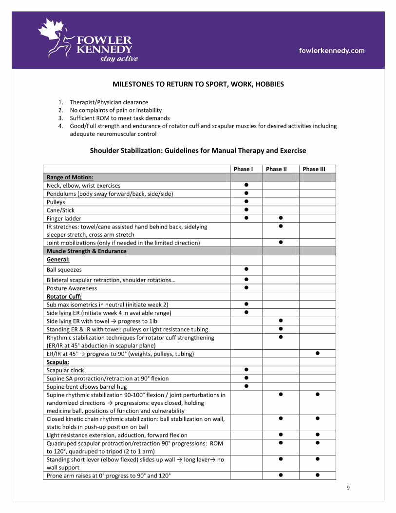
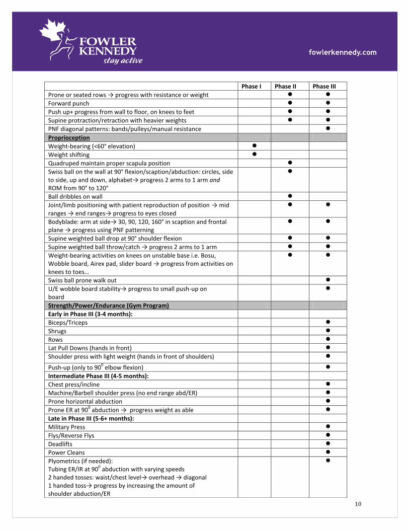
Shoulder Stabilization: Guidelines for Manual Therapy and Exercise
Phase I Phase II Phase III
Range of Motion:
Neck, elbow, wrist exercises
Pendulums (body sway forward/back, side/side)
Pulleys
Cane/Stick
Finger ladder
IR stretches: towel/cane assisted hand behind back, sidelying sleeper stretch, cross arm stretch
Joint mobilizations (only if needed in the limited direction)
Muscle Strength & Endurance
General:
Ball squeezes
Bilateral scapular retraction, shoulder rotations…
Posture Awareness
Rotator Cuff:
Sub max isometrics in neutral (initiate week 2)
Side lying ER (initiate week 4 in available range)
Side lying ER with towel → progress to 1lb
Standing ER & IR with towel: pulleys or light resistance tubing
Rhythmic stabilization techniques for rotator cuff strengthening (ER/IR at 45° abduction in scapular plane)
ER/IR at 45° → progress to 90° (weights, pulleys, tubing)
Scapula:
Scapular clock
Supine SA protraction/retraction at 90° flexion
Supine bent elbows barrel hug
Supine rhythmic stabilization 90-100° flexion / joint perturbations in randomized directions → progressions: eyes closed, holding medicine ball, positions of function and vulnerability
Closed kinetic chain rhythmic stabilization: ball stabilization on wall, static holds in push-up position on ball
Light resistance extension, adduction, forward flexion
Quadruped scapular protraction/retraction 90° progressions: ROM to 120°, quadruped to tripod (2 to 1 arm)
Standing short lever (elbow flexed) slides up wall → long lever→ no wall support
Prone arm raises at 0° progress to 90° and 120°
10
Phase I Phase II Phase III
Prone or seated rows → progress with resistance or weight
Forward punch
Push up+ progress from wall to floor, on knees to feet
Supine protraction/retraction with heavier weights
PNF diagonal patterns: bands/pulleys/manual resistance
Proprioception
Weight-bearing (<60° elevation)
Weight shifting
Quadruped maintain proper scapula position
Swiss ball on the wall at 90° flexion/scaption/abduction: circles, side to side, up and down, alphabet→ progress 2 arms to 1 arm and ROM from 90° to 120°
Ball dribbles on wall
Joint/limb positioning with patient reproduction of position → mid ranges → end ranges→ progress to eyes closed
Bodyblade: arm at side→ 30, 90, 120, 160° in scaption and frontal plane → progress using PNF patterning
Supine weighted ball drop at 90° shoulder flexion
Supine weighted ball throw/catch → progress 2 arms to 1 arm
Weight-bearing activities on knees on unstable base i.e. Bosu, Wobble board, Airex pad, slider board → progress from activities on knees to toes…
Swiss ball prone walk out
U/E wobble board stability→ progress to small push-up on board
Strength/Power/Endurance (Gym Program)
Early in Phase III (3-4 months):
Biceps/Triceps
Shrugs
Rows
Lat Pull Downs (hands in front)
Shoulder press with light weight (hands in front of shoulders)
Push-up (only to 900 elbow flexion)
Intermediate Phase III (4-5 months):
Chest press/incline
Machine/Barbell shoulder press (no end range abd/ER)
Prone horizontal abduction
Prone ER at 900 abduction → progress weight as able
Late in Phase III (5-6+ months):
Military Press
Flys/Reverse Flys
Deadlifts
Power Cleans
Plyometrics (if needed): Tubing ER/IR at 90
0 abduction with varying speeds
2 handed tosses: waist/chest level→ overhead → diagonal 1 handed toss→ progress by increasing the amount of shoulder abduction/ER
11
References
1. Cicak N, Bilic R, Delimar D. Hill-Sachs lesion in recurrent shoulder dislocation: sonographic detection. J Ultrasound Med 1998; 17:557-60. 2. Snyder SJ, Karzel RP, Del Pizzo W, Ferkel RD, Friedman MJ. SLAP lesions of the shoulder. Arthroscopy 1990; 6:274-9. 3. Parentis MA, Mohr KJ, ElAttrache NS. Disorders of the superior labrum: review and treatment guidelines. Clin Orthop Relat Res 2002; (400):77-87. 4. Maurer SG, Rosen JE, Bosco JA,3rd. SLAP lesions of the shoulder. Bull Hosp Jt Dis 2003; 61:186-92. 5. Blackburn TA, Guido JA. Rehabilitation after Ligamentous and Labral Surgery of the Shoulder: Guiding Concepts. J Athl Train 2000; 35:373-81. 6. Roth CA, Bartolozzi AR, Ciccotti MG, Wetzler MJ, Gillespie MJ, Snyder-Mackler L, Santare MH. Failure properties of suture anchors in the glenoid and the effects of cortical thickness. Arthroscopy 1998; 14:186-91. 7. Abboud JA, Soslowsky LJ. Interplay of the static and dynamic restraints in glenohumeral instability. Clin Orthop Relat Res 2002; (400):48-57. 8. Grana WA, Buckley PD, Yates CK. Arthroscopic Bankart suture repair. Am J Sports Med 1993; 21:348-53. 9. Kim SH, Ha KI, Cho YB, Ryu BD, Oh I. Arthroscopic anterior stabilization of the shoulder: two to six-year follow-up. J Bone Joint Surg Am 2003; 85-A:1511-8. 10. Hayes K, Callanan M, Walton J, Paxinos A, Murrell GA. Shoulder instability: management and rehabilitation. J Orthop Sports Phys Ther 2002; 32:497-509. 11. Burkhart SS. Arthroscopic treatment of massive rotator cuff tears. Clinical results and biomechanical rationale. Clin Orthop Relat Res 1991; (267):45-56. 12. Ludewig PM, Hoff MS, Osowski EE, Meschke SA, Rundquist PJ. Relative balance of serratus anterior and upper trapezius muscle activity during push-up exercises. Am J Sports Med 2004; 32:484-93. 13. Burkhart SS, Morgan CD, Kibler WB. The disabled throwing shoulder: spectrum of pathology Part I: pathoanatomy and biomechanics. Arthroscopy 2003; 19:404-20. 14. Lukasiewicz AC, McClure P, Michener L, Pratt N, Sennett B. Comparison of 3-dimensional scapular position and orientation between subjects with and without shoulder impingement. J Orthop Sports Phys Ther 1999; 29:574,83; discussion 584-6. 15. Endo K, Ikata T, Katoh S, Takeda Y. Radiographic assessment of scapular rotational tilt in chronic shoulder impingement syndrome. J Orthop Sci 2001; 6:3-10. 16. Schenkman M, Rugo de Cartaya V. Kinesiology of the shoulder complex. J Orthop Sports Phys Ther 1987; 8:438-50. 17. Bourne DA, Choo AM, Regan WD, MacIntyre DL, Oxland TR. Three-dimensional rotation of the scapula during functional movements: an in vivo study in healthy volunteers. J Shoulder Elbow Surg 2007; 16:150-62. 18. McClure PW, Michener LA, Sennett BJ, Karduna AR. Direct 3-dimensional measurement of scapular kinematics during dynamic movements in vivo. J Shoulder Elbow Surg 2001; 10:269-77. 19. Barden JM, Balyk R, Raso VJ, Moreau M, Bagnall K. Dynamic upper limb proprioception in multidirectional shoulder instability. Clin Orthop Relat Res 2004:181-9. 20. Myers JB, Ju YY, Hwang JH, McMahon PJ, Rodosky MW, Lephart SM. Reflexive muscle activation alterations in shoulders with anterior glenohumeral instability. Am J Sports Med 2004; 32:1013-21. 21. Aydin T, Yildiz Y, Yanmis I, Yildiz C, Kalyon TA. Shoulder proprioception: a comparison between the shoulder joint in healthy and surgically repaired shoulders. Arch Orthop Trauma Surg 2001; 121:422-5. 22. Myers JB, Wassinger CA, Lephart SM. Sensorimotor contribution to shoulder stability: effect of injury and rehabilitation. Man Ther 2006; 11:197-201. 23. Potzl W, Thorwesten L, Gotze C, Garmann S, Steinbeck J. Proprioception of the shoulder joint after surgical repair for Instability: a long-term follow-up study. Am J Sports Med 2004; 32:425-30. 24. Fremerey R, Bosch U, Freitag N, Lobenhoffer P, Wippermann B. Proprioception and EMG pattern after capsulolabral reconstruction in shoulder instability: a clinical and experimental study. Knee Surg Sports Traumatol Arthrosc 2006; 14:1315-20.
12
25. Fremerey R, Bosch U, Lobenhoffer P, Wippermann B. Joint position awareness and sports activity after capsulolabral reconstruction in the overhead athlete. Int J Sports Med 2006; 27:648-52. 26. Smith J, Dahm DL, Kaufman KR, Boon AJ, Laskowski ER, Kotajarvi BR, Jacofsky DJ. Electromyographic activity in the immobilized shoulder girdle musculature during scapulothoracic exercises. Arch Phys Med Rehabil 2006; 87:923-7. 27. Myers JB, Lephart SM. The Role of the Sensorimotor System in the Athletic Shoulder. J Athl Train 2000; 35:351-63. 28. Wise MB, Uhl TL, Mattacola CG, Nitz AJ, Kibler WB. The effect of limb support on muscle activation during shoulder exercises. J Shoulder Elbow Surg 2004; 13:614-20.