article 4 treatment of accommodative and vergence ... 4 treatment of accommodative and vergence...
TRANSCRIPT
165 Optometry & Visual Performance Volume 3 | Issue 3 | 2015, June
Article 4 Treatment of Accommodative and Vergence Dysfunction in Traumatic Brain Injury (TBI): A Case Report
Anna Griffith, OD, Chico, California
ABSTRACT
Background: Accommodative insufficiency (AI), accommodative infacility, and convergence insufficiency (CI) are some of the most common visual problems following traumatic brain injury (TBI). In light of the increased prevalence of TBIs in modern-day combat, it is important for clinicians to be aware of the associated visual symptoms and methods of treatment. The mechanism of progressive neural damage in TBI as well as the neural-anatomical pathways of accommodation and vergence will be reviewed in the case report presented here. Important considerations when treating patients with TBI will also be discussed.
Case Report: This case report describes the diagnosis, management, and treatment of accommodative and vergence dysfunction in a 33-year-old veteran with a history of multiple TBIs incurred during combat in Afghanistan. The veteran was diagnosed with asymmetric accommodative insufficiency and infacility and gross convergence insufficiency, and he had decreased depth perception. Five in-office vision therapy sessions were conducted over the course of a month and a half, with daily practice at home, followed by maintenance activities and tapering of vision training. Treatment restored the patient’s visual clarity, comfort, and fine stereopsis, enabling him to pursue his goal of returning to school for engineering.
Conclusion: Vision therapy improves visual function and symptoms from TBI in many patients. An increase in TBI due to modern-day warfare has increased the awareness of and the need for recognition and treatment of visual problems. Most, if not all, communities have a need for vision care for patients with TBI from car crashes, accidents, sports injuries, and concussion. Prompt diagnosis and treatment of visual dysfunction is critical to improving quality of life, ability to work towards vocational goals, and progress of other rehabilitation therapies which require varied visual tasks.
Keywords: accommodative infacility, accommodative insufficiency, convergence insufficiency, post trauma vision syndrome, traumatic brain injury, vergence
IntroductionAccommodative insufficiency (AI), accommodative infa-
cility, and convergence insufficiency (CI) are some of the most common visual problems following traumatic brain injury (TBI).1,2 Every year there are 1.7 million brain injuries in the U.S.3 Over five million Americans with TBI, approximately 2% of the population, need assistance with activities of daily living.4 Methods of modern-day warfare such as the use of land mines, rocket-propelled grenades (RPGs), and improvised explosive devices (IEDs) have greatly increased the incidence of TBI within the armed forces. Nearly 150,000 members of Operation New Dawn, Operation Iraqi Freedom, and Operation Enduring Freedom had a TBI in the last five years (2009-2013).5 Enhanced protective equipment keeps vital organs intact and saves lives,6,7 but it cannot prevent coup-contrecoup forces on the brain. Advances in combat medicine and air evacuation have increased the survival rate of military personal incurring blast injury. Many of these veterans will need acute and long-term care. Rehabilitation of TBI is recognized as a national priority in light of recent combat in Iraq and Afganistan.8
Motor vehicle accidents, collisions, and falling are the other common causes of TBI. Children aged 0-4 years, adolescents aged 15-19 years, and adults aged 65 years and older are most
likely to sustain a TBI.3 TBI rates for males are higher than for females.
TBI commonly results in significant visual problems. Individuals with TBI are more likely to have AI10 and/or accom-modative infacility (41%),11 vergence dysfunction (56%),11 CI,1 and version dysfunction (51%).11 Other visual diagnoses that occur fairly frequently following head trauma include strabismus, cranial nerve palsies,1,11-15 vertical deviations,8 visual field defects,8,13 photosensitivity,13 and dry eye.8
TBI is known to cause diffuse axonal injury (DAI) due to coup-contrecoup force.14,15 This shearing force often damages many areas of the brain, including the cranial nerves. The high frequency of oculomotor and binocular vision problems following TBI11 is not surprising due to the fact that three of the cranial nerves orchestrate fine oculomotor control, and another regulates visual-vestibular function.1
There is also evidence of secondary injury, a biochemical cascade triggered by the primary injury that occurs for weeks or months following the initial trauma.17,18 The traumatic forces from the primary injury cause massive nerve conduction disruptions, which alter the relationship of cerebral blood flow and cerebral glucose metabolism.17 Ischemia and hypoxia result, which lead to metabolic imbalances within cells that alter cell membrane permeability. Consequently, there is a toxic
Volume 3 | Issue 3 | 2015, June Optometry & Visual Performance 166
inflow of calcium and outflow of potassium which activates protease enzymes. The enzymes disrupt cell structure and create excessive neurotransmitter activity, which kills neural cells. Progressive loss of neuron function erodes established pathways that automatically control the complex sensorimotor components of the visual process, ambient-focal functioning, and efficient integrated information processing.17
TBI can be classified as mild, moderate, or severe based on the Glasgow Coma Scale, length of coma, alteration of consciousness, and post-traumatic amnesia.18 Forty percent of patients with mild TBI experience visual problems.19 However, mild TBIs are not always identified.19 Though injured, these patients may be able to walk away from the incident, but secondary damage continues to occur, and subsequent visual symptoms may arise. Post-Trauma Vision Syndrome (PTVS) has been used as an umbrella term for the visual symptoms following TBI.20 Common symptoms include blurry vision, diplopia, eyestrain, fatigue, headaches, reading difficulty, and avoidance of near tasks.10,21 In several studies, the most common symptom was related to reading (50-90%).16,22
Symptoms manifest from one or more of the frequent visual diagnoses associated with TBI: AI,10 accommodative infacility,10 version dysfunction,11 vergence dysfunction, strabismus, cranial nerve palsies,1,11-15 vertical deviation,8 visual field defect,8,13 photosensitivity,13 and dry eye.8 Clinical findings suggesting accommodative dysfunction include reduced accommodative amplitude, reduced positive relative accommodation, increased lag of accommodation, slow accommodative facility, visual fatigue, and restricted fusional ranges at near related to accommodative problems.10 AI is defined as decreased amplitude of accommodation significantly below age-expected norms.23,24 Studies of patients with mild TBI found the incidence of AI to be 10% to 36%.10,22,25-28 Accommodative infacility is a condition in which the patient has difficulty changing the accommodative response level, such as when looking from near to far or far to near. Accommodative infacility is diagnosed when the patient is unable monocularly to clear both +2.00 and -2.00 lenses quickly enough to complete 10 cycles in one minute.2
The neural pathway for accommodation travels through many areas of the brain. The retinal signal is sent through the LGN to the primary visual cortex. Here, blur is detected. Signals are also transmitted to parieto-temporal areas and the cerebellum for processing. Signals go to the Edinger-Westphal nucleus in the midbrain, where a motor command is sent through CN III to the ciliary nerve and ciliary muscle, which changes the shape of the lens for accommodation.10 All parts of the pathway must function well to deliver a clear visual image, which is often not the case in patients with traumatic brain injury.23,29,30
Clinical findings suggesting vergence dysfunction include a receded near point of convergence, reduced vergence ranges, and slow vergence facility. Vergence dysfunction can cause symptoms of diplopia, movement of words or lines of
text, and intermittent blur.1 The overall delayed and slowed vergence response and rapid fatigue of patients with TBI contribute to these symptoms. A recent study by Szymanowicz et al. shows that the primary neural deficit in the mild TBI patient is the pulse–the dynamic velocity of both convergence and divergence.1 Initiation of vergence movements is delayed. This latency suggests a processing delay in the afferent visual pathways.31 During vergence eye movements, neural firing occurs in the midbrain, pons,32-34 cerebellum,34,35 areas of the cerebral cortex,34,36 parietal lobes,34,37 and primary visual cortex.38 Thus, it is not surprising that DAI occurring in traumatic brain injury affects vergence response and efficiency.
Case ReportA 33-year-old white male combat veteran presented to our
clinic on December 7, 2011 with complaints of intermittent blur at distance and near that was worse in the right eye. He used to “breeze through books,” but after his multiple TBIs, he reported extreme eyestrain that prevented him from reading more than a chapter at a time. He also experienced asthenopia following two hours of working on the computer. 3-D movies induced nausea, and he was not able to appreciate the 3-D effect. He was also extremely photosensitive, had mild balance problems, and was occasionally hypersensitive to movement and noise. Other complaints included difficulty finding items in crowded drawers or on crowded shelves.
The patient’s last eye examination was less than one year prior. He had worn a single vision myopic prescription since he was a teenager and wore contact lenses until he began training in dusty environments prior to his TBIs. While in active duty
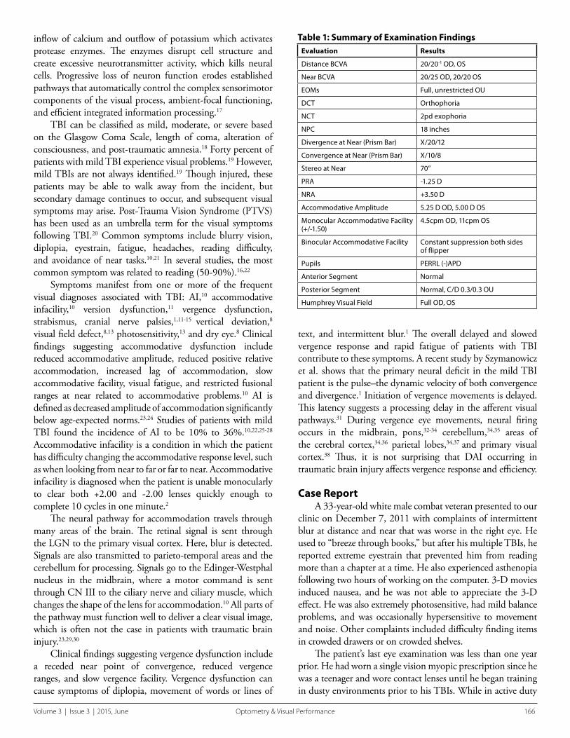
Table 1: Summary of Examination FindingsEvaluation Results
Distance BCVA 20/20-1 OD, OS
Near BCVA 20/25 OD, 20/20 OS
EOMs Full, unrestricted OU
DCT Orthophoria
NCT 2pd exophoria
NPC 18 inches
Divergence at Near (Prism Bar) X/20/12
Convergence at Near (Prism Bar) X/10/8
Stereo at Near 70”
PRA -1.25 D
NRA +3.50 D
Accommodative Amplitude 5.25 D OD, 5.00 D OS
Monocular Accommodative Facility (+/-1.50)
4.5cpm OD, 11cpm OS
Binocular Accommodative Facility Constant suppression both sides of flipper
Pupils PERRL (-)APD
Anterior Segment Normal
Posterior Segment Normal, C/D 0.3/0.3 OU
Humphrey Visual Field Full OD, OS
167 Optometry & Visual Performance Volume 3 | Issue 3 | 2015, June
in Afghanistan from 2007 to 2009, he suffered three blasts requiring medical intervention. These blasts caused temporary loss of vision, hearing, and memory and diminished cognitive function. Although sensory status improved and some of his cognitive ability returned, he still struggled with decreased memory capacity. His medical history indicated brain contusions and blood clots, hypertension, migraines, gastroesophageal reflux disease, and post-traumatic stress disorder (PTSD) with flashbacks and violent dreams. He was taking Diazepam, Docusate, Mirtazapine, Omeprazole, Promethazine, Sumatriptan, Verapamil, and Hydrocodone 10/Acetaminophen, and he was using a nicotine patch and resin complex to quit smoking. Smoking history was half a pack a day for ten years. The patient was oriented to time, place, and person. A caregiver accompanied him to his appointment.
A summary from the patient’s exam is found in Table 1. The patient’s NRA suggested that he was over-corrected, which was confirmed with a careful binocularly-balanced refraction. However, the visual acuity was unchanged with the manifest (-2.25 -0.50 x 035 OD and -2.00 DS OS), and when this was demonstrated to the patient, it was strongly rejected in favor of his habitual (-2.50 -0.75 x 030 OD and -2.50 DS OS).
Monocular accommodative facility testing was attempted with the standard +/-2.00 accommodative flipper, but the
patient struggled, so the power was dropped to +/-1.50 to obtain a sense of his accommodative function.
A vision rehabilitation program was designed to increase amplitude and facility of accommodation, to improve gross convergence, to minimize current symptoms, and to attain the patient’s goals to read comfortably and return to school for electrical engineering. One-hour in-office sessions were to be scheduled weekly, with 30 minutes of daily exercises to be done at home. The therapy would begin with activities to improve accommodative amplitude and facility monocularly and then binocularly. Vergence therapy would begin with gross convergence, then in-instrument smooth vergence followed by jump vergence, and finally vergence activities in free space. The patient was motivated and willing to pursue training. An accommodative rock card, +/-1.50 flipper lenses, and an eye patch were dispensed to the patient for monocular accommodative facility training, five minutes per eye each day, timed in one-minute intervals to monitor progress. Monocular sustained flipper reading was also prescribed for five minutes each eye, twice per day. The patient was instructed to flip the lenses after each paragraph. A follow-up appointment was scheduled for a tint evaluation for the patient’s extreme photophobia and for further evaluation of visual efficiency and function.
Follow-up #1Due to various circumstances, the patient did not return
until January 26th, 2012, eight weeks after the evaluation. Homework compliance was good. On average, the patient did each activity five days per week, but he often did the exercises even longer than prescribed. A variety of absorptive filters (Solar 3s) were trialed with the patient. Medium grey and plum were preferred, but the patient decided to keep his habitual grey tint. The Motor Free Visual Perception Test–Third Edition (MVPT-3) was administered to evaluate visual perceptual skill status. The patient scored 56 points, placing him in the 32nd percentile for his age, an 18-year-old age equivalent. Results suggested a relative deficit in form constancy (ability to choose the same form when it is in a different orientation) and figure-ground (ability to distinguish an object of interest amidst a cluttered background). His deficit in figure-ground perception provided a probable explanation for the patient’s subjective report of having difficulty finding things on a crowded shelf or in a messy drawer and needing to maintain absolute organization in his apartment.
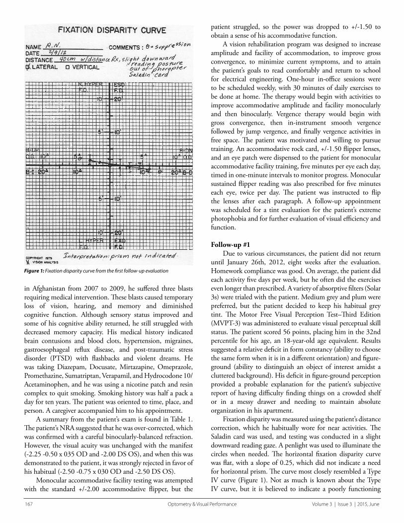
Fixation disparity was measured using the patient’s distance correction, which he habitually wore for near activities. The Saladin card was used, and testing was conducted in a slight downward reading gaze. A penlight was used to illuminate the circles when needed. The horizontal fixation disparity curve was flat, with a slope of 0.25, which did not indicate a need for horizontal prism. The curve most closely resembled a Type IV curve (Figure 1). Not as much is known about the Type IV curve, but it is believed to indicate a poorly functioning
Figure 1: Fixation disparity curve from the first follow-up evaluation
Volume 3 | Issue 3 | 2015, June Optometry & Visual Performance 168
binocular system, with compromised fusional vergence reflexes. Speculation is that these patients use conscious will to align the eyes rather than relying on normal accommodative/vergence reflex alignment. It is believed that vision therapy may help convert a Type IV curve to a normal Type I curve. The vertical associated phoria was 1 prism diopter right hyperphoria. Prism trial of 1∆ BD OD improved stereo acuity to 20 arc seconds. Stereo acuity had been measured as 30 arc seconds before prism demonstration, which was improved from 70 arc seconds two months prior when therapy was initiated. The patient subjectively reported that the circles had more depth with prism, but this feeling of subjective improvement was the same with both 1∆ BU and 1∆ BD OD. With 1∆ BU OD, the patient also reported increased clarity and stability with vertical Maddox rod testing and distance viewing, but he preferred 1∆ BD OD at near. Due to this inconsistency, prism was not prescribed, but it might be considered again at a later time if results were consistent and repeatable.
The patient completed the Convergence Insufficiency Symptom Survey (CISS) with a score of 45. A study of validity and reliability of the CISS with adults aged 19-30 years found that good discrimination (sensitivity=97.8%, specificity=87%) was obtained using a score of 21 or higher.39 Although our patient’s age was slightly above the upper age range listed in this study, his high score suggested a clinically significant level of visual discomfort. A follow-up appointment was scheduled for one week, and the patient was to continue accommodative facility and sustained paragraph reading with the +/-1.50 flippers every day at home.
Follow-up #2The patient returned on February 2nd, 2012 to begin in-
office rehabilitative training. Treatment consisted of monocular accommodative rock (MAR) with +/-2.00 flippers, near-far Hart chart, Brock string, and vectograms. Performance results were as follows. Right eye monocular accommodative facility was 7cpm (cycles per minute) for the first minute and 6.5 cpm for the second minute. For the left eye, first-minute facility was 9 cpm, and second-mnute facility was 6 cpm. These values are consistent with accommodative infacility and fatigue. Subjectively, the patient felt a little disoriented. The (-) side of the lenses was more difficult in each case. A 10/10 Hart chart was placed on the wall six feet away from the patient. Habitual distance correction was worn, and one eye was covered with an eye patch. The patient was instructed to hold a small near Hart chart as close to the eye as he could without blur, read the first row of letters at near, then the first row at distance, then the second row at near, etc. Performance was consistent with accommodative insufficiency and infacility. The patient could only hold the near chart at 10-12 inches from the eye. With the right eye, the patient accomplished the task in 3 minutes and 28 seconds, and he reported difficulty shifting focus from distance to near. The left eye was not timed, but comparable difficulty was demonstrated with the task; however, with this
eye, the patient reported shifting focus from near to distance was more difficult than the reverse.
The best break values achieved on quoit vectogram were 2∆ BI and 2∆ BO, with both recoveries at the ortho position. Brock string fusion was limited; the patient saw double when viewing a bead closer than arm’s length away. With encouragement and proprioceptive feedback, the patient was able to improve fusional distance on the Brock string from arm’s length to six inches, but only with considerable effort and discomfort. When viewing the Brock string, he exhibited normal physiological diplopia by seeing two crossing strings. Additionally, he almost always perceived the cross at the bead or just in front. Relative to what is expected, this suggested an abnormal accommodative vergence response.
Later in the session, the patient was able to mobilize voluntary convergence to about twelve inches from his nose without tactile support. For home training, flipper power was increased to +/-2.00 for MAR. Brock string was prescribed for five minutes twice a day, as well as one completion of the Hart chart near-far rock for each eye.
Follow-up #3The patient’s third clinic treatment session was one week
later on February 9th, 2012. Compliance with two of the three prescribed home treatment activities was excellent. The patient reported only having worked with the Brock string twice because it made him dizzy. The Developmental Eye Movement Test (DEM) was administered to assess saccadic/fixation function. The patient made two errors, and the horizontal time to vertical time ratio was in the 7th percentile for his age.
In-office performance with the quoit vectogram had not improved from session #2 (2 BI and 2 BO breaks with recoveries at the ortho position). Spirangle was substituted for quoit to provide more central visual detail. At first, spirangle base-in ranges were very limited, but with practice, these increased to “off the chart” for the break and 9∆ for the recovery. The best base-out break achieved was 10∆ with recovery at 4∆. For home training, monocular Hart chart saccades were combined with near-far rock. The patient was instructed to complete the entire chart twice per day with each eye. Loose lens rock with -3.00 and +2.25 lenses was prescribed for five minutes daily with each eye. A monocular accommodative rock sheet with 20/40 size words was provided as the near target. Brock string was modified to require additional bead fixations (5 on string), with emphasis first on vergence accuracy and then speed, while constantly monitoring for suppression.
Follow-up #4The patient returned two weeks later on February 23rd,
2012, reporting visual improvement. He was now “seeing things he knew he wouldn’t have seen before” such as a jackrabbit on the side of the road when he was driving. He also reported reading more with increased comfort. He had read half a novel in only a few days. Performance on MAR
169 Optometry & Visual Performance Volume 3 | Issue 3 | 2015, June
with +/-2.00 flippers was 18 cycles per minute with both the right and left eye. Near-far Hart chart with the near chart held at 10 inches was completed in 2:31 with the right eye, shaving nearly a minute off of his original time. The task was completed in 2:38 with the left eye and the near chart held at 8 inches. Vergence was then trained with Vision Builder computer software with Randot targets. The patient achieved fusion with 21∆ BO and 11∆ BI and achieved a jump vergence score of 20.8. Subjectively, he reported that he never would have been able to see the targets without his training. This activity had not been done previously, so there are no objective findings to corroborate his conclusion. Vertical fixation disparity was re-measured as 0.50∆ BD OD. The patient was instructed to continue near-far Hart chart saccades, loose lens rock, and Brock string.
Follow-up #5The following week, on March 1st, 2012, the patient
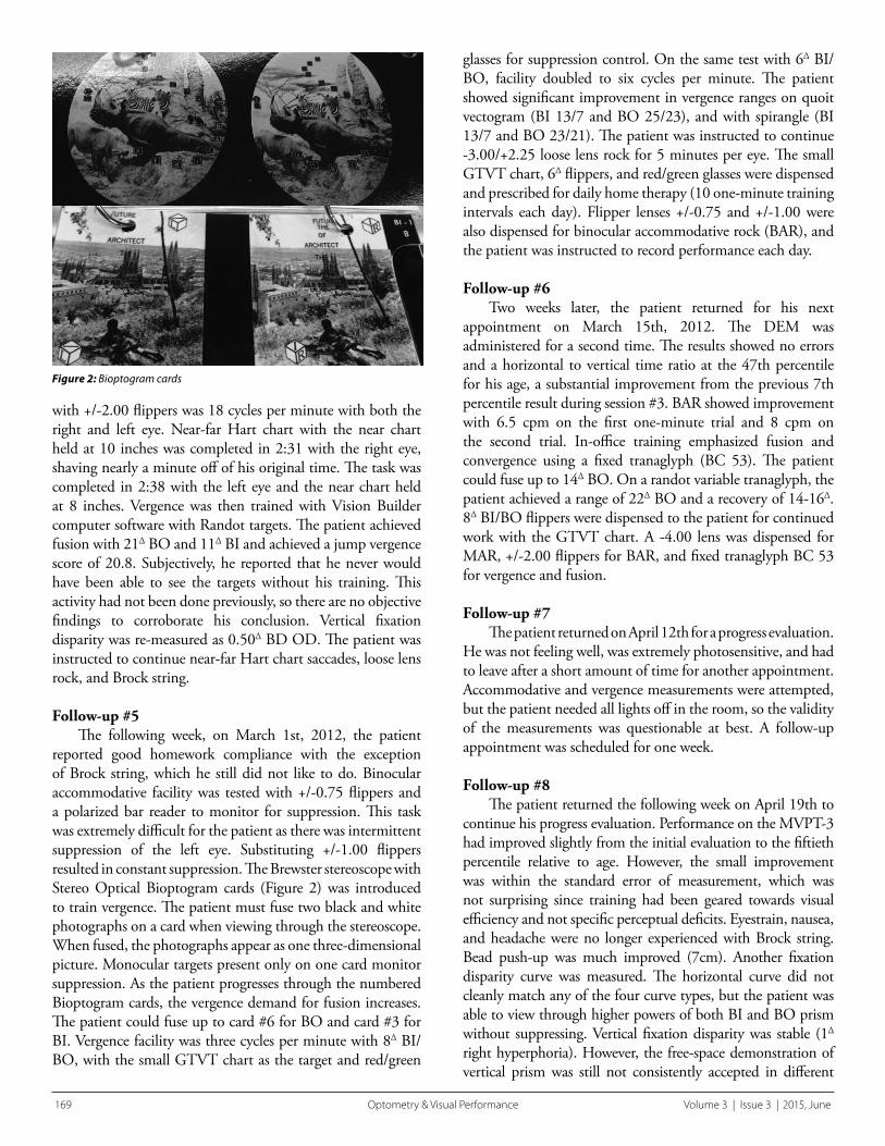
reported good homework compliance with the exception of Brock string, which he still did not like to do. Binocular accommodative facility was tested with +/-0.75 flippers and a polarized bar reader to monitor for suppression. This task was extremely difficult for the patient as there was intermittent suppression of the left eye. Substituting +/-1.00 flippers resulted in constant suppression. The Brewster stereoscope with Stereo Optical Bioptogram cards (Figure 2) was introduced to train vergence. The patient must fuse two black and white photographs on a card when viewing through the stereoscope. When fused, the photographs appear as one three-dimensional picture. Monocular targets present only on one card monitor suppression. As the patient progresses through the numbered Bioptogram cards, the vergence demand for fusion increases. The patient could fuse up to card #6 for BO and card #3 for BI. Vergence facility was three cycles per minute with 8∆ BI/BO, with the small GTVT chart as the target and red/green
glasses for suppression control. On the same test with 6∆ BI/BO, facility doubled to six cycles per minute. The patient showed significant improvement in vergence ranges on quoit vectogram (BI 13/7 and BO 25/23), and with spirangle (BI 13/7 and BO 23/21). The patient was instructed to continue -3.00/+2.25 loose lens rock for 5 minutes per eye. The small GTVT chart, 6∆ flippers, and red/green glasses were dispensed and prescribed for daily home therapy (10 one-minute training intervals each day). Flipper lenses +/-0.75 and +/-1.00 were also dispensed for binocular accommodative rock (BAR), and the patient was instructed to record performance each day.
Follow-up #6Two weeks later, the patient returned for his next
appointment on March 15th, 2012. The DEM was administered for a second time. The results showed no errors and a horizontal to vertical time ratio at the 47th percentile for his age, a substantial improvement from the previous 7th percentile result during session #3. BAR showed improvement with 6.5 cpm on the first one-minute trial and 8 cpm on the second trial. In-office training emphasized fusion and convergence using a fixed tranaglyph (BC 53). The patient could fuse up to 14∆ BO. On a randot variable tranaglyph, the patient achieved a range of 22∆ BO and a recovery of 14-16∆. 8∆ BI/BO flippers were dispensed to the patient for continued work with the GTVT chart. A -4.00 lens was dispensed for MAR, +/-2.00 flippers for BAR, and fixed tranaglyph BC 53 for vergence and fusion.
Follow-up #7The patient returned on April 12th for a progress evaluation.
He was not feeling well, was extremely photosensitive, and had to leave after a short amount of time for another appointment. Accommodative and vergence measurements were attempted, but the patient needed all lights off in the room, so the validity of the measurements was questionable at best. A follow-up appointment was scheduled for one week.
Follow-up #8The patient returned the following week on April 19th to
continue his progress evaluation. Performance on the MVPT-3 had improved slightly from the initial evaluation to the fiftieth percentile relative to age. However, the small improvement was within the standard error of measurement, which was not surprising since training had been geared towards visual efficiency and not specific perceptual deficits. Eyestrain, nausea, and headache were no longer experienced with Brock string. Bead push-up was much improved (7cm). Another fixation disparity curve was measured. The horizontal curve did not cleanly match any of the four curve types, but the patient was able to view through higher powers of both BI and BO prism without suppressing. Vertical fixation disparity was stable (1∆ right hyperphoria). However, the free-space demonstration of vertical prism was still not consistently accepted in different
Figure 2: Bioptogram cards
Volume 3 | Issue 3 | 2015, June Optometry & Visual Performance 170
viewing conditions. The patient was educated that vertical prism increased his depth perception and might reduce his visual symptoms. However, the patient did not perceive the benefit to be great and desired to return to wearing contact lenses. We tinted a trial pair of contact lenses to decrease his photophobia indoors and scheduled a contact lens fitting for the next week.
Follow-up #9The patient returned on May 3rd for a contact lens fitting
and completion of his progress evaluation. He had bought a 3-D TV since his last appointment and felt good about the improvement in his vision and focusing ability. Before the contact lens fitting, binocular accommodative facility was measured at 9 cpm. Frequency 55 lens material is conducive to tinting, so the patient was fitted in Frequency 55/Flat/14.4/ -2.50 in both eyes. Corrected visual acuity in both eyes was 20/20-1. The lenses were centered, moved 0.25 mm with blink, and were comfortable. The patient demonstrated proper insertion and removal of the contact lenses, was educated on proper lens care, and was instructed to discontinue contact lens wear immediately and seek care if redness, pain, or discharge occurred. Vergence facility with 8∆ BI/BO was 4.5 cpm. For home training, the patient was instructed to emphasize vergence facility with the GTVT chart. A homework maintenance program of vergence facility and accommodation exercises was prescribed 20-30 minutes per day, three days per week. The patient was scheduled for a follow-up in two weeks.
Follow-up #10The patient returned on May 17th. He reported that all of
his visual symptoms were improving and that he could read for several hours now without blur or eyestrain. He admitted only doing the maintenance activities once or twice in the last two weeks. With the contact lens trials, he was seeing 20/30 with his right eye initially (20/20-1 later in the exam) and 20/20-1 with his left eye. The contact lenses fit well and did not have any deposits. Examination of the anterior segment and fluorescein evaluation of the cornea was unremarkable. Spherical over-refraction was plano in the right eye and +0.50 DS in the left. The spherical lens OD did not correct for the patient’s
astigmatism. It is possible that the patient was squinting to see more clearly in the initial evaluation. Squinting would have provided maximum benefit if the axis of astigmatism was 180 degrees instead of 30 degrees but may have still helped the patient see more clearly. Though fit and ocular health appeared normal, perhaps the right contact lens should have been taken out, lubricated, re-inserted, and visual acuity re-checked, but the decision was made to attempt to provide the same correction as his spectacle prescription including astigmatism. Stereoacuity with contact lenses was 20”. Vergence facility with 8∆ BI/BO was 8.5 cpm, and binocular accommodative facility was 9 cpm. Maintenance homework of binocular accommodative rock and binocular extended reading with +/-2.00 flippers was prescribed for 20 minutes three days per week. A contact lens follow-up appointment was scheduled for one week, as well as a one-month follow-up to evaluate visual function. In the meantime, contact lenses would be tinted at the Pacific University College of Optometry. Given the initial two-line acuity decrement in the right eye, it was decided to trial a toric lens OD at the next visit. The prescription in the left eye would also be reduced by 0.25 diopter.
Follow-up #11Due to various issues, the patient was next seen on June
21st. He had discontinued his trial contact lenses after a month’s wear. A Frequency 55 Toric 8.4/14.4/-2.25 -0.75 x 020 was fit on the right eye. Due to a lack of flat base curve trial lenses, a Frequency 55 STP/14.4/-2.25 was used for the left eye. The patient reported that this lens felt heavier. Over-refraction of the left eye varied, and best-corrected visual acuity OS was 20/25+2. Comfort, fit, and vision was excellent for the right eye. With the contact lenses, PRA was -2.50/-2.00, and NRA was +3.50/+3.00. Monocular minus lens to blur for the right eye was 3.75D/3.25D and for the left eye was 5.75D/5.25D. Near divergence was 23/24/7 and near convergence was x/34/26. Distance divergence was measured as x/8/2, and convergence was 23/28/16. The patient was encouraged to continue maintenance homework with an emphasis on accommodative exercises for the right eye.
Treatment SummaryThe initial evaluation took two office visits. In-office vision
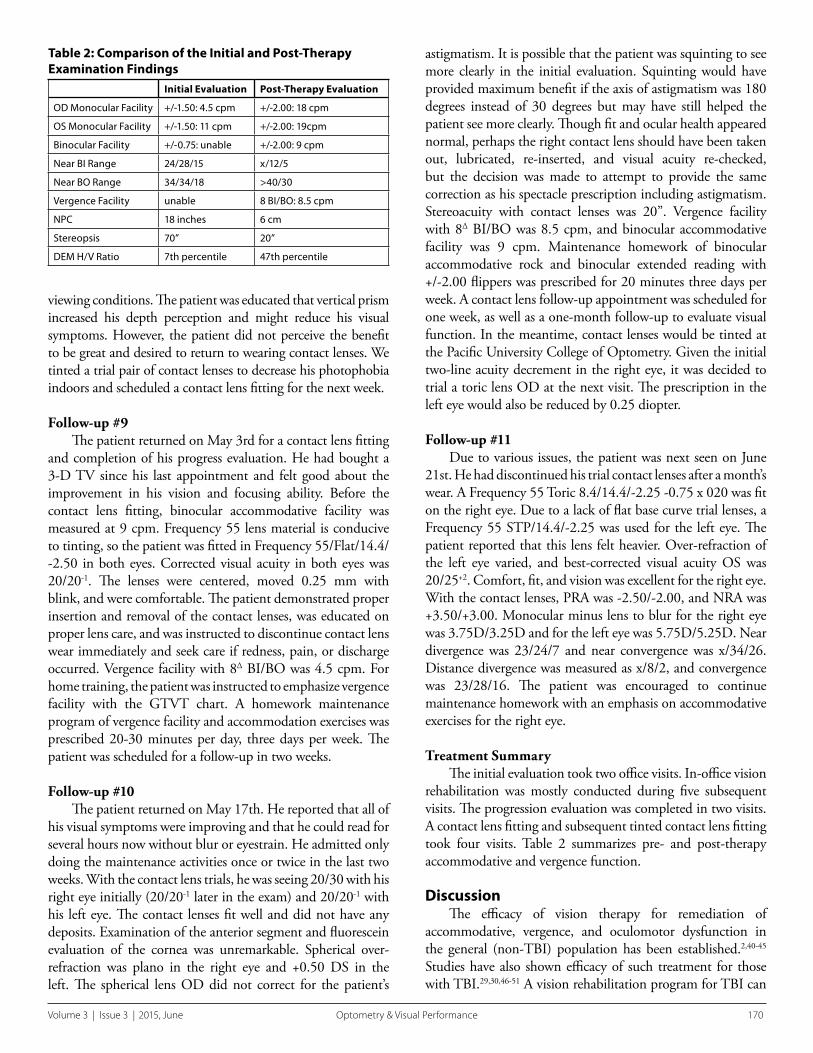
rehabilitation was mostly conducted during five subsequent visits. The progression evaluation was completed in two visits. A contact lens fitting and subsequent tinted contact lens fitting took four visits. Table 2 summarizes pre- and post-therapy accommodative and vergence function.
DiscussionThe efficacy of vision therapy for remediation of
accommodative, vergence, and oculomotor dysfunction in the general (non-TBI) population has been established.2,40-45
Studies have also shown efficacy of such treatment for those with TBI.29,30,46-51 A vision rehabilitation program for TBI can
Table 2: Comparison of the Initial and Post-Therapy Examination Findings
Initial Evaluation Post-Therapy Evaluation
OD Monocular Facility +/-1.50: 4.5 cpm +/-2.00: 18 cpm
OS Monocular Facility +/-1.50: 11 cpm +/-2.00: 19cpm
Binocular Facility +/-0.75: unable +/-2.00: 9 cpm
Near BI Range 24/28/15 x/12/5
Near BO Range 34/34/18 >40/30
Vergence Facility unable 8 BI/BO: 8.5 cpm
NPC 18 inches 6 cm
Stereopsis 70” 20”
DEM H/V Ratio 7th percentile 47th percentile
171 Optometry & Visual Performance Volume 3 | Issue 3 | 2015, June
follow similar training concepts that are used with traditional therapy patients; however, there are special considerations when working with patients who have had a TBI.2 Speech and cognitive impairments can lengthen the training period. Fatigue, depression, and memory deficits can also impact the efficacy of therapy. Performance often fluctuates, with good and bad days throughout rehabilitation. Nausea is more common with procedures. Though there are cases that proceed quickly, in general, training usually takes a longer period of time. Depending on the case, there may be organic neurological damage that will not permit a full visual recovery. However, in the majority of cases, visual rehabilitation can improve or eliminate visual symptoms. Prompt diagnosis and treatment of visual dysfunction is critical to improving activities of daily living and quality of life and working towards re-establishing vocational and avocational goals. Improving visual symptoms can also speed the progress of other rehabilitation therapies such as physical, occupational, speech, and cognitive therapies which require varied visual tasks.52,53
Though this patient’s blurry vision was due to accommo-dative insufficiency, infacility, and gross convergence insufficiency, other causes should be ruled out. Differential diagnosis of accommodative dysfunction following TBI includes accommodative paralysis, damage to the ciliary muscle, dry eye, a cranial nerve palsy, pseudo-myopia, and vergence dysfunction. Pharmacological side effects of medication, illness, and stress should also be considered as possible contributors to accommodative dysfunction.
Initial observation of the patient did not reveal obvious ptosis or a “down and out” eye suggestive of a third nerve palsy. A patient with third nerve involvement may notice monocular blur at near.54 The patient’s extraocular motilities and pupils were normal, making even a partial third nerve palsy an unlikely diagnosis. Slit lamp exam did not indicate dry eye. Accommodative paralysis or ciliary body damage was unlikely because the patient did have the ability to accommodate 5-6 diopters with each eye.
Medication side effects must always be considered when there are visual symptoms. The patient was taking a number of medications with potential visual side effects. Blurred vision is a possible side effect of Diazepam,55 an anti-anxiety medication. Drowsiness, a common side effect of hydrocodone,56 can affect visual stamina. Blurred vision is a less commonly reported side effect of Mirtazapine.57 Though it hasn’t been determined to be a causal relationship, blurred vision has been reported with Verapamil.58 Blurred vision can be a less common but severe side effect of nicotine patches.55 Blurred vision and double vision have been reported with Omeprazole. These reports include events observed in open and uncontrolled studies, reported voluntarily after the approval of Omeprazole, so the role of the medication as a causal factor of blurry or double vision cannot be unequivocally determined.
Post-traumatic stress disorder (PTSD) is known to affect accommodative function. The visual pathway within the brain has interconnections with the hypothalamus and amygdala, which are key players in the limbic system. Emotion associated with memory is generated in the limbic system.59 The hypothalamus controls neuropolypeptides that affect multiple ocular components responsible for clear vision, including the ciliary body muscle.59 Though PTSD and possible medication side effects may be contributing to the patient’s symptoms, they are not necessarily the sole cause of the symptoms. Visual rehabilitation may still improve function.
Basic vision training principals were employed during the visual rehabilitation program. Training began monocularly in order to build the accommodative amplitude and efficiency of each eye individually. The monocular stage also allows the equalization of visual efficiency between the two eyes. Training monocular skills first makes subsequent binocular accommodation and vergence training more effective and successful.2 A visual rehabilitation program should be customized for the individual patient and modified as needed; however, a general progression of training procedures is effective for most patients. Vergence training begins with smooth vergence, a gradual increase in vergence demand, with the goal of increasing vergence ranges. Next, jump vergence is trained with methods that stimulate quick jumps in vergence demand. Training in free space and in different fields of gaze expands the training to more natural viewing conditions. Free space exercises are often prescribed as maintenance homework for a period of time following the more intense visual training program consisting of weekly office visits and daily home exercises. Training time is tapered, and the patient is monitored for decrease in performance.
It should be noted that out of all the accommodative and vergence measures summarized in Table 1, the divergence range at near was the only parameter that did not improve and even decreased. This illustrates a well-known aspect of vergence training: it is much easier to train convergence than divergence. Even with both divergence and convergence training, the patient’s convergence improved, seemingly at the expense of some divergence ability.
ConclusionThis case demonstrates the role and necessity of
rehabilitative vision training for patients with TBI-associated visual symptoms within the practice of optometry. Eye care professionals should be aware of the visual dysfunctions and symptoms commonly associated with TBI in order to provide appropriate care. Measures of accommodative amplitude, accommodative flipper facility, oculomotor function, near point of convergence, and relative vergence ranges can all easily be taken in any clinical setting to readily detect accommodative and vergence dysfunction in patients with TBI. Ongoing wars oversees will likely continue to add to the great number of individuals with traumatic brain
Volume 3 | Issue 3 | 2015, June Optometry & Visual Performance 172
injury and those needing optometric treatment of TBI-associated visual dysfunction.
References1. Thiagarajan P, Ciuffreda KJ, Ludlam DP. Vergence dysfunction in mild traumatic
brain injury (mTBI): A review. Ophthalmic Phsiol Opt 2011;31:456-68.
2. Scheiman M, Wick B. Heterophoric, accommodative, and eye movement disorders. In: Scheiman M, Wick B, eds. Clinical management of binocular vision, 2nd ed. Philadelphia: Lippincott, Williams, and Wilkins, 2002.
3. Traumatic Brain Injury in the United States: Emergency Department Visits, Hospitalizations, and Deaths, 2002-2006. http://www.cdc.gov/TraumaticBrainInjury/tbi_ed.html Last Accessed September 30, 2014.
4. Thurmann D, Alverson C, Dunn K, Guerro J, et al. Traumatic brain injury in the United States: a public health perspective. J Head Trauma Rehab 1999;14:602-15.
5. Fischer H. A Guide to U.S. Military Casualty Statistics: Operation New Dawn, Operation Iraqi Freedom, and Operation Enduring Freedom. 2014. http://1.usa.gov/1P6ndMY. Last Accessed September 30, 2014.
6. Cooper G. Protection of the lung from blast overpressure by thoracic stress wave decouplers. J Trauma 1996;40:105S.
7. Wood G, Panzer M, Shridharani J, Matthews K, et al. Attenuation of blast pressure behind ballistic protective vests. Inj Prev 2012;doi: 10.1136/injuryprev-2011-040277.
8. Warden D. Mild TBI during the Iraq and Afghanistan wars. J Head Trauma Rehabil 2006;21:398-402.
9. Leslie S. Myopia and accommodative insufficiency associated with moderate head trauma. Opt Vis Dev 2009;40:25-31.
10. Green W, Ciuffreda K, Thiagarajan P, Szymanowicz D, et al. Static and dynamic aspects of accommodation in mild traumatic brain injury: A review. Optometry 2010;81:129-36.
11. Ciuffreda KJ, Kapoor N, Rutner D, et al. Occurrence of oculomotor dysfunctions in acquired brain injury: a retrospective analysis. Optometry 2007;78:155-61.
12. Goodrich GL, Kirby J, Cockerham G, Ingalla SP, et al. Visual function in patients of a polytrauma rehabilitation center: a descriptive study. J Rehab Res Dev 2007;44:929-36.
13. Lew HL, Poole JH, Vanderploeg RD, Goodrich GL, et al. Program development and defining characteristics of returning military in a VA polytrauma network site. J Rehab Res Dev 2007;44:1027-34.
14. Stelmack JA, Frith T, Koevering DV, Rinne S, et al. Visual function in patients followed at a Veterans affairs polytrauma network site: an electronic medical record review. Optometry 2009;80:419-24.
15. Brahm KD, Wilgenburg HM, Kirby J, Ingalla S, et al. Visual impairment and dysfunction in combat-injured servicemembers with traumatic brain injury. Optom Vis Sci 2009;86:817-25.
16. Kappor N, Ciuffreda K. Vision disturbances following traumatic brain injury. Curr Treat Opt Neurology 2002;4:271-80.
17. Meythaler JM, Peduzzi JD, Eleftheriouw E, Novak TA. Current concepts: diffuse axonal injury-associated traumatic brain injury. Arch Phys Med Rehabil 2001;82:1461-71.
18. Greve MW, Zink BJ. Pathophysiology of traumatic brain injury. Mt Sinai J Med 2009;76:97-104.
19. Werner C, Engelhard K. Pathophysiology of traumatic brain injury. Br J Anaesth 2007;99:4-9.
20. Center for Neuro Skills. Traumatic Brain Injury, Veterans Health Initiative: Department of Veterans Affairs, Employee Education System http://bit.ly/1P6nijI. January 2004. Accessed May 23, 2012.
21. Sherer M, Struchen MA, Yablon SA, Wang Y, et al. Comparison of indices of traumatic brain injury severity: Glasgow Coma Scale, length of coma and post-traumatic amnesia. J Neurol Neurosurg Psychiatry 2008;79:678-85.
22. Goodrich GL, Kirby J, Cockerham G, et al. Visual function in patients of a polytrauma rehabilitation center: A descriptive study. J Rehab Research Dev 2007;44:929-36.
23. Leslie S. Accommodation in acquired brain injury. In: Suchoff IB, Kapoor N, Ciuffreda KJ, eds. Visual and Vestibular Consequences of Acquired Brain Injury. Santa Ana, CA: Optometric Extension Program Foundation, 2001:56-76.
24. Suchoff IB, Kapoor N, Ciuffreda KJ. An overview of acquired brain injury and optometric implications. In: Suchoff IB, Kapoor N, Ciuffreda KJ, eds. Visual and Vestibular Consequences of Acquired Brain Injury. Santa Ana, CA: Optometric Extension Program Foundation, 2001:1-9.
25. Al-Qurainy IA. Convergence insufficiency and failure of accommodation following midfacial trauma. Brit J Oral Maxillofac Surg 1995;33:71-5.
26. Gianutsos R, Ramsey G, Perlin RR. Rehabilitative optometric services for survivors of acquired brain injury. Arch Phys Med Rehabil 1988;69:573-8.
27. Suchoff IB, Kapoor N, Waxman R, et al. The occurrence of ocular and visual dysfunctions in an acquired brain-injured patient sample. J Am Optom Assoc 1999;5:301-8.
28. Ciuffreda KJ, Kapoor N, Rutner D, et al. Occurrence of oculomotor dysfunctions in acquired brain injury: A retrospective analysis. Optometry 2007;78:155-61.
29. Ciuffreda KJ, Rutner D, Kapoor N, et al. Vision therapy for oculomotor dysfunctions in acquired brain injury: A retrospective analysis. Optometry 2008;79:18-22.
30. Scheiman M, Gallaway M. Vision therapy to treat binocular vision disorders after acquired brain injury: Factors affecting prognosis. In: Suchoff IB, Kapoor N, Ciuffreda KJ, eds. Visual and Vestibular Consequences of Acquired Brain Injury. Santa Ana, CA: Optometric Extension Program Foundation, 2001:89-113.
31. Stark L, Kenyon RV, Krishnan VV, Ciuffreda KJ. Disparity vergence: a proposed name for a dominant component of binocular vergence eye movements. Am J Optom Physiol Opt 1980;57:606-9.
32. Rambold H, Neumann G, Helmchen C. Vergence deficits in pontine lesions. Neurology 2004;62:1850-3.
33. Rambold H, Sander T, Neumann G, Helmchen C. Palsy of fast and slow vergence by pontine lesions. Neurology 2005;63:338-40.
34. Gamlin PD. Neural mechanisms for the control of vergence eye movements. Ann NY Acad Sci 2002;956:264-72.
35. Westheimer G, Blair SM. Oculomotor defects in cerebelectomized monkeys. Invest Ophthalmol Vis Sci 1973;12:618-20.
36. Gamblin PD, Yoon K. An area for vergence eye movement in primate frontal cortex. Nature 2000;407:1003-7.
37. Hasebe H, Oyamada H, Kinomura S. Human cortical areas activated in relation to vergence eye movements – a PET study. Neuroimage 1999;10:200-8.
38. Trotter Y, Celebrini S, Stricanne B, Thorpe S, et al. Neural processing of stereopsis as a function of viewing distance in primate cortical area V1. J Neurophysiol 1996;76:2872-85.
39. Rouse MW, Borsting EJ, Mitchell GL, Scheiman M, et al. Convergence Insufficiency Treatment Trial Group. Validity of the revised convergence insufficiency symptom survey in adults. Ophthalmic Physiol Opt 2004;24:384-90.
40. Griffin JR, Grisham JD. Binocular anomalies: diagnosis and vision therapy, 4th ed. Boston: Butterworth-Heinemann, 2002.
41. Ciuffreda KJ. The scientific basis for and efficacy of optometric vision therapy in non-strabismic accommodative and vergence disorders. Optometry 2002;73:735-62.
42. Cooper J, Selenow A, Ciuffreda KJ, Feldman J. Reduction of asthenopia in patients with convergence insufficiency after fusional vergence training. Am J Optom Physiol Opt 1983;60:982-9.
43. North RV, Henson DB. The effect of orthoptic treatment upon the vergence adaptation mechanism. Optom Vis Sci 1992;69:294-9.
173 Optometry & Visual Performance Volume 3 | Issue 3 | 2015, June
44. Scheiman M, Mitchell GL, Cotter S, et al. A randomized clinical trial of vision therapy/orthoptics vs. pencil pushups for the treatment of convergence insufficiency in young adults. Optom Vis Sci 2005;82:583-95.
45. Brautaset RL, Jennings AJ. Effects of orthoptic treatment on the CA/C and AC/A ratios in convergence insufficiency. Invest Ophthalmol Vis Sci 2006;47:2876-80.
46. Candler R. Some observations on orthoptic treatment following head injury. Br Orthopt J 1944;2:56-62.
47. Cohen AH. Optometric management of binocular dysfunctions secondary to head trauma: case reports. J Am Optom Assoc 1992;63:569-75.
48. Hellerstein LF, Freed S. Rehabilitative optometric management of a traumatic brain injury patient. J Behav Optom 1994;5:143-8.
49. Ludlam WM. Rehabilitation of traumatic brain injury with associated visual dysfunction – a case report. Neuro Rehabil 1996;6:183-92.
50. Berne SA. Visual therapy for the traumatic brain-injured. J Optom Vis Dev 1990;21:13-6.
51. Krohel GB, Kristan RW, Simon JW, Barrows NA. Post-traumatic convergence insufficiency. Ann Ophthalmol 1986;18:101-4.
52. Grosswasser Z, Cohen M, Blankstein E. Polytrauma associated with traumatic brain injury: incidence, nature, and impact on rehabilitation outcome. Brain Injury 1990;4:161-6.
53. Reding MJ, Potes E. Rehabilitation outcome following initial unilateral hemispheric stroke: life table analysis approach. Stroke 1988;19:135-8.
54. Nicotine Patch. http://bit.ly/1c5erfO. Last Accessed March 4, 2015.
55. Chugh JP, Prachi J, Chouhan RS. Third Nerve Palsy: An Overview. Indian Journal of Clinical Practice 2012;22:12.
56. www.rxlist.com. Last Accessed March 4, 2015.
57. Antidepressants for Tension Headaches – Topic Overview. http://wb.md/1Ip6f70. Last Accessed March 4, 2015.
58. Covera HS. http://bit.ly/1ALf44Q. Last Accessed March 4, 2015.
59. Trachtman JN. Post-traumatic stress disorder and vision. Optometry 2010;81:240-52.
Correspondence regarding this article should be emailed to Anna Griffith, OD at [email protected]. All statements are the author’s personal opinions and may not reflect the opinions of the the representative organizations, ACBO or OEPF, Optometry & Visual Performance, or any institution or organization with which the author may be affiliated. Permission to use reprints of this article must be obtained from the editor. Copyright 2015 Optometric Extension Program Foundation. Online access is available at www.acbo.org.au, www.oepf.org, and www.ovpjournal.org.
Griffith A. Treatment of accommodative and vergence dysfunction in traumatic brain injury (TBI): A case report. Optom Vis Perf 2015;3(3):165-73.
The online version of this article contains digital enhancements.