article in press - core.ac.uk · research article...
TRANSCRIPT
E X P E R I M E N T A L C E L L R E S E A R C H X X ( 2 0 0 9 ) X X X – X X X
YEXCR-08141; No. of pages: 13; 4C: 3, 4, 6, 7, 8, 9
ava i l ab l e a t www.sc i enced i r ec t . com
www.e l sev i e r. com/ loca te /yexc r
ARTICLE IN PRESS
Research Article
Engraftment ofmesenchymal stem cells into dystrophin-deficientmice is not accompanied by functional recovery
Eun Ji Ganga, Radbod Darabia,c, Darko Bosnakovskia,c, Zhaohui Xua, Kristine E. Kammb,Michael Kybaa,c, Rita C.R. Perlingeiroa,c,⁎aDepartment of Developmental Biology, University of Texas Southwestern Medical Center, 5323 Harry Hines Blvd, Dallas 75390-9133, Texas, USAbDepartment of Physiology, University of Texas Southwestern Medical Center, 5323 Harry Hines Blvd, Dallas 75390-9133, Texas, USAcLillehei Heart Institute, Department of Medicine, University of Minnesota, 312 Church St. S.E., Minneapolis 55455, Minnesota, USA
A R T I C L E I N F O R M A T I O N
⁎ Corresponding author. Lillehei Heart InstituMN, USA. Fax: +612 624 8118.
E-mail address: [email protected] (R.C.R. P
0014-4827/$ – see front matter © 2009 Elseviedoi:10.1016/j.yexcr.2009.05.009
Please cite this article as: E.J. Gang, et al., Eby functional recovery, Exp. Cell Res. (2009
A B S T R A C T
Article Chronology:
Received 16 February 2009Revised version received 5 May 2009Accepted 6 May 2009
Mesenchymal stem cell preparations have been proposed for muscle regeneration in
musculoskeletal disorders. Although MSCs have great in vitro expansion potential and possessthe ability to differentiate into several mesenchymal lineages, myogenesis has proven to be muchmore difficult to induce. We have recently demonstrated that Pax3, the master regulator of theembryonic myogenic program, enables the in vitro differentiation of a murine mesenchymal stemcell line (MSCB9-Pax3) into myogenic progenitors. Here we show that injection of these cells intocardiotoxin-injured muscles of immunodeficient mice leads to the development of muscle tumors,resembling rhabdomyosarcomas. We then extended these studies to primary humanmesenchymal stem cells (hMSCs) isolated from bone marrow. Upon genetic modification with alentiviral vector encoding PAX3, hMSCs activated the myogenic program as demonstrated byexpression of myogenic regulatory factors. Upon transplantation, the PAX3-modified MSCs did notgenerate rhabdomyosarcomas but rather, resulted in donor-derived myofibers. These were found
at higher frequency in PAX3-transduced hMSCs than in mock-transduced MSCs. Nonetheless,neither engraftment of PAX3-modified or unmodified MSCs resulted in improved contractility.Thus these findings suggest that limitations remain to be overcome beforeMSC preparations resultin effective treatment for muscular dystrophies.
© 2009 Elsevier Inc. All rights reserved.
Keywords:
Mesenchymal stem cellsPax3Muscle differentiationDystrophinTransplantationTumors
Introduction
Muscular dystrophies are genetically and clinically heterogeneousdisorders characterized by progressiveweakness and degenerationof the skeletal muscles that control movement. Among thesedisorders, Duchenne muscular dystrophy (DMD) is the mostcommon, affecting 1 out of every 3500 live born boys. DMD is anX-linked recessive muscle disorder caused by mutations in the
te, University of Minnesota
erlingeiro).
r Inc. All rights reserved.
ngraftment of mesenchym), doi:10.1016/j.yexcr.200
dystrophin gene, resulting in the absence of dystrophin in skeletalmuscle and other tissues [1–3]. Current treatment involvespharmacological control of inflammation which amelioratessymptoms but does not cure or extend the life expectancy ofaffected patients.
Bone marrow transplantation is at the vanguard of cellulartherapies and it has beenwidely and successfully used in clinics forthe treatment of congenital, malignant, and degenerative diseases
, 4-124 Nils Hasselmo Hall, 312 Church St. S.E., Minneapolis 55455,
al stem cells into dystrophin-deficient mice is not accompanied9.05.009

2 E X P E R I M E N T A L C E L L R E S E A R C H X X ( 2 0 0 9 ) X X X – X X X
ARTICLE IN PRESS
of the bone marrow (BM). During the last decade, a flurry ofcontroversial studies has exploited the plasticity of bone marrowcells and their therapeutic potential on non-hematological dis-orders [4–9], including muscular dystrophies [10–15]. Ferrari et al.were the first to document that BM cells had the ability to migratefrom the circulation into cardiotoxin-injured muscles, and partici-pate in skeletal muscle regeneration. These results were furtherconfirmed by several investigators that applied BM transplantationtomdx mice, an animal model for DMD [11–13]. Despite the initialenthusiasm, quantification of engraftment revealed that thenumber of donor-derived myofibers in transplanted mice wasrather low (<1%) [11,16], leading to questioning of the biologicaland therapeutic relevance of these findings. As observedwith othercell lineages, it is possible that the low level engraftment observedinmuscle tissues results solely fromcell fusion rather than fromBMplasticity [17–22]. It is also plausible to assume that a cellpopulation with limited myogenic potential was transplanted inthese experiments, given that most reports described above madeuse of whole BM, which is highly heterogeneous.
BM is well-known as a source of hematopoietic stem cells, but italso contains a stromal cell population, known as mesenchymalstem cells or marrow stromal cells (MSCs), that is endowed withthe ability to self-renew and differentiate into multiple mesench-ymal lineages, including bone, fat, cartilage, and connective tissue[23–25]. Experimental approaches involving induction of MSCswith 5-azacytidine [26], co-culture of MSCs with myoblasts [27] ormyoblast-conditioned medium [28], and more recently activatedNotch [29], suggest that skeletal muscle progenitors can also beobtained from MSCs. Although MSCs represent less than 0.01% ofthe BMmononuclear cell fraction, they are easily expanded in vitroand thus, amenable to genetic manipulation. We have recentlydemonstrated that by overexpressing Pax3, the master regulator ofthe myogenic program, a mesenchymal stem cell line can bespecifically reprogrammed to differentiate into the myogeniclineage [30]. These results led us to test their myogenic potentialin vivo as well as to investigate whether PAX3 is able to reprogramprimary human MSCs, and whether resulting reprogrammed cellsare capable of promoting muscle regeneration in vivo.
Materials and methods
MSC isolation and culture
Human BM samples were purchased from Cambrex. hMSCs wereisolatedaccording to previously describedprotocol [25]. Briefly, cells
Table 1 – Specific human sense and anti-sense primers for quanti
Gene Primer sense sequence
hMYOD CACTCCGGTCCCAAATGTAGhMYF5 GATTCACAGCCTCGAACTCChMYOGENIN TAGCAGGGGCCTCCTAAGChMHC ATGGCAGGTCTGGATGAAAChGAPDH CATCTTCCAGGAGCGAGAT
MHC, myosin heavy chain type IIb.
Please cite this article as: E.J. Gang, et al., Engraftment of mesenchymby functional recovery, Exp. Cell Res. (2009), doi:10.1016/j.yexcr.200
were separated from 10 to 20 ml of marrow diluted with fourvolumes of Minimum Essential Medium Alpha Medium (α-MEM,Invitrogen) containing 10% FBS (Atlanta Biologicals) by centrifuga-tion at 900 g for 15 min. Cells were washed with cold Dulbecco'sphosphate-buffered saline (DPBS) once and plated at 2×10⁶ cellsper cm2 inMSCexpansionmediumconsistingof low-glucoseDMEMsupplemented with 40% MCDB-201 (Invitrogen), 10% FBS, 1×insu-lin–transferrin–selenium (ITS; Sigma), 1×linoleic-acid–bovine-serum-albumin (Sigma), 10−8 M dexamethasone (Sigma), 10−4 Mascorbic acid 2-phosphate (Sigma), 50 U/ml penicillin/streptomy-cin (Invitrogen), 10 ng/ml of hPDGF-BB (Peprotech), and 10 ng/mlof hEGF (Peprotech). After 72 h of culture, suspended cells wereremoved and replaced with fresh media. Medium was changedevery 3 days thereafter. hMSCs grew as colonies which weredetached with 0.1% trypsin–EDTA and sub-cultured at a density of4–8×10³ cells per cm2.
Lentiviral infection and purification of GFP+ mesenchymalstem cells
The multiple cloning site (BglII–EcoRI–NotI–SalI) of pBS was PCR-amplified and cloned into pGEM-T (Promega) to generate pGEM-BS. pGEMiresGFP was generated by inserting 1.3 kb SalI-digestediresGFP fragment from pMSCViresGFP [31] into the SalI site ofpGEM-BS. The lentiviral vector pSAMwas generated by inserting a1.3 kb BglII/NdeI-digested and filled-in iresGFP fragment frompGEMiresGFP into the BamHI/EcoRI-digested and filled-in cloningsite of FUGW [32].We refer to this vector as pSAM. Lentiviral vectorpSAM-Pax3 was created by sub-cloning the cDNA for Pax3obtained from pSPORT-Pax3 (BC048699, Open Biosystems) intopSAM as an EcoR I/BamH I digested and BamH I filled-in fragment.This vector was co-transfected with packaging and coat proteinconstructs Δ8.91 and pVSVG, respectively, into 293T cells using theFuGENE 6 transfection reagent (Roche) as recommended by themanufacturer. Virus-containing supernatant was collected 48 hafter transfection and filtered through a 0.45 μm filter before use.MSC cells were suspended in 3 ml of viral supernatant containing10 ng/ml hPDGF-BB, 10 ng/ml hEGF, and 8 μg/ml polybrene(Sigma). Cells were then transferred to 6-well plates andcentrifuged at 2500 rpm for 90 min at 33 °C on a Hettichcentrifuge. After a 4 h-incubation at 37 °C with 5% CO2 in air,supernatant was removed and changed with fresh MSC expansionmedium as described above. Two days following infection, cellswere harvested and sorted based on the gating of GFP+ on the FITCchannel. GFP+ hMSCs weremaintained in MSC expansionmediumfor further in vitro and in vivo studies.
tative real-time PCR analysis.
Primer anti-sense sequence Product size (bp)
TTCCCTGTAGCACCACACAC 180TGAAGCCTTCTTCGTCCTGT 125CTCTGGTCCCCTGCTTTACC 193CCAAGGACCCTTCAAGATCA 172TGCAAATGAGCCCCAGCCTT 114
al stem cells into dystrophin-deficient mice is not accompanied9.05.009

Fig. 1 – Development of rhabdomyosarcoma following the transplantation of MSCB9-Pax3 cells. (A) MSCB9-Pax3 or MSCB9-Vector(MSCB9-Vec) cells were injected into cardiotoxin-injured TAmuscles of Rag2−/− γc−/− mice. Contralateral TA muscles were used ascontrols. Engrafted cells (LacZ+) were detected at 4-week (i. upper two panels) and 8-week post-transplantation (i. lower twopanels) by X-gal staining. These latter samples were also examined by H&E staining (ii. lower panel). (B) Immunofluorescentstaining ofmuscle cryosections at 4 weeks post-transplantation forMyoD andMyogenin (stainingwith Cy3 in red); direct GFP signal(green); merge for GFP and MyoD or Myogenin (cells expressing both are seen in yellow/orange); DAPI staining in blue, and mergefor DAPI and MyoD or Myogenin (cells expressing both are seen in purple). Pictures were taken at 200× (A.i and B) and 600× (A.ii).Scale bar is 100 μm.
3E X P E R I M E N T A L C E L L R E S E A R C H X X ( 2 0 0 9 ) X X X – X X X
ARTICLE IN PRESS
Please cite this article as: E.J. Gang, et al., Engraftment of mesenchymal stem cells into dystrophin-deficient mice is not accompaniedby functional recovery, Exp. Cell Res. (2009), doi:10.1016/j.yexcr.2009.05.009

4 E X P E R I M E N T A L C E L L R E S E A R C H X X ( 2 0 0 9 ) X X X – X X X
ARTICLE IN PRESS
Western blot
Western blots were performed using the following antibodies:mouse monoclonal anti-Pax3 (R and D Systems), and mousemonoclonal anti-actin (Sigma).
FACS analysis of GFP+ cells
Pax3- or Vector control-transduced MSCs at passage 2 weretrypsinized with 0.1% Trypsin–EDTA and washed with blockingbuffer (DPBS with 2% FBS), suspended in the same buffercontaining 0.25 μg/106 cells of Fc block (Pharmingen) and placedon ice for 5 min. Allophycocyanin (APC)- or Phycoerythrin (PE)-conjugated mouse antibodies to human markers including CD73,CD44, CD90, CD45, CD34 (Pharmingen), CD105 (Serotec), and
Fig. 2 – Activation of the myogenic program in PAX3-expressing hMsorted cells in hMSCs transduced with PAX3 or vector control. GFP iwere sorted according to represented gates and cultured in MSC ex100 μm. (B) Immunofluorescence for PAX3 (Cy3, red) in hMSC-PAXtaken at 400×. Scale bar is 100 μm. (C) Western blot analysis for PAanalysis by real-time PCR for myogenic-specific markers in hMSC-PGAPDH (y axis). Error bars indicate standard errors from 3 indepen
Please cite this article as: E.J. Gang, et al., Engraftment of mesenchymby functional recovery, Exp. Cell Res. (2009), doi:10.1016/j.yexcr.200
CD56 (NCAM; BioLegend) were added at 1 μg/106 cells andincubated at 4 °C for 20 min before washing with blocking buffer.As matching isotype controls, APC- or PE-conjugated mouse anti-IgG antibodies (Pharmingen) were used. Cells were then analyzedon FACS Aria (Becton-Dickinson) after addition of propidiumiodide (Pharmingen) to exclude dead cells.
Myogenic induction
For myogenic differentiation, cells were cultured for up to 3 weeksin several muscle inductive conditions as follows: LG-DMEMsupplemented with 10% FBS, bFGF (10 ng/ml; Peprotech),neuregulin (200 ng/ml; Peprotech), hPDGF-AA (5 ng/ml; Pepro-tech), and forskolin (FSK, 5 μM; Calbiochem); LG-DMEM supple-mented with 2% horse serum (HS, Atlanta Biologicals) and 1×ITS,
SCs. (A) FACS profile for GFP expression and morphology ofs indicated on x axis, and autofluorescence on y axis. GFP+ cellspansion medium. Pictures were taken at 200×. Scale bar is3 and hMSC-Vector cells. DAPI staining in blue. Pictures wereX3 in hMSC-PAX3 and hMSC-Vector cells. (D) Gene expressionAX3 and hMSC-Vector cells. Transcripts were normalized todent experiments.
al stem cells into dystrophin-deficient mice is not accompanied9.05.009
5E X P E R I M E N T A L C E L L R E S E A R C H X X ( 2 0 0 9 ) X X X – X X X
ARTICLE IN PRESS
and LG-DMEM supplemented with 10% FBS and 5-azacytidine(5 μM; Sigma); as controls, we also evaluated cells that weremaintained in MSC expansion medium (with and without growthfactors) and α-MEM supplemented with 16.5% FBS.
RNA isolation and real-time quantitative PCR analysis
Total RNAwas isolated using Trizol (Invitrogen) as recommended bythemanufacturer. First strand cDNAwas produced using Superscript
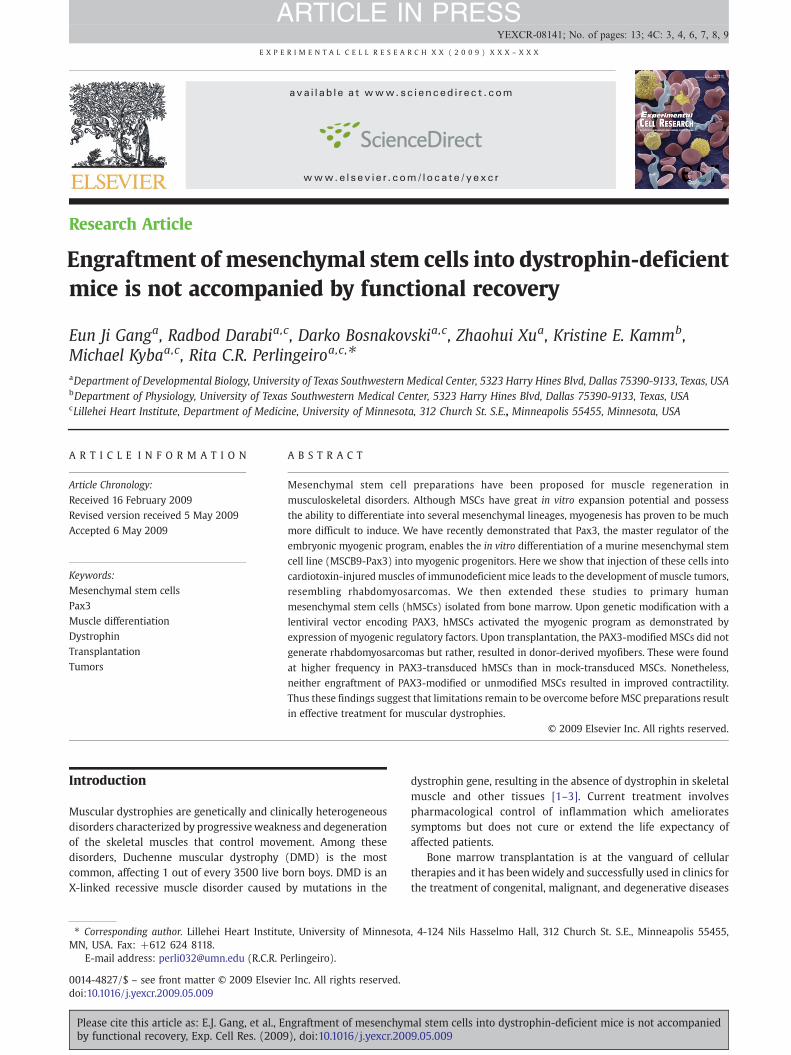
Fig. 3 – Characterization of hMSCs-PAX3. Flow cytometric analysesisotype or secondary control staining profile (thin line) versus specthe fraction of cells that express a given surface antigen. This analy
Please cite this article as: E.J. Gang, et al., Engraftment of mesenchymby functional recovery, Exp. Cell Res. (2009), doi:10.1016/j.yexcr.200
II reverse transcriptase (Invitrogen) with Oligo dT. For gene expres-sion assays, 2 μl of cDNA from reverse-transcription of total RNA as atemplate was amplified using SYBR Green Master Mix reagent(Applied Biosystems) and 7500 Real-Time PCR System (AppliedBiosystem). The muscle-specific primers for human MYOD, MYF5,MYOGENIN, MHC, GAPDH and PCR product sizes are described inTable 1. For PAX3, a probe setwas acquired fromApplied Biosystems.The data are reported as a ratio of absolute mRNA copy number ofeach specific gene to the absolute copy number of GAPDH.
of hMSC-PAX3 and hMSC-Vector cells at passage 2. Plots showific antibody staining profile (thick line). Percentages representsis was performed twice.
al stem cells into dystrophin-deficient mice is not accompanied9.05.009
6 E X P E R I M E N T A L C E L L R E S E A R C H X X ( 2 0 0 9 ) X X X – X X X
ARTICLE IN PRESS
Animals and transplantation
Animal experiments were carried out according to protocolsapproved by the UT Southwestern Medical Center InstitutionalAnimal Care and Use Committee and met US National Institutes ofHealth guidelines for the humane care of animals. Six- to eight-week-old Rag2−/− γc−/− immunodeficient mice (Taconic labora-tories) and C57 BL/10ScSN-Dmdmdx/j (X-linked muscular dystro-phy; Jackson Laboratory) were used as recipients for thetransplantation study. One day before transplantation, 75 μl ofcardiotoxin (10 μM, Sigma) was injected into both tibialis anterior(TA)muscles of eachmouse to inducemuscle injury. 24h later, Pax3-or Vector-transducedMSCB9 or primary humanmesenchymal stemcells were injected (1×10⁶ in 50 μl of phosphate-buffered saline)into the right TAmuscles of each group. As control, 50 μl of PBSwereinjected in the left TA muscle of animals. For immuno-suppression,mdx mice received a daily dose of 5 mg/kg FK 506 (Tacrolimus;Sigma) intra-peritoneally (IP) from the day before cell injection untilthe time of euthanasia (4 weeks after transplantation).
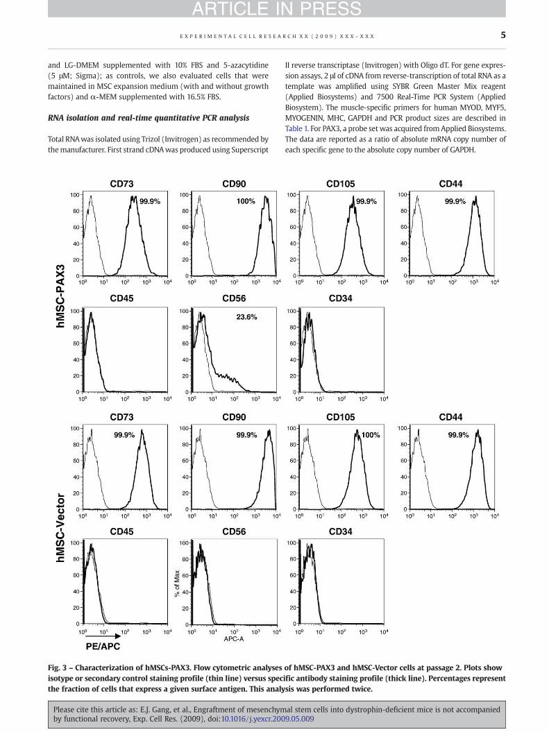
Fig. 4 – Effects of several induction protocols on the myogenic diffhMSC-PAX3 and hMSC-Vector cells after 4 weeks under several myoexpression profile by real-time PCR analysis. Transcripts were normfrom 4 independent experiments. (C) Immunofluorescent stainingcells grown under expansion (#1) or myogenic induction (#2, bFGFArrows indicate nuclei within the myotube. All photographs were taMHC+ in these hMSC-PAX3 cultures. Error bars indicate standard e
Please cite this article as: E.J. Gang, et al., Engraftment of mesenchymby functional recovery, Exp. Cell Res. (2009), doi:10.1016/j.yexcr.200
Immunofluorescent staining of cultured cells and tissuesections
TA muscles were isolated, embedded in OCT compound (Tissue-Tek) and immediately frozen in isopentane cooled in liquidnitrogen. Serial cryostat sections (8–12 μm) were collected. Forimmunofluorescence staining, cells cultured on slides and tissuecryosections were fixed using 4% paraformaldehyde/PBS (acetonewas applied for dystrophin staining), permeabilized with 0.5%Triton X-100 (Sigma), and blocked with 10% goat serum (or 3%BSA), and then incubated with primary antibodies including GFP(Molecular Probes), Pax3 (R and D Systems), MyoD (both from BDBiosciences), MHC (Developmental Studies Hybridoma Bank), anddystrophin (Abcam). Cy2 and Cy3 (Jackson ImmunoresearchLaboratories) secondary antibodies were used. DAPI (4,6-diami-dino-2-phenylindole; Fluka) was used to counter-stain nuclei.Nonspecific isotype control antibodies were used as negativecontrols. X-gal staining was performed for detection of LacZ+ cells.Sections were fixed with 0.2% glutaraldehyde in washing buffer
erentiation of hMSCs. (A) Representative morphologies ofgenic induction protocols and, (B) their respective genealized to GAPDH (y axis). Error bars indicate standard errorsfor MYOD and MHC (Cy3, red) in hMSC-PAX3 and hMSC-Vectorand neuregulin) medium. Cells are co-stained with DAPI (blue).ken at 200×. Scale bar is 100 μm. (D) Percentage of MYOD+ andrrors from 2 independent experiments.
al stem cells into dystrophin-deficient mice is not accompanied9.05.009

Fig. 4 (continued).
7E X P E R I M E N T A L C E L L R E S E A R C H X X ( 2 0 0 9 ) X X X – X X X
ARTICLE IN PRESS
(5mMEGTA, 2mMMgCl2, 0.1 M sodium phosphate buffer, pH 7.3),rinsed with washing buffer and incubated overnight at 37 °Cin X-gal solution (2 mM 5-bromo-4-chloro-3-indolyl-b-D-galacto-pyranoside/dimethyl-formamide, 4 mM K3Fe(CN)6, 4 mM K4Fe(CN)6U3H2O and 1 mM MgCl2 in washing buffer. Sections werethen counterstained with Hematoxylin and Eosin (H&E) staining,as described [33].
Please cite this article as: E.J. Gang, et al., Engraftment of mesenchymby functional recovery, Exp. Cell Res. (2009), doi:10.1016/j.yexcr.200
Muscle preparation for mechanical studies
For the measurement of contractile properties, the mice wereanaesthetizedwith sodiumpentobarbitone (70mg/kg I.P.) and intacttibialis anterior (TA) muscles were dissected and placed in anexperimental organ bath filled with mammalian Ringer solutioncontaining (mM):NaCl 120.5;NaHCO3 20.4; glucose 10;KCl 4.8; CaCl2
al stem cells into dystrophin-deficient mice is not accompanied9.05.009
8 E X P E R I M E N T A L C E L L R E S E A R C H X X ( 2 0 0 9 ) X X X – X X X
ARTICLE IN PRESS
1.6; MgSO4 1.2; NaH2PO4 1.2; pyruvate 1.0, adjusted to pH 7.4. Thechamber was perfused continuously with 95% O2–5% CO2 andmaintained at a temperature of 25 °C. The muscles were stimulatedbyanelectric fieldgeneratedbetween twoplatinumelectrodesplacedlongitudinally on either side of the muscle (square wave pulses 25 V,0.2 ms in duration, 150 Hz). Muscles were adjusted to the optimumlength (Lo) for the development of isometric twitch force and a 5minrecovery period was allowed between stimulations. Optimal musclelength (Lo) and stimulation voltage (25 V) were determined frommicromanipulation of muscle length and a series of twitch contrac-tions that produced maximum isometric twitch force. In brief, afterdetermination of optimal muscle length (Lo) and measurement ofmaximum isometric tetanic force, total muscle fiber cross-sectionalarea (CSA)was calculated bydividingmusclemass (milligram)by theproduct of fiber length (millimeter) and 1.06mg/mm3, the density ofmammalian skeletal muscle. Specific force (sF0) was determined bynormalizing maximum isometric tetanic force to CSA.
Statistical analysis
Differences between samples were assessed by using the Student'stwo-tailed t test for independent samples.
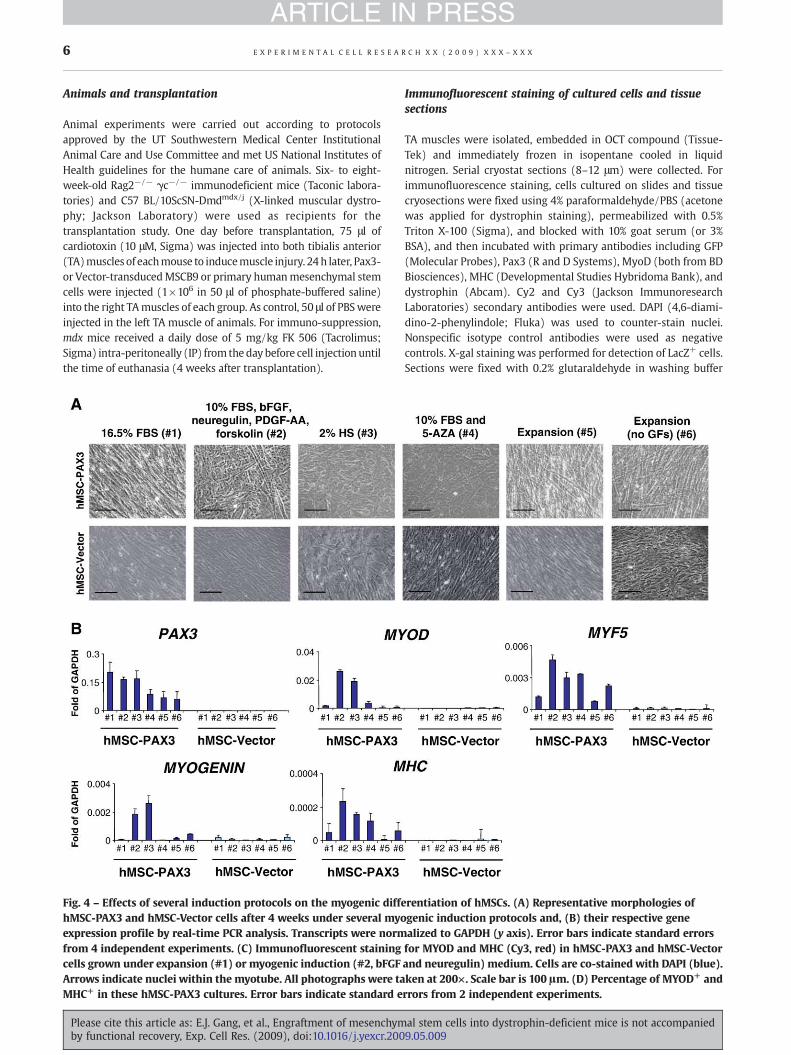
Fig. 5 – Engraftment of hMSCs is improved in the presence ofPAX3. Human mesenchymal stem cell preparations transducedwith PAX3 or Vector only were transplanted into tibialisanterior (TA) muscles from mdx mice that had been previouslyinjured with cardiotoxin. (A) Dystrophin expression (in red) inwild-type mice (top row) and untreated mdx mice (secondrow). Third and fourth rows represent detection of dystrophin+
muscle fibers in mdx mice 4 weeks after transplantation withhMSC-PAX3 and hMSC-Vector cells, respectively. Scale bar is100 μm. (B) Percentage of Dystrophin+ myofibers per field intransplanted muscles. For each group, 80 high power fields(200×) were selected and photographed at random areas, andmyofiber number was recorded. The number in the parenthesisindicates the number of mice per group. ⁎P< 0.01.
Results
Rhabdomyosarcomas upon transplantation of MSCB9-Pax3cells
We have previously shown that Pax3 enables the commitment of amurinemesenchymal stem cell line toward themyogenic lineage invitro [30]. We transplanted these cells into cardiotoxin-injuredmuscles of immunodeficient mice, and observed tumors arise by4 weeks post-transplantation. These tumors were of donor origin,as evidenced by LacZ staining and GFP fluorescence (Figs. 1A, B).H&E staining of these cryosections revealed cell death (necrosis) inabundance as well as a large number of small rounded cellscharacterized by large nuclei and little cytoplasm (Fig. 1A.ii, lowerleft panel). Pathological analyses of these samples classified thesetumors as rhabdomyosarcomas. Indeed these very poorly differ-entiated neoplasms were positive for MyoD and Myogenin(Fig. 1B), a profile also characteristic of rhabdomyosarcoma[34–36]. Rhabdomyosarcomas (RMS) represent the most commonsoft tissue tumors affecting children and young adolescents, andcan be sub-divided in twomain categories, embryonal and alveolar.The latter is generallymore aggressive and can be identified inmostinstances by the presence of the fusion oncogene Pax3/Forkhead orPax7/Forkehead resulting from reciprocal chromosomal transloca-tions [37,38]. Although we did not observe the development oftumors inmice injectedwithMSCB9-Vector control cells at 4weeks(Fig. 1A.i) or at 8 weeks (Supplementary Fig. 1) after thetransplantation, these cells were found at increasing levels asmononuclear cells in recipient muscles over time (SupplementaryFig. 1), suggesting they may eventually develop a tumor.
PAX3 promotes the activation of the myogenic program inprimary hMSCs
To assess whether the generation of rhabdomyosarcoma in vivowas due specifically to Pax3 or a combinatory effect of Pax3 with
Please cite this article as: E.J. Gang, et al., Engraftment of mesenchymby functional recovery, Exp. Cell Res. (2009), doi:10.1016/j.yexcr.200
the use of MSCB9, an immortalized cell line [39], we investigatednext the ability of Pax3 to reprogram primary mesenchymal stemcells into myogenic progenitors, and whether these cells in vivowould develop rhabdomyosarcoma or produce muscle engraft-ment. For this purpose, we transduced Pax3 into primary mouseMSC preparations. Although these cells were amenable to
al stem cells into dystrophin-deficient mice is not accompanied9.05.009

9E X P E R I M E N T A L C E L L R E S E A R C H X X ( 2 0 0 9 ) X X X – X X X
ARTICLE IN PRESS
infection and MyoD could be detected in Pax3-transduced cultures(data not shown), after isolation, transduction, and sorting, cellswere left with limited proliferative potential, with or withoutPax3. This was probably due to the fact that cells were approachingsenescence.
Based on these results, we decided to transduce primaryhuman MSC preparations, which are known to grow much betterthan their mouse counterparts. Experiments were conducted withMSC preparations obtained from two independent human BMsamples. Two days following lentiviral infection, both hMSC-PAX3and control vector-transduced hMSC were FACS sorted for GFP+
cells (Fig. 2A, upper panel). In the presence of MSC expansionmedium, both sorted cell fractions, hMSC-PAX3 and hMSC-Vector,proliferated at a similar rate and presented no apparent morpho-logical differences (Fig. 2A, lower panel). Expression of PAX3 byhMSC-PAX3 but not hMSC-Vector cells was confirmed by immu-nofluorescent staining and Western blot analysis (Figs. 2B, C).Gene expression analyses for myogenic regulatory factors (MRFs)demonstrated up-regulation of MYOD in hMSC-PAX3 cultures(Fig. 2D). MYF5 and MYOGENIN, as well as MYOSIN HEAVY CHAIN(MHC), a marker of terminal muscle differentiation, were barelydetectable (Fig. 2D) under these expansion culture conditions.Both PAX3-transduced and control MSC preparations werenegative for CD45 and CD34, while maintaining high expressionlevels of CD73, CD90, CD105, and CD44, a profile characteristic ofMSCs (Fig. 3). The only antigen found to be expressed differentiallyin hMSC-PAX3 was CD56, a known marker for myogenicprogenitors. While undetectable in hMSC-Vector cells, CD56 wasup-regulated in PAX3-transduced MSCs (23.6%; Fig. 3).
To test their muscle differentiation potential in vitro, wecultured hMSC-PAX3 and hMSC-Vector cells under several muscleinductive conditions (#2, 10% FBS, bFGF, neuregulin, PDGF-AA and
Fig. 6 – MSCs fail to promote in vivo functional recovery. Values showmice per group±SEM. (A) Representative example of force tracingtransplanted mdx mice. Blue and red lines show force tracing from(control), respectively. Muscles at L0 were stimulated for 2 s with a 1recorded. (B) Effect of cell transplantation on specific force (sF0:F0 n(E) Number of myofibers per field in analyzed muscles. For each grphotographed and myofiber number was recorded in a blinded fas
Please cite this article as: E.J. Gang, et al., Engraftment of mesenchymby functional recovery, Exp. Cell Res. (2009), doi:10.1016/j.yexcr.200
forskolin) [29]; #3, 2% HS [40], and #4, 10% FBS and 5-azacitidinefor up to 4 weeks [26] (Fig. 4), along with control conditions,including basic MSC medium (#1, 16.5% FBS) [39], completeexpansion medium (#5, 10% FBS and PDGF-BB/EGF) [39], andexpansion medium without growth factors (#6). This lattercondition was investigated to assess whether growth factorwithdrawal would allow the differentiation of hMSC-PAX3 andhMSC-Vector cells into myogenic cells. From all these conditions,myogenic differentiation was more prominent with mediumcontaining bFGF, neuregulin, PDGF-AA and forskolin, and theinduction medium containing 2% HS (conditions 2 and 3,respectively), as evidenced by gene expression analysis whichrevealed up-regulation of MYOD, MYF5, MYOGENIN, and to a lesserextent, MHC (Fig. 4B). Irrespective of the induction cultureconditions, myogenic regulatory factors were scarcely detectablein hMSCs-Vector control cells. These results were corroborated byimmunofluorescent staining for MYOD and MHC comparinghMSC-PAX3 to hMSCs-Vector cells (Fig. 4C), in the presence ofproliferation (16.5% FBS, #1) and induction medium containingbFGF, neuregulin, PDGF-AA, and forskolin (#2). The inductivedifferentiation condition (#2) resulted in the generation ofMYOD+
and MHC+ cells (23.84%±3.22% and 4.5%%±1.7%, respectively),with a fusion index of 3.1%±1.8%.
PAX3-modified primary human MSCs do not generaterhabdomyosarcomas in vivo
To test the myogenic differentiation potential of hMSC-PAX3 cellsin vivo, we initially transplanted these cells as well as hMSC-Vectorcontrol cells into the TA muscles of CTX-injured Rag2−/− γc−/−
immunodeficient mice. At 8 weeks post-transplantation ratherthan rhabdomyosarcoma, as previously observed with MSCB9-
n are the results of 3 independent experiments on a total of 14in TA muscles from hMSC-Vector- (left) and hMSC-PAX3- (right)muscles that had received cell transplantation or PBS50 Hz, 25 V, 0.2 ms square pulse and isometric tetanic force wasormalized to CSA). (C, D) Average weight and CSA, respectively.oup, 10 high power fields (100×) selected at random werehion by 2 independent investigators.
al stem cells into dystrophin-deficient mice is not accompanied9.05.009
10 E X P E R I M E N T A L C E L L R E S E A R C H X X ( 2 0 0 9 ) X X X – X X X
ARTICLE IN PRESS
Pax3 cells (Fig. 1), we detected the presence of GFP+ muscle fibers(data not shown). The fact that PAX3-transduced primary humanMSCs did not result in rhabdomyosarcoma formation in vivosuggests that Pax3 per se does not lead tomuscle tumor, onlywhenused in combination with an immortalized cell line.
To evaluate whether these cells have the ability to restoredystrophin expression in mdx mice, we injected either PAX3-transduced or Vector control MSCs into CTX-injured TA muscles.As an internal control, contralateral TA muscle was injected withthe same volume (50 μl) of PBS. Four weeks later, dystrophinexpressionwas observed in both groups receiving cell transplanta-tion, although more clusters of dystrophin+ myofibers weredetected in hMSC-PAX3 than their vector control group (11% versus7%; Figs. 5A, B).
Restored dystrophin expression is not accompanied byfunctional recovery
Finally, to assess whether cell engraftment would result in animprovement in muscle function, we measured the contractileproperties of TA muscles from mdx mice following the transplan-tation of hMSC-PAX3 or hMSC-Vector cultures. Both groupsshowed similar isometric tetanic force (Fig. 6A) and specificforce (Fig. 6B) when compared to their control counterparts.Accordingly, therewere no changes inmuscleweight (Fig. 6C), CSA(Fig. 6D), or numbers of myofibers between PBS- and cell-transplanted muscles (Fig. 6E). These results suggest that engraft-ment by MSCs is not sufficient to trigger functional improvementof dystrophic muscles.
Discussion
A growing body of evidence suggests that cell-based therapiesmight be beneficial for the treatment of neuromuscular conditions.Encouraging results have been obtained following the transplanta-tion of several stem cell preparations [29,41–48] into mousemodels of muscular dystrophy. Although most of these studiescomprise cell populations isolated directly from muscle tissues,such as satellite cells andmesoangioblasts, stromal cells from bonemarrow [29] and adipose tissues [46,47] have also been attributedwith in vivo muscle regenerative potential.
In terms of cells isolated from bone marrow, in vivo skeletalmuscle regeneration has been observed with human MSCsgenetically manipulated with activated Notch [29]. Engraftmentof these human cells was observed following their intramuscularor intravenous injection into immunosuppressed rats as well asinto immunodeficient dystrophic (mdx-nude) mice previouslyinjured with cardiotoxin (CTX) [29]. The ratio of donor-derivedmyofibers (GFP+) in engrafted muscles varied between 14.7% and50.9%. Although muscle function was not investigated, with suchlevels of chimerism, one would expect improvement in musclefunction following the transplantation of these cells. The mechan-ism bywhich activated Notch acts in this study is unclear since thisgene is well recognized for its repressive effect on myogenicdetermination [49–52].
Wehavedemonstrated thatPax3, anessential transcription factorfor embryonic muscle specification [53–56], enables the generationof early skeletal muscle progenitors from differentiating mouseembryonic stem cells [57]. Upon local and systemic transplantation
Please cite this article as: E.J. Gang, et al., Engraftment of mesenchymby functional recovery, Exp. Cell Res. (2009), doi:10.1016/j.yexcr.200
into mdx mice, these cells produce substantial engraftment of adultmyofibers, which are endowed with superior contractile function[57]. In order to assess whether Pax3 would enable the reprogram-ming of adult cells towards the myogenic lineage, we began byextending this approach to a mesenchymal stem cell line (MSCB9)derived from adult mouse bone marrow [39]. As recently reported,Pax3 allowed the activation of the myogenic program in these cells,which occurred at the expense of their differentiation into fat, bone,and cartilage tissues [30]. This effect appears cell type-selectivesince overexpression of Pax3 in endothelial cells failed to promotethe activation of the myogenic program [30].
Here we show that transplantation of Pax3-transduced MSCB9cell preparations into CTX-injured skeletal muscles of immunode-ficient mice leads to the development of muscle tumors,resembling rhabdomyosarcomas [34–36]. Most alveolar rhabdo-myosarcomas (ARMS) are characterized by the presence of atranslocation-mediated fusion of Pax3 or Pax7 to a Fkhr gene, t(2;13) and t(1;13), respectively. Pax3/Fkhr fusion gene is morecommon, being detected in 55%–75% of ARMS cases [37,38,58,59].Nevertheless, both Pax3/Fkhr and Pax7/Fkhr fusion proteins arestrong transcriptional activators and are thought to play a crucialrole in the origin of ARMS by interfering with the muscledevelopment program. Since transplantation of Pax3-transducedprimary MSCs did not generate tumors, our studies demonstratethat ARMS-like tumors were not solely due to Pax3 but acombination of Pax3 and mutations acquired by MSCB9, animmortalized cell line with karyotypic abnormalities [39]. Inter-estingly, successful generation of a mouse model for ARMS wasobtained only when combining conditional expression of Pax3/Fkhr with Ink4a/ARF and Trp53 loss of function [36]. Previoustransgenic mice expressing only Pax3/Fkhr showed aberrantmyogenesis but no tumor formation [60,61]. Additional mutationsprobably play a role in ARMS by providing a growth advantage toaffected cells.
Our in vitro data with primary human MSCs show that PAX3indeed has the ability to activate the myogenic program in MSCs,although to a lesser extent than observed with the MSCB9 cell line[30]. Importantly, transplantation of PAX3-transduced primaryhuman MSC preparations produces muscle engraftment, ratherthan muscle tumors. However engraftment into mdx mice is notaccompanied by functional recovery, as evidenced by measuringthe contractility force of engrafted muscles. It is possible that thelevels of engraftment obtained here (10.8+3.6%) are not enoughto trigger functional improvement. As discussed above, high levelsof engraftment have been obtained following the transplantationof human MSCs transduced with activated Notch into CTX-injuredimmunosuppressed rats and mdx-nude mice [29] but to date,functional measurements have not been documented. It may alsobe the case that human-derived myofibers have impaired functionin the mouse environment. Nonetheless, reports of improvementin muscle function following the transplantation of specific humancell populations have been described using the scid/mdx mousemodel [48,62].
A recent study by Rose et al. has shown that although bonemarrow-derived MSCs can be induced to adopt a cardiac musclephenotype, by expressing cardiac-specific makers, they still retainthe MSC phenotype and fail to become functional cardiomyocytesin vitro [63]. The samemay be the case here since PAX3-transducedhMSCs still maintain a MSC phenotype (Fig. 3), despite expressionof MYOD and CD56. These results suggest that the presence of
al stem cells into dystrophin-deficient mice is not accompanied9.05.009
11E X P E R I M E N T A L C E L L R E S E A R C H X X ( 2 0 0 9 ) X X X – X X X
ARTICLE IN PRESS
Dystrophin+ myofibers in transplanted mice may result exclu-sively from fusion of donor cells with resident myofibers ratherthan from the reprogramming of MSCs into myogenic progenitors.This hypothesis is reinforced by the lack of evidence that hMSC-Pax3 cells contribute to the satellite cell compartment in vivo(persona). If this is the case, it is possible that multiple cellinjections might be necessary in order to trigger functionalimprovement of dystrophic muscles by MSC therapy. Thus furtherdevelopment is necessary in order to overcome the limitationsassociated with the therapeutic application of MSC preparationsfor muscular dystrophies.
Acknowledgments
This work was supported by the Dr. Bob and Jean SmithFoundation. We thank Diego H. Castrillon for pathology expertise.The monoclonal antibody to MHC was obtained from theDevelopmental Studies Hybridoma Bank developed under theauspices of the NICHD and maintained by the University of Iowa.
Appendix A. Supplementary data
Supplementary data associated with this article can be found, inthe online version, at doi:10.1016/j.yexcr.2009.05.009.
R E F E R E N C E S
[1] E.P. Hoffman, R.H.J. Brown, L.M. Kunkel, Dystrophin: the proteinproduct of the Duchenne muscular dystrophy locus, Cell 51(1987) 919–928.
[2] M. Koenig, E.P. Hoffman, C.J. Bertelson, A.P. Monaco, C. Feener, L.M.Kunkel, Complete cloning of the Duchenne muscular dystrophy(DMD) cDNA and preliminary genomic organization of the DMDgene in normal and affected individuals, Cell 50 (1987) 509–517.
[3] R. Nawrotzki, D.J. Blake, K.E. Davies, The genetic basis ofneuromuscular disorders. Trends Genet. 12 (1996) 294–298.
[4] B.E. Petersen, W.C. Bowen, K.D. Patrene, W.M. Mars, A.K. Sullivan,N. Murase, S.S. Boggs, J.S. Greenberger, J.P. Goff, Bone marrow as apotential source of hepatic oval cells, Science 284 (1999)1168–1170.
[5] T.R. Brazelton, F.M. Rossi, G.I. Keshet, H.M. Blau, From marrow tobrain: expression of neuronal phenotypes in adult mice, Science290 (2000) 1775–1779.
[6] T. Ito, A. Suzuki, M. Okabe, E. Imai, M. Hori, Application of bonemarrow-derived stem cells in experimental nephrology, Exp.Nephrol. 9 (2001) 444–450.
[7] D. Orlic, J. Kajstura, S. Chimenti, I. Jakoniuk, S.M. Anderson, B. Li,J. Pickel, R. McKay, B. Nadal-Ginard, D.M. Bodine, A. Leri,P. Anversa, Bone marrow cells regenerate infarcted myocardium,Nature 410 (2001) 701–705.
[8] E. Lagasse, H. Connors, M. Al-Dhalimy, M. Reitsma, M. Dohse,L. Osborne, X. Wang, M. Finegold, I.L. Weissman, M. Grompe,Purified hematopoietic stem cells can differentiate intohepatocytes in vivo, Nat. Med. 11 (2000) 1229–1234.
[9] D. Hess, L. Li, M. Martin, S. Sakano, D. Hill, B. Strutt, S. Thyssen, D.A.Gray, M. Bhatia, Bone marrow-derived stem cells initiatepancreatic regeneration, Nat. Biotechnol. 21 (2003) 763–770.
[10] G. Ferrari, G. Cusella-De Angelis, M. Coletta, E. Paolucci, A.Stornaiuolo, G. Cossu, F. Mavilio, Muscle regeneration by bonemarrow-derived myogenic progenitors, Science 279 (1998)1528–1530.
Please cite this article as: E.J. Gang, et al., Engraftment of mesenchymby functional recovery, Exp. Cell Res. (2009), doi:10.1016/j.yexcr.200
[11] E. Gussoni, Y. Soneoka, C.D. Strickland, E.A. Buzney, M.K. Khan,A.F. Flint, L.M. Kunkel, R.C. Mulligan, Dystrophin expression in themdx mouse restored by stem cell transplantation, Nature 401(1999) 390–394.
[12] R.E. Bittner, C. Schofer, K. Weipoltshammer, S. Ivanova, B. Streubel,E. Hauser, M. Freilinger, H. Hoger, A. Elbe-Burger, F. Wachtler,Recruitment of bone-marrow-derived cells by skeletal andcardiac muscle in adult dystrophic mdx mice, Anat. Embryol.(Berl). 199 (1999) 391–396.
[13] S. Fukada, Y. Miyagoe-Suzuki, H. Tsukihara, K. Yuasa, S. Higuchi,S. Ono, K. Tsujikawa, S. Takeda, H. Yamamoto, Muscleregeneration by reconstitution with bone marrow or fetal livercells from green fluorescent protein-gene transgenic mice, J. CellSci. 115 (2002) 1285–1293.
[14] M. Abedi, D.A. Greer, B.M. Foster, G.A. Colvin, J.A. Harpel,D.A. Demers, J. Pimentel, M.S. Dooner, P.J. Quesenberry, Criticalvariables in the conversion of marrow cells to skeletal muscle,Blood 106 (2005) 1488–1494.
[15] P. Bossolasco, S. Corti, S. Strazzer, C. Borsotti, R. Del Bo,F. Fortunato, S. Salani, N. Quirici, F. Bertolini, A. Gobbi, G.L.Deliliers, G. Pietro Comi, D. Soligo, Skeletal muscle differentiationpotential of human adult bone marrow cells. Exp. Cell Res. 295(2004) 66–78.
[16] G. Ferrari, A. Stornaiuolo, F. Mavilio, Failure to correct murinemuscular dystrophy, Nature 411 (2001) 1014–1015.
[17] M. Alvarez-Dolado, R. Pardal, J.M. Garcia-Verdugo, J.R. Fike,H.O. Lee, K. Pfeffer, C. Lois, S.J. Morrison, A. Alvarez-Buylla, Fusionof bone-marrow-derived cells with Purkinje neurons,cardiomyocytes and hepatocytes, Nature 425 (2003) 968–973.
[18] R.F. Castro, K.A. Jackson, M.A. Goodell, C.S. Robertson, H. Liu,H.D. Shine, Failure of bone marrow cells to transdifferentiate intoneural cells in vivo, Science 297 (2002) 1299.
[19] J.M. Nygren, S. Jovinge, M. Breitbach, P. Säwén, W. Röll,J. Hescheler, J. Taneera, B.K. Fleischmann, S.E. Jacobsen, Bonemarrow-derived hematopoietic cells generate cardiomyocytes ata low frequency through cell fusion, but not transdifferentiation,Nat. Med. 10 (2004) 494–501.
[20] X. Wang, H. Willenbring, Y. Akkari, Y. Torimaru, M. Foster,M. Al-Dhalimy, E. Lagasse, M. Finegold, S. Olson, M. Grompe,Cell fusion is the principal source of bone-marrow-derivedhepatocytes, Nature 422 (2003) 897–901.
[21] L.B. Balsam, A.J. Wagers, J.L. Christensen, T. Kofidis, I.L. Weissman,R.C. Robbins, Haematopoietic stem cells adopt maturehaematopoietic fates in ischaemic myocardium, Nature 428(2004) 668–673.
[22] C.E. Murry, M.H. Soonpaa, H. Reinecke, H. Nakajima, H.O.Nakajima, M. Rubart, K.B. Pasumarthi, J.I. Virag, S.H. Bartelmez,V. Poppa, G. Bradford, J.D. Dowell, D.A. Williams, L.J. Field,Haematopoietic stem cells do not transdifferentiate intocardiac myocytes in myocardial infarcts, Nature 428 (2004)664–668.
[23] R.F. Pereira, K.W. Halford, M.D. O'Hara, D.B. Leeper, B.P. Sokolov,M.D. Pollard, O. Bagasra, D.J. Prockop, Cultured adherent cellsfrom marrow can serve as long-lasting precursor cells for bone,cartilage, and lung in irradiated mice, Proc. Natl. Acad. Sci. U. S. A.92 (1995) 4857–4861.
[24] D.J. Prockop, Marrow stromal cells as stem cells fornonhematopoietic tissues, Science 276 (1997) 71–74.
[25] M.F. Pittenger, A.M. Mackay, S.C. Beck, R.K. Jaiswal, R. Douglas,J.D. Mosca, M.A. Moorman, D.W. Simonetti, S. Craig, D.R. Marshak,Multilineage potential of adult human mesenchymal stem cells,Science 284 (1999) 143–147.
[26] S.T. Wakitani, S. Caplan, A.I., , Myogenic cells derived from ratbone marrow mesenchymal stem cells exposed to 5-azacytidine,Muscle Nerve 18 (1995) 1417–1426.
[27] T. Saito, J.E. Dennis, D.P. Lennon, R.G. Young, A.I. Caplan, Myogenicexpression of mesenchymal stem cells within myotubes of mdxmice in vitro and in vivo, Tissue Eng. 1 (1995) 327–343.
[28] J. Chan, K. O'Donoghue, M. Gavina, Y. Torrente, N. Kennea, H.
al stem cells into dystrophin-deficient mice is not accompanied9.05.009
12 E X P E R I M E N T A L C E L L R E S E A R C H X X ( 2 0 0 9 ) X X X – X X X
ARTICLE IN PRESS
Mehmet, H. Stewart, D.J. Watt, J.E. Morgan, N.M. Fisk, Galectin-1induces skeletal muscle differentiation in human fetalmesenchymal stem cells and increases muscle regeneration,Stem Cells 24 (2006) 1879–1891.
[29] M. Dezawa, H. Ishikawa, Y. Itokazu, T. Yoshihara, M. Hoshino,S. Takeda, C. Ide, Y. Nabeshima, Bone marrow stromal cellsgenerate muscle cells and repair muscle degeneration, Science309 (2005) 314–317.
[30] E.J. Gang, D. Bosnakovski, T. Simsek, K. To, R.C.R. Perlingeiro, Pax3activation promotes the differentiation of mesenchymal stemcells toward the myogenic lineage, Exp. Cell Res. (2008) doi:10.1016/j.yexcr.2008.02.016.
[31] M. Kyba, R.C. Perlingeiro, G.Q. Daley, HoxB4 confers definitivelymphoid-myeloid engraftment potential on embryonic stem celland yolk sac hematopoietic progenitors, Cell 109 (2002) 29–37.
[32] C. Lois, E.J. Hong, S. Pease, E.J. Brown, D. Baltimore, Germlinetransmission and tissue-specific expression of transgenesdelivered by lentiviral vectors, Science 295 (2002) 868–872.
[33] M. Wijker, N.V. Morgan, S. Herterich, C.G. van Berkel, A.J. Tipping,H.J. Gross, J.J. Gille, G. Pals, M. Savino, C. Altay, S. Mohan, I. Dokal,J. Cavenagh, J. Marsh, M. van Weel, J.J. Ortega, D. Schuler,E. Samochatova, M. Karwacki, A.N. Bekassy, M. Abecasis, W. Ebell,M.L. Kwee, T. de Ravel, C.G. Mathew, Heterogeneous spectrum ofmutations in the Fanconi anaemia group A gene, Eur. J. Hum.Genet. 7 (1999) 52–59.
[34] J. Khan, R. Simon, M. Bittner, Y. Chen, S.B. Leighton, T. Pohida,P.D. Smith, Y. Jiang, G.C. Gooden, J.M. Trent, P.S. Meltzer, Geneexpression profiling of alveolar rhabdomyosarcoma with cDNAmicroarrays, Cancer Res. 58 (1998) 5009–5013.
[35] N.J. Sebire, M. Malone, Myogenin and MyoD1 expression inpaediatric rhabdomyosarcomas, J. Clin. Pathol. 56 (2003)412–416.
[36] C. Keller, B.R. Arenkiel, C.M. Coffin, N. El-Bardeesy, R.A. DePinho,M.R. Capecchi, Alveolar rhabdomyosarcomas in conditional Pax3:Fkhr mice: cooperativity of Ink4a/ARF and Trp53 loss of function,Genes Dev. 18 (2004) 2614–2626.
[37] N. Galili, R.J. Davis, W.J. Fredericks, S. Mukhopadhyay,F.J.r. Rauscher, B.S. Emanuel, G. Rovera, F.G. Barr, Fusion of a forkhead domain gene to PAX3 in the solid tumour alveolarrhabdomyosarcoma, Nat. Genet. 5 (1993) 230–235.
[38] R.J. Davis, C.M. D'Cruz, M.A. Lovell, J.A. Biegel, F.G. Barr,Fusion of PAX7 to FKHR by the variant t(1;13)(p36;q14)translocation in alveolar rhabdomyosarcoma, Cancer Res. 54(1994) 2869–2872.
[39] E.J. Gang, D. Bosnakovski, C.A. Figueiredo, J.W. Visser, R.C.R.Perlingeiro, SSEA-4 identifies mesenchymal stem cells from bonemarrow, Blood 109 (2007) 1743–1751.
[40] L.A. Megeney, B. Kablar, K. Garrett, J.E. Anderson, M.A. Rudnicki,MyoD is required for myogenic stem cell function in adult skeletalmuscle, Genes Dev. 10 (1996) 1173–1183.
[41] D. Montarras, J. Morgan, C. Collins, F. Relaix, S. Zaffran, A. Cumano,T. Partridge, M. Buckingham, Direct isolation of satellite cells forskeletal muscle regeneration, Science 309 (2005) 2064–2067.
[42] R.I. Sherwood, J.L. Christensen, I.M. Conboy, M.J. Conboy,T.A. Rando, I.L. Weissman, A.J. Wagers, Isolation of adult mousemyogenic progenitors: functional heterogeneity of cells withinand engrafting skeletal muscle, Cell 119 (2004) 543–554.
[43] Z. Qu-Petersen, B. Deasy, R. Jankowski, M. Ikezawa, J. Cummins,R. Pruchnic, J. Mytinger, B. Cao, C. Gates, A. Wernig, J. Huard,Identification of a novel population of muscle stem cells in mice:potential for muscle regeneration, J. Cell Biol. 157 (2002)851–864.
[44] M. Sampaolesi, Y. Torrente, A. Innocenzi, R. Tonlorenzi,G. D'Antona, M.A. Pellegrino, R. Barresi, N. Bresolin, M.G. DeAngelis, K.P. Campbell, R. Bottinelli, G. Cossu, Cell therapy ofalpha-sarcoglycan null dystrophic mice through intra-arterialdelivery of mesoangioblasts, Science 301 (2003) 487–492.
[45] M. Cerletti, S. Jurga, C.A. Witczak, M.F. Hirshman, J.L. Shadrach,L.J. Goodyear, A.J. Wagers, Highly efficient, functional engraftment
Please cite this article as: E.J. Gang, et al., Engraftment of mesenchymby functional recovery, Exp. Cell Res. (2009), doi:10.1016/j.yexcr.200
of skeletal muscle stem cells in dystrophic muscles, Cell 134(2008) 37–47.
[46] N.M. Vieira, C.R.J. Bueno, V. Brandalise, L.V. Moraes, E. Zucconi,M. Secco, M.F. Suzuki, M.M. Camargo, P. Bartolini, P.C. Brum, M.Vainzof, M. Zatz, Sjl dystrophic mice express a significant amountof human muscle proteins following systemic delivery of humanadipose-derived stromal cells without immunosuppression, StemCells (2008) [electronic publication ahead of print].
[47] A.M. Rodriguez, D. Pisani, C.A. Dechesne, C. Turc-Carel, J.Y.Kurzenne, B.Wdziekonski, A. Villageois, C. Bagnis, J.P. Breittmayer,H. Groux, G. Ailhaud, C. Dani, Transplantation of a multipotent cellpopulation from human adipose tissue induces dystrophinexpression in the immunocompetent mdx mouse, J. Exp. Med.201 (2005) 1397–1405.
[48] Y. Torrente, M. Belicchi, M. Sampaolesi, F. Pisati, M. Meregalli,G. D'Antona, R. Tonlorenzi, L. Porretti, M. Gavina, K. Mamchaoui,M.A. Pellegrino, D. Furling, V. Mouly, G.S. Butler-Browne,R. Bottinelli, G. Cossu, N. Bresolin, Human circulating AC133(+)stem cells restore dystrophin expression and ameliorate functionin dystrophic skeletal muscle, J. Clin. Invest. 114 (2004) 182–195.
[49] R. Kopan, J.S. Nye, H. Weintraub, The intracellular domain ofmouse Notch: a constitutively activated repressor of myogenesisdirected at the basic helix–loop–helix region of MyoD,Development 120 (1994) 2385–2396.
[50] J. Wilson-Rawls, J.D. Molkentin, B.L. Black, E.N. Olson, Activatednotch inhibits myogenic activity of the MADS-Box transcriptionfactor myocyte enhancer factor 2C, Mol. Cell. Biol. 19 (1999)2853–2862.
[51] D. Nofziger, A. Miyamoto, K.M. Lyons, G. Weinmaster, Notchsignaling imposes two distinct blocks in the differentiation ofC2C12 myoblasts, Development 126 (1999) 1689–1702.
[52] T. Kitamura, Y.I. Kitamura, Y. Funahashi, C.J. Shawber, D.H.Castrillon, R. Kollipara, R.A. DePinho, J. Kitajewski, D. Accili,A Foxo/Notch pathway controls myogenic differentiation andfiber type specification, J. Clin. Invest. 117 (2007) 2477–2485.
[53] L. Kassar-Duchossoy, E. Giacone, B. Gayraud-Morel, A. Jory,D. Gomes, S. Tajbakhsh, Pax3/Pax7 mark a novel population ofprimitive myogenic cells during development, Genes Dev. 19(2005) 1426–1431.
[54] F. Relaix, D. Rocancourt, A. Mansouri, M. Buckingham,A Pax3/Pax7-dependent population of skeletal muscle progenitorcells, Nature 435 (2005) 948–953.
[55] M. Goulding, A. Lumsden, A.J. Paquette, Regulation of Pax-3expression in the dermomyotome and its role in muscledevelopment, Development 120 (1994) 957–971.
[56] J.P. Tremblay, S. Dietrich, M. Mericskay, F.R. Scubert, Z. Li, D. Paulin,A crucial role for Pax3 in the development of the hypaxialmusculature and the long-range migration of muscle precursors,Dev. Biol. 203 (1998) 49–61.
[57] R. Darabi, K. Gehlbach, R.M. Bachoo, S. Kamath, M. Osawa,K.E. Kamm, M. Kyba, R.C. Perlingeiro, Functional skeletal muscleregeneration from differentiating embryonic stem cells. Nat. Med.14 (2008) 134–143.
[58] F.G. Barr, N. Galili, J. Holick, J.A. Biegel, G. Rovera, B.S. Emanuel,Rearrangement of the PAX3 paired box gene in the paediatric solidtumour alveolar rhabdomyosarcoma, Nat. Genet. 3 (1993) 113–117.
[59] P.H. Sorensen, J.C. Lynch, S.J. Qualman, R. Tirabosco, J.F. Lim,H.M. Maurer, J.A. Bridge, W.M. Crist, T.J. Triche, F.G. Barr,PAX3-FKHR and PAX7-FKHR gene fusions are prognosticindicators in alveolar rhabdomyosarcoma: a report from thechildren's oncology group, J. Clin. Oncol. 20 (2002) 2672–2679.
[60] I. Lagutina, S.J. Conway, J. Sublett, G.C. Grosveld, Pax3-FKHRknock-in mice show developmental aberrations but do notdevelop tumors, Mol. Cell. Biol. 22 (2002) 7204–7216.
[61] F.G. Finckenstein, E. Davicioni, K.G. Osborn, W.K. Cavenee,K.C. Arden, M.J. Anderson, Transgenic mice expressingPAX3-FKHR have multiple defects in muscle development,including ectopic skeletal myogenesis in the developing neuraltube, Transgenic Res. 15 (2006) 595–614.
al stem cells into dystrophin-deficient mice is not accompanied9.05.009

13E X P E R I M E N T A L C E L L R E S E A R C H X X ( 2 0 0 9 ) X X X – X X X
ARTICLE IN PRESS
[62] A. Dellavalle, M. Sampaolesi, R. Tonlorenzi, E. Tagliafico,B. Sacchetti, L. Perani, A. Innocenzi, B.G. Galvez, G. Messina,R. Morosetti, S. Li, M. Belicchi, G. Peretti, J.S. Chamberlain,W.E. Wright, Y. Torrente, S. Ferrari, P. Bianco, G. Cossu, Pericytes ofhuman skeletal muscle are myogenic precursors distinct fromsatellite cells, Nat. Cell Biol. 9 (2007) 255–267.
Please cite this article as: E.J. Gang, et al., Engraftment of mesenchymby functional recovery, Exp. Cell Res. (2009), doi:10.1016/j.yexcr.200
[63] R.A. Rose, H. Jiang, X. Wang, S. Helke, J.N. Tsoporis, N. Gong,S.C. Keating, T.G. Parker, P.H. Backx, A. Keating, Bonemarrow-derived mesenchymal stromal cells expresscardiac- specific markers, retain the stromal phenotype and donot become functional cardiomyocytes in vitro, Stem Cells 26(2008) 2884–2892.
al stem cells into dystrophin-deficient mice is not accompanied9.05.009