article in press - webnodefiles.face-categorization-lab.webnode.com/200000684-af156b00e5/... ·...
TRANSCRIPT

model 5
YNIMG-04237; No. of pages: 17; 4C: 7, 8, 10, 12
www.elsevier.com/locate/ynimg
ARTICLE IN PRESS
NeuroImage xx (2007) xxx–xxx
Understanding the functional neuroanatomy ofacquired prosopagnosia
Bettina Sorger,a,b,⁎ Rainer Goebel,a,b,d Christine Schiltz,c and Bruno Rossionc,⁎
aDepartment of Cognitive Neuroscience, Maastricht University, The NetherlandsbMaastricht Brain Imaging Center (M-BIC), Maastricht, The NetherlandscUnité Cognition and Développement and Laboratoire de Neurophysiologie, Université catholique de Louvain, BelgiumdF.C. Donders Centre for Cognitive Neuroimaging, Nijmegen, The Netherlands
Received 29 May 2006; revised 26 September 2006; accepted 27 September 2006
One of the most remarkable disorders following brain damage isprosopagnosia, the inability to recognize faces. While a number ofcases of prosopagnosia have been described at the behavioral level, thefunctional neuroanatomy of this face recognition impairment, and thusthe brain regions critically involved in normal face recognition, hasnever been specified in great detail. Here, we used anatomical andfunctional magnetic resonance imaging (fMRI) to present the detailedfunctional neuroanatomy of a single case of acquired prosopagnosia(PS; Rossion, B., Caldara, R., Seghier, M., Schuller, A.-M.,Lazeyras, F., Mayer, E., 2003a. A network of occipito-temporalface-sensitive areas besides the right middle fusiform gyrus isnecessary for normal face processing. Brain 126, 2381–95; Rossion,B., Joyce, C.A., Cottrell, G.W., Tarr, M.J., 2003b. Early lateraliza-tion and orientation tuning for face, word, and object processing in thevisual cortex. Neuroimage 20, 1609–24) with normal object recogni-tion. First, we clarify the exact anatomical location and extent of PS’lesions in relation to (a) retinotopic cortex, (b) face-preferring regions,and (c) other classical visual regions. PS’ main lesion – most likelycausing her prosopagnosia – is localized in the posterior part ofthe right ventral occipitotemporal cortex. This lesion causes a left
Abbreviations: BA(s), Brodmann area(s); BFRT, Benton Face Recogni-tion Test; BOLD, blood oxygenation level-dependent; BORB, BirminghamObject Recognition Battery; EPI, echo-planar imaging; ‘FFA’, ‘fusiformface area’; (f)MRI, (functional) magnetic resonance imaging; hMT+/V5,human middle temporal cortex; LOC, lateral occipital complex (dLOC—dorsal, vLOC—ventral); MFG, middle fusiform gyrus; MTG, middletemporal gyrus; ‘OFA’, ‘occipital face area’; PET, position emissiontomography; ‘PPA’, ‘parahippocampal place area’; PFC, prefrontal cortex;ROI(s), region(s) of interest; STS, superior temporal sulcus; ‘VWFA’,‘visual word form area’; WRMT, Warrington Recognition Memory Test.⁎ Corresponding authors. B. Sorger is to be contacted at Department of
Cognitive Neuroscience, Faculty of Psychology, Maastricht University,Postbus 616, 6200 MDMaastricht, The Netherlands. Fax: +31 43 388 4125.B. Rossion, Unité Cognition and Développement and Laboratoire deNeurophysiologie, Université catholique de Louvain, 10 Place cardinalMercier, 1348 Louvain-la-Neuve, Belgium. Fax: +32 10 47 37 74.
E-mail addresses: [email protected] (B. Sorger),[email protected] (B. Rossion).
Available online on ScienceDirect (www.sciencedirect.com).
1053-8119/$ - see front matter © 2006 Elsevier Inc. All rights reserved.doi:10.1016/j.neuroimage.2006.09.051
Please cite this article as: Sorger, B., et al., Understanding the functional neuroneuroimage.2006.09.051
superior paracentral scotoma, as frequently observed in cases ofprosopagnosia.
While the borders of the early visual areas in the left hemispherecould be delineated well, the extensive posterior right-sided lesionhampered a full specification of the cortical representation of the leftvisual field. Using multiple scanning runs, face-preferring activationwas detected within the right middle fusiform gyrus (MFG) in the so-called ‘fusiform face area’ (‘FFA’), but also in the left inferior occipitalgyrus (left ‘OFA’), and in the right posterior superior temporal sulcus(STS). The dorsal part of the lateral occipital complex (LOC) and thehuman middle temporal cortex (hMT+/V5+) were localized bilater-ally. The color-preferring region V4/V8 was localized only in the lefthemisphere. In the right hemisphere, the posterior lesion spared theventral part of LOC, a region that may be critical for the preservedobject recognition abilities of the patient, and the restriction of herdeficit to the category of faces. The presumptive functions of bothstructurally damaged and preserved regions are discussed and newhypotheses regarding the impaired and preserved abilities of thepatient during face and non-face object processing are derived.
Fine-grained neurofunctional analyses of brain-damaged singlecases with isolated recognition deficits may considerably improve ourknowledge of the brain regions critically involved in specific visualfunctions, such as face recognition.© 2006 Elsevier Inc. All rights reserved.
Keywords: Prosopagnosia; Face recognition; Object recognition; Motionperception; Color perception; Visual cortex; Retinotopy; Functionalmagnetic resonance imaging; Perimetry
Introduction
Prosopagnosia is defined as the inability to recognize individualfaces, usually following brain damage, despite intact intellectual andcognitive function and preserved low-level visual processing. Thecondition of this spectacular impairment was firstly described in the19th century (Quaglino et al., 1867; Wilbrand, 1892; Wigan, 1944)and introduced as a term by Bodamer in 1947 (Ellis and Florence,1990). Despite the relative rarity of this disorder, several detailed
anatomy of acquired prosopagnosia, NeuroImage (2007), doi:10.1016/j.

1 We will use the (admittedly animistic) expression “face-preferring”rather than “face-sensitive” or “face-specific” because there are many face-sensitive brain regions (e.g., LOC, even hMT+/V5), and no regionresponding exclusively to faces has been found so far.
2 B. Sorger et al. / NeuroImage xx (2007) xxx–xxx
ARTICLE IN PRESS
studies of prosopagnosic patients have been reported in the literaturein the 1970s and 1980s, aiming at correlating clinical aspects andthe localization of the lesions causing this face recognitionimpairment (e.g., Lhermitte et al., 1972; Meadows, 1974; Benton,1980; Damasio et al., 1982). However, more recent case studies ofacquired prosopagnosia (e.g., Schweich and Bruyer, 1993; Farahet al., 1995; Clarke et al., 1997; Henke et al., 1998; Gauthier et al.,1999; Dixon et al., 1998; Barton et al., 2002; 2004; Joubert et al.,2003; Bukach et al., 2006) have concentrated exclusively on thebehavioral aspects of the deficit. For instance, the double disso-ciation reported between the recognition of facial expression andfacial identity (e.g., Bruyer et al., 1983; Tranel et al., 1988), or lip-reading and face recognition (Campbell et al., 1986), has helped todistinguish between different face processing sub-functions in acognitive model of face processing (Bruce and Young, 1986). Otherstudies have tested prosopagnosic patients to address the question ofthe interaction between semantic and visual features during facerecognition (Dixon et al., 1998), to determine which facial cues canno longer be processed efficiently in such cases (Caldara et al., 2005;Bukach et al., 2006) or to document the functional distinctionbetween configural and featural processing of faces (e.g., Boutsenand Humphreys, 2002; Barton et al., 2002; Joubert et al., 2003;Sergent and Signoret, 1992). Perhaps most significantly, the study ofacquired prosopagnosia at the functional level has largely con-tributed to the theoretical debate about the modularity of faceprocessing (e.g., see Barton and Cherkasova, 2005; Farah et al.,1995; Gauthier et al., 1999; Henke et al., 1998; Sergent and Signoret,1992).
The shift of interest from a neuroanatomo-clinical perspective toa cognitive approach of face processing impairments in the literaturecoincides with the advent of functional neuroimaging studies of faceprocessing in the healthy human brain, initiated by Justine Sergentand her colleagues in the early 1990s (Sergent et al., 1992, 1994).These and subsequent studies have led to a relatively precisemapping of the cortical network of brain regions involved in normalface processing in humans (for a review, see Haxby et al., 2000).Because neuroimaging in healthy subjects alone is unable to indicatewhether the regions activated during face processing are critical forthe successful perception and recognition of faces, the preciseidentification of the lesions causing prosopagnosia is still inprinciple of great interest to understand the neural basis of faceprocessing. However, using only the lesion method (i.e., correlatingneuroanatomical and clinical outcomes) as a means to establish theneuroanatomical basis of prosopagnosia is associated with a numberof weaknesses (e.g., Sergent et al., 1992). First, even though thecritical regions underlying a face perception deficit are thought toencompass the lingual, fusiform, and parahippocampal gyri with aright-hemisphere dominance, there is a large amount of variabilityin terms of the extent and the location of the lesions amongprosopagnosic patients (Bouvier and Engel, 2006; Barton et al.,2002; Sergent and Signoret, 1992; Damasio et al., 1982; Meadows,1974). Second, brain regions which may appear structurally intactand thus not considered to be critically associated with the impairedfunction(s) in a patient may in fact be functionally depressed becausethey do not receive normal inputs from lesioned regions. Forinstance, Sergent and Signoret (1992) reported a case of prosopag-nosia with no structural damage to the right parahippocampal gyrus.Yet, this region was functionally depressed, as observed with posi-tron emission tomography (PET), most likely because it wasdeprived of normal inputs from lesioned parts of the cortex (see alsoMichel et al., 1989). Third, the prosopagnosic disorder is generally
Please cite this article as: Sorger, B., et al., Understanding the functional neuroneuroimage.2006.09.051
associated with important perceptual deficits (Barton et al., 2004),most notably color processing (central achromatopsia, see Bouvierand Engel, 2006; Zeki, 1990; Damasio et al., 1980; Meadows,1974), and object recognition impairments (e.g., Clarke et al., 1997;Damasio et al., 1982; Levine and Calvanio, 1989; Gauthier et al.,1999), hampering the precise correlation of the lesions’ locationwiththe functional face recognition impairments. These limitations ofneuroanatomo-clinical correlation studies based on structuralimaging alone, coupled with the rarity of the prosopagnosic deficit,may explain why the lesions leading to face processing impairmentshave not yet been specified with great accuracy. In fact, the preciselocalization of the lesioned regions with respect to particular gyriand sulci to retinotopic cortical areas and to other functionallydefined higher visual regions is still lacking in the current literature.
Taking these issues into account, in the present paper we report adetailed neuroanatomical and neurofunctional analysis of a singlecase of prosopagnosia, PS, a patient presenting a deficit restricted tothe recognition of individual faces following brain damage. Our firstgoal was to help clarify the neural basis of face recognition impair-ments in general. Our second objective was aimed at complementingprevious data collected on this pure case of prosopagnosia (Rossionet al., 2003a; Caldara et al., 2005; Schiltz et al., 2006) to provideinformation about the functional and neuroanatomical aspects of herface recognition impairment. Clarifying in detail the functionalneuroanatomy of the visual system of this patient may contribute to abetter understanding of the nature of the identified deficits andgenerate new hypotheses regarding the impaired and preservedabilities of the patient during face and non-face object processing.
The prosopagnosic patient PS investigated in the present study isof particular interest because she presents a massive impairment atrecognizing faces and yet has largely preserved low-level visionand intact object recognition abilities (Rossion et al., 2003a;Schiltz et al., 2006). Structural magnetic resonance imaging (MRI)of PS revealed lesions to the left middle ventral cortex (mainlyfusiform gyrus) and the right inferior occipital cortex but displayed astructurally intact right middle fusiform gyrus (MFG). In ourprevious functional MRI (fMRI) experiments, we disclosed a prefe-rential activation to faces as compared to objects in the right MFG(‘fusiform face area’, ‘FFA’, Kanwisher et al., 1997) of the patient(Rossion et al., 2003a; Schiltz et al., 2006).
In the present report, we complement these findings by definingprecisely (a) the neuroanatomical basis of her face recognitionimpairments, i.e., the locations and the extent of her lesions withrespect to well-known human gyri and sulci as well as to the func-tionally mapped retinotopic cortex, and (b) the functional neuroa-natomy of the visual cortex, in particular relative to the regionsinvolved in face and object perception. More precisely, we identifythe exact anatomical locations of the patient’s lesions in relation to:(1) early retinotopic visual areas (e.g., Engel et al., 1994), (2) face-preferring1 visual brain regions in the fusiform gyrus (‘FFA’;Kanwisher et al., 1997), the occipital cortex (‘occipital face area’,‘OFA’; Gauthier et al., 2000), and in the posterior part of thesuperior temporal sulcus (STS; e.g., Puce et al., 1998), and (3) non-face-preferring higher visual brain regions, i.e., object-preferringlateral occipital complex (LOC; Malach et al., 1995), house-/place-preferring ‘parahippocampal place area’ (‘PPA’; Epstein and
anatomy of acquired prosopagnosia, NeuroImage (2007), doi:10.1016/j.

3B. Sorger et al. / NeuroImage xx (2007) xxx–xxx
ARTICLE IN PRESS
Kanwisher, 1998), motion-preferring human middle temporalcortex (hMT+/V5; Zeki et al., 1991), and the color-preferring areaV4/V8 (Hadjikhani et al., 1998). The importance of such aneurofunctional 2 (vs. merely neuroanatomical) lesions’ definitionis reinforced by the high inter-individual variability of the locationof high-level visual regions (Culham and Kanwisher, 2001).
Material and methods
Subjects
Patient PSPS’ clinical history and functional deficits have already been
described in detail (see Rossion et al., 2003a) and will be thereforeonly briefly summarized here. PS is a 56 years old (born in 1950)woman working as a kindergarten teacher, who sustained a closedhead injury in 1992. Conventional MRI revealed extensive lesionsof the left mid-ventral (mainly fusiform gyrus) and the right inferioroccipital cortex. Furthermore, a minor damage to the left posteriorcerebellumwas localized. Despite these multiple, partially extensivebrain lesions and the initial pronounced cognitive deficits followingthe accident, PS recovered extremely well after medical treatmentand neuropsychological rehabilitation (Mayer et al., 1999; Mayerand Rossion, in press). Her only continuing complaint remains aprofound difficulty in recognizing faces, including those of herfamily as well as her own face. To determine a person’s identity, sheusually relies on contextual information and non-face cues such asthe person’s voice, posture, or gait, etc. But she may also use sub-optimal facial cues such as the mouth or the external contour of theface (Caldara et al., 2005). The Benton Face Recognition Test(BFRT, Benton and van Allen, 1972) ranks her as highly impaired(score: 27/54) and the Warrington Recognition Memory Test(WRMT; Warrington, 1984) for faces characterizes her assignificantly less accurate than controls (score: 18/25). PS doesnot present any difficulty in recognizing objects, even at asubordinate level (Rossion et al., 2003a; Schiltz et al., 2006). PS’visual field is almost full (small left paracentral scotoma), and hervisual acuity is good (0.8 for both eyes as tested in August 2003).The performance of PS on standard clinical and neuropsychologicaltests of visual perception and recognition is summarized in Table 1.
Control participantsIn addition to PS, two sex-matched healthy volunteers (BS and
CG [34 and 37 years old], normal vision) participated in theneuroimaging experiments. Normal subjects were tested only forillustration of the locations of the regions of activation in the normalbrain, not for direct comparisons (i.e., of fMRI signal magnitudes)with the patient (see Rossion et al., 2003a; Schiltz et al., 2006).Younger control subjects were chosen mainly for practical reasons.It should be noted that there is no evidence for age-relateddifferences of brain activation at least for the face-preferring regionsinvestigated here (e.g., Brodtmann et al., 2003; Schiltz et al., 2006).
PS as well as the control participants gave their informed writtenconsent prior to the fMRI experiments. The study was conducted inconformity to the Declaration of Helsinki and was approved by theEthics Committee of the Medical Department of the University ofLouvain. The subjects’ handedness was evaluated by the EdinburghInventory (Oldfield, 1971). All subjects were strongly right-handed.
2 In this paper, the term ‘neurofunctional’ refers to functional imagingstudies.
Please cite this article as: Sorger, B., et al., Understanding the functional neuroneuroimage.2006.09.051
Perimetry
The patient’s visual fields were mapped using a combination ofdynamic and static perimetry. PS was seated at a Tübingen perimeter(Oculus, Wetzlar, FRG), and fixated a central 30′ red spot. Fixationwas controlled by means of an infrared sensitive video cameraprojecting an enlarged image of the tested eye onto a video screenpositioned in the experimenter’s field of view. The two eyes weretested separately, with the untested eye covered by a patch. On a lowphotopic white background (10 cd/m2), a circular white 116′ targetof 320 cd/m2 luminance was moved slowly from the peripherytowards fixation, or from unseeing regions (such as the blind spot)towards the normal field. The patient was asked to press a bell buttonwhenever the stimulus appeared in her field of view. In addition tomapping the borders at closely spaced positions in the centralportion of the field out to 30° eccentricity, the same stimulus waspresented for 200 ms to further delineate small defective regionswithin the field. Again, PS pressed the bell button whenever shedetected the briefly presented stimulus.
Additional tests of low-level vision
Color perception/discriminationPS was tested before (Rossion et al., 2003a) with the Ishihara
plates, commonly used to test achromatopsics, and she scored in thelower range (5 mistakes) in this task which requires to segregateisoluminant colored circles to identify the letters they form. Here wecomplement this information by reporting the Farnsworth–Munsell100-Hue test (Farnsworth, 1957), which requires to arrange coloreddisks that continuously vary in hue. This test is commonly used toassess color deficits (see Bouvier and Engel, 2006), and cerebralachromats perform at chance levels (e.g., Heywood et al., 1996). Theworst performing 5% of the normal population score between 80 and195 depending upon age (for normative data see Kinnear andSahraie, 2002).
Stimuli, designs, and tasks of the fMRI experiments
Retinotopic mapping experimentsThe responsiveness and delineation of the early visual areas V1,
V2, V3, VP, V3A, and V4v – the terminology of the early visualareas used here follows that of Felleman and Van Essen (1991) –were investigated with retinotopic mapping scans (cf. Sereno et al.,1995; Goebel et al., 2003). For eccentricity mapping, a ring-shapedconfiguration of black and white contrast-reversing (8 Hz)checkers was presented centered around an orange-colored fixationpoint. The ring started with a radius of approximately 1° visualangle and expanded to a radius of 12° within 96 s. For polar anglemapping, the same pattern was configured as a wedge subtending22.5° in polar angle with the tip at the fixation point. The wedgestarted at the left horizontal meridian and rotated clockwise for afull cycle of 360° within 96 s. Each retinotopic mappingexperiment consisted of eight repetitions of full expansions andfull rotations, respectively. The subjects were instructed to look atthe fixation point while attentively recognizing the changinglocation of the stimulus presented.
Motion perception experimentMotion-preferring cortex was identified by comparing the
hemodynamic responses during presentation of a moving stimulus(moving plaids) and a stationary control stimulus (static plaids). The
anatomy of acquired prosopagnosia, NeuroImage (2007), doi:10.1016/j.

Table 1Results of PS' ophthalmologic and neuropsychological examination
A. Basic/perceptual visual processingAcuity 0.8 (bilaterally)Contrast sensitivity
• Contrast sensitivity test (Nicolet) OKColor perception/discrimination
• Ishihara plates 12/17 (lower range)• Farnsworth–Munsell 100-Hue test 145 (lower range)
B. Low-level visual processingBenton line orientation OKBORB: Object copying (test 1) OKBORB: Line length (test 2) OKBORB: Size (test 3) OKBORB: Orientation (test 4) OKBORB: Gap position (test 5) OKBORB: Overlapping shapes (test 6) OK (slowed for 3 subtests)BORB: Minimal feature match (test 7) OKBORB: Foreshortened views (test 8) OK
C. Visual object recognitionBORB: Object decision task (test 10) OKBORB: Item match (class recognition,
test 11)OK
BORB: Semantic association (test 12) OKObject decision task (computer-aided) OK (objects: 946 ms,
non-objects: 1626 ms)Object naming (colorized Snodgrass and
Vanderwart [1980] drawings by Rossionand Pourtois [2004])
OK
D. Short term visual memoryTest de la ruche (French) OK
E. Long term visual memoryDoors test OKRey complex drawing OK
F. Visual face processingBFRT 27/54 (strongly impaired)WRMT for faces 18/25 (impaired, percentile 3)
G. ReadingReading OK (slightly slowed down,
as tested in June 2005)
H. Visual imageryBORB: Object drawings (test 9) OK
I. Reaction timePhasic alert Slow, percentile 5
Table adapted from Rossion et al., 2003a.
4 B. Sorger et al. / NeuroImage xx (2007) xxx–xxx
ARTICLE IN PRESS
monochrome stimuli were generated using the Direct X graphicslibrary (see Castelo-Branco et al., 2002). The moving stimuliconsisted of two square-wave gratings moving in oppositedirections with a velocity of 8°/s in a circular aperture. The staticcontrol stimulus was composed of two overlaid square-wavegratings. Each stimulation block lasted for 20 s and was repeatedten times within the whole functional run. The two experimentalconditions were interchangeably presented. Subjects were in-structed to pay attention to the stimuli throughout the wholeexperiment.
Please cite this article as: Sorger, B., et al., Understanding the functional neuroneuroimage.2006.09.051
Color perception experimentIn order to define color-preferring regions, brain responses to color
and gray-scale perception were compared. During each of the (sixcolored, six gray-scale) stimulation blocks, 24 pictures of 750 msduration were shown resulting in a block length of 18 s. The colorstimuli were colored patterns made up of a set of small rectangles ofdifferent physically isoluminant colors and sizes. The gray-scaleversions of the pictures had comparable saturation and intensity butwith no variation in hue. The two experimental conditions wereinterchangeably presented and separated by a fixation period of 9 s.The task of the subject was to look attentively at the stimuli whilefixating on the small cross, which was centered in the visual scene.
Higher level vision experiments
Localization of face-preferring regions. The stimuli, experimen-tal design, and task were the same as used by Rossion et al., 2003a.In a block design, epochs of face and object presentations (18 s)were counterbalanced and separated by baseline epochs (fixationcross, 9 s). Within one functional run, each experimental conditionwas presented six times. In each epoch, 24 stimuli were presentedfor 750 ms, without any offset, but a small shift of position(20 pixels). The subjects’ task was to detect (and indicate by buttonpress) an immediate repetition of the same face or object image(one-back matching task). Normal subjects performed twofunctional runs. In order to maximize the possibility to detect allface-preferring regions in PS by increasing the statistical power, wealso used the data of identical experiments that had been performedpreviously and reported in Schiltz et al. (2006; 2 localizer runs).Thus, data of six functional runs (including 4 new face localizerrecordings) were included in the statistical analysis.
Localization of object-preferring regions. According to thefunctional definition of object-preferring cortex (Malach et al.,1995), object photos and scrambled versions of these were presentedusing a block design alternating stimulation of 30 s (three objectblocks and three scrambled object blocks, counterbalanced) andfixation periods of 20 s (seven blocks only presenting a smallfixation cross on a black background). The scrambling of each photowas realized by tessellating the object images into little squares of10×10 pixels. Within each stimulation block, 45 different photos(252×252 pixel gray-scale images) of common objects/scrambledobjects were foveally presented with a duration of 666 ms perstimulus. The subject’s task was to fixate and attentively follow thevisual stimulation.
Localization of house-preferring regions. The design and thesubject’s task of this experiment were exactly the same as in theprevious one (Localization of object-preferring regions). Only thecontent of the stimulation conditions had to be adjusted followingEpstein and Kanwisher (1998). Accordingly, photographs of housesand faces were presented during the two experimental conditionblocks.
Stimulus presentation in the scanner
All stimulus images were generated by a personal computer andprojected onto a frosted screen located at the end of the scanner bore(at the side of the subjects’ head) with a liquid crystal display (LCD)projector (SV-6011, Avotec, Inc., Stuart, USA [measurements inBrussels]; VPL-PX21, Sony, Tokyo, Japan [measurements in
anatomy of acquired prosopagnosia, NeuroImage (2007), doi:10.1016/j.

5B. Sorger et al. / NeuroImage xx (2007) xxx–xxx
ARTICLE IN PRESS
Nijmegen]; PLC-XT11, Sanyo North America Corporation, SanDiego, USA [measurements in Maastricht]). The subjects viewedthe stimuli via a mirror mounted to the head coil at an angle of ~45°.
Anatomical and functional MRI
The MRI units used were commercial MRI scanners withmagnetic field strengths of 1.5 T (Gyroscan Intera, Philips, Best, TheNetherlands [measurements in Brussels]) and 3 T (Magnetom Trio,Siemens AG, Erlangen, Germany [measurements in Nijmegen];Siemens Allegra, Siemens AG, Erlangen, Germany [measurementsin Maastricht]) provided with standard quadrature birdcage headcoils. Each subject underwent three MR sessions in total.
Anatomical measurementsAt each session, one three-dimensional (3D) T1-weighted data
set encompassing the whole brain was acquired for every subjectafter initial positioning images had been obtained. For precise ana-tomical reference, a magnetization-prepared rapid acquisitiongradient echo (MPRAGE) sequence (scan parameters: repetitiontime [TR]=8.1 ms, echo time [TE]=3.7 ms, flip angle [FA]=8°,field of view [FOV]=256×256 mm2, matrix size=256×256,number of slices=200, slice thickness=1 mm, no gap, total scantime=12 min and 49 s) was used in one of the sessions. Thissequence especially facilitates later cortex segmentation. In theremaining sessions, a faster MPRAGE sequence with differing scanparameters (TR=30 ms, TE=4.6 ms, FA=30°, FOV=220×220 mm2, matrix size=256×212, number of slices=110, slicethickness=1.5 mm, no gap, total scan time=9 min and 14 s) wasapplied.
Functional measurementsIn order to determine the functional regions of interest (ROIs),
repeated single-shot echo-planar imaging (EPI) was performedusing the blood oxygenation level-dependent (BOLD) effect as anindirect marker of local neuronal activity (Ogawa et al., 1990). Thescan parameters of the functional sequence used were: TE=50 ms,FA=90°, matrix size=64×64, FOV=250×250 mm2, sliceorder=descending, interleaved. Information about the other scanparameters (TR, number of slices, slice thickness etc.), whichvaried over the different experiments, is provided in Table 2.
Table 2Scan parameters of the functional measurements varying over the experiments and
Experiment Areas/regionsto be defined
Place/field strength
Retinotopic mappingeccentricity mapping V1, V2, V3A, VP, V4v Brussels, 1.5 T (PSpolar mapping Nijmegen, 3.0 T
Localization of face-preferringregions
‘FFA’ Nijmegen, 3.0 T (PSTS Maastricht, 3.0 T‘OFA’
Localization of other classicalvisual regions
hMT+/V5 Brussels, 1.5 T (PSNijmegen, 3.0 T
LOC Brussels, 1.5 TNijmegen, 3.0 (CG
‘PPA’ Brussels, 1.5 T (PSNijmegen, 3.0 T
V4/V8 Nijmegen, 3.0 T (PMaastricht, 3.0 T
Please cite this article as: Sorger, B., et al., Understanding the functional neuroneuroimage.2006.09.051
Analysis of anatomical and functional dataThe analysis of the anatomical and functional data sets (exclu-
sively single subject analysis) was performed using BrainVoyagerQX (Version 1.6; Brain Innovation, Maastricht, The Netherlands).
Prior to the regression analysis, the functional data sets weresubjected to a series of preprocessing operations. To excludescanner-related signal drifts, a linear trend removal was performed.Temporal high-pass filtering was applied to remove temporalfrequencies lower than 3 cycles per run. Small interscan headmovements, which altogether not exceeded a translation of 3 mm ora rotation of 3°, were corrected for by a rigid body algorithm rotatingand translating each functional volume in 3D space. To enable thecomparison between subjects, all anatomical as well as thefunctional volumes were spatially normalized (Talairach transfor-mation; Talairach and Tournoux, 1988). Subsequently, the func-tional data were analyzed using multiple regression modelsconsisting of predictors, which corresponded to the particularexperimental conditions of each experiment. Following Boynton etal. (1996), the predictor time courses used were generated on thebasis of a linear model of the relation between neural activity andhemodynamic response. To compare the BOLD responses duringthe experimental conditions, general linear model (GLM) contrastswere computed resulting in t-maps. Signal differences with athreshold of p<0.05 (one-tailed, Bonferroni-corrected for multiplecomparisons) were considered as significant. For each subject, theregions of interest were individually localized by appropriatecontrasts for localizing (a) motion-preferring regions: moving plaidsvs. static plaids; (b) color-preferring regions: colored vs. gray-scalepatterns; (c) face-preferring regions: face pictures vs. object pictures;(d) object-preferring regions: object pictures vs. scrambled objectpictures; (e) house-/place-preferring regions: house pictures vs. facepictures. Talairach coordinates (centers of gravity of the ROIs) werecalculated for all relevant activation clusters.
Retinotopic maps were created based on a cross-correlationanalysis (for details, see Linden et al., 1999; Goebel et al., 2003).The eccentricity and polar angle represented by a given cortical sitewere determined by finding the lag value maximizing the cross-correlation. The obtained lag values at each voxel, corresponding tothe eccentricity or polar angle of optimal stimulation, were encodedin pseudocolors. Voxels were included into the statistical map if theobtained cross-correlation coefficient r was >0.4 (p<0.0001,
subjects
Numberof runs
Numberof slices
Slice thickness Numberof volumes
TR (ms)
) 1 28 3 mm 268 20001
S) 2 28 5 mm 111 3000
) 1 28 4 mm 200 2000
2 28 4 mm 160 2000)) 2 28 4 mm 160 2000
S) 2 28 4 mm 160 2000
anatomy of acquired prosopagnosia, NeuroImage (2007), doi:10.1016/j.

6 B. Sorger et al. / NeuroImage xx (2007) xxx–xxx
ARTICLE IN PRESS
uncorrected). In order to detect weak activity within and surroundingthe lesioned or denervated regions, retinotopicmapping experimentswere also analyzed with a lowered correlation threshold of r>0.2.The boundaries of retinotopic cortical areas V1, V2, V3, VP, V3Aand V4v were estimated based on the polar angle mapping results.
For anatomical reference, the statistical maps computed weresuperimposed on the 3D T1-weighted scans. Moreover, followingcortex segmentation, inflation, and flattening, the functionallydefined early visual areas as well as the lesions (PS) were projectedon the patient’s and control subjects’ 3D representations of thewhite–gray matter border and/or on cortical flat maps.
Results
Perimetry
PS’ visual field plots are shown in Fig. 1. They demonstrate fullfields for the moving high contrast target (top). A small homon-ymous paracentral scotoma is seen in the upper left quadrant(highlighted in red). Static perimetry of the central 60° revealedadditional regions of blindness for the right eye (bottom, indicatedby black dots). As the patient responded to the stimulus when it waspresented at the corresponding retinal positions of the left eye, thedefects are of prechiasmatic origin (most likely damages of the righteye’s retina due to a small hemorrhage following the accident).
Low-level vision
Color perception/discriminationPS’ error score at the Farnsworth–Munsell 100-Hue test was 145.
Considering her age, PS’ score corresponds to a low normaldiscrimination (95% error score for 51–55 years old is 154). WhilePS had a tendency for more errors in the yellow/blue direction thanin the red/green direction, the magnitude of errors was not unusualfor color normals over age 40.
Anatomy
Patient PSThe precise neuroanatomically based delineation of PS’
lesions was undertaken by using different visual representationsof the anatomical information obtained: (a) reconstructed T1-weighted MR images (Fig. 2), (b) 3D visualizations of the white–gray matter boundary of both hemispheres (Fig. 3), and (c)further processed visualizations in the form of cortical flat maps(Fig. 4).
The major lesion of PS (size: ∼8830 mm3) is located in theposterior part of the right-sided ventral occipitotemporal cortex(center of gravity in Talairach space: 26, −80, −14), partiallydamaging the inferior occipital, lingual, and posterior fusiformgyri, but sparing entirely the parahippocampal and the anterior/mid-fusiform gyri. Another smaller right-hemisphere lesion(∼1900 mm3) is situated more anteriorly and laterally withinthe middle temporal gyrus (62, −30, −14), sparing the right MFG.A third, left-sided lesion (∼7084 mm3) encompasses mainly theposterior and mid-fusiform gyrus and parts of the inferior occipitaland lingual gyri (−39, −59, −15). Finally, the anatomical imagesalso demonstrated a posterior medial lesion (∼3724 mm3) of theleft cerebellar hemisphere (−13, −69, −33), which can partially beseen in Fig. 2 (see two bottom boxes). The locations of PS’lesions can most clearly be visualized on 3D cortex representa-
Please cite this article as: Sorger, B., et al., Understanding the functional neuroneuroimage.2006.09.051
tions of the gray matter boundary (Fig. 3) and cortical flat maps(Fig. 4).
Control subjectsThe high-resolution T1-weighted images of both control
subjects showed no structural brain abnormalities (not shown).
Functional results
Prosopagnosic patient PSThe delineation of the central and peripheral parts of the right
visual field could be defined well within the left hemisphere. In theright hemisphere, the representation of the left peripheral visual fieldis entirely preserved. However, the cortical representation of the cen-tral part of the left upper visual hemifield could not be specified at allbecause the posterior lesion concerns this area of retinotopic cortex.
The borders of early visual areas in the left hemisphere could bedelineated well. However, in accordance with the results of theeccentricity mapping, a precise delineation of the corticalrepresentations within the right hemisphere could not beperformed. Yet, sensitivity to the different visual quadrants couldbe observed (Fig. 5A). The representation of the central part of theleft lower visual field is substantially smaller compared to therepresentation of the right lower visual field within the lefthemisphere (Fig. 5B).
Face-preferring activation was replicated in the right ‘FFA’(Rossion et al., 2003a; Schiltz et al., 2006), but could also beshown for a left inferior occipital region (possibly ‘OFA’) andwithin the right posterior STS. ‘PPA’, dorsal LOC (dLOC), andhMT+/V5 could be localized bilaterally. The anterior/ventral partof LOC – vLOC – could only be specified within the righthemisphere, and area V4/V8 was observed only in the left occipitalcortex (see Fig. 6A).
Control subjects CG and BSThe delineation of the early visual areas succeeded for both
control subjects in either hemisphere. Fig. 5C exemplarilydemonstrates the normal topography of the retinotopic borders inthe right hemisphere of one control subject (BS) and the delineationof the central and peripheral aspect of the left visual field. Thefunctional regions ‘FFA’, STS, ‘OFA’, ‘PPA’, vLOC, dLOC, V4/V8and hMT+/V5 could be precisely defined for the control subjects CGand BS. The ROIs defined for subject BS are presented onanatomical MR slices in Fig. 6B.
Table 3 provides Talairach coordinates, Brodmann areas (BAs),t-values, and cluster sizes of all activation sites determined both forPS and the control subjects.
Discussion
The goals of this paper were to help clarify the critical functionalneuroanatomy of face processing in general and to provide extensiveinformation about the structure and function of the visual cortex in asingle case of prosopagnosia with a deficit restricted to facerecognition (PS; Rossion et al., 2003a; Caldara et al., 2005; Schiltzet al., 2006).
Prosopagnosia is extremely rare, occurring in less than 1% ofbrain-damaged patients (Sergent and Villemure, 1989). Forexample, Zihl and Von Cramon (1986) did not find any pureprosopagnosics in a series of 258 patients suffering from posteriorbrain lesions of different origins (closed head injury, encephalitis,
anatomy of acquired prosopagnosia, NeuroImage (2007), doi:10.1016/j.

Fig. 2. PS' lesions on reconstructed T1-weighted MR images. The locations of the bilateral but asymmetrical cerebral lesions (indicated in red) are shown onrepresentative anatomical MRI slices (green: y=−63, magenta: x=26, yellow: y=−77, orange: x=−40, blue: y=−31, turquoise: x=61). The main lesion issituated in the posterior part of the right ventral occipitotemporal cortex. A smaller lesion is located within the right middle temporal gyrus. A third lesionencompasses the medial part of the left ventral occipitotemporal cortex. Note also the posterior medial lesion of the left cerebellar hemisphere visible in the twobottom boxes (not marked in red) (remarks: R—right hemisphere, L—left hemisphere).
Fig. 1. Results of the perimetry of patient PS. Plots of PS' visual fields for the left (OS) and the right eye (OD) which are based on a combination of static anddynamic perimetry using a white 116°, 320 cd/m2 stimulus on a white 10 cd/m2 background. Dynamic perimetry was used to map the borders, the blind spots (ingreen), and the small paracentral homonymous scotoma in the upper left quadrant (in red) that is barely visible in the upper plots. The central regions ±30°(bottom) were mapped with 200 ms presentations of the same stimulus. Only heteronymous circumscribed defects were registered in the right eye (indicated byblack dots) (remarks: white dots indicate successful light detection, red regions/black dots indicate missed light detection).
7B. Sorger et al. / NeuroImage xx (2007) xxx–xxx
ARTICLE IN PRESS
Please cite this article as: Sorger, B., et al., Understanding the functional neuroanatomy of acquired prosopagnosia, NeuroImage (2007), doi:10.1016/j.neuroimage.2006.09.051

Fig. 3. PS' lesions visualized on 3D representations of the gray matter boundary. The figure shows 3D cortex reconstructions following the segmentation of thegray matter boundary. The lesioned regions are displayed in red so that the 3D extent of all lesions becomes apparent (remarks: A=posterior, B=ventral,C=ventrolateral view).
8 B. Sorger et al. / NeuroImage xx (2007) xxx–xxx
ARTICLE IN PRESS
tumors, surgical lesions for epilepsy, hemorrhages, and cerebralinfarcts). If prosopagnosia is considered as one symptom amongother visual and neuropsychological defects, the frequency is higher(for example 6% in the study of Hécaen and Angerlergues (1962)concerning 382 patients with posterior cerebral lesions of differentorigins).
Moreover, there are only a few reports with detailed neurolo-gical information (e.g., Damasio et al., 1982; Barton et al., 2002)and no articles with defining the lesions leading to face processingimpairments with respect to retinotopic areas and high-level visualregions (Michel et al., 1989).
Because a deficit restricted to the category of faces isextremely rare (e.g., De Renzi, 1986; Sergent and Signoret,1992), a particular interest of the present patient report consistsof the fact that PS presents a massive prosopagnosia whilehaving remarkably preserved other visual functions and objectrecognition abilities (Rossion et al., 2003a; Schiltz et al., 2006).Indeed, whereas acquired prosopagnosic patients are generallyfound to be impaired at subordinate judgments of non-facecategories (e.g., Damasio et al., 1982) – especially when responsetimes are taken into account (Gauthier et al., 1999; Laeng andCaviness, 2001) – PS does not present such difficulties (Schiltz et al.,2006).
Fig. 4. PS' lesions shown on cortical flat maps. The patient's lesions are displayMarking the lesions' boundaries in red makes the lesions' locations with respect to wthis visualization demonstrates which anatomical structures are affected by the lesibrain surface folding, dark gray=concave brain surface folding; anatomically base
Please cite this article as: Sorger, B., et al., Understanding the functional neuroneuroimage.2006.09.051
Notwithstanding the rarity of prosopagnosia, there is a largeamount of variability with regard to the extent and localization of thelesions in prosopagnosic patients (e.g., Meadows, 1974; Damasioet al., 1982; Michel et al., 1989; Sergent and Signoret, 1992;Barton et al., 2002; Bouvier and Engel, 2006). This precludesdirect generalizations from the described lesion pattern of the patientPS to other cases. Nonetheless, the detailed anatomical andfunctional findings may help to answer hitherto unresolved ques-tions concerning the critical brain regions involved in face pro-cessing and their interactions.
Our detailed investigation of the brain-damaged prosopagnosicpatient PS has allowed us to go several steps further than previousstudies in clarifying the neurofunctional anatomy of face (andobject) processing. First, the localization of the critical lesion ofprosopagnosia in this case and many others (Bouvier and Engel,2006) is defined here at a sulcal/gyral level on segmented andflattened cortex representations. Second, besides replicating theactivation of the right ‘FFA’ for faces (Rossion et al., 2003a), themeta-analysis of face localizers in the patient disclosed preferentialactivation for faces in the right STS and in the left ‘OFA’ for thefirst time. These observations have several implications for ourunderstanding of the functional relationships between ‘face areas’in the healthy human brain. Third, the description of the lesions
ed on cortical flat maps created following cortex inflation, and flattening.ell-known human gyri (labeled) and sulci (not labeled) clearer. Furthermore,ons (remarks: A=left, B=right hemisphere; light gray on flat map=convexd calcarine cut).
anatomy of acquired prosopagnosia, NeuroImage (2007), doi:10.1016/j.

3 In 42 (82.4%) out of 51 subjects investigated recently by our groupusing a 1.5T scanner (see Rossion et al., 2003a,b, Schiltz et al., 2006;Schiltz and Rossion, 2006), the right ‘OFA’ could be determined. Amongthese subjects were two age- and sex-matched controls (concerning PS),both showing right-sided face-preferring activity in ‘OFA’.
9B. Sorger et al. / NeuroImage xx (2007) xxx–xxx
ARTICLE IN PRESS
with respect to retinotopic visual areas allows a better under-standing of the impaired and preserved visual functions of thepatient. Finally, and perhaps most importantly, we localized anormal activation for object shapes, the LOC, in both the dorsal(both hemispheres) and ventral stream (right hemisphere). Theventral LOC in the right hemisphere is located next to the posteriorlesion. The observation of an LOC region that is structurally andfunctionally intact is in line with the preserved object recognitionand discrimination abilities of the patient. These findings will bediscussed in turn.
The neuroanatomy of prosopagnosia
The critical role of the right inferior occipital cortexDespite the inter-individual variability in terms of extent and
localization of the lesions subtending prosopagnosia, it is generallyaccepted that the critical sites concern the lingual, fusiform andparahippocampal gyri, with right-hemisphere dominance (Meadows,1974; Michel et al., 1989; Sergent and Signoret, 1992). There is ageneral consensus that a lesion of the right hemisphere is necessary tocause prosopagnosia, even though one left-handed patient has beenreported with a lesion restricted to the left hemisphere (see Mattsonet al., 2000). However, the question whether a right-hemisphere lesionalone is sufficient to cause prosopagnosia, or if a bilateral lesion isalways necessary, has proven to be more difficult to resolve (Damasioet al., 1982; Michel et al., 1989). This debate originated from theobservation of a large proportion of defects in the left superior part ofthe visual field associated with prosopagnosia, supporting the viewthat prosopagnosia could follow a unilateral right-hemisphere lesion(see Hécaen and Angerlergues, 1962; Meadows, 1974; and morerecently Goldsmith and Liu, 2001; Bouvier and Engel, 2006). PS’condition is a typical example of such an association of deficits. Eventhough the localization of visual defects could not be taken as decisiveevidence for the necessity of a right-hemisphere lesion (Damasio et al.,1982), during the past 20 years, several cases of prosopagnosia havebeen described with a lesion limited to the right hemisphere (DeRenzi, 1986; Landis et al., 1998; 3 cases inBarton et al., 2002;Marottaet al., 2001: case 2; Sergent and Signoret, 1992: cases 1 and 2; Uttneret al., 2002; Wada and Yamamoto, 2001), most of them followingright posterior cerebral artery infarct. Sergent and Villemure (1989)also reported the case of a right-hemispherectomized patient sufferingfrom prosopagnosia, whose intellectual and cognitive functions wereotherwise almost normal.
It should be noted that in the absence of functional neuroimagingdata in such cases one cannot exclude that the posterior right-hemisphere lesion causes a functional depression of face responsesin the left hemisphere, possibly contributing to the face recognitionimpairment. Here, such responses could not be found in the leftMFG because it was also damaged. Yet, interestingly, we observedfor the first time a clear response to faces in the left inferior occipitalcortex of the patient, that we labeled “left ‘OFA’” despite its ratherposterior and medial location as compared to control subjects (cf.Table 4). It should also be noted that, given the etiology of the lesion(closed head injury), one cannot exclude that theremight also beminorbrain damage such as axonal shearing or other (micro-)lesions notdetectable with the spatial resolution of the imaging methods used inour study which may contribute to the face processing deficit in PS, inaddition to this dominant right inferior occipital damage.
The dominance of right-hemisphere lesions as a generator forprosopagnosia is in line with the established right-hemispheredominance for face processing in general (e.g., Hillger and Koenig,
Please cite this article as: Sorger, B., et al., Understanding the functional neuroneuroimage.2006.09.051
1991; Kanwisher et al., 1997; McCarthy et al., 1997; Rossion et al.,2000, 2001; Sergent et al., 1992; Zangenehpour and Chaudhuri,2005; Le Grand et al., 2003; Schiltz and Rossion, 2006). Mostinterestingly, a recent meta-analysis shows that the maximal spatialoverlap between the lesions of several cases of prosopagnosia withsufficiently documented anatomical report concerns the regionlocated in the right inferior occipital gyrus—posterior to thefusiform gyrus (Bouvier and Engel, 2006), exactly where the maindamage is found in PS’ brain (Figs. 2–6). This strongly suggeststhat the damage to the right inferior occipital gyrus was critical incausing PS’ prosopagnosic deficit.
Neuroimaging studies of healthy subjects consistently reportface-preferring activation in this right inferior occipital region(‘OFA’, Haxby et al., 2000), as was also found in the controlparticipants of this study (cf. Table 3)3. The level and extent of face-preferring activation in this posterior brain region are generallyweaker than in the right MFG, the latter being dubbed for this reasonby some authors as a ‘face module’ for face perception (‘FFA’,Kanwisher et al., 1997; McCarthy et al., 1997). However, both thelesion overlap method (Bouvier and Engel, 2006) and our detailedanatomical data in a pure case of prosopagnosia suggest that theright ‘OFA’ is a critical region for the recognition of individualfaces. Admittedly, a lesion restricted to the right ‘FFA’ could havecomparable or greater effects. However, the critical role of theright ‘FFA’ in causing prosopagnosia is currently unknownbecause the prosopagnosic patients that have been reported withright fusiform lesions also appear to have lateral occipital damagein the same hemisphere (e.g., Barton et al., 2002; Wada andYamamoto, 2001). Moreover, even if the lesion concerned theright ‘FFA’ only, the patients may not show any activation forfaces in the right inferior occipital cortex in the absence of a right‘FFA’ (see the discussion in Rossion et al., 2003a; Schiltz et al.,2006).
The contribution of the left MFG/mid-ventrotemporal cortexIn the left hemisphere, PS’ lesion encompasses the fusiform
gyrus, where face-preferring responses have also been observed inthe healthy human brain (including in this study, see Table 3). Thelesion is symmetric to her anatomically preserved right fusiformgyrus (‘FFA’, see above, Figs. 2–6). So far, no patient withprosopagnosia has been reported in the literature whose lesions arerestricted to the left hemisphere, except the left-handed patientalready mentioned (Mattson et al., 2000). However, a left-sideddefect in addition to right-hemisphere lesions may be important incertain cases (Ettlin et al., 1992). Thus, we cannot completely ruleout that the left-sided lesion plays an additional role in theprosopagnosic deficit reported here.
The fact that there exists only a single reported case ofprosopagnosia following a lesion restricted to the left hemisphereis remarkable, given that the left hemisphere also contributes tonormal face processing, as evidenced by lateralized visual fieldexperiments (e.g., Hillger and Koenig, 1991), and more recently byneuroimaging (e.g., Sergent et al., 1992; Haxby et al., 2000) andevent-related potential (e.g., Rossion et al., 2003b) studies. Faceprocessing performed in the left hemisphere may not be critical but
anatomy of acquired prosopagnosia, NeuroImage (2007), doi:10.1016/j.

10 B. Sorger et al. / NeuroImage xx (2007) xxx–xxx
ARTICLE IN PRESS
rather complementary to a massively right localized function in thenormal brain. Alternatively, a lesion in the left occipitotemporalcortex may cause only subtle face processing deficits that are usually
Please cite this article as: Sorger, B., et al., Understanding the functional neuroneuroimage.2006.09.051
not detected by conventional neuropsychological tests, perhapsbecause left unilateral brain-damaged patients present other, morespectacular symptoms (e.g., pure alexia, see Farah, 1990).
anatomy of acquired prosopagnosia, NeuroImage (2007), doi:10.1016/j.
11B. Sorger et al. / NeuroImage xx (2007) xxx–xxx
ARTICLE IN PRESS
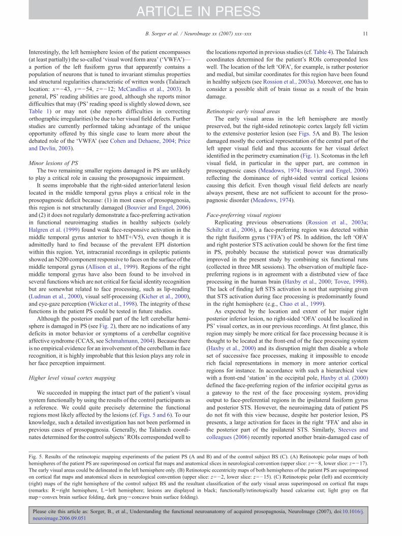
Interestingly, the left hemisphere lesion of the patient encompasses(at least partially) the so-called ‘visual word form area’ (‘VWFA’)—a portion of the left fusiform gyrus that apparently contains apopulation of neurons that is tuned to invariant stimulus propertiesand structural regularities characteristic of written words (Talairachlocation: x=−43, y=−54, z=−12; McCandliss et al., 2003). Ingeneral, PS’ reading abilities are good, although she reports minordifficulties that may (PS’ reading speed is slightly slowed down, seeTable 1) or may not (she reports difficulties in correctingorthographic irregularities) be due to her visual field defects. Furtherstudies are currently performed taking advantage of the uniqueopportunity offered by this single case to learn more about thedebated role of the ‘VWFA’ (see Cohen and Dehaene, 2004; Priceand Devlin, 2003).
Minor lesions of PSThe two remaining smaller regions damaged in PS are unlikely
to play a critical role in causing the prosopagnosic impairment.It seems improbable that the right-sided anterior/lateral lesion
located in the middle temporal gyrus plays a critical role in theprosopagnosic deficit because: (1) in most cases of prosopagnosia,this region is not structurally damaged (Bouvier and Engel, 2006)and (2) it does not regularly demonstrate a face-preferring activationin functional neuroimaging studies in healthy subjects (solelyHalgren et al. (1999) found weak face-responsive activation in themiddle temporal gyrus anterior to hMT+/V5), even though it isadmittedly hard to find because of the prevalent EPI distortionwithin this region. Yet, intracranial recordings in epileptic patientsshowed anN200 component responsive to faces on the surface of themiddle temporal gyrus (Allison et al., 1999). Regions of the rightmiddle temporal gyrus have also been found to be involved inseveral functions which are not critical for facial identity recognitionbut are somewhat related to face processing, such as lip-reading(Ludman et al., 2000), visual self-processing (Kicher et al., 2000),and eye-gaze perception (Wicker et al., 1998). The integrity of thesefunctions in the patient PS could be tested in future studies.
Although the posterior medial part of the left cerebellar hemi-sphere is damaged in PS (see Fig. 2), there are no indications of anydeficits in motor behavior or symptoms of a cerebellar cognitiveaffective syndrome (CCAS, see Schmahmann, 2004). Because thereis no empirical evidence for an involvement of the cerebellum in facerecognition, it is highly improbable that this lesion plays any role inher face perception impairment.
Higher level visual cortex mapping
We succeeded in mapping the intact part of the patient’s visualsystem functionally by using the results of the control participants asa reference. We could quite precisely determine the functionalregions most likely affected by the lesions (cf. Figs. 5 and 6). To ourknowledge, such a detailed investigation has not been performed inprevious cases of prosopagnosia. Generally, the Talairach coordi-nates determined for the control subjects’ROIs correspondedwell to
Fig. 5. Results of the retinotopic mapping experiments of the patient PS (A andhemispheres of the patient PS are superimposed on cortical flat maps and anatomicaThe early visual areas could be delineated in the left hemisphere only. (B) Retinotopon cortical flat maps and anatomical slices in neurological convention (upper slic(right) maps of the right hemisphere of the control subject BS and the resultant(remarks: R=right hemisphere, L= left hemisphere; lesions are displayed in bmap=convex brain surface folding, dark gray=concave brain surface folding).
Please cite this article as: Sorger, B., et al., Understanding the functional neuroneuroimage.2006.09.051
the locations reported in previous studies (cf. Table 4). The Talairachcoordinates determined for the patient’s ROIs corresponded lesswell. The location of the left ‘OFA’, for example, is rather posteriorand medial, but similar coordinates for this region have been foundin healthy subjects (see Rossion et al., 2003a). Moreover, one has toconsider a possible shift of brain tissue as a result of the braindamage.
Retinotopic early visual areasThe early visual areas in the left hemisphere are mostly
preserved, but the right-sided retinotopic cortex largely fell victimto the extensive posterior lesion (see Figs. 5A and B). The lesiondamaged mostly the cortical representation of the central part of theleft upper visual field and thus accounts for her visual defectidentified in the perimetry examination (Fig. 1). Scotomas in the leftvisual field, in particular in the upper part, are common inprosopagnosic cases (Meadows, 1974; Bouvier and Engel, 2006)reflecting the dominance of right-sided ventral cortical lesionscausing this deficit. Even though visual field defects are nearlyalways present, these are not sufficient to account for the proso-pagnosic disorder (Meadows, 1974).
Face-preferring visual regionsReplicating previous observations (Rossion et al., 2003a;
Schiltz et al., 2006), a face-preferring region was detected withinthe right fusiform gyrus (‘FFA’) of PS. In addition, the left ‘OFA’and right posterior STS activation could be shown for the first timein PS, probably because the statistical power was dramaticallyimproved in the present study by combining six functional runs(collected in three MR sessions). The observation of multiple face-preferring regions is in agreement with a distributed view of faceprocessing in the human brain (Haxby et al., 2000; Tovee, 1998).The lack of finding left STS activation is not that surprising giventhat STS activation during face processing is predominantly foundin the right hemisphere (e.g., Chao et al., 1999).
As expected by the location and extent of her major rightposterior inferior lesion, no right-sided ‘OFA’ could be localized inPS’ visual cortex, as in our previous recordings. At first glance, thisregion may simply be more critical for face processing because it isthought to be located at the front-end of the face processing system(Haxby et al., 2000) and its disruption might then disable a wholeset of successive face processes, making it impossible to encoderich facial representations in memory in more anterior corticalregions for instance. In accordance with such a hierarchical viewwith a front-end ‘station’ in the occipital pole, Haxby et al. (2000)defined the face-preferring region of the inferior occipital gyrus asa gateway to the rest of the face processing system, providingoutput to face-preferential regions in the ipsilateral fusiform gyrusand posterior STS. However, the neuroimaging data of patient PSdo not fit with this view because, despite her posterior lesion, PSpresents, a large activation for faces in the right ‘FFA’ and also inthe posterior part of the ipsilateral STS. Similarly, Steeves andcolleagues (2006) recently reported another brain-damaged case of
B) and of the control subject BS (C). (A) Retinotopic polar maps of bothl slices in neurological convention (upper slice: z=−8, lower slice: z=−17).ic eccentricity maps of both hemispheres of the patient PS are superimposede: z=−2, lower slice: z=−15). (C) Retinotopic polar (left) and eccentricityclassification of the early visual areas superimposed on cortical flat mapslack; functionally/retinotopically based calcarine cut; light gray on flat
anatomy of acquired prosopagnosia, NeuroImage (2007), doi:10.1016/j.

Fig. 6. Functional regions of interest of the patient PS (A) and the control subject BS (B). Functionally defined visual regions of the patient PS (A) and the controlsubject BS (B) are superimposed on T1-weighted anatomical MRI slices (remarks: R=right hemisphere, L= left hemisphere).
12 B. Sorger et al. / NeuroImage xx (2007) xxx–xxx
ARTICLE IN PRESS
prosopagnosia, DF, with bilateral inferior occipital lesions and yetbilateral face-preferring activations in ‘FFA’ and STS. Together,these observations suggest that, in healthy subjects, anterior face-related activations in both the fusiform gyrus and STS may also beelicited independently of the contribution of the ipsilateral inferioroccipital cortex. While the patient DF, reported by Steeves andcollaborators (2006), has bilateral inferior occipital lesions, onecannot fully exclude that PS’ face-preferring response in the rightfusiform gyrus or STS originates from inputs of the left hemisphere(e.g., from the left ‘OFA’). However, this is unlikely because right‘FFA’ activation can be found without activation of the left ‘OFA’in healthy subjects and in PS (e.g., Rossion et al., 2003a; Schiltzet al., 2006).
Finally, it might seem surprising that a large number of face-preferring regions (right STS and ‘FFA’, left ‘OFA’) are stronglyactive in the brain of a patient who is largely unable to recognizefaces. However, these regions, albeit presenting a preferentialresponse to faces, might not process faces adequately. In agreementwith this view, more recent neuroimaging data of patient PSindicate that the right ‘FFA’ activation, albeit being normal at aquantitative level, is qualitatively different compared to controlsubjects: whereas healthy subjects show a decreased BOLD
Please cite this article as: Sorger, B., et al., Understanding the functional neuroneuroimage.2006.09.051
response when being presented with blocks or pairs of identicalfaces compared to different faces, PS’ ‘FFA’ shows the same levelof activation for same and different faces (Schiltz et al., 2006). Thissuggests that the right ‘FFA’ (possibly also the STS) is notfunctionally intact, perhaps missing reentrant connections of thedamaged ipsilateral ‘OFA’ (see discussion in Rossion et al., 2003a;Schiltz et al., 2006; Steeves et al., 2006). It should be stressed herethat looking at a face initiates a multitude of different processes,besides identity processing. Although slowed down, PS is still ableto detect faces and categorize them according to their gender, age,and expression (Rossion et al., 2003a). Her ability to discriminateindividual faces is also clearly impaired, but is much better thanchance level, especially if she is given unlimited time (Rossion et al.,2003a; Schiltz et al., 2006). Thus, the different regions showing apreferential response for faces may still play a role in her preservedface perception abilities (see alsoMarotta et al., 2001). Alternatively,PS may rely on other intact higher visual regions which do notprocess faces preferentially, such as the vLOC (see below).
Following the detailed mapping of the visual cortex of thepatient, these questions will be addressed in future experiments,combining functional neuroimaging and psychophysical investiga-tions to correlate brain activity patterns and behavioral outcome.
anatomy of acquired prosopagnosia, NeuroImage (2007), doi:10.1016/j.

Table 3Talairach locations, t-values and cluster sizes of the functionally defined key regions
p<0.05 (1-sided, Bonferroni-corrected), voxel size: 1 mm3, × region lesioned. *p<0.00002, — region could not be detected, BA=Brodmann area, colorednumbers of BAs indicate an affection by lesioning. (For interpretation of the references to colour in this table legend, the reader is referred to the webversion of this article.)
13B. Sorger et al. / NeuroImage xx (2007) xxx–xxx
ARTICLE IN PRESS
Non-face-preferring higher visual brain regionsThe bilateral ‘PPA’ (Epstein et al., 1999), dLOC, and hMT+/V5
(Sunaert et al., 1999) could be localized bilaterally and are entirelyspared by the lesions. Previous studies have shown that localizeddamage to the region of the ‘PPA’ is associated with topographicalagnosia and impairment in scene recognition (Epstein et al., 2001).Although PS has not been tested on scene categorization orrecognition, or topographical orientation, she never complained of
Table 4Talairach coordinates of the functional key regions' determined in previous studie
*These authors differentiate the two subdivisions of LOC as follows: (a) a caudapart (LOa/pFs, here vLOC) (see e.g., Grill-Spector et al., 2001).
Please cite this article as: Sorger, B., et al., Understanding the functional neuroneuroimage.2006.09.051
any corresponding difficulties. Self-reports and observations ineveryday life rather suggest that her abilities might be aboveaverage. She perfectly recognizes places previously visited andnever depends on other people to find her way home, her work place,friends’ places, the hospital, a hotel, etc. Given the sparing of theneural substrates of topographical orientation as identified in thepresent study, it can be predicted that she will perform normally incorresponding tests. The total preservation of her occipitoparietal
s
l dorsal lateral occipital part (LO, here dLOC) and (b) a posterior fusiform
anatomy of acquired prosopagnosia, NeuroImage (2007), doi:10.1016/j.

14 B. Sorger et al. / NeuroImage xx (2007) xxx–xxx
ARTICLE IN PRESS
visual system (the ‘dorsal stream’), and in particular hMT+/V5and the dorsal part of the lateral occipital complex (dLOC) (Fig.6A), is also in agreement with her preserved movement vision andher normal ability to reach and grasp objects. As for the occipito-temporal visual system (the ‘ventral stream’), we could define V4/V8 (Tootell and Hadjikhani, 2001) within the left hemisphereonly. PS’ color perception ability may only be mildly impaired forcentral vision (Table 1), and complementary tests performed hereclearly confirm that she is not achromatopsic. However, becauseof the lack of defining right V4/V8, PS may be worse atprocessing color stimuli in her left visual field. Similarly, damageof the left vLOC does not seem to have a major effect on her non-face object recognition abilities, but this has to be tested withlateralized presentations. Even though PS may possibly have moredifficulties recognizing object shapes flashed in her right visualfield, the presence of the left scotoma and possibly low-level visualdifficulties in the left visual field may balance any hemisphericdifference out.
Most interestingly, the right occipital inferior lesion that isthought to be critical in causing PS’ prosopagnosia spared entirelythe right vLOC (Fig. 6A), a region which is directly correlated withobject perception (e.g., Grill-Spector et al., 1998, 2000; Avidan etal., 2002; James et al., 2000). The role of the LOC in thediscrimination of individual objects is also supported by fMRIadaptation (Grill-Spector et al., 2006, 2001) or repetition-suppres-sion (Henson and Rugg, 2003) studies, showing a larger response inthis region to novel objects than to repeated objects (Avidan et al.,2002; Grill-Spector et al., 1999; Sayres and Grill-Spector, 2006).Accordingly, the sparing of the vLOC demonstrated here mayaccount for PS’ preserved object recognition and discriminationabilities (Rossion et al., 2003a; Schiltz et al., 2006). In contrast to PS,the patient DF, first reported by Milner et al. (1991), is massivelyimpaired at object recognition following lesions of the main part ofthe vLOC in both hemispheres (James et al., 2003). Thus, while thepatient DF presents a bilateral lesion including both the vLOC andthe ‘OFA’ and is impaired at both object and face recognition, thepatient PS’ lesion damaged the right ‘OFA’ but spared the rightvLOC, resulting in a deficit restricted to the category of faces. Takentogether, these observations suggest that prosopagnosia is mostoften associated with object recognition deficits (visual agnosia)because of right inferior occipital and occipitotemporal lesionsdamaging both the vLOC and the ‘OFA’. However, a lesionrestricted to the right ‘OFA’, sparing the right vLOC, may lead to anisolated deficit at face recognition, as observed for the patient PS.This observation of a normal vLOC activation in PS’ brain, next tothe posterior right hemisphere lesion, also underscores the importantrole that fMRI (compared to conventional structural MRI) can playin revealing the functional integrity of brain regions that might bepotentially damaged (James et al., 2003; Rossion et al., 2003a;Schiltz et al., 2006; Steeves et al., 2006).
Conclusions
We presented for the first time a detailed study on the functionalneuroanatomy of the visual system in a prosopagnosic patient. Ourdata reinforce the critical involvement of the right inferior occipitalgyrus (‘OFA’) in causing prosopagnosia (Bouvier and Engel,2006), while supporting the view that a lesion of this region doesnot interrupt the preferential processing of faces in ipsilateralhigher level visual regions such as the ‘FFA’ (Rossion et al., 2003a;Schiltz et al., 2006) and the STS. Besides the precise definition of
Please cite this article as: Sorger, B., et al., Understanding the functional neuroneuroimage.2006.09.051
the lesions in this single case, the observation of a number offunctionally intact visual regions, such as the vLOC, correspondswell with the remarkably well preserved visual functions of thepatient – including object recognition – and opens the way forfurther clarification of the neural basis of her face and objectprocessing abilities. Detailed neurofunctional reports of singlecases as reported here should be particularly important to clarifyfurther the neural basis of prosopagnosia and to provide invaluableinformation about the functional neuroanatomy of face processingin the healthy human brain.
Acknowledgments
We are extremely grateful to PS for her patience and highmotivation during the experiments performed in connectionwith this study. We thank Petra Stoerig for performing theperimetry examination of PS and providing Fig. 1. Further-more, we would like to thank Nikolaus Kriegeskorte for hishelp in fMRI data acquisition, Steve Shevell for his help ininterpreting PS’ score at the color Farnsworth–Munsell 100-Huetest, Xavier Golay for optimizing the acquisition of anatomicalimages, as well as Judith Peters, Joel Reithler, and two anonymousreviewers for their enriching comments on a previous version of thispaper.
This researchwas supported byMaastricht University and a grantARC 01/06-267, (Communauté Française de Belgique—Actions deRecherche Concertées). Bruno Rossion is supported by the BelgianNational Foundation for Scientific Research (FNRS).
References
Allison, T., Puce, A., Spencer, D.D., McCarthy, G., 1999. Electrophysio-logical studies of human face perception. I: Potentials generated inoccipitotemporal cortex by face and non-face stimuli. Cereb. Cortex 9,415–430.
Avidan, G., Harel, M., Hendler, T., Ben-Bashat, D., Zohary, E., Malach, R.,2002. Contrast sensitivity in human visual areas and its relationship toobject recognition. J. Neurophysiol. 87, 3102–3116.
Barton, J.J., Cherkasova, M.V., 2005. Impaired spatial coding withinobjects but not between objects in prosopagnosia. Neurology 65,270–274.
Barton, J.J.S., Press, D.Z., Keenan, J.P., O’Connor, M., 2002. Lesions of thefusiform face area impair perception of facial configuration inprosopagnosia. Neurology 58, 71–78.
Barton, J.J., Cherkasova, M.V., Press, D.Z., Intriligator, J.M., O’Connor, M.,2004. Perceptual functions in prosopagnosia. Perception 33, 939–956.
Benton, A.L., 1980. The neuropsychology of facial recognition. Am.Psychol. 35, 176–186.
Benton, A.L., van Allen, M., 1972. Prosopagnosia and facial discrimination.J. Neurol. Sci. 15, 167–172.
Bodamer, J., 1947. Die Prosop-agnosie. Arch. Psychiatr. Nervenkr. 179,6–54.
Boutsen, L., Humphreys, G.W., 2002. Face context interferes with local partprocessing in a prosopagnosic patient. Neuropsychologia 40,2305–2313.
Bouvier, S.E., Engel, S.A., 2006. Behavioral deficits and cortical damageloci in cerebral achromatopsia. Cereb. Cortex 16, 183–191.
Boynton, G.M., Engel, S.A., Glover, G.H., Heeger, D.J., 1996. Linearsystems analysis of functional magnetic resonance imaging in humanV1. J. Neurosci. 16, 4207–4221.
Brodtmann, A., Puce, A., Syngeniotis, A., Darby, D., Donnan, G., 2003. Thefunctional magnetic resonance imaging hemodynamic response to facesremains stable until the ninth decade. NeuroImage 20, 520–528.
anatomy of acquired prosopagnosia, NeuroImage (2007), doi:10.1016/j.

15B. Sorger et al. / NeuroImage xx (2007) xxx–xxx
ARTICLE IN PRESS
Bruce, V., Young, A., 1986. Understanding face recognition. Br. J. Psychol.77, 305–327.
Bruyer, R., Laterre, C., Seron, X., Feyereisen, P., Strypstein, E., Pierrard, E.,Rectem, D., 1983. A case of prosopagnosia with some preserved covertremembrance of familiar faces. Brain Cogn. 2, 257–284.
Bukach, C.M., Bub, D.N., Gauthier, I., Tarr, M.J., 2006. Perceptual expertiseeffects are not all or none: spatially limited perceptual expertise for facesin a case of prosopagnosia. J. Cogn. Neurosci. 18, 48–63.
Caldara, R., Schyns, P., Mayer, E., Smith, M., Gosselin, F., Rossion, B.,2005. Does prosopagnosia take the eyes out from faces? Evidence for adefect in the use of diagnostic facial information in a brain-damagedpatient. J. Cogn. Neurosci. 17, 1652–1666.
Campbell, R., Landis, T., Regard, M., 1986. Face recognition and lipreading.A neurological dissociation. Brain 109, 509–521.
Castelo-Branco,M.,Formisano,E.,Backes,W.,Zanella,F.,Neuenschwander,S., Singer, W., Goebel, R., 2002. Activity patterns in human motion-sensitive areas depend on the interpretation of global motion. Proc. Natl.Acad. Sci. U. S. A. 99, 13914–13919.
Chao, L.L., Martin, A., Haxby, J.V., 1999. Are face-responsive regionsselective only for faces? NeuroReport 10, 2945–2950.
Clarke, S., Lindemann, A., Maeder, P., Borruat, F.X., Assal, G., 1997. Facerecognition and postero-inferior hemispheric lesions. Neuropsychologia35, 1555–1563.
Cohen, L., Dehaene, S., 2004. Specialization within the ventral stream: thecase for the visual word form area. NeuroImage 22, 466–476.
Culham, J.C., Kanwisher, N.G., 2001. Neuroimaging of cognitive functionsin human parietal cortex. Curr. Opin. Neurobiol. 11, 157–163.
Damasio, A., Yamada, T., Damasio, H., Corbett, J., McKee, K., 1980.Central achromatopsia: behavioral, anatomic, and physiologic aspects.Neurology 30, 1064–1071.
Damasio, A.R., Damasio, H., Van Hoesen, G.W., 1982. Prosopagnosia:anatomic basis and behavioral mechanisms. Neurology 32, 331–341.
De Renzi, E., 1986. Prosopagnosia in two patients with CT scan evidenceof damage confined to the right hemisphere. Neuropsychologia 24,385–389.
Dixon,M.J., Bub, D.N., Arguin, M., 1998. Semantic and visual determinantsof face recognition in a prosopagnosic patient. J. Cogn. Neurosci. 10,362–376.
Ellis, H.D., Florence, M., 1990. Bodamer’s (1947) paper on prosopagnosia.Cogn. Neuropsychol. 7, 81–105.
Engel, S.A., Rumelhart, D.E., Wandell, B.A., Lee, A.T., Glover, G.H.,Chichilnisky, E.J., Shadlen, M.N., 1994. fMRI of human visual cortex.Nature 369, 525.
Epstein, R., Kanwisher, N., 1998. A cortical representation of the localvisual environment. Nature 392, 598–601.
Epstein, R., Harris, A., Stanley, D., Kanwisher, N., 1999. The parahippo-campal place area: recognition, navigation, or encoding? Neuron 23,115–125.
Epstein, R., DeYoe, E.A., Press, D.Z., Rosen, A.C., Kanwisher, N., 2001.Neuropsychological evidence for a topographical learning mechanism inparahippocampal cortex. Cogn. Neuropsychol. 18, 481–508.
Ettlin, T.M., Beckson, M., Benson, D.F., Langfitt, J.T., Amos, E.C., Pineda,G.S., 1992. Prosopagnosia: a bihemispheric disorder. Cortex 28,129–134.
Farah, M.J., 1990. Visual Agnosia: Disorders of Object Recognition andWhat They Tell us About Normal Vision. MIT Press, Cambridge.
Farah, M.J., Levinson, K.L., Klein, K.L., 1995. Face perception and within-category discrimination in prosopagnosia. Neuropsychologia 33,661–674.
Farnsworth, D., 1957. The Farnsworth–Munsell 100-Hue test for theexamination of color vision. Munsell Color Company, Baltimore, MD.
Felleman, D.J., Van Essen, D.C., 1991. Distributed hierarchical processingin the primate cerebral cortex. Cereb. Cortex 1, 1–47.
Gauthier, I., Behrmann, M., Tarr, M.J., 1999. Can face recognitionreally be dissociated from object recognition? J. Cogn. Neurosci. 11,349–370.
Gauthier, I., Tarr, M.J., Moylan, J., Skudlarski, P., Gore, J.C., Anderson,
Please cite this article as: Sorger, B., et al., Understanding the functional neuroneuroimage.2006.09.051
A.W., 2000. The fusiform “face area” is part of a network that processesfaces at the individual level. J. Cogn. Neurosci. 12, 495–504.
Goebel, R., Muckli, L., Kim, D.-S., 2003. The visual system. In: Paxinos,G., Mai, J.K. (Eds.), The Human Nervous System. Elsevier AcademicPress, San Diego, pp. 1280–1305.
Goldsmith, Z.G., Liu, G.T., 2001. Facial recognition and prosopagnosia:past and present concepts. Neuro-Ophthalmology 25, 177–192.
Grill-Spector, K., Kushnir, T., Edelman, S., Itzchak, Y., Malach, R., 1998.Cue-invariant activation in object-related areas of the human occipitallobe. Neuron 21, 191–202.
Grill-Spector, K., Kushnir, T., Edelman, S., Avidan, G., Itzchak, Y., Malach,R., 1999. Differential processing of objects under various viewingconditions in the human lateral occipital complex. Neuron 24, 187–203.
Grill-Spector, K., Kushnir, T., Hendler, T., Malach, R., 2000. The dynamicsof object-selective activation correlate with recognition performance inhumans. Nat. Neurosci. 3, 837–843.
Grill-Spector, K., Kourtzi, Z., Kanwisher, N., 2001. The lateral occipitalcomplex and its role in object recognition. Vision Res. 41,1409–1422.
Grill-Spector, K., Henson, R., Martin, A., 2006. Repetition and thebrain: neural models of stimulus-specific effects. Trends Cogn. Sci.10, 14–23.
Hadjikhani, N., Liu, A.K., Dale, A.M., Cavanagh, P., Tootell, R.B.H., 1998.Retinotopy and color sensitivity in human visual cortical area V8. Nat.Neurosci. 1, 235–241.
Halgren, E., Dale, A.M., Sereno, M.I., Tootell, R.B., Marinkovic, K., Rosen,B.R., 1999. Location of human face-selective cortex with respect toretinotopic areas. Hum. Brain Mapp. 7, 29–37.
Haxby, J.V., Hoffman, E.A., Gobbini, M.I., 2000. The distributed humanneural system for face perception. Trends Cogn. Sci. 4, 223–233.
Hécaen, H., Angerlergues, R., 1962. Agnosia for faces (prosopagnosia).Arch. Neurol. 7, 92–100.
Henke, K., Schweinberger, S.R., Grigo, A., Klos, T., Sommer, W., 1998.Specificity of face recognition: recognition of exemplars of non-faceobjects in prosopagnosia. Cortex 34, 289–296.
Henson, R.N., Rugg, M.D., 2003. Neural response suppression, haemody-namic repetition effects, and behavioral priming. Neuropsychologia 41,263–270.
Heywood, C.A., Nicholas, J.J., Cowey, A., 1996. Behavioural andelectrophysiological chromatic and achromatic contrast sensitivity inan achromatopsic patient. J. Neurol. Neurosurg. Psychiatry 60,638–643.
Hillger, L.A., Koenig, O., 1991. Separable mechanisms in face processing:evidence from hemispheric specialization. J. Cogn. Neurosci. 3, 42–58.
James, T.W., Humphrey, G.K., Gati, J.S., Menon, R.S., Goodale, M.A.,2000. The effects of visual object priming on brain activation before andafter recognition. Curr. Biol. 10, 1017–1024.
James, T.W., Culham, J., Humphrey, G.K., Milner, A.D., Goodale, M.A.,2003. Ventral occipital lesions impair object recognition but not object-directed grasping: an fMRI study. Brain 126, 2643–2675.
Joubert, S., Felician, O., Barbeau, E., Sontheimer, A., Barton, J.J., Ceccaldi,M., Poncet, M., 2003. Impaired configurational processing in a case ofprogressive prosopagnosia associated with predominant right temporallobe atrophy. Brain 126, 2537–2550.
Kanwisher, N., McDermott, J., Chun, M.M., 1997. The fusiform facearea: a module in human extrastriate cortex specialized for faceperception. J. Neurosci. 17, 4302–4311.
Kicher, T.T.J., Senior, C., Phillips, M.L., Benson, P.J., Bullmore, E.T.,Brammer, M., Simmons, A., Williams, S.C., Bartels, M., David, A.S.,2000. Towards a functional neuroanatomy of self processing: effects offaces and words. Cogn. Brain Res. 10, 133–144.
Kinnear, P.R., Sahraie, A., 2002. New Farnsworth–Munsell 100 hue testnorms of normal observers for each year of age 5–22 and for age decades30–70. Br. J. Ophthalmol. 86 (12), 1408–1411.
Laeng, B., Caviness, V.S., 2001. Prosopagnosia as a deficit in encodingcurved surface. J. Cogn. Neurosci. 13, 556–576.
Landis, T., Regard, M., Bliestle, A., Kleihues, P., 1998. Prosopagnosia and
anatomy of acquired prosopagnosia, NeuroImage (2007), doi:10.1016/j.

16 B. Sorger et al. / NeuroImage xx (2007) xxx–xxx
ARTICLE IN PRESS
agnosia for noncanonical views. An autopsied case. Brain 111,1287–1297.
Le Grand, R., Mondloch, C.J., Maurer, D., Brent, H.P., 2003. Expert faceprocessing requires visual input to the right hemisphere during infancy.Nat. Neurosci. 6, 1108–1112.
Levine, D.N., Calvanio, R., 1989. Prosopagnosia: a defect in visualconfigural processing. Brain Cogn. 10, 149–170.
Lhermitte, F., Chain, F., Escourolle, R., Ducarne, B., Pillon, B., 1972.Anatomoclinical study of a case of prosopagnosia. Rev. Neurol. 126,329–346.
Linden, D.E.J., Kallenbach, U., Heinecke, A., Singer, W., Goebel, R.,1999. The myth of upright vision. A psychophysical and functionalimaging study of adaptation to inverting spectacles. Perception 28,469–481.
Ludman, C.N., Summerfield, A.Q., Hall, D., Elliott, M., Foster, J., Hykin,J.L., Bowtell, R., Morris, P.G., 2000. Lip-reading ability and patterns ofcortical activation studied using fMRI. J. Audiol. 34, 225–230.
Malach, R., Reppas, J.B., Benson, R.R., Kwong, K.K., Jiang, H., Kennedy,W.A., Ledden, P.J., Brady, T.J., Rosen, B.R., Tootell, R.B., 1995.Object-related activity revealed by functional magnetic resonanceimaging in human occipital cortex. Proc. Natl. Acad. Sci. U. S. A. 92,8135–8139.
Mattson, A.J., Levin, H.S., Grafman, J., 2000. A case of prosopagnosiafollowing moderate closed head injury with left hemisphere focal lesion.Cortex 36, 125–137.
Marotta, J.J., Genovese, C.R., Behrmann, M., 2001. A functional MRI studyof face recognition in patients with prosopagnosia. NeuroReport 12,1581–1587.
Mayer, E., Rossion, B., in press. Prosopagnosia. In: Godefroy, O.,Bogousslavsky, J. (Eds.). The cognitive neurology of stroke: CambridgeUniversity Press.
Mayer, E., Fistarol, P., Valenza, N., 1999. Prise en charge neuropsycho-logique d’une patiente prosopagnosique. In: Azouvi, P., Perrier, D.,Van der Linden, M. (Eds.), La Reeducation en Neuropsychologie:Etudes de Cas. Solal, Marseille.
McCandliss, B.D., Cohen, L., Dehaene, S., 2003. The visual word formarea: expertise for reading in the fusiform gyrus. Trends Cogn. Sci. 7,293–299.
McCarthy, M., Puce, A., Gore, J.C., Allison, T., 1997. Face-specificprocessing in the human fusiform gyrus. J. Cogn. Neurosci. 9, 605–610.
Meadows, J.C., 1974. The anatomical basis of prosopagnosia. J. Neurol.Neurosurg. Psychiatry 37, 489–501.
Michel, F., Poncet, M., Signoret, J.L., 1989. Les lésions responsables de laprosopagnosie sont-elles toujours bilatérales? Rev. Neurol. 145,764–770.
Milner, A.D., Perrett, D.I., Johnston, R.S., Benson, P.J., Jordan, T.R.,Heeley, D.W., Bettucci, D., Mortara, F., Mutani, R., Terazzi, E., 1991.Perception and action in ‘visual form agnosia’. Brain 114, 405–428.
Ogawa, S., Lee, T.M., Kay, A.R., Tank, D.W., 1990. Brain magneticresonance imaging with contrast dependent on blood oxygenation. Proc.Natl. Acad. Sci. U. S. A. 87, 9868–9872.
Oldfield, R.C., 1971. The assessment and analysis of handedness: theEdinburgh inventory. Neuropsychologia 9, 97–113.
Price, C.J., Devlin, J.T., 2003. The myth of the visual word form area.NeuroImage 19, 473–481.
Puce, A., Allison, T., Bentin, S., Gore, J.C., McCarthy, G., 1998.Temporal cortex activation in humans viewing eye and mouth move-ments. J. Neurosci. 18, 2188–2199.
Quaglino, A., Borelli, G.B., Della Sala, S., Young, A.W., 2003. Quaglino’s1867 case of prosopagnosia. Cortex 39, 440–533.
Rossion, B., Pourtois, G., 2004. Revisiting Snodgrass and Vanderwart’sobject pictorial set: the role of surface detail in basic-level objectrecognition. Perception 33, 217–236.
Rossion, B., Dricot, L., Devolder, A., Bodart, J.M., Crommelinck, M.,De Gelder, B., Zoontjes, R., 2000. Hemispheric asymmetries forwhole-based and part-based face processing in the human fusiformgyrus. J. Cogn. Neurosci. 12, 793–802.
Please cite this article as: Sorger, B., et al., Understanding the functional neuroneuroimage.2006.09.051
Rossion, B., Schiltz, C., Robaye, L., Pirenne, D., Crommelinck, M.,2001. How does the brain discriminate familiar and unfamiliar faces?: aPET study of face categorical perception. J. Cogn. Neurosci. 13,1019–1034.
Rossion, B., Caldara, R., Seghier, M., Schuller, A.-M., Lazeyras, F., Mayer,E., 2003a. A network of occipito-temporal face-sensitive areas besidesthe right middle fusiform gyrus is necessary for normal face processing.Brain 126, 2381–2395.
Rossion, B., Joyce, C.A., Cottrell, G.W., Tarr, M.J., 2003b. Earlylateralization and orientation tuning for face, word, and objectprocessing in the visual cortex. NeuroImage 20, 1609–1624.
Sayres, R., Grill-Spector, K., 2006. Object-selective cortex exhibitsperformance-independent repetition suppression. J. Neurophysiol. 95,995–1007.
Schiltz, C., Rossion, B., 2006. Faces are represented holistically in thehuman occipito-temporal cortex. NeuroImage 32, 1385–1394.
Schiltz, C., Sorger, B., Caldara, R., Ahmed, F., Mayer, E., Goebel, R.,Rossion, B., 2006. Impaired face discrimination in acquired prosopag-nosia is associated with abnormal response to individual faces in theright middle fusiform gyrus. Cereb. Cortex 16, 574–586.
Schweich, M., Bruyer, R., 1993. Heterogeneity in the cognitive manifesta-tions of prosopagnosia: the study of a group of single cases. Cogn.Neuropsychol. 10, 529–547.
Sereno, M.I., Dale, A.M., Reppas, J.B., Kwong, K.K., Belliveau, J.W.,Brady, T.J., Rosen, B.R., Tootell, R.B., 1995. Borders of multiple visualareas in humans revealed by functional magnetic resonance imaging.Science 268, 889–893.
Sergent, J., Signoret, J.-L., 1992. Varieties of functional deficits inprosopagnosia. Cereb. Cortex 2, 375–388.
Sergent, J., Villemure, J.G., 1989. Prosopagnosia in a right hemispher-ectomized patient. Brain 112, 975–995.
Sergent, J., Otha, S., MacDonald, B., 1992. Functional neuroanatomy offace and object processing. A positron emission tomography study.Brain 115, 15–36.
Sergent, J., MacDonald, B., Zuck, E., 1994. Structural and functionalorganisation of knowledge about faces and proper names: apositron emission tomography study. In: Umiltà, C., Moscovitch,M. (Eds.), Attention and Performance XV. MIT Press, Cambridge,pp. 203–208.
Schmahmann, J.D., 2004. Disorders of the cerebellum: ataxia, dysmetria ofthought, and the cerebellar cognitive affective syndrome. J. Neuropsy-chiatry Clin. Neurosci. 16, 367–378.
Snodgrass, J.G., Vanderwart, M., 1980. A standardized set of 260 pictures:norms for name agreement, image agreement, familiarity, and visualcomplexity. J. Exp. Psychol. 6, 174–215.
Steeves, J.K., Culham, J.C., Duchaine, B.C., Pratesi, C.C., Valyear, K.F.,Schindler, I., Humphrey, G.K., Milner, A.D., Goodale, M.A., 2006. Thefusiform face area is not sufficient for face recognition: evidence from apatient with dense prosopagnosia and no occipital face area. Neurop-sychologia 44, 594–609.
Sunaert, S., Van Hecke, P., Marchal, G., Orban, G.A., 1999. Motion-responsive regions of the human brain. Exp. Brain Res. 127, 355–370.
Talairach, G., Tournoux, P., 1988. Co-Planar Stereotaxic Atlas of the HumanBrain. Thieme, New York.
Tootell, R.B., Hadjikhani, N., 2001. Where is ‘dorsal V4’ in human visualcortex? Retinotopic, topographic and functional evidence. Cereb. Cortex11, 298–311.
Tovee, M.J., 1998. Face processing: getting by with a little help from itsfriends. Curr. Biol. 8, R317–R320.
Tranel, D., Damasio, A.R., Damasio, H., 1988. Intact recognition of facialexpression, gender, and age in patients with impaired recognition of faceidentity. Neurology 38, 690–696.
Uttner, I., Bliem, H., Danek, A., 2002. Prosopagnosia after unilateral rightcerebral infarction. J. Neurol. 249, 933–935.
Wada, Y., Yamamoto, T., 2001. Selective impairment of facial recognitiondue to a haematoma restricted to the right fusiform and lateral occipitalregion. J. Neurol. Neurosurg. Psychiatry 71, 254–257.
anatomy of acquired prosopagnosia, NeuroImage (2007), doi:10.1016/j.

17B. Sorger et al. / NeuroImage xx (2007) xxx–xxx
ARTICLE IN PRESS
Warrington, E., 1984. Warrington Recognition Memory Test. WesternPsychological Services, Los Angeles.
Wicker, B., Michel, F., Henaff, A.M., Decety, J., 1998. Brain regions involvedin the perception of gaze: a PET study. NeuroImage 8, 221–227.
Wigan, A.L., 1844. A New View of Insanity: The Duality of the Mind.Longman, London.
Wilbrand, H., 1892. Ein Fall von Seelenblindheit und Hemianopsie mitSectionsbefund. Dtsch. Z. Nervenheilkd. 2, 361–387.
Zangenehpour, S., Chaudhuri, A., 2005. Patchy organization and asym-
Please cite this article as: Sorger, B., et al., Understanding the functional neuroneuroimage.2006.09.051
metric distribution of the neural correlates of face processing in monkeyinferotemporal cortex. Curr. Biol. 15, 993–1005.
Zeki, S., 1990. A century of cerebral achromatopsia. Brain 113 (6),1721–1777.
Zeki, S., Watson, J.D., Lueck, C.J., Friston, K.J., Kennard, C., Frackowiak,R.S., 1991. A direct demonstration of functional specialization in humanvisual cortex. J. Neurosci. 11, 641–649.
Zihl, J., Von Cramon, D., 1986. Visual field rehabilitation in the corticallyblind? J. Neurol. Neurosurg. Psychiatry 49, 965–966.
anatomy of acquired prosopagnosia, NeuroImage (2007), doi:10.1016/j.