articulations chapter 9. classification table 9–1
Post on 21-Dec-2015
227 views
TRANSCRIPT
Articulations
Chapter 9
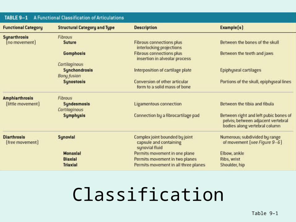
ClassificationTable 9–1

Functional Classification of Joints
• Synarthroses (singular = synarthrosis)– Immovable joints
• Amphiarthroses (singular = amphiarthrosis)– Slightly movable joints
• Diarthroses (singular = diarthrosis)– Freely movable joints

Structural Classification of Joints• Fibrous
• no joint cavity, bones held together with collagen fibers
• Cartilagnous• no joint cavity, bones held together with cartilage
• Synovial• have a “synovial” cavity, bones held together with an enclosed capsule & ligaments
•Synostosis• Conversion of other joints to solid bone mass
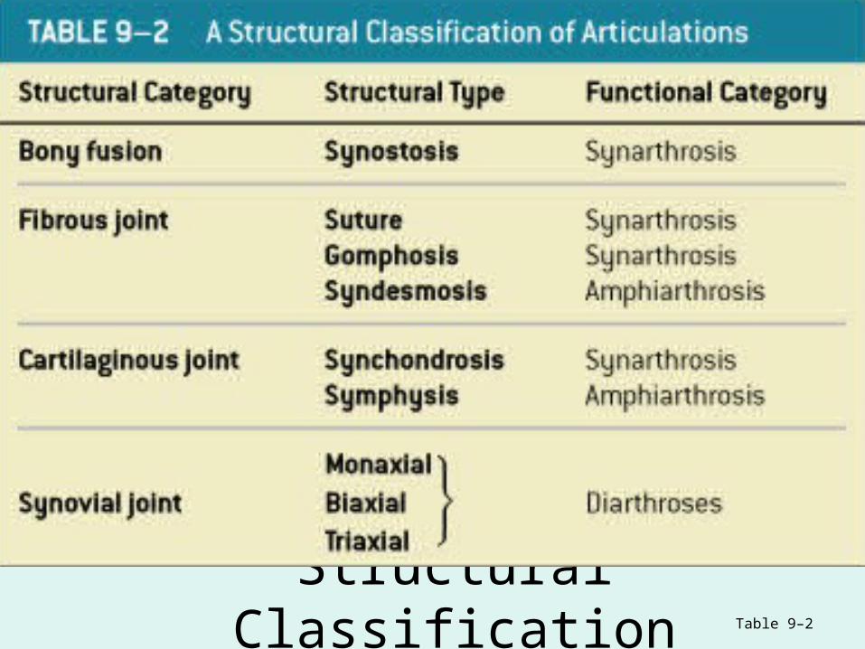
Structural ClassificationTable 9–2
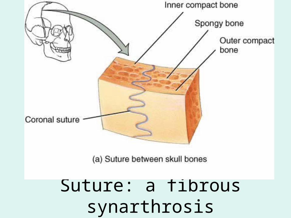
Suture: a fibrous synarthrosis
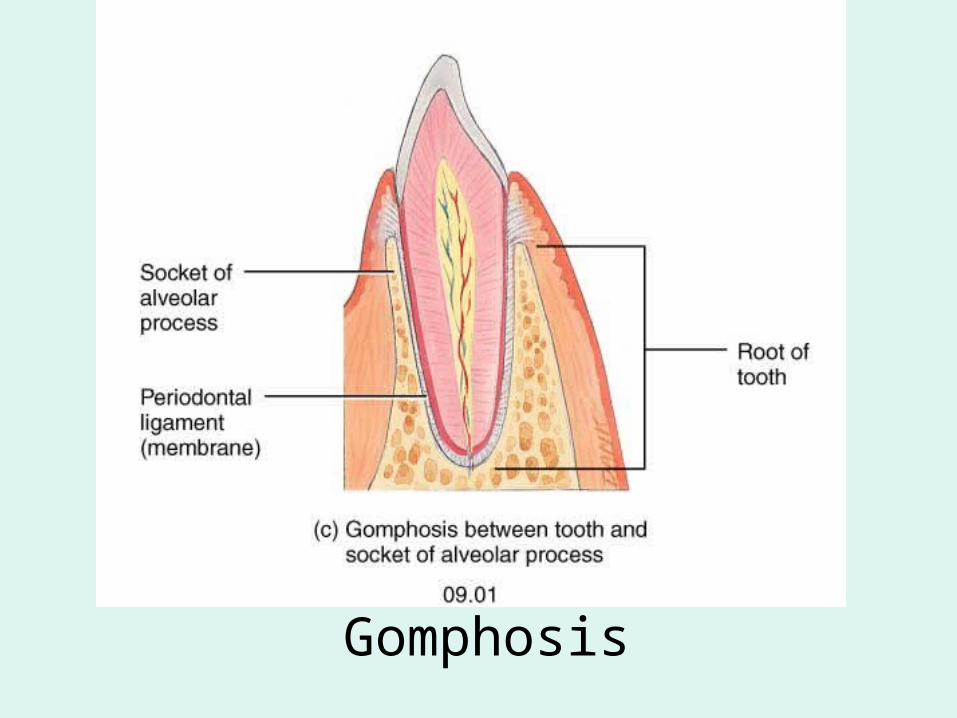
Gomphosis
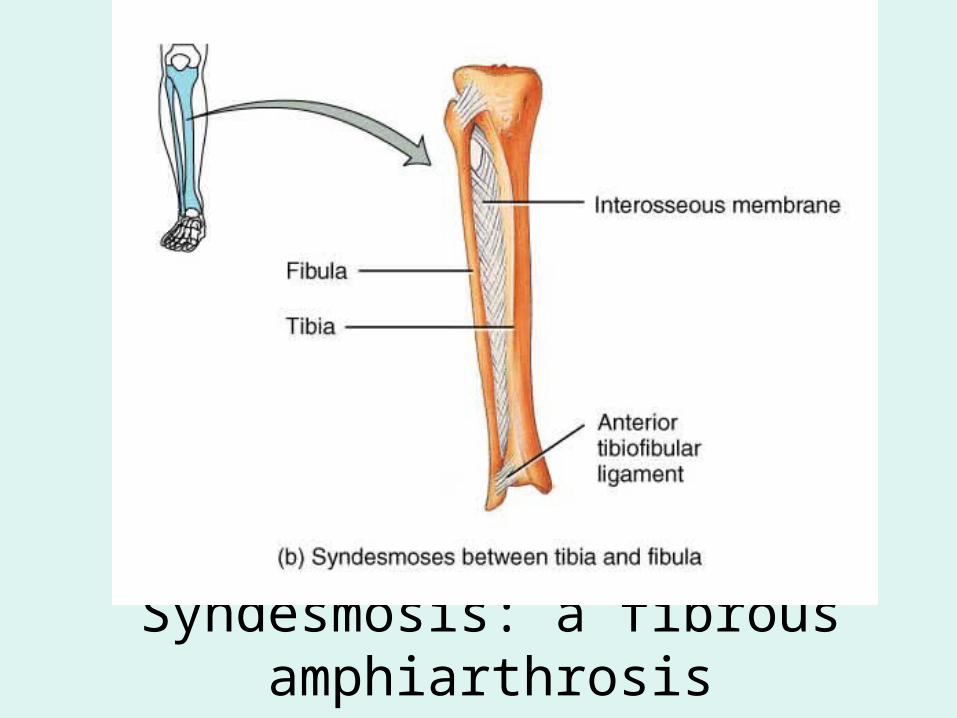
Syndesmosis: a fibrous amphiarthrosis

An amphiarthrotic synchondrosis
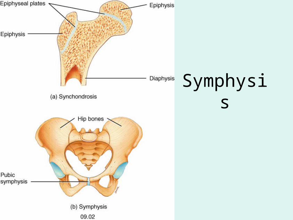
Symphysis
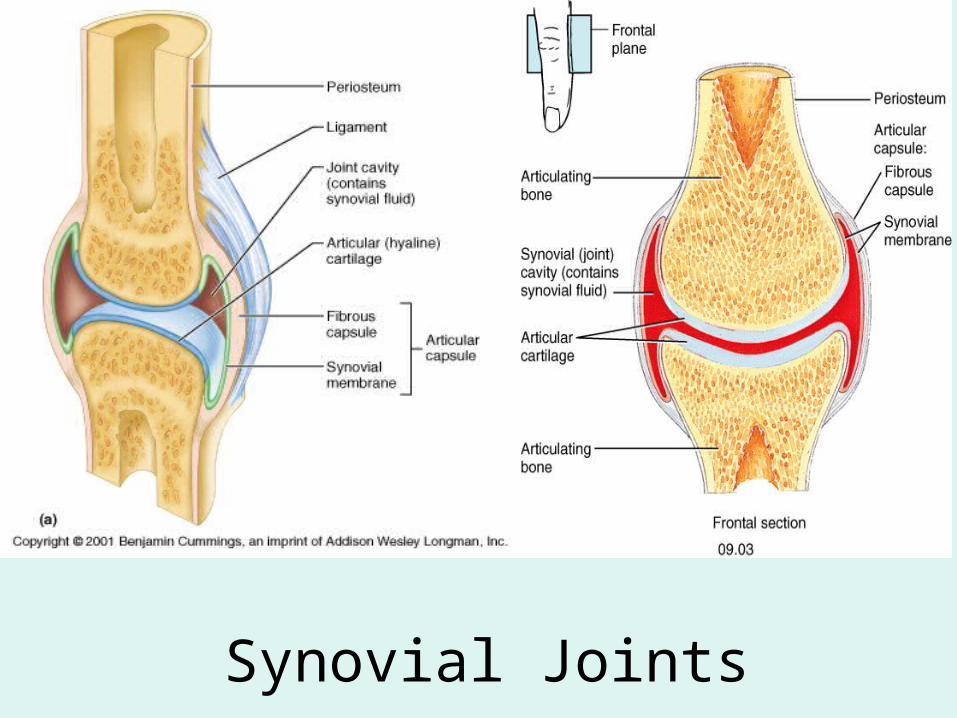
Synovial Joints
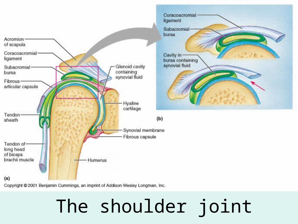
The shoulder joint
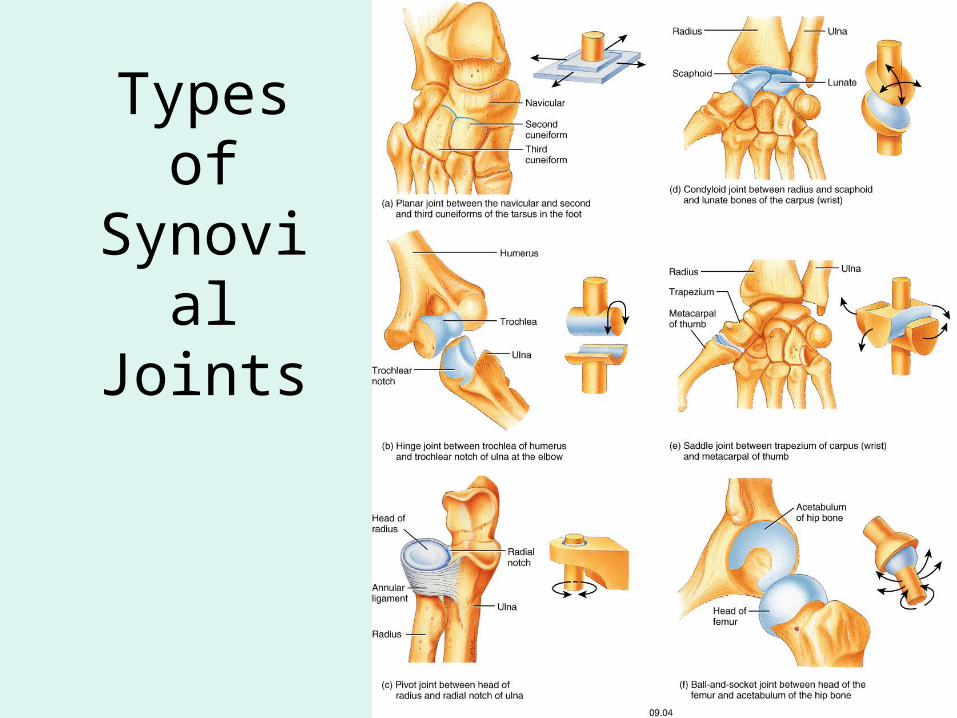
Types of Synovial
Joints

Linear (non-axial) Motion
• Pencil maintains vertical orientation, but changes position
Figure 9–2a, b
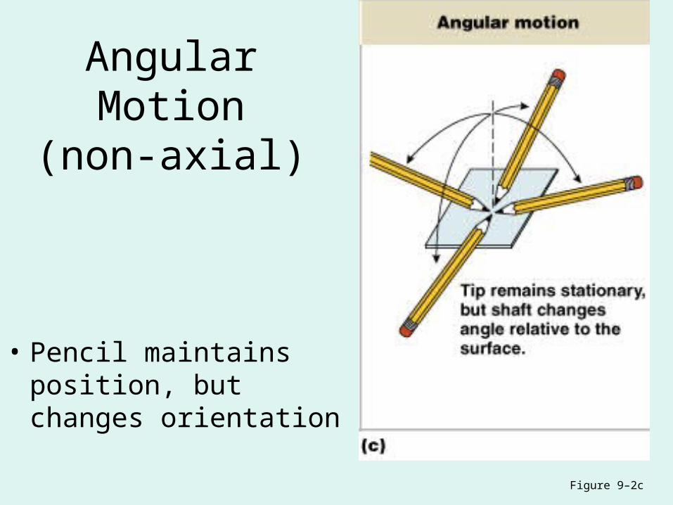
Angular Motion(non-axial)
• Pencil maintains position, but changes orientation
Figure 9–2c

Circumduction(Multiaxial)
• Circular angular motion
Figure 9–2d
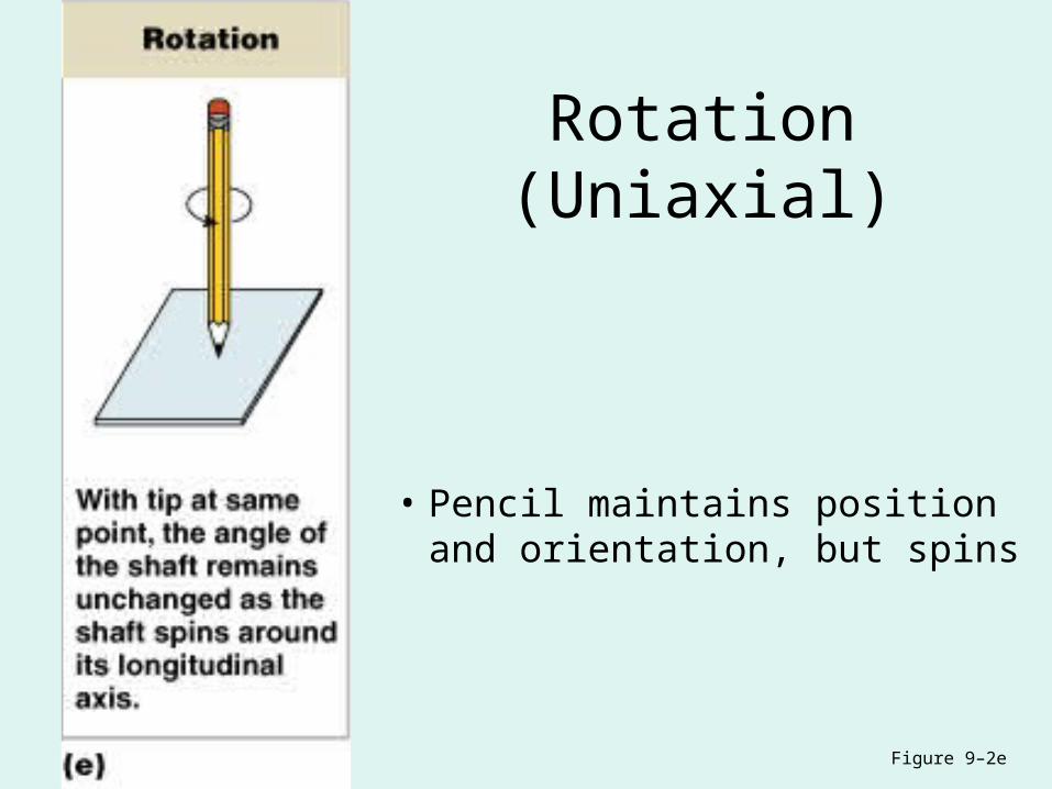
Rotation(Uniaxial)
• Pencil maintains position and orientation, but spins
Figure 9–2e
Planes (Axes) of Dynamic Motion
• Monaxial or uniaxial (1 axis)
• Biaxial (2 axes)
• Triaxial or multiaxial (3 axes)
Types of Movements Possible at Synovial Joints

Gliding

Flexion
Figure 9–3a
Flexion
• Angular motion
• Anterior–posterior plane
• Reduces angle between elements

Lateral Flexion
• Bends vertebral column from side to side
Figure 9–5f
Extension
• Angular motion
• Anterior–posterior plane
• Increases angle between elements
Hyperextension
• Angular motion
• Extension past anatomical position
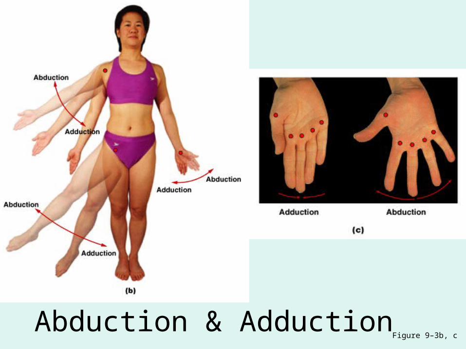
Abduction & AdductionFigure 9–3b, c
Abduction
• Angular motion
• Frontal plane
• Moves away from longitudinal axis
Adduction
• Angular motion
• Frontal plane
• Moves toward longitudinal axis
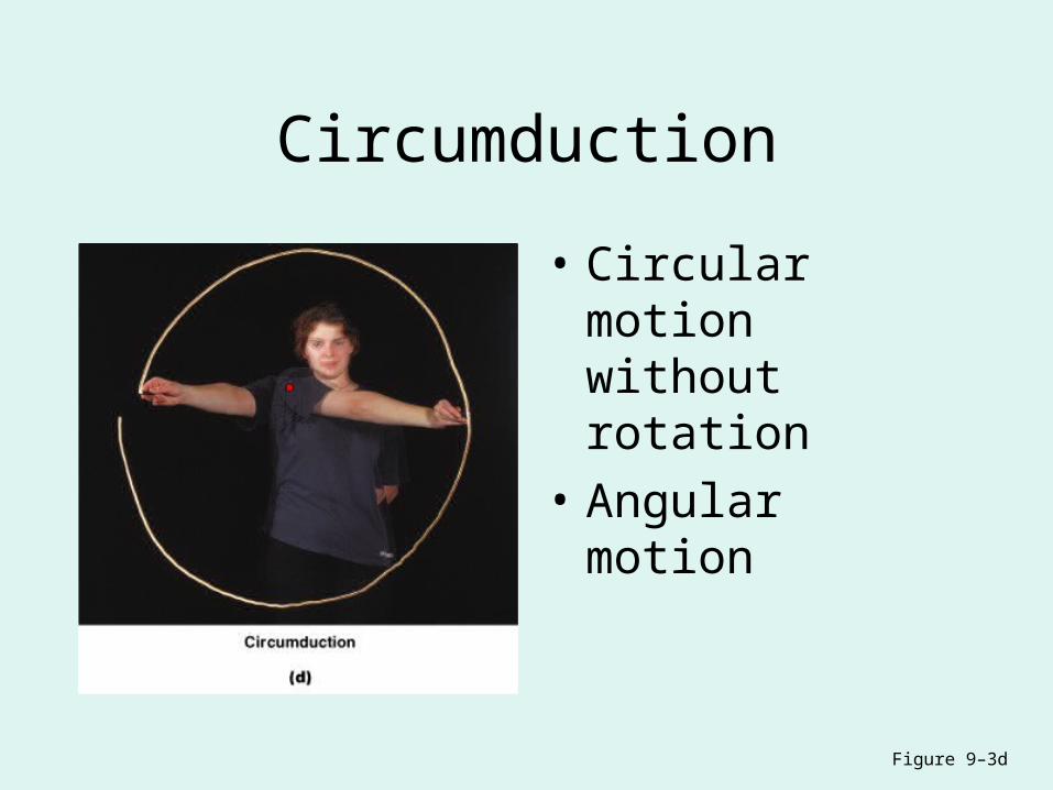
Circumduction
• Circular motion without rotation
• Angular motion
Figure 9–3d
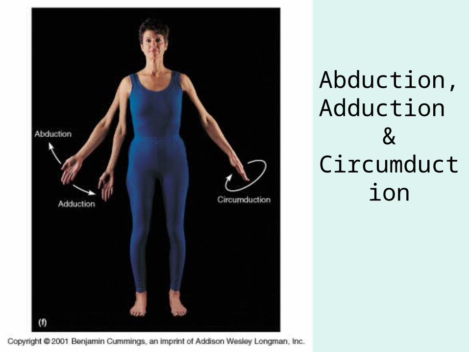
Abduction,Adduction
&Circumduction
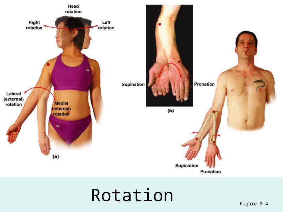
RotationFigure 9–4

Rotation
• Direction of rotation from anatomical position
• Relative to longitudinal axis of bodyLeft or right rotation
• Medial rotation (inward rotation): – rotates toward axis
• Lateral rotation (outward rotation): – rotates away from axis
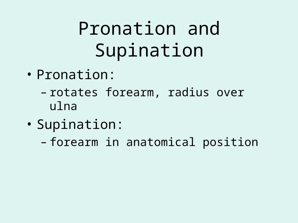
Pronation and Supination
• Pronation:– rotates forearm, radius over ulna
• Supination:– forearm in anatomical position
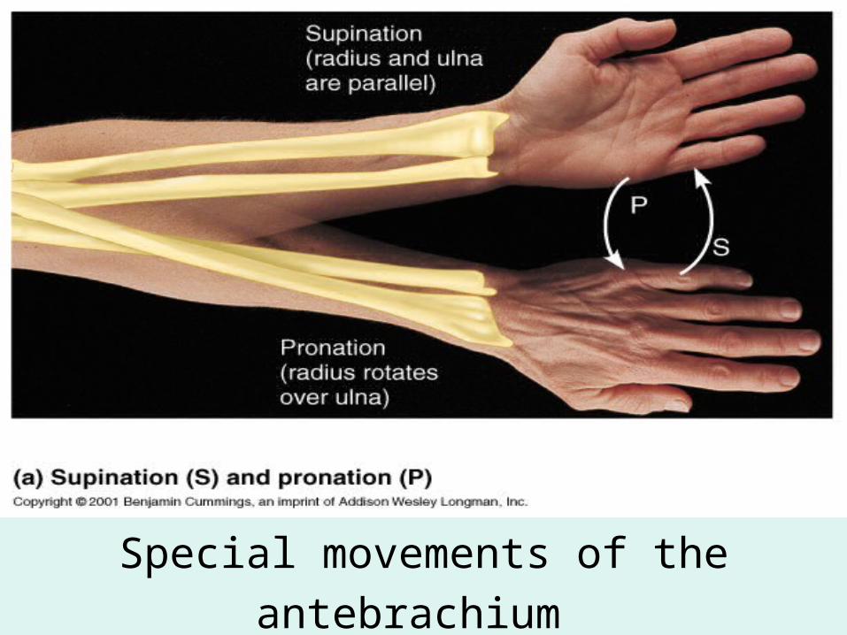
Special movements of the antebrachium
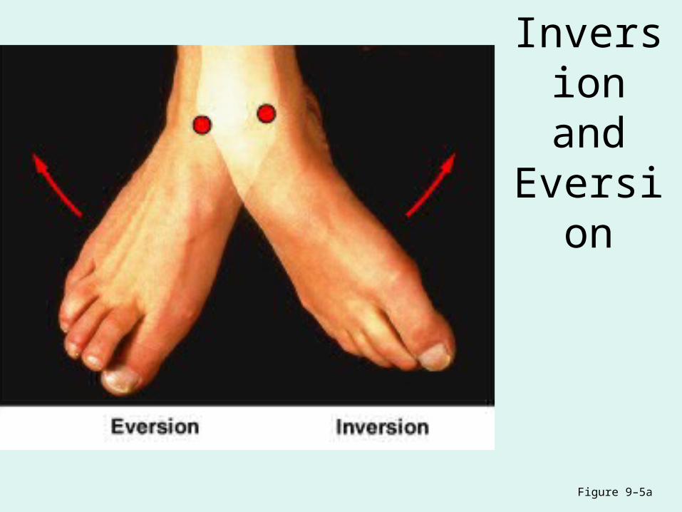
Inversion and
Eversion
Figure 9–5a
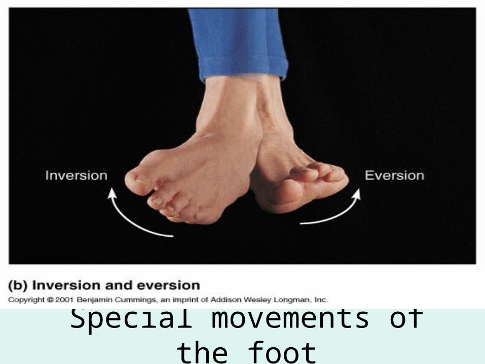
Special movements of the foot
Inversion and Eversion
• Inversion:– twists sole of foot medially
• Eversion:– twists sole of foot laterally
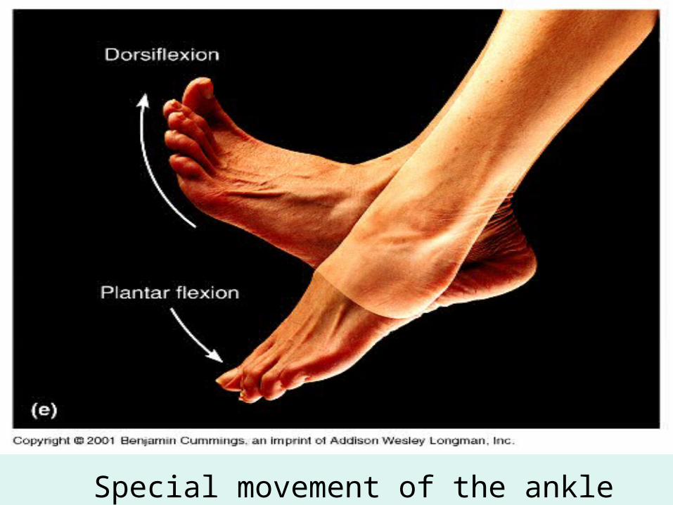
Special movement of the ankle
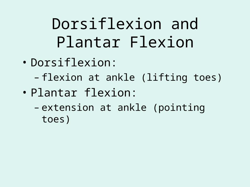
Dorsiflexion and Plantar Flexion
• Dorsiflexion: – flexion at ankle (lifting toes)
• Plantar flexion:– extension at ankle (pointing toes)
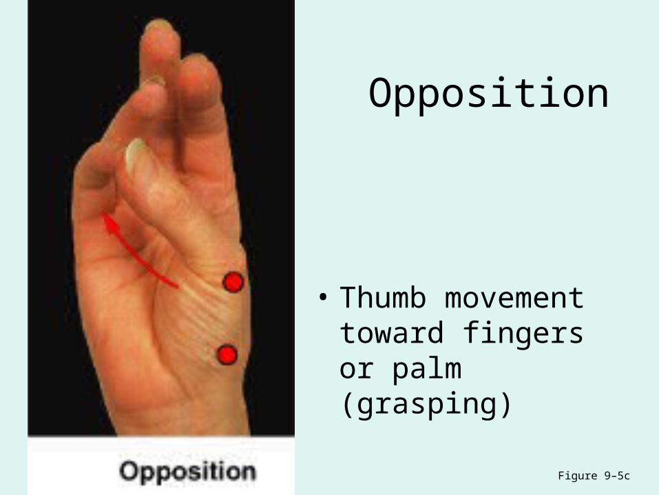
Opposition
• Thumb movement toward fingers or palm (grasping)
Figure 9–5c
Protraction and Retraction
• Protraction: – moves anteriorly– in the horizontal plane (pushing forward)
• Retraction: – opposite of protraction– moving anteriorly (pulling back)
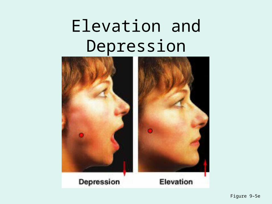
Elevation and Depression
Figure 9–5e
Elevation and Depression
• Elevation: – moves in superior direction (up)
• Depression:– moves in inferior direction (down)

Types synovial joints
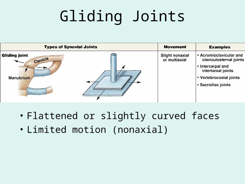
Gliding Joints
• Flattened or slightly curved faces
• Limited motion (nonaxial)
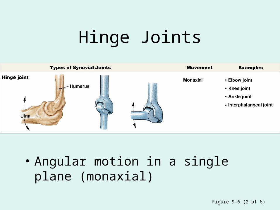
Hinge Joints
• Angular motion in a single plane (monaxial)
Figure 9–6 (2 of 6)
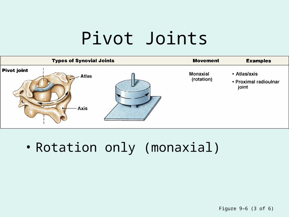
Pivot Joints
• Rotation only (monaxial)
Figure 9–6 (3 of 6)
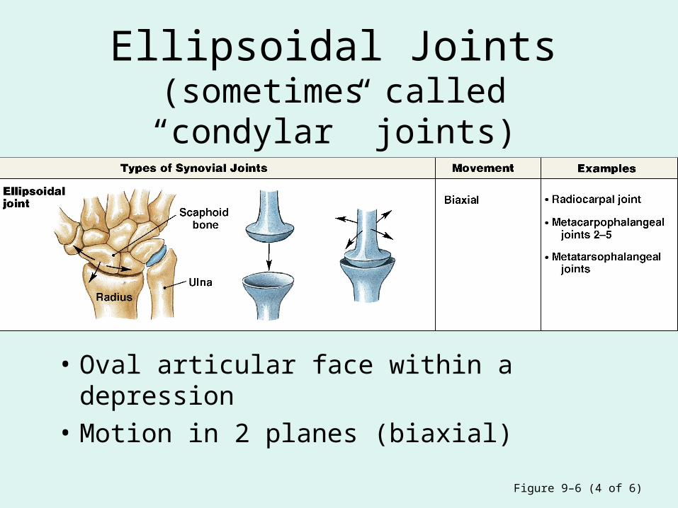
Ellipsoidal Joints(sometimes called “condylar” joints)
• Oval articular face within a depression
• Motion in 2 planes (biaxial)
Figure 9–6 (4 of 6)
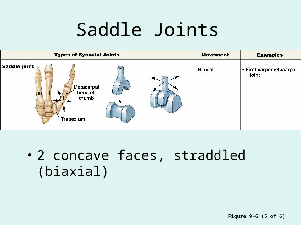
Saddle Joints
• 2 concave faces, straddled (biaxial)
Figure 9–6 (5 of 6)
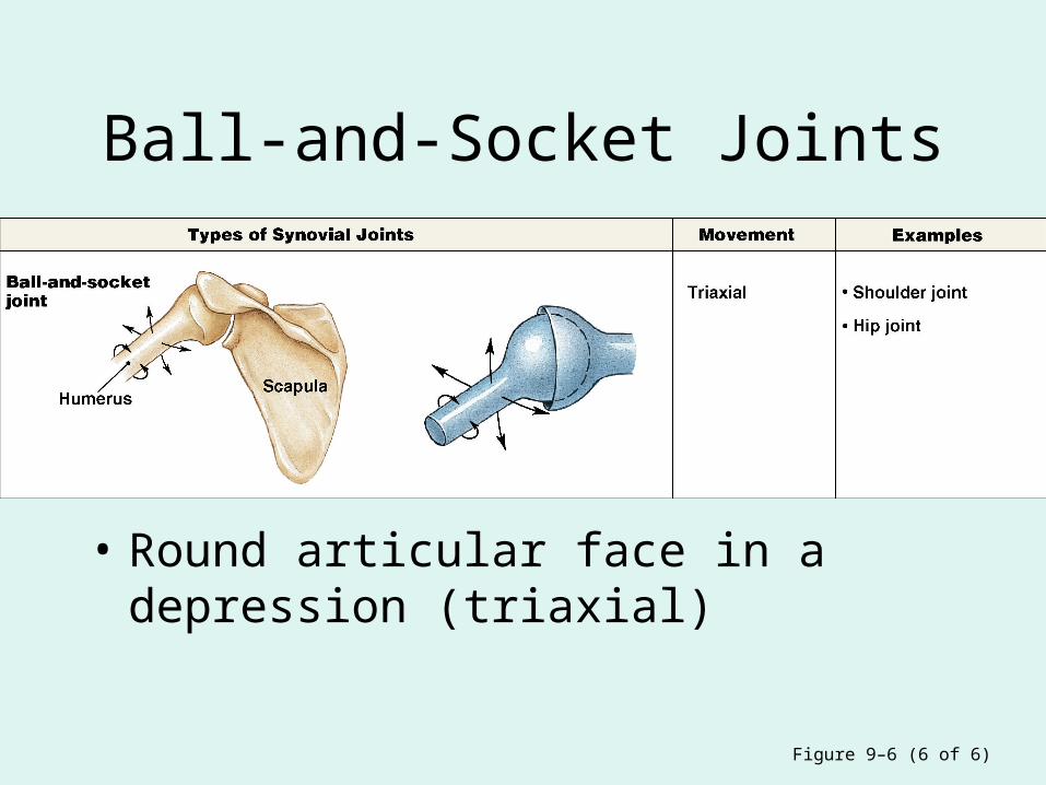
Ball-and-Socket Joints
• Round articular face in a depression (triaxial)
Figure 9–6 (6 of 6)
Structural Details of Some Synovial Joints
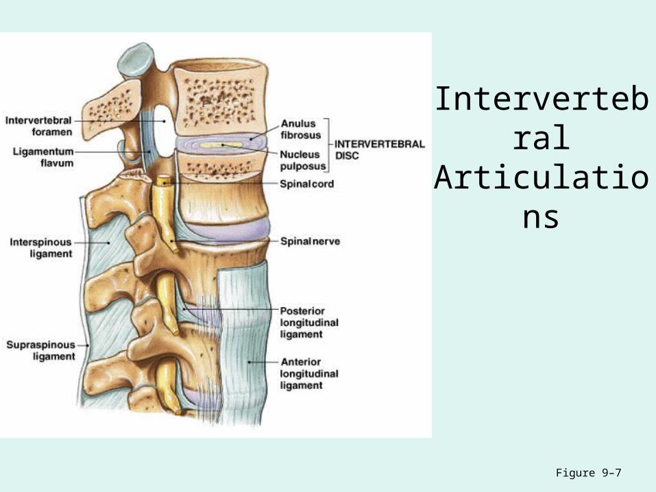
Intervertebral Articulations
Figure 9–7

Intervertebral Articulations
• C2 to L5 spinal vertebrae articulate:
– at inferior and superior articular processes (gliding joints)
– between adjacent vertebral bodies (symphyseal joints)
Disc Structure
• Anulus fibrosus:– tough outer layer– attaches disc to vertebrae
• Nucleus pulposus:– elastic, gelatinous core – absorbs shocks
Verterbral Joints
• Also called symphyseal joints
• As vertebral column moves:– nucleus pulposus shifts– disc shape conforms to motion
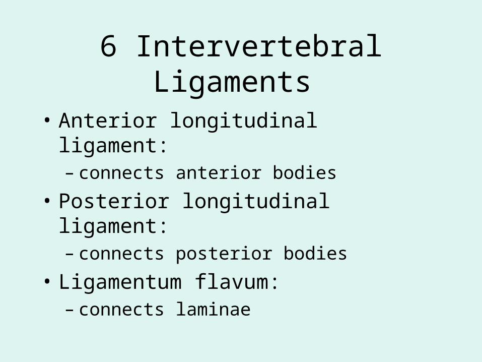
6 Intervertebral Ligaments
• Anterior longitudinal ligament:– connects anterior bodies
• Posterior longitudinal ligament:– connects posterior bodies
• Ligamentum flavum:– connects laminae

6 Intervertebral Ligaments
• Interspinous ligament: – connects spinous processes
• Supraspinous ligament:– connects tips of spinous processes (C7 to
sacrum)
• Ligamentum nuchae:– continues supraspinous ligament (C7 to skull)
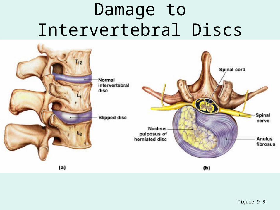
Damage to Intervertebral Discs
Figure 9–8
Damage to Intervertebral Discs
• Slipped disc:– bulge in anulus fibrosus – invades vertebral canal
• Herniated disc:– nucleus pulposus breaks through anulus
fibrosus– presses on spinal cord or nerves
Movements of the Vertebral Column
• Flexion:– bends anteriorly
• Extension:– bends posteriorly
• Lateral flexion:– bends laterally
• Rotation
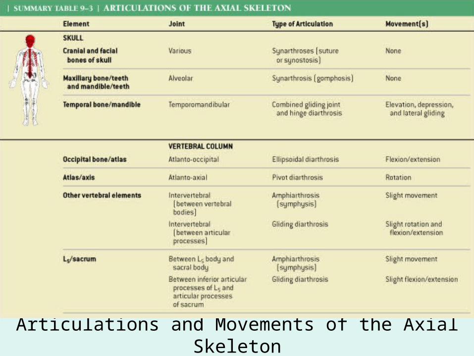
Articulations and Movements of the Axial Skeleton
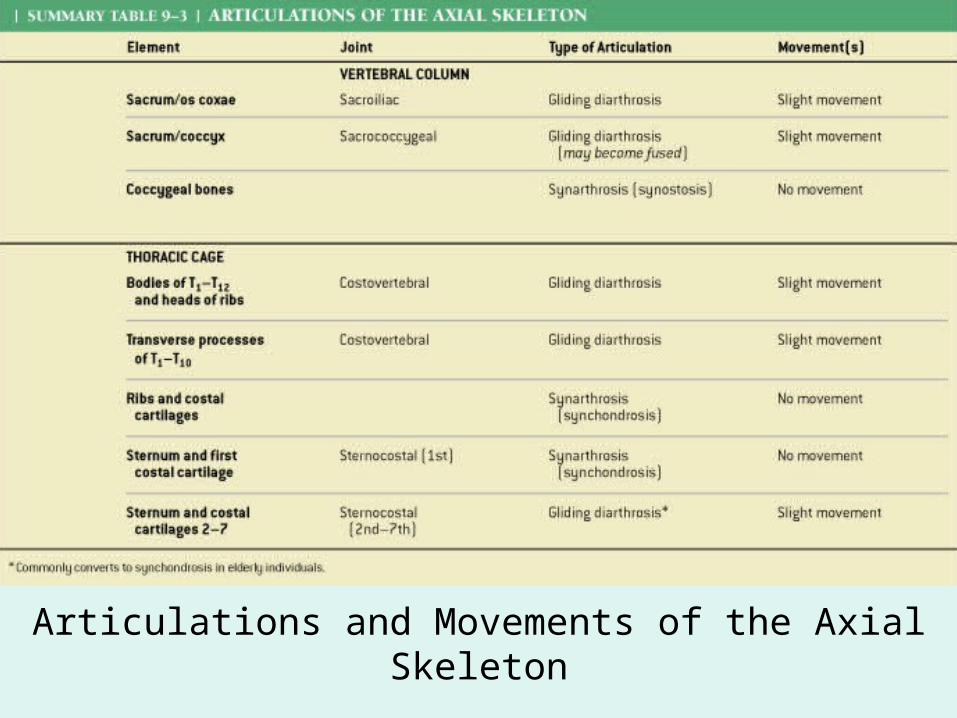
Articulations and Movements of the Axial Skeleton

The Shoulder
Joint
Figure 9–9a
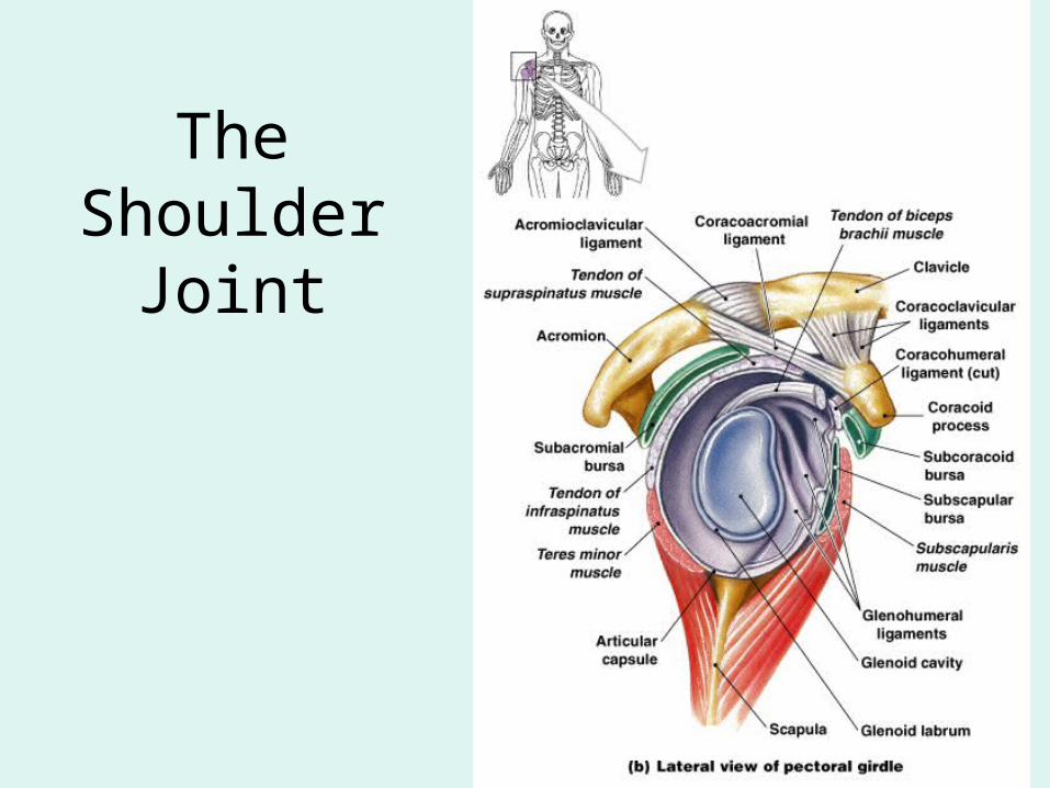
The Shoulder Joint
Figure 9–9b
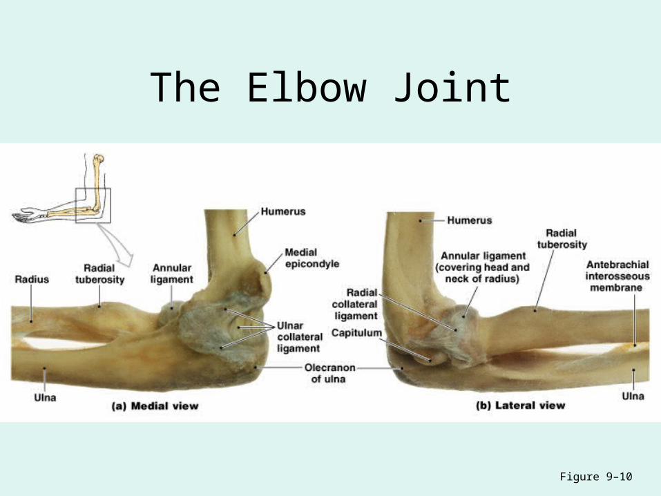
The Elbow Joint
Figure 9–10
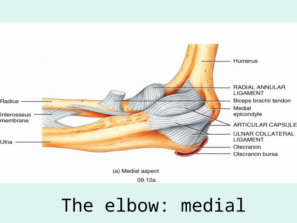
The elbow: medial
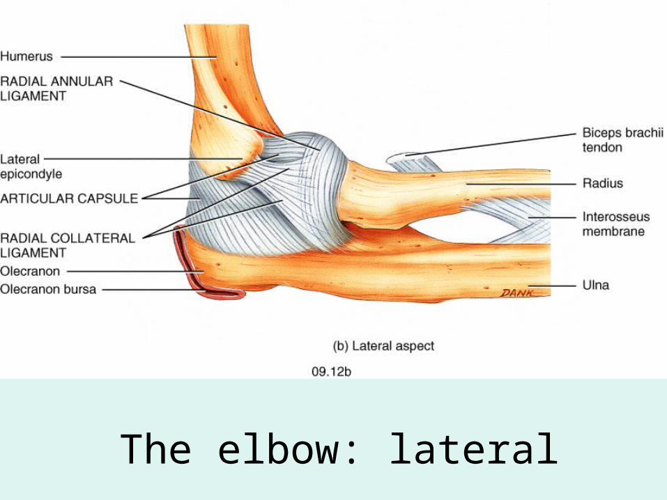
Fig. 09.12b
The elbow: lateral

The Hip JointFigure 9–11a
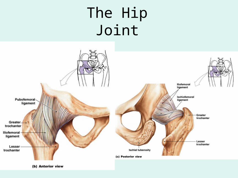
The Hip Joint

The Knee Joint
Figure 9–12a, b
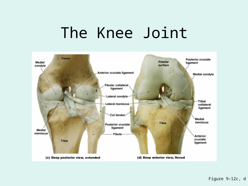
The Knee Joint
Figure 9–12c, d
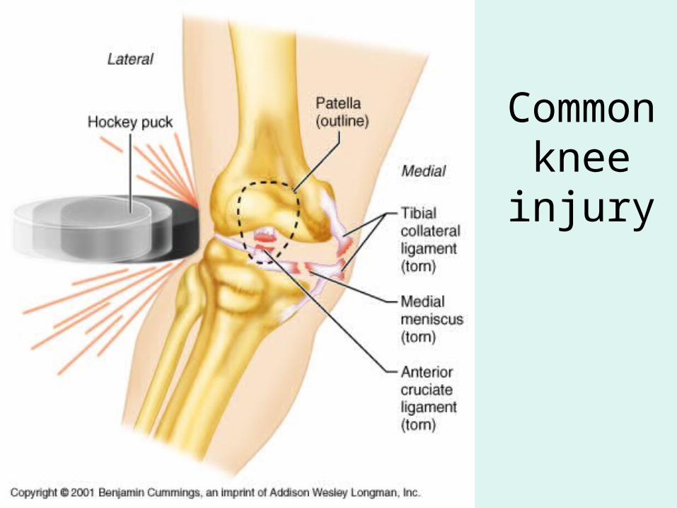
Common knee injury
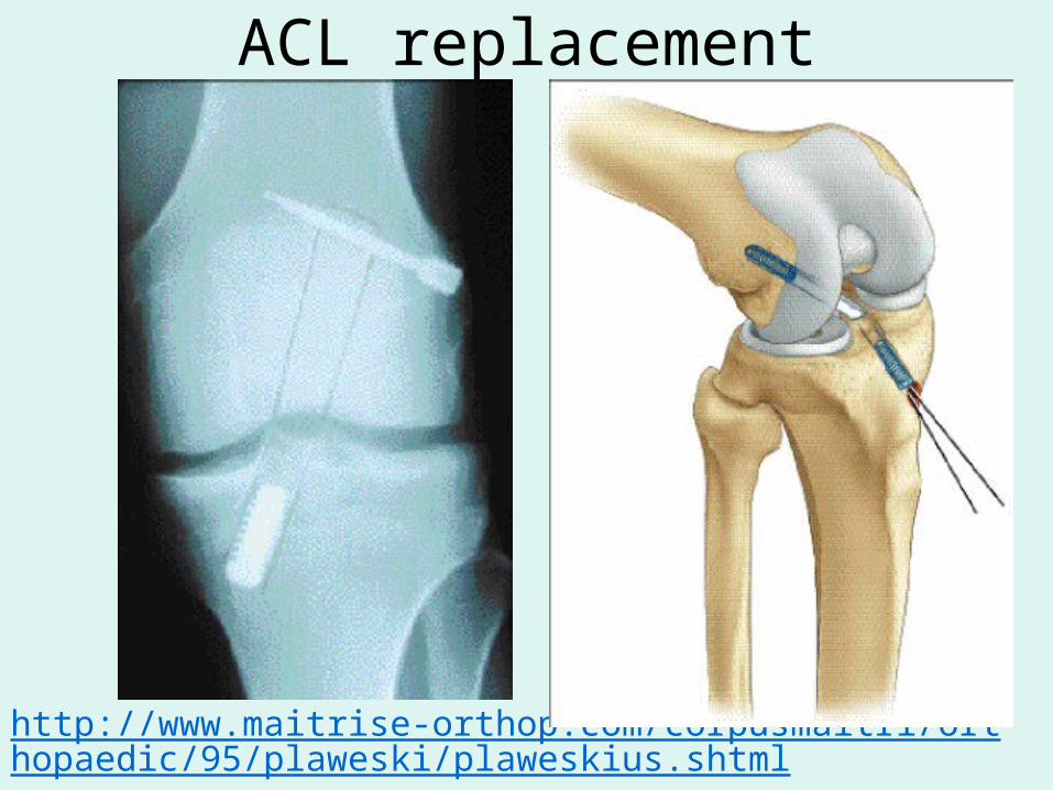
ACL replacement
http://www.maitrise-orthop.com/corpusmaitri/orthopaedic/95/plaweski/plaweskius.shtml
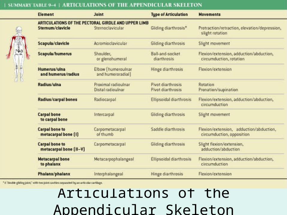
Articulations of the Appendicular Skeleton
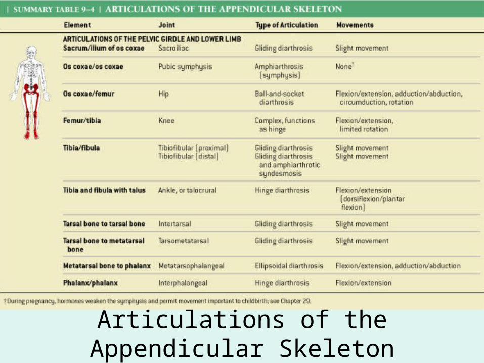
Articulations of the Appendicular Skeleton
Rheumatism• A pain and stiffness of skeletal and
muscular systems
Arthritis• All forms of rheumatism that damage
articular cartilages of synovial joints
Osteoarthritis
• Caused by wear and tear of joint surfaces, or genetic factors affecting collagen formation
• Generally in people over age 60

Rheumatoid Arthritis
• An inflammatory condition
• Caused by infection, allergy, or autoimmune disease
• Involves the immune system
Gouty Arthritis
• Occurs when crystals (uric acid or calcium salts):– form within synovial fluid– due to metabolic disorders
Joint Immobilization
• Reduces flow of synovial fluid
• Can cause arthritis symptoms
• Treated by continuous passive motion (therapy)
Bones and Aging
• Bone mass decreases
• Bones weaken
• Increases risk of hip fracture, hip dislocation, or pelvic fracture

No Mas