ascites as the initial characteristic manifestation in a … · gastric lymphoma accounts for 3%-5%...
TRANSCRIPT

1707
Introduction
Gastric lymphoma accounts for 3%-5% of all malignant tumors of the stomach1, which inclu-des primary tumor and disseminated lymphoma. Approximately 10% of non-Hodgkin’s lympho-mas involve the gastrointestinal tract at the time of initial evaluation2. Primary gastrointestinal lymphomas account for one-fourth of malignant lymphomas: the stomach and the small bowel are the most frequently involved sites. Gastric diffu-se large B-cell lymphoma (DLBCL) is the most commonly high malignant type in primary ga-stric lymphoma, yet the etiology for most cases of gastric lymphoma is unknown, although genetic and infections agent have been implicated.
So far, CD8-positive has been previously de-scribed in very few isolated case reports. Nogu-chi et al3 reported the case of gastric CD8+ DLBL with HTLV-1, and Toyama et al4 reported the first case of primary splenic CD8-positive DLBL. Pri-mary diffuse large B-cell lymphoma with ascites is very rare. Zenda et al5 reported a DLBCL case initially manifested as a form of effusion lympho-ma with lymphoma cells detected in ascites but only minimum solid tumor component. Overall, CD8-positive DLBCL with ascites as the initial major characteristics is very rarely reported.
Case Presentation A 78-year-old woman was admitted to Wuxi
Traditional Chinese Medicine Hospital on 15 March 2011 with complaints of abdominal disten-sion, scanty urine, fatigue, anorexia. She had no history of viral hepatitis, tuberculosis or schisto-somiasis. On physical examination, there found no enlarged superficial lymph nodes, abdominal tenderness or rebound tenderness, a palpable liver
Abstract. – CASE PRESENTATION: Diffuse large B-cell lymphoma (DLBCL) is the most com-mon lymphoid malignancy and the most common type of non-Hodgkin’s lymphomas, the stomach is the most common extranodal site. Gastric DL-BCL is often characterized by epigastric pain and vomiting. We report a case of a 78-year-old female patient with gastric diffuse large B-cell lympho-ma (DLBCL) with high CD8 level which was initial-ly manifested with ascites of unknown origin. The patient was admitted with a chief complaint of ab-dominal distension and scanty urine over the last twenty days, while without anorexia and fatigue until 15 March. She had no history of viral hepati-tis, tuberculosis, schistosomiasis.
RESULTS: Laboratory data revealed normal aminotransferases and bilirubin levels, but se-rum lactate dehydrogenase, CA125, ascitic fluid lactate dehydrogenase, ascitic fluid lymphocytes increased. The ascitic fluid was yellow-colored with 98.5% lymphocytes. Stool occult blood test was positive. Upper gastrointestinal endosco-py performed a few days later revealed multiple gastric crateriform ulcers, and Helicobacter py-lori was detected in the biopsy specimen. Pe-ripheral blood CD8+ was increased by 51%. Pa-thology test showed lymphocytes with atypical hyperplasia, and immunohistochemistry test re-sulted CD20+, CD10-, CD79α+, κ+, bcl-6+, Ki-67+ (approximately 95%), λ-, bcl-2-, CD3-, CD43-. Im-munoglobulin gene (Ig) clonal rearrangement showed IgH: FR1 (+), FR2 (+), FR3(-), Igk: VJ(+), Vkde (+) in lymphoma tissue.
CONCLUSIONS: The features of histopatholo-gy and immunohistochemistry of the tissue con-firmed diffuse large B-cell lymphoma (DLBL). The patient received an uncompleted CHOP pro-gram combined with H. pylori eradication. How-ever, the patient deceased due to disease devel-opment sixteen days later after the diagnosis.
Key Words:Primary gastric diffuse large B-cell lymphoma,
CD8-positive, Initial characteristic manifestation, Ascites.
European Review for Medical and Pharmacological Sciences 2016; 20: 1707-1711
K.-X. ZHAO1, G.-Z. DAI1, J.-F. ZHU2
1Department of Gastroenterology, Wuxi Traditional Chinese Medicine Hospital, Jiangsu, China 2Department of Gastroenterology, Wuxi Traditional Chinese Medicine Hospital, Jiangsu, China
Corresponding Author: Kexue Zhao, MD; e-mail: [email protected]
Ascites as the initial characteristic manifestation in a patient withprimary gastric CD8-positive diffuselarge B-cell lymphoma

K.-X. Zhao, G.-Z. Dai, J.-F. Zhu
1708
and a palpable spleen. There was an exposure of varication in the abdominal wall, in combination with a distended abdomen with shifting dullness. Edema was found in the patient’s lower limbs, especially the left limb. The patient was afebrile, and cardiac pulse and blood pressure were wi-thin normal range. Blood routine test showed that white blood cell (WBC) was 5.96x10⁹/L (80.4% neutrophil, 13% lymphocyte), red blood cell (RBC) 3.30x10¹²/L, hemoglobin (Hb) 100 g/L, and platelet (PLT) 305x109/L. Prothrombin time was 16.5 s. Serologic studies for HIV and HCV were negative, and HBeAb and HBcAb were positive. Liver function tests were: lactate dehy-drogenase 746 IU/L (normal 105-245 IU/L), cho-linesterase 4215 U/L (normal 4500-13000 U/L), total protein 53.5 g/L (60-85 g/L), albumin 29.3 g/L (35-55 g/L), prealbumin 0.102 g/L (0.20-0.40 g/L). Other parameters were within normal range. Renal function and electrolytes levels were nor-mal. Ascites was drained with abdominocentesis and tested: yellow-colored, turbid, tumor cell (-), mesothelial cell (+), lymphocytes (+), Rivalta test (+), white blood cell 18.75x10⁹/L, red blood cell 3.5x10⁹/L, neutrophil 1.5%, lymphocyte 98.5%, lactate dehydrogenase 4483 IU/L (normal 105-245 U/L), adenosine deaminase 64.9 U/L (nor-mal 0-24 U/L), alpha fetal protein (AFP) (-), car-cino-embryonic antigen (CEA) (-), carbohydrate antigen 19-9 (CA199) (-). Bacteria culture result of the ascites was negative. Serology tumor mar-ker test results were: alpha fetal protein (AFP) 2.54 ng/ml (normal 0-13.4 ng/ml), carcino-em-bryonic antigen (CEA) 0.789 ng/ml (normal 0-10 ng/ml), carbohydrate antigen 153 (CA153) 13.41 U/ml (normal 0-25 U/ml), carbohydrate antigen 19-9 (CA199) 5.59 U/ml (normal 0-27 U/ml), and carbohydrate antigen 72-4 (CA72.4) 0.534 U/ml (normal 0-6.9 U/ml), and an increased carbohy-drate antigen (CA125) 566.9 U/ml (normal 0-27 U/ml). The PPD (purified protein derivative) skin test and tubercle bacillus smear were negative, Doppler ultrasound showed that there was no thromboembolism in the limbs. Serum cellular immune function: CD16+: 27% (normal 12.5-29.2%), CD3+: 68% (normal 60.7-77.2%), CD19+: 3% (normal 5-14.7%), CD4+: 16% (normal 27.3-42.6%), Th/Ts: 0.31 (normal 0.9-2.2), CD8+: 51% (normal 19.2-30.5%).
A current-generation multislice helical com-puted tomography (CT) scan using intravenous contrast material called compound meglumine in chest and abdomen showed a suspicion of ascen-ding colon cancer which maybe metastasized to
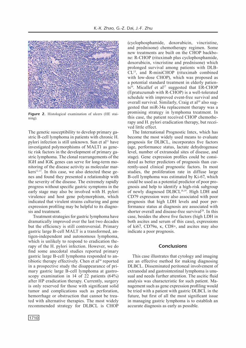
kidneys and adrenal glands, little pleural effusion in the left lower thorax, small hemangioma in the left liver, gallbladder stones, cholecystitis massive ascites, but no enlarged lymph nodes. Three sto-ol routine tests showed that the color was yellow, shape and properties was soft, yeasts were found in the first stool sample, occult blood was negati-ve. After taking live combined Bifidobacterium lactobacillus, and Enterococcus (420 mg, q8h) for three days, yeasts were no longer found in the se-cond stool sample, and occult blood was slightly positive. Occult blood turned negative without acid inhibitors or hemostatic. Upper gastrointesti-nal endoscopy was performed on March 23, mul-tiple gastric crateriform ulcers (fundus, body, and antrum of the stomach, about 1.5x1.5 cm multiple ulcers, surrounding with elevated crater-like mu-cosa) were found (Figure 1). A biopsy was taken from the border of the antrum and gastric fundus ulcers shew lymphocytes with atypical hyperpla-sia (Figure 2). H. pylori was detected from antral biopsy specimens using urease testing. Immu-nohistochemistry detected expression of CD20+, CD10-, CD79+, κ+, bcl-6+, Ki-67+ (approximately 95%), λ-, bcl-2-, MUM1-, CD3-, CD43- in lym-phoma tissue. The results of clonal rearrangement of the immunoglobulin (Ig) gene in lymphoma tissue were IgH: FR1 (+), FR2 (+), FR3 (-), Igk: VJ (+), Vkde (+). The results of pathology, im-munohistochemistry, gene rearrangements were consistent with diffusive large B-cell lymphoma. The comprehensive analysis confirmed diagnose of diffusive large B-cell lymphoma. Furthermore, capsule endoscopy did not detect tumors, or an-giomas, ulcer or bleeding. Colonoscopy showed
Figure 1. Panendoscopy: multiple crater-like gastric ulcers observed under upper gastrointestinal endoscopy (1.5 cm x 1.5 cm) was suspected to be malignancy.

Ascites as the initial characteristic manifestation in a patient with primary gastric CD8-positive DLBL
1709
numerous diverticula in the caecus, ulcer in the ileocecal valve, and biopsy pathology of ileocecal valve showed chronic inflammation with local dy-splastic changes of local lymphocytes.
After she had been admitted to the hospital, she received palliative and symptomatic treatments, including salt restriction and diuretics. On the fi-fth day of admission, she had a moderate fever (37.8○C), with no chills, rigors, abdominal pain or cough. Blood routine test showed WBC 9.25x10⁹/L (neutrophil 88.41%, lymphocyte 7.02%), RBC 3.08x10¹²/L, Hb 92 g/L, PLT 363.0x10⁹/L, and cul-tures of blood were negative. Epstein-Barr virus detection was negative. The patient refused a sur-gical treatment and a CHOP program at first. But after being told that triple therapy might provide a better prognosis, she took esomeprazole (20 mg, q12h), amoxicillin (1 g, q12h), and clarithromycin (500 mg, q12h). The treatment only lasted one day due to severe nausea and vomiting. On March 26, the renal function tests found that urea nitrogen (BUN) increased to 11.51 mmol/L (normal 2.1-7.9 mmol/L), creatinine (Cr) increased to 425.9 μmol/L (normal 40.0-115 µmol/L), and uric acid (UA) was 783 µmol/L (normal 150-440 µmol/L). Then the patient decided to try CHOP program. Three days later the condition deteriorated. She decided to go home for supportive care. She died sixteen days later after being diagnosed with dif-fusive large B-cell lymphoma.
Discussion
Diffuse large B-cell lymphoma presenting as peritoneal carcinomatosis is rare, without defi-nite demographic characteristics. Usually, ga-stric diffuse large B-cell lymphoma (DLBCL) presents with most symptoms such as epigastric pain, vomiting, melena and fever. To the best of our knowledge, ascites is rarely reported as one of the manifestations of this disease at an advan-ced stage. Lack of specific symptoms often delays the diagnosis. The diagnosis mainly bases on ga-stro-biopsy, histological, immunohistochemical and genetic examination. Gastric diffuse large B-cell lymphoma with ascites as initial characte-ristic feature and CD8-positive is extremely rare.
It is well known that liver cirrhosis is the un-derlying cause of ascites in at least 80% of patien-ts, but other factors (e.g., heart failure, constricti-ve pericarditis, nephrotic syndrome, tuberculous peritonitis, peritoneal malignancy, and pancreatic duct leak) may be involved. In this case, all the
above causes were eliminated. Because of the increased serum lactate dehydrogenase, CA125, ascitic fluid lactate dehydrogenase, ascitic fluid lymphocytes and intermittent slight positive of occult blood, gastroscopy was performed. The endoscopy revealed multiple crateriform gastric ulcers measuring 1.5 cm x 1.5 cm in the fun-dus, body, and antrum of the stomach. H. pylo-ri was also detected in antral biopsy specimens. The results of immunohistochemistry showed CD20+, CD10-, CD79α+, κ+, bcl-6+, Ki-67+ (ap-proximately 95%) in the biopsy tissue. And the immunoglobulin (Ig) gene clonal rearrangement demonstrated IgH: FR1 (+), FR2 (+), Igk: VJ (+), Vkde (+). So the diagnosis of gastric diffuse large B-cell lymphoma (DLBCL) was affirmed. Me-anwhile, peripheral blood CD8 was highly incre-ased. This was the first reported case of primary gastric CD8-positive DLBL with ascites, which suggested that physician may consider the possi-bility of gastric DLBCL in patients with similar symptoms, such as ascites. We reviewed literatu-re reporting rare DLBCL cases and only find few hits about lymphomatosis with ascites. Weng and Wu6 reported one case of lymphoma presenting as peritoneal lymphomatosis with ascites, which was diagnosed with similar tests and also presen-ted with ascites; the patients also died soon after diagnosis.
For most cases of gastric lymphoma, etiology and pathogenesis are so far poorly understood and genetic and infections factors may be implicated. Helicobacter pylori (H. pylori) plays a role in the development of most mucosa-associated lym-phoid tissue (MALT) lymphoma in the stomach, which may be similarly implicated in the deve-lopment of DLBCL, but DLBCL rarely complete alleviates after eradication therapy alone7. Chro-nic inflammation may enhance the probability of malignant transformation via B cell proliferation in response to H. pylori-mediated by tumor-infil-trating T cells. One study8 founds that in patients who develop gastric lymphomas in response to H. pylori, virulent strains expressing CagA protein are preferentially associated with diffuse large B-cell lymphoma. MALT lymphoma may tran-sform to gastric DLBCL, but mechanisms still re-mains poorly understood. According to the WHO classification, low-grade MALT lymphoma with focal high-grade component constituted by “solid or sheet-like proliferations of transformed cells” were included as diffuse large B-cell lymphoma9. Craig et al10 elucidates a novel Myc- and FoxP1-de-pendent pathway of malignant transformation.
K.-X. Zhao, G.-Z. Dai, J.-F. Zhu
1710
The genetic susceptibility to develop primary ga-stric B-cell lymphoma in patients with chronic H. pylori infection is still unknown. Sun et al11 have investigated polymorphisms of MALT1 as gene-tic risk factors in the development of primary ga-stric lymphoma. The clonal rearrangements of the IGH and IGK genes can serve for long-term mo-nitoring of the disease activity as molecular mar-kers12,13. In this case, we also detected these ge-nes and found they presented a relationship with the severity of the disease. The extremely rapidly progress without specific gastric symptoms in the early stage may also be involved with H. pylori virulence and host gene polymorphism, which indicated that virulent strains culturing and gene expression profiling may be helpful to its diagno-sis and treatment.
Treatment strategies for gastric lymphoma have dramatically improved over the last two decades but the efficiency is still controversial. Primary gastric large B-cell MALT is a transformed, an-tigen-independent and autonomous lymphoma, which is unlikely to respond to eradication the-rapy of the H. pylori infection. However, we do find some anecdotal studies reported primary gastric large B-cell lymphoma responded to an-tibiotic therapy effectively. Chen et al14 reported in a prospective study the disappearance of pri-mary gastric large B-cell lymphoma at gastro-scopy examination in 14 of 22 patients (64%) after HP eradication therapy. Currently, surgery is only reserved for those with significant solid tumor and complications such as perforation, hemorrhage or obstruction that cannot be trea-ted with alternative therapies. The most widely recommended strategy for DLBCL is CHOP
(cyclophosphamide, doxorubicin, vincristine, and prednisone) chemotherapy regimen. Some new treatments are built on the CHOP backbo-ne: R-CHOP (rituximab plus cyclophosphamide, doxorubicin, vincristine and prednisone) which prolonged survival among patients with DLB-CL15, and R-miniCHOP (rituximab combined with low-dose CHOP), which was proposed as a potential standard treatment in elderly patien-ts16. Micallef et al17 suggested that ER-CHOP (Epratuzumab with R-CHOP) is a well-tolerated schedule with improved event-free survival and overall survival. Similarly, Craig et al10 also sug-gested that miR-34a replacement therapy was a promising strategy in lymphoma treatment. In this case, the patient received CHOP chemothe-rapy and H. pylori eradication therapy, but recei-ved little effect.
The International Prognostic Intex, which has become the most widely used means to evaluate prognosis for DLBCL, incorporates five factors (age, performance status, lactate dehydrogenase level, number of extranodal sites of disease, and stage). Gene expression profiles could be consi-dered as better predictors of prognosis than cur-rently-used clinical prognostic factors. In most studies, the proliferation rate in diffuse large B-cell lymphoma was estimated by Ki-67, which could be used as a potential predictor of poor pro-gnosis and help to identify a high-risk subgroup of newly diagnosed DLBCL18,19. High LDH and CD79 expression were also associated with poor prognosis that high LDH levels and poor per-formance status at diagnosis are associated with shorter overall and disease-free survival20. In this case, besides the above five factors (high LDH in both ascites and serum of this case), expressions of ki67, CD79α, κ, CD8+, and ascites may also indicate a poor prognosis.
Conclusions
This case illustrates that cytology and imaging are an effective method for making diagnosing DLBCL. Disseminated peritoneal involvement of extranodal and gastrointestinal lymphoma is unu-sual and needs further attention. The ascitic fluid analysis was characteristic for such patient. Ma-nagement such as gene expression profiling would be tried with a patient with gastric DLBCL in the future, but first of all the most significant issue in managing gastric lymphoma is to establish an accurate diagnosis as early as possible.
Figure 2. Histological examination of ulcers (HE stai-ning).
Ascites as the initial characteristic manifestation in a patient with primary gastric CD8-positive DLBL
1711
References
1) Ferrucci PF, Zucca e. Primary gastric lymphoma pathogenesis and treatment: what has changed over the past 10 years? Br J Haematol 2007; 136: 521-538.
2) Kim Y, cho o, Song S, Lee h, rhim h, Koh B. Perito-neal lymphomatosis: CT findings. Abdom Imaging 1998; 23: 87-90.
3) noguchi m, SaKamoto n, oKumura K, Sato n, Yamada K, iSoBe Y, Kato a, oShimi K. CD8-positive diffuse large B-cell lymphoma. Rinsho Ketsueki 2000; 41: 591-595.
4) toYama t, KuBuKi Y, SaSaKi h, hidaKa t, oKamoto m, SuZuKi m, tSuBouchi h. Primary splenic CD8-posi-tive diffuse large B-cell lymphoma. Rinsho Ket-sueki 2001; 42: 1187-1191.
5) Zenda t, tominaga K, choto S, oKada t, KaneKo S, minato h. Diffuse large B-cell lymphoma initially manifested by massive ascites and a small ga-stric lesion, clinically mimicking primary effusion lymphoma (PEL) in the abdominal cavity: a case report and review of the literature on Japanese PEL patients. Nihon Shokakibyo Gakkai Zasshi 2007; 104: 1772-1780.
6) Weng Sc, Wu cY. Lymphoma presenting as peri-toneal lymphomatosis with ascites. J Chin Med Assoc 2008; 71: 646-650.
7) huSSeLL t, iSaacSon Pg, craBtree Je, SPencer J. Heli-cobacter pylori-specific tumour-infiltrating T cells provide contact dependent help for the growth of malignant B cells in low-grade gastric lymphoma of mucosa-associated lymphoid tissue. J Pathol 1996; 178: 122-127.
8) deLchier Jc, Lamarque d, LevY m, tKouB em, co-Pie-Bergman c, deForgeS L, chaumette mt, haioun c. Helicobacter pylori and gastric lymphoma: high seroprevalence of CagA in diffuse large B-cell lymphoma but not in low-grade lymphoma of mu-cosa-associated lymphoid tissue type. Am J Ga-stroenterol 2001; 96: 2324-2328.
9) tohda S. Overview of lymphoid neoplasms in the fourth edition of the WHO classification. Rinsho Byori 2012; 60: 560-564.
10) craig vJ, cogLiatti SB, imig J, renner c, neuen-SchWander S, rehrauer h, SchLaPBach r, dirnhoFer S, tZanKov a, muLLer a. Myc-mediated repression of microRNA-34a promotes high-grade transfor-mation of B-cell lymphoma by dysregulation of FoxP1. Blood 2011; 117: 6227-6236
11) Sun cm, Luan cF. Overexpression of microRNA-21 in peripheral blood mononuclear cells of patients with B-cell non-Hodgkin’s lymphoma is associa-ted with disease stage and treatment outcome. Eur Rev Med Pharmacol Sci 2015; 19: 3397-3402.
12) KrSKova L, hiLSKa i, KoPecKa P, SouKuP J, Kodet r. Mo-lecular diagnosis of malignant B-cell lymphoma:
detection of clonality and a brief review of pro-blems with our results. Casopis lekaru ceskych 2004; 143: 191-194
13) Kido S, miYaZaKi K, toKunaga o. The relationship between primary gastric B-cell lymphoma and immunoglobulin heavy chain (IgH) gene rear-rangement--a histopathological study of primary gastric lymphomas. Pathol Res Pract 2003; 199: 647-658.
14) chen Lt, Lin Jt, tai JJ, chen gh, Yeh hZ, Yang SS, Wang hP, Kuo Sh, Sheu BS, Jan cm, Wang Wm, Wang te, Wu cW, chen cL, Su iJ, Whang-Peng J, cheng aL. Long-term results of anti-Helicobacter pylori therapy in early-stage gastric high-grade transformed MALT lymphoma. J Natl Cancer Inst 2005; 97: 1345-1353.
15) LenZ g, Wright g, dave SS, Xiao W, PoWeLL J, Zhao h, Xu W, tan B, goLdSchmidt n, iqBaL J, voSe J, BaSt m, Fu K, WeiSenBurger dd, greiner tc, armitage Jo, KYLe a, maY L, gaScoYne rd, connorS Jm, troen g, hoLte h, KvaLoY S, diericKX d, verhoeF g, deLaBie J, SmeLand eB, JareS P, martineZ a, LoPeZ-guiLLermo a, montSerrat e, camPo e, BraZieL rm, miLLer tP, rimSZa Lm, cooK Jr, PohLman B, SWeetenham J, tuBBS rr, FiSher ri, hart-mann e, roSenWaLd a, ott g, muLLer-hermeLinK hK, Wrench d, LiSter ta, JaFFe eS, WiLSon Wh, chan Wc, Staudt Lm, Lymphoma/Leukemia Molecular Profiling P. Stromal gene signatures in large-B-cell lympho-mas. N Engl J Med 2008; 359: 2313-2323.
16) PeYrade F, Jardin F, thieBLemont c, thYSS a, emiLe JF, caStaigne S, coiFFier B, haioun c, BoLogna S, FitouSSi o, LePeu g, Fruchart c, BordeSSouLe d, BLanc m, deLarue r, Janvier m, SaLLeS B, andre m, Fournier m, gauLard P, tiLLY h, Groupe d’Etude des Lympho-mes de l’Adulte i. Attenuated immunochemothe-rapy regimen (R-miniCHOP) in elderly patients older than 80 years with diffuse large B-cell lym-phoma: a multicentre, single-arm, phase 2 trial. Lancet Oncol 2011; 12: 460-468.
17) micaLLeF in, maurer mJ, WiSeman ga, niKcevich da, Kurtin PJ, cannon mW, PereZ dg, Soori gS, LinK BK, haBermann tm, WitZig te. Epratuzumab with rituximab, cyclophosphamide, doxorubicin, vincri-stine, and prednisone chemotherapy in patients with previously untreated diffuse large B-cell lym-phoma. Blood 2011; 118: 4053-4061.
18) chaBot-richardS dS, martin dr, mYerS oB, cZuch-LeWSKi dr, hunt Ke. Quantitative image analysis in the assessment of diffuse large B-cell lymphoma. Mod Pathol 2011; 24: 1598-1605.
19) gaudio F, giordano a, Perrone t, PaStore d, curci P, deLia m, naPoLi a, de’ riSi c, SPina a, ricco r, LiSo v, SPecchia g. High Ki67 index and bulky disease remain significant adverse prognostic factors in patients with diffuse large B cell lymphoma before and after the introduction of rituximab. Acta Hae-matol 2011; 126: 44-51.
20) medina-Franco h, germeS SS, maLdonado cL. Pro-gnostic factors in primary gastric lymphoma. Ann Surg Oncol 2007; 14: 2239-2245.