asfa meeting 2015 san antonio robin willis rn … · asfa meeting 2015 san antonio robin willis rn...
TRANSCRIPT
ASFA Meeting 2015 San Antonio
Robin Willis RN BSN HP
Red Cell Exchange Overview

Conflict of Interest
• No Disclosures

Overview
• Indications for Red Cell Exchange
• Procedural Goals
• Vascular Access
• Case Studies
• Isovolemic Hemodilution
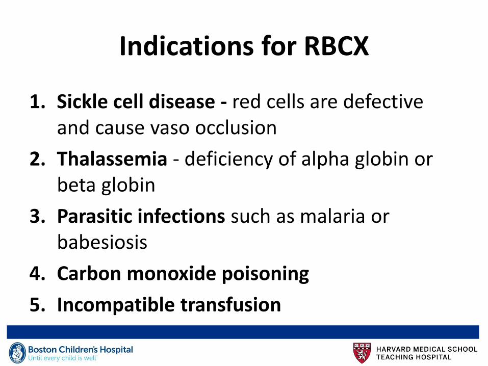
Indications for RBCX
1. Sickle cell disease - red cells are defective and cause vaso occlusion
2. Thalassemia - deficiency of alpha globin or beta globin
3. Parasitic infections such as malaria or babesiosis
4. Carbon monoxide poisoning
5. Incompatible transfusion
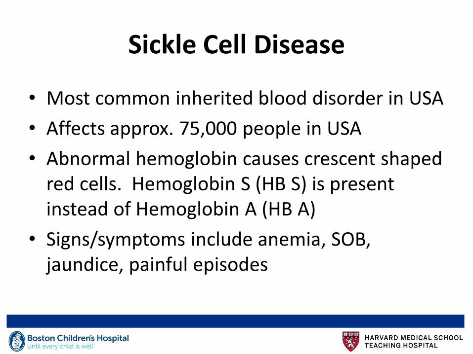
Sickle Cell Disease
• Most common inherited blood disorder in USA
• Affects approx. 75,000 people in USA
• Abnormal hemoglobin causes crescent shaped red cells. Hemoglobin S (HB S) is present instead of Hemoglobin A (HB A)
• Signs/symptoms include anemia, SOB, jaundice, painful episodes
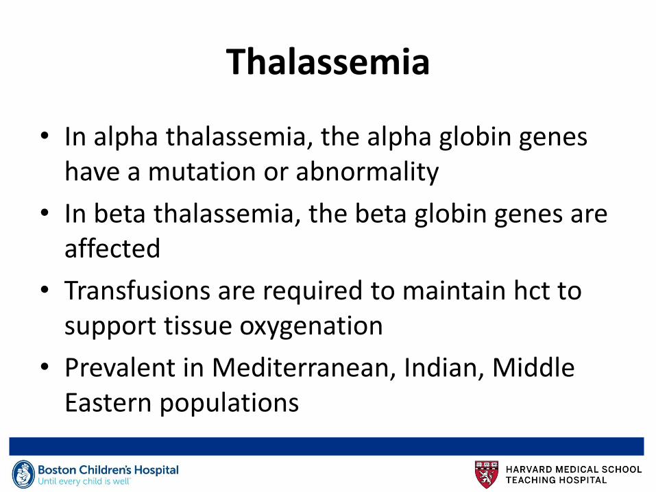
Thalassemia
• In alpha thalassemia, the alpha globin genes have a mutation or abnormality
• In beta thalassemia, the beta globin genes are affected
• Transfusions are required to maintain hct to support tissue oxygenation
• Prevalent in Mediterranean, Indian, Middle Eastern populations

Parasitic Infections
• Malaria- spread by mosquitoes infected with the Plasmodium Falciparum parasite - intraerythrocytic parasite (no longer indicated by the CDC)
• Babesiosis- spread by ticks infected with the protozoan parasite Babesia – intraerythrocytic parasite

Carbon Monoxide Poisoning
• Carbon Monoxide has an affinity for the heme molecule – RBC’s become saturated with CO
• Signs/Symptoms – most common symptoms - headache, dizziness, weakness, upset stomach, vomiting, chest pain, and confusion
• RBCX - removes the carbon dioxide saturated red cells and replaces with healthy blood donor cells
Incompatible Transfusion
• Example – Rh + red blood cells are transfused to Rh - recipient
• Goal - Remove Rh incompatible cells are removed from the recipient

Procedural Goals
• Removal of unhealthy cells with simultaneous replacement of healthy donor red blood cells
• Diminish pain episodes
• Stabilize hematocrit
• Improve respiratory status
• Maintain even fluid balance
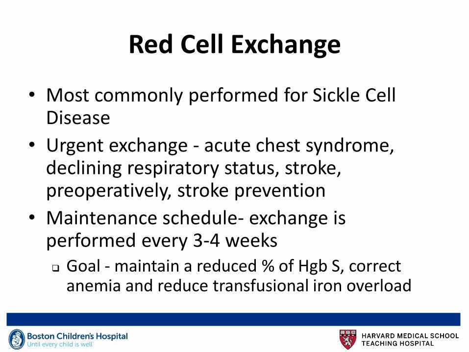
Red Cell Exchange
• Most commonly performed for Sickle Cell Disease
• Urgent exchange - acute chest syndrome, declining respiratory status, stroke, preoperatively, stroke prevention
• Maintenance schedule- exchange is performed every 3-4 weeks Goal - maintain a reduced % of Hgb S, correct
anemia and reduce transfusional iron overload
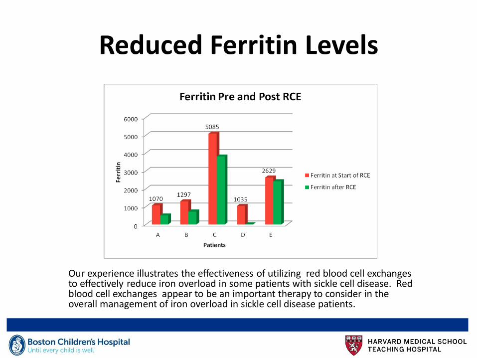
Reduced Ferritin Levels
Our experience illustrates the effectiveness of utilizing red blood cell exchanges to effectively reduce iron overload in some patients with sickle cell disease. Red blood cell exchanges appear to be an important therapy to consider in the overall management of iron overload in sickle cell disease patients.
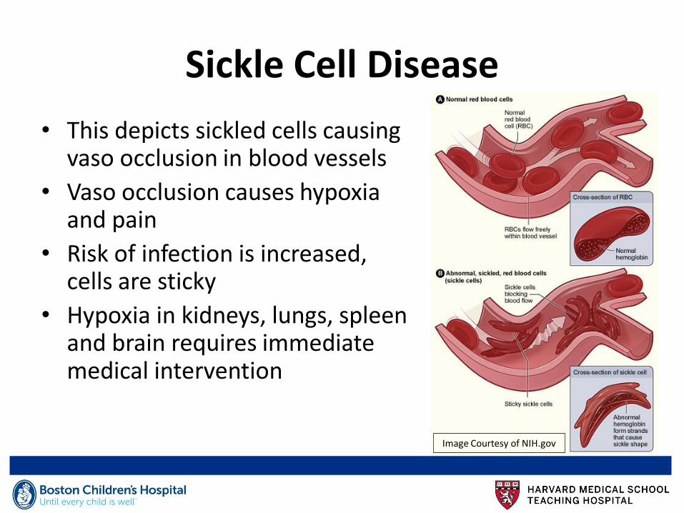
Sickle Cell Disease
• This depicts sickled cells causing vaso occlusion in blood vessels
• Vaso occlusion causes hypoxia and pain
• Risk of infection is increased, cells are sticky
• Hypoxia in kidneys, lungs, spleen and brain requires immediate medical intervention
Image Courtesy of NIH.gov

Complications of SCD
• As the cells sickle and congregate in the vessel, blood flow is blocked
• Vaso occlusive events can lead to:
Stroke
Acute chest syndrome
MSOF (Multi System Organ Failure)
Cholestatic Crisis (Bilirubin over 50)

Managing Sickle Cell Crisis
• Oxygen therapy to improve respiratory status
• IV fluids for hydration
• Antibiotics for potential/actual infection
• Opioids for pain management
• Red blood cell transfusion for anemia

Indications for RBC Exchange
• Declining respiratory status/↑WOB
• Anemia
• Increased percentage of HgS
• Pain crisis

Vascular Access
• Peripheral
Draw line 16 or 17 GA fistula needle
Return Line 18GA – 20GA angiocatheter
• Central
Central Venous Catheter (CVC) -Double Lumen Apheresis Catheter
Port - Vortex double lumen or single lumen port (one on each side of upper chest)

Temporary
Femoral
Medcomp 7F x 10 cm Pt ‹ 25 kg
Medcomp 9 F x 12 cm Pt › 25 kg
Temporary Subclavian/Internal Jugular
Mahurkar 8F x 12 cm (smaller pts) Quinton 8 F x 15 cm (smaller pts)
Mahurkar 10 F x 15 cm (small adults) (Quinton) 10 F x 19.5 cm (small adults)
Mahurkar 11.5 F x 13.5 cm (adult pts) (Quinton) 11.5 F x 16 cm (adult pts) 11.5 F x 19.5 cm (adult pts)
DL Central Venous Catheters Guidelines for Apheresis Treatments
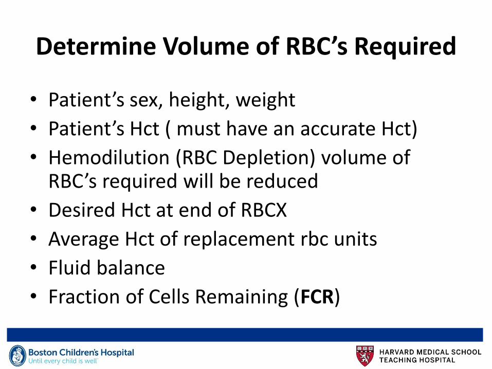
Determine Volume of RBC’s Required
• Patient’s sex, height, weight
• Patient’s Hct ( must have an accurate Hct)
• Hemodilution (RBC Depletion) volume of RBC’s required will be reduced
• Desired Hct at end of RBCX
• Average Hct of replacement rbc units
• Fluid balance
• Fraction of Cells Remaining (FCR)
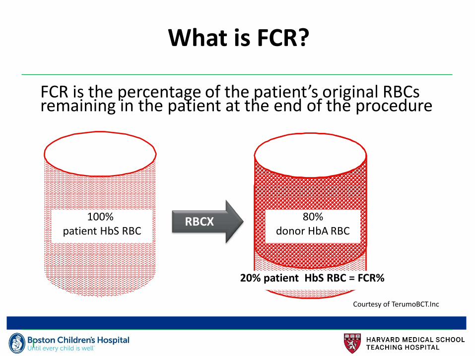
What is FCR?
FCR is the percentage of the patient’s original RBCs remaining in the patient at the end of the procedure
RBCX
20% patient HbS RBC = FCR%
80% donor HbA RBC
100% patient HbS RBC
Courtesy of TerumoBCT.Inc
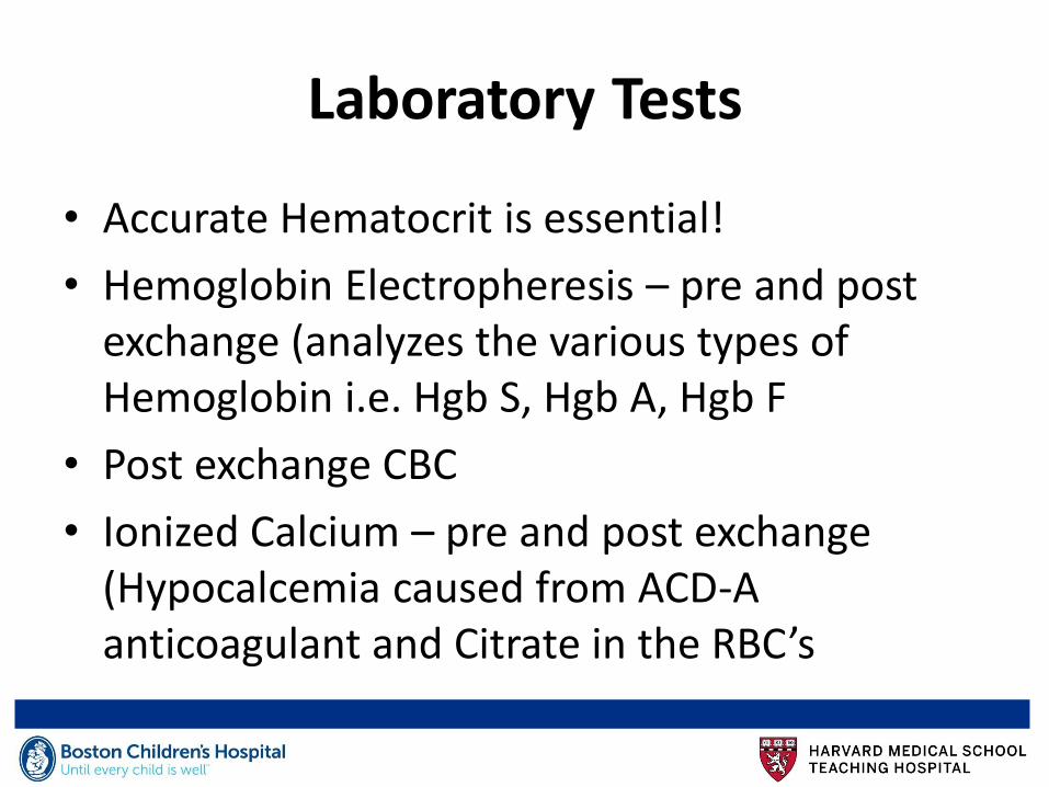
Laboratory Tests
• Accurate Hematocrit is essential!
• Hemoglobin Electropheresis – pre and post exchange (analyzes the various types of Hemoglobin i.e. Hgb S, Hgb A, Hgb F
• Post exchange CBC
• Ionized Calcium – pre and post exchange (Hypocalcemia caused from ACD-A anticoagulant and Citrate in the RBC’s
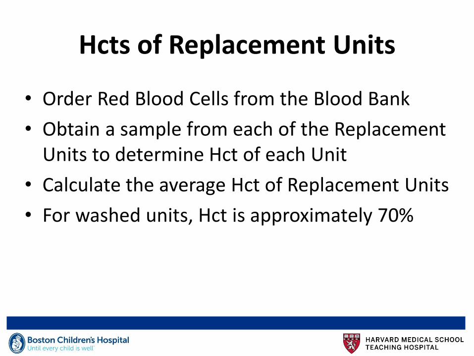
Hcts of Replacement Units
• Order Red Blood Cells from the Blood Bank
• Obtain a sample from each of the Replacement Units to determine Hct of each Unit
• Calculate the average Hct of Replacement Units
• For washed units, Hct is approximately 70%
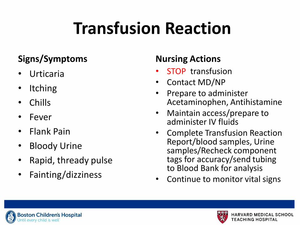
Transfusion Reaction
Signs/Symptoms
• Urticaria
• Itching
• Chills
• Fever
• Flank Pain
• Bloody Urine
• Rapid, thready pulse
• Fainting/dizziness
Nursing Actions • STOP transfusion • Contact MD/NP • Prepare to administer
Acetaminophen, Antihistamine • Maintain access/prepare to
administer IV fluids • Complete Transfusion Reaction
Report/blood samples, Urine samples/Recheck component tags for accuracy/send tubing to Blood Bank for analysis
• Continue to monitor vital signs
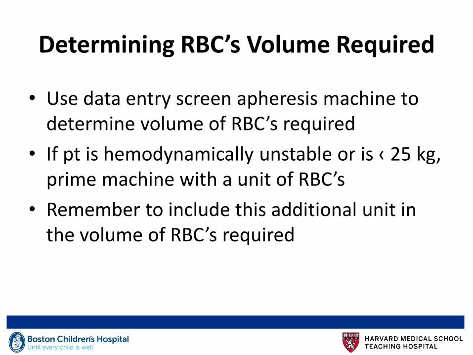
Determining RBC’s Volume Required
• Use data entry screen apheresis machine to determine volume of RBC’s required
• If pt is hemodynamically unstable or is ‹ 25 kg, prime machine with a unit of RBC’s
• Remember to include this additional unit in the volume of RBC’s required
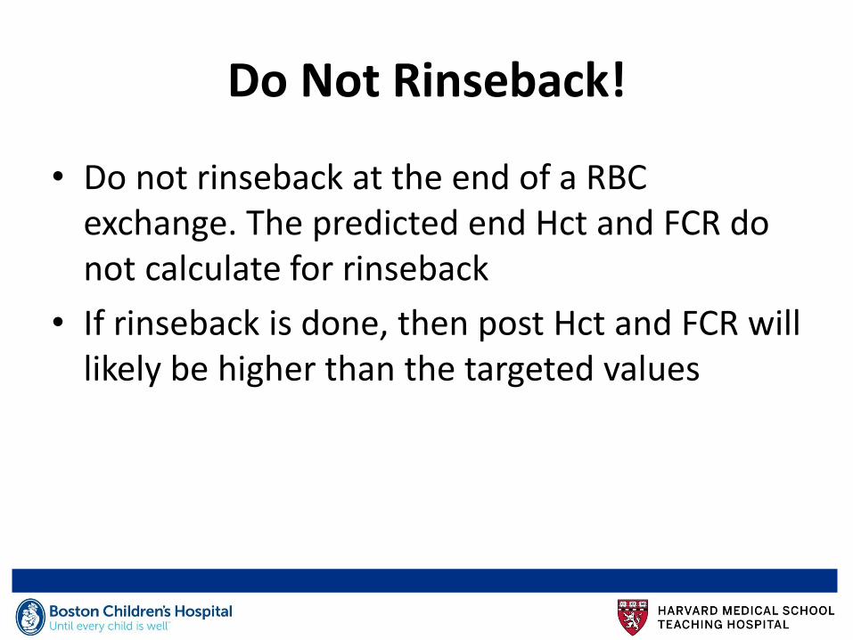
Do Not Rinseback!
• Do not rinseback at the end of a RBC exchange. The predicted end Hct and FCR do not calculate for rinseback
• If rinseback is done, then post Hct and FCR will likely be higher than the targeted values

Isovolemic Hemodilution
• Two part process in the RBC Exchange
• 1st Phase - RBC Depletion- red blood cells are removed with isovolemic replacement with 0.9% NaCl to reach the desired Hct
• 2nd Phase – Immediately following the RBC Depletion, RBC Exchange is initiated replacing with RBC’s

Advantages of Isovolemic Hemodilution
• Reduces the volume of RBC’s required for the RBCX
• Increases the efficiency of the RBCX by promoting further reduction of the Hgb S
• May increase the time interval between scheduled RBC exchanges

Case Study #1
• Male, 5’10” (178 cm) 75 kg (TBV 5082 mLs)
• Hct 26%
• End hct 30%
• Average hct of replacement units is 62%
• FCR 25%
• Fluid balance 100%
• Replace volume = 3106 mLs
• Replace volume with hemodilution = 2861mLs
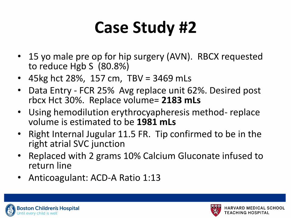
Case Study #2
• 15 yo male pre op for hip surgery (AVN). RBCX requested to reduce Hgb S (80.8%)
• 45kg hct 28%, 157 cm, TBV = 3469 mLs • Data Entry - FCR 25% Avg replace unit 62%. Desired post
rbcx Hct 30%. Replace volume= 2183 mLs • Using hemodilution erythrocyapheresis method- replace
volume is estimated to be 1981 mLs • Right Internal Jugular 11.5 FR. Tip confirmed to be in the
right atrial SVC junction • Replaced with 2 grams 10% Calcium Gluconate infused to
return line • Anticoagulant: ACD-A Ratio 1:13

Review of Case Study # 2 Post RBCX
• Flow rates 40-47 mLs/minute
• Run time 92 minutes
• Replace volume 2183 mLs
• Hct 30.0% Hgb S 21.4%
• Post RBC exchange - FCR 26.5%
• Plt count reduced by 59%

Case Study #3
• 3 yo 20.2 kg female with increased WOB/chest pain/abdominal pain. RLL infiltrate, hypoxia
• Hct 22.7% HgbS 46.1%
• Temporary (non tunneled) Left Subclavian Apheresis Catheter 7.0 FR, tip confirmed to be in upper SVC
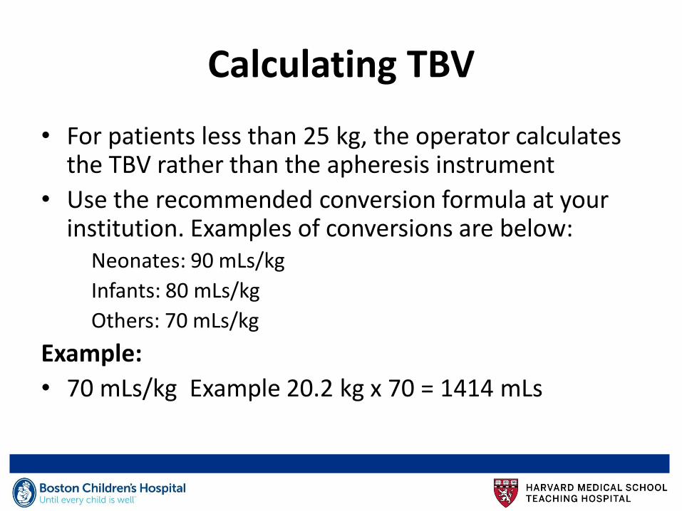
Calculating TBV
• For patients less than 25 kg, the operator calculates the TBV rather than the apheresis instrument
• Use the recommended conversion formula at your institution. Examples of conversions are below: Neonates: 90 mLs/kg
Infants: 80 mLs/kg
Others: 70 mLs/kg
Example:
• 70 mLs/kg Example 20.2 kg x 70 = 1414 mLs

Benefits of Blood Priming
• Maintains hemodynamic stability
• Maintains isovolemia
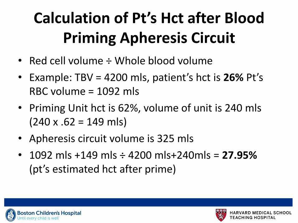
Calculation of Pt’s Hct after Blood Priming Apheresis Circuit
• Red cell volume ÷ Whole blood volume
• Example: TBV = 4200 mls, patient’s hct is 26% Pt’s RBC volume = 1092 mls
• Priming Unit hct is 62%, volume of unit is 240 mls (240 x .62 = 149 mls)
• Apheresis circuit volume is 325 mls
• 1092 mls +149 mls ÷ 4200 mls+240mls = 27.95% (pt’s estimated hct after prime)
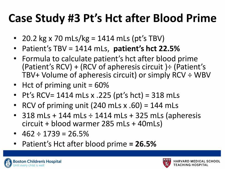
• 20.2 kg x 70 mLs/kg = 1414 mLs (pt’s TBV) • Patient’s TBV = 1414 mLs, patient’s hct 22.5% • Formula to calculate patient’s hct after blood prime
(Patient’s RCV) + (RCV of apheresis circuit )÷ (Patient’s TBV+ Volume of apheresis circuit) or simply RCV ÷ WBV
• Hct of priming unit = 60% • Pt’s RCV= 1414 mLs x .225 (pt’s hct) = 318 mLs • RCV of priming unit (240 mLs x .60) = 144 mLs • 318 mLs + 144 mLs ÷ 1414 mLs + 325 mLs (apheresis
circuit + blood warmer 285 mLs + 40mLs) • 462 ÷ 1739 = 26.5% • Patient’s Hct after blood prime = 26.5%
Case Study #3 Pt’s Hct after Blood Prime
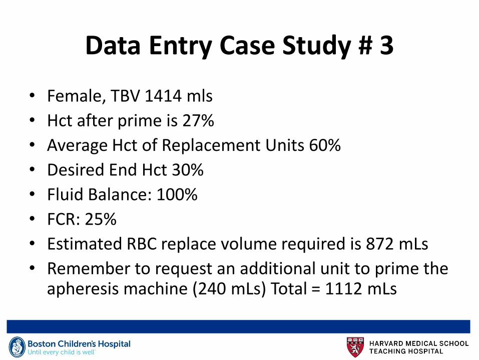
Data Entry Case Study # 3
• Female, TBV 1414 mls
• Hct after prime is 27%
• Average Hct of Replacement Units 60%
• Desired End Hct 30%
• Fluid Balance: 100%
• FCR: 25%
• Estimated RBC replace volume required is 872 mLs
• Remember to request an additional unit to prime the apheresis machine (240 mLs) Total = 1112 mLs
Thank You!
Acknowledgements
Apheresis Nurses
• Cheryl Pacheco
• Phaedra Truglia
• Lauren Salter
• Jennifer Strykowski
• Jill Dattilio
Physicians
• John Manis
• Steven Sloan
• Rick Kaufman
• Li Chai
• William Savage
Questions