asian archives of anaesthesiology and resuscitationaaarnaccm.com/issue 146.pdfcorrespond : asian...
TRANSCRIPT
146Asian Archives of Anaesthesiology and Resuscitation
1971-2012
CONTENTS
The Official Journal of “Anaesthesiology and Resuscitation Research Forum”
Volume 74 No. 2 April - June 2012
GUIDELINES TO CONTRIBUTORS7
(i)
2
1
3
4
6
5
Published and Printed by Dir. Prof. U.C.Verma on behalf of Asian Archives of Anaesthesiology and Resuscitation,
Office Address : Room No.: 306-309, Department of Anaesthesia,3rd Floor, BL Taneja Block, MAMC and LN Hospital, New Delhi
Mobile No.: 09646121503, 09868399699 E-mail : [email protected]
Typeset and Printed at Creative Offset Press, 131 Patperganj Industrial Area, Delhi -110092, Ph : 9136434848
NERVE STIMULATOR GUIDED SUPRASCAPULAR BLOCK IN
ADHESIVE CAPSULITIS: A NOVEL TECHNIQUE
Shailendra Singh Chauhan
2194
COMPARATIVE EVALUATION OF ATTENUATION OF POST- EXTUBATION
HYPERDYNAMIC RESPONSES WITH SINGLE DOSE DEXMEDETOMIDINE OR
ESMOLOL : A DOUBLE BLIND, RANDOMIZED, CONTROLLED TRIAL
Vansh Priya, P. S Malviya, L.S Mishra
2199
EASE OF FIBREOPTIC INTUBATION WITH TWO DIFFERENT
CONCENTRATION OF PROPOFOL AND KETAMINE
2206
Rajiv Gautam, B.K. Raw, L.S. Mishra, Gaurav Misra
CO-AMOXICLAV PROPHYLAXIS IN LSCS CAUSING ANAPHYLAXIS AND
INTRAUTERINE FETAL DEATH
Prasoon Gupta, Ranvinder Kaur, Lalita Chaudhary, Aruna Jain
2210
Maninder Kaur, Ruchi Gupta, Kuljit Singh Aujla, Chiteshwar Walia, Lakshmi Mahajan,
Nayyaamat K Sandhu
A STUDY TO EVALUATE THE EFFICACY OF PROSEAL LARYNGEAL MASK AIRWAY
AND LARYNGEAL TUBE SUCTION IN PATIENTS FOR SURGERY UNDER GENERAL
ANAESTHESIA WITH CONTROLLED VENTILATION
2215
A COMPARATIVE STUDY OF THE EFFECTS OF EPIDURAL BUPIVACAINE AND
BUPIVACAINE – FENTANYL MIXTURE IN LOWER ABDOMINAL SURGERY
2225
2236
www.aaarnaccm.com
Geeta Karki, Lalit Singh, S Moied Ahmed

Correspond : Asian Archives of Anaesthesiology and Resuscitation, Office Address :
Department of Anaesthesia,3rd Floor, BL Taneja Block, MAMC and LN Hospital, New Delhi
Mobile No.: 9868378740, 9871741419, 9868399699, 9646121503 E-mail: [email protected]
Room No. 306- 309,
(ii)
ASIAN ARCHIVES OF ANAESTHESIOLOGY AND RESUSCITATIONOFFICE BEARERS FOR 2010-2013
EDITORIAL BOARD
Editor-in-chief
Dir. Prof. U.C. Verma
Co-Editors
Dir. .Prof Baljit Singh
Executive DirectorDr. Yashwant Singh
Dir. r .P of R.S. Rautela
Dr. Manpreet Singh
Founder Member(Late) Prof. W.E. Sporel(Late) Prof. N.P. Singh(Late) Prof. S.D. Gupta
1. Dr. T.C.K. Brown Dept. of Anaesthesia Royal Childrens’ Hospital Melbourne 3502 (Australia)
2. Dr. Rashid M. Khan Sr. Consultant, Khoula Hospital, Muscat OMAN
3. Dr. Michael J.A. Parr MBBS, MRCP, FRCA, FANZCA Specialist in Intensive Care, Liverpool Hospital. Lecturer in Intensive Care, Anaesth and Emergency Medicine Intensive & Critical Care Medicine
MEMBERS (FOREIGN)
Exempted from Income Tax under Section 35 of Income Tax Act 1961 vide letter No. 1231 (F.N. DG/IT/E/ND/-81/35 (i),
(22)/90/-IT (E) of 26-10-94 from Dept. of Revenue, Min. of Finance, Govt. of India (1.4.93-31.396)
(iii)
Cheque/Cash.......................... Cheque No ............................... Date .................................. Amount ...............................
Office Address:306- 309,DEPARTMENT OF ANAESTHESIA 3RD FLOOR, BL TANEJA BLOCKMAMC and LN HOSPITAL,NEW DELHI,[email protected]
President - Dir. Prof. U.C. VermaVice President - Dir. Prof. Baljit SinghG. Secretary - Dr. Manpreet SinghJt. Secretary - Dir. Prof. R.S. RautelaTreasurer - Dr. Yashwant Singh
1. Prof. (Dir.) Rajiv Chawla, New Delhi
2. Prof. (Dir.) Deepak K. Tempe, New Delhi
3. Dr. S.C. Parakh, Hyderabad
4. Dr. Pramod Kumar, Jam Nagar
5. Prof. Dilip Pawar, New Delhi
6. Dr. V.P. Kumar, New Delhi
7. Dr. S.C. Manchanda, New Delhi
8. Dr. (Col.) S.K. Chadha, New Delhi
9. Prof. L.D. Mishra, Varanasi
10. Prof. H.C. Chandola, Allahabad
11. Prof. Shahjahan Bano, Aligarh
12. Dr. Lalit Maini, New Delhi
13. Prof. A.M. Hashia, Solan
14. Prof. Mridula Pawar, New Delhi
15. Dr. Sunila Sharma, New Delhi
16. Prof. S.M. Ahmad, Aligarh
17. Dr. Dheeraj Kapoor, Chandigarh
18. Prof. Lakesh Anand, Chandigarh
19. Dr. Deepak Thapa, Chandigarh
20. Prof. S.K. Malhotra, Chandigarh

Asian Archives of Anaesthesiology And Resuscitation 2194 Volume 74 No. 2 April (A-J) 2012
Shailendra Singh Chauhan
MD, Pain physician
Consultant Anaesthesiology & Pain Medicine, Jabalpur Pain & Spine Clinic, JABALPUR
NERVE STIMULATOR GUIDED SUPRASCAPULAR BLOCK IN
ADHESIVE CAPSULITIS: A NOVEL TECHNIQUE
Shailendra Singh Chauhan
Adhesive capsulitis, also known as 'Frozen
Shoulder' is characterized by spontaneous onset
of gradually progressive shoulder pain and
restriction of active and passive glenohumeral 1,2
movement in all planes. The current consensus
definition of American Shoulder and Elbow
Surgeons is “ a condition of uncertain etiology
characterized by significant restriction of both
active and passive shoulder motion that occurs in
the absence of a known intrinsic shoulder 3disorder” Relieving pain and restoring of normal
shoulder function are the common aims of various
methods used in the treatment of adhesive 4-7capsulitis. To achieve this goal, therapeutic
exercises are the most important part of the
treatment program. The most important factor
preventing active exercise is pain. Suprascapular
nerve block is effective for the management of 8-shoulder pain resulting from adhesive capsulitis.
10 There are various techniques to block 11
suprascapular nerve. These techniques either
depend on costly instruments or are blind &
technically difficult. This technique is simple &
effectively blocks suprascapular nerve as the
drug is injected at the point nearest to the nerve
guided by motor nerve stimulator.
EPIDEMIOLOGY & CLASSIFICATION
It is a common condition affecting around 2% to 55% of general population. It most commonly
affects women aged between 40 & 60 years often
presenting bilaterally.It has not been reported to
have any predilection with race.
Adhesive capsulitis is usually classified as
primary or secondary.It is called Primary if no
findings on history or examination explain the
onset of disease,may be related to hormonal or
immunologic changes.Secondary develops due
to stiffness & immobility caused by some trauma
or surgery.
The present study was conducted to evaluate
clinical efficacy of nerve stimulator guided
suprascapular nerve block techniques in patients
with adhesive capsulitis.
MATERIAL & METHODS
Eighty patients [male: female/55:25], in age group
[40 -65 years] suffering from adhesive capsulitis
were studied over a period of one year. All these
patients received supra-scapular nerve block via
above mentioned technique.The patients who
had pain & stiffness in shoulders [one or both] for
at least 4 weeks, aggravated by movements, who
had restricted active & passive range of
movements[ROM] at gleno-humeral joint,who
had sleep disturbance with increased pain when
sleeping on the affected site,were included.The
patients with no history of recent trauma & no
previous injection in the involved shoulder,no
history of allergy to local anaesthetics & steroids,
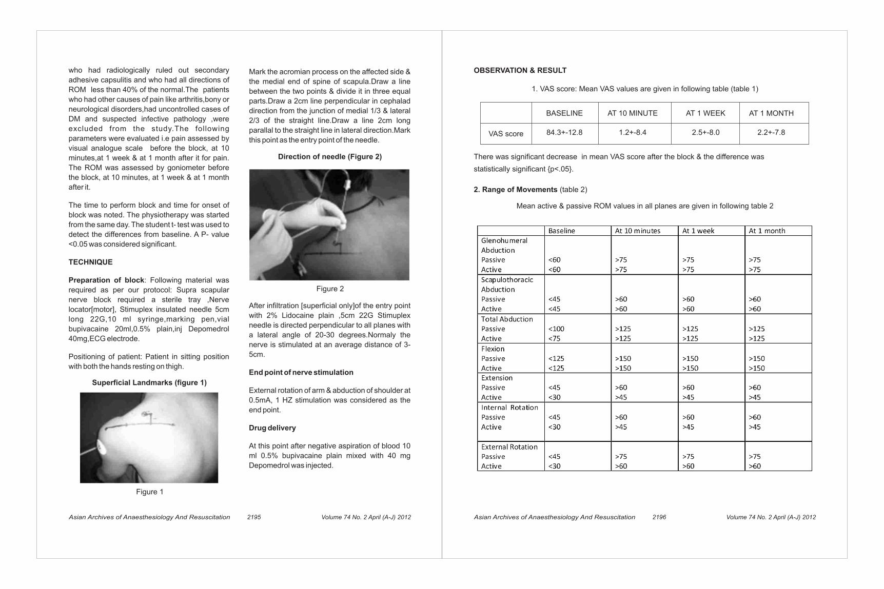
Mark the acromian process on the affected side &
the medial end of spine of scapula.Draw a line
between the two points & divide it in three equal
parts.Draw a 2cm line perpendicular in cephalad
direction from the junction of medial 1/3 & lateral
2/3 of the straight line.Draw a line 2cm long
parallal to the straight line in lateral direction.Mark
this point as the entry point of the needle.
Asian Archives of Anaesthesiology And Resuscitation 2195 Volume 74 No. 2 April (A-J) 2012
Superficial Landmarks (figure 1)
Figure 1
Figure 2
Direction of needle (Figure 2)
After infiltration [superficial only]of the entry point
with 2% Lidocaine plain ,5cm 22G Stimuplex
needle is directed perpendicular to all planes with
a lateral angle of 20-30 degrees.Normaly the
nerve is stimulated at an average distance of 3-
5cm.
End point of nerve stimulation
External rotation of arm & abduction of shoulder at
0.5mA, 1 HZ stimulation was considered as the
end point.
Drug delivery
At this point after negative aspiration of blood 10
ml 0.5% bupivacaine plain mixed with 40 mg
Depomedrol was injected.
Asian Archives of Anaesthesiology And Resuscitation 2196 Volume 74 No. 2 April (A-J) 2012
OBSERVATION & RESULT
1. VAS score: Mean VAS values are given in following table (table 1)
BASELINE AT 10 MINUTE AT 1 WEEK AT 1 MONTH
VAS score 84.3+-12.8 1.2+-8.4 2.5+-8.0 2.2+-7.8
There was significant decrease in mean VAS score after the block & the difference was
statistically significant {p<.05}.
2. Range of Movements (table 2)
Mean active & passive ROM values in all planes are given in following table 2
who had radiologically ruled out secondary
adhesive capsulitis and who had all directions of
ROM less than 40% of the normal.The patients
who had other causes of pain like arthritis,bony or
neurological disorders,had uncontrolled cases of
DM and suspected infective pathology ,were
excluded from the study.The following
parameters were evaluated i.e pain assessed by
visual analogue scale before the block, at 10
minutes,at 1 week & at 1 month after it for pain.
The ROM was assessed by goniometer before
the block, at 10 minutes, at 1 week & at 1 month
after it.
The time to perform block and time for onset of
block was noted. The physiotherapy was started
from the same day. The student t- test was used to
detect the differences from baseline. A P- value
<0.05 was considered significant.
TECHNIQUE
Preparation of block: Following material was
required as per our protocol: Supra scapular
nerve block required a sterile tray ,Nerve
locator[motor], Stimuplex insulated needle 5cm
long 22G,10 ml syringe,marking pen,vial
bupivacaine 20ml,0.5% plain,inj Depomedrol
40mg,ECG electrode.
Positioning of patient: Patient in sitting position
with both the hands resting on thigh.

techniques which provide comparable results are
either costly or are not widely available. This
technique is very effective, less time consuming
and easy to use.
To determine the minimum effective dosage for
the nerve block, more comparative studies are
needed. Proven effective in adhesive capsulitis in
the present study, we believe the motor nerve
locator guided technique can also be used in
appropriate cases of shoulder pain caused by
pathologies other than adhesive capsulitis.
REFERENCES
1. Reeves B. The natural history of frozen shoulder syndrome. Scand J Rheumatol.
1975;4:193–196
2. Rizk TE, Pinals RS. Frozen shoulder. Semin
Arthritis Rheum. 1982;11:440–452
3. Zuckerman etal.Definition and classification
of frozen shoulder: a consensus approach.J
Shoulder Elbow Surg 1994;3:S72
4. Hill JJ .Manipulation in the treatement of
frozen shoulder.Orthopaedics 1988;11:1255-
60
5. Murnaghan JP.Adhesive capsulitis of
shoulder: Current concepts and treat-
ment. Orthopaedics 1988;11:153-8
6. Rizk TE .Adhesive capsulitis [frozen
shoulder]:A new approach to its manage-
ment. Arch Phys Med Rehabil 1983;64:29-33
7. Older MWJ .Distention arthrography of the
shoulder joint Can J Surg 1976;19:203-7
8. Bonica JJ, Buckley FP. Regional analgesia
with local anesthetics. In: Bonica JJ editors.
The management of pain. Philadelphia: : Lea
& Febiger; 1990;p. 1883–1966
There was significant increase in range of
movements after the block & the difference was
statistically significant {p<.05}.Average onset of
time of block was 8.5 minutes & range was from 6
to 10 minutes.
Average time taken to perform block was 6.2
minutes. No significant adverse effects were
noted
DISCUSSION
12
9-14
Shoulder pain and restriction of glenohumeral
movements are the main clinical findings in
adhesive capsulitis. Considering the functional
disability, the most important components of the
treatment are pain relief and therapeutic
exercises for early mobilization.
A simple and effective regional nerve block
method for shoulder pain is the suprascapular
nerve block.Nerve locator guided technique was
chosen for more accuracy, easy availability &
presence of motor f ibres innervat ing
supraspinatus & infraspinatus muscle in
suprascapular nerve.
The most appropriate site is around the
suprascapular notch, in which the nerve can also
be located easily.That decided the entry point of
needle insertion.
Various suprascapular nerve block techniques
have been described by several investigators.
This type of approach is easy,delivers drug in near
viscinity of nerve hence more effective than other
blind techniques and decreases the risk of
pneumothorax.
CONCLUSION
This new motor nerve locator guided technique
enables more accurate placement of needle tip
near nerve thus a more successful nerve block as
compared to classical blind technique.Other
12. Wolf JM.Influence of comorbidity on self
assessment instruments scores of patients
with idiopathic adhesive capsulitis.J Bone
Joint Surg Am 2002;84:1167-73
13. Parris WC. Suprascapular nerve block: a
safer technique. Anesthesiology. 1990;
72:580–581
14. Bulgen DY, Binder AI, Hazleman BL, Dutton J,
Roberts S. Frozen shoulder: prospective
clinical study with an evaluation of three
treatment regimens. Ann Rheum Dis.
1984;43:353–360
9. Brown DE, James DC, Roy S. Pain relief by
suprascapular nerve block in gleno-humeral
arthritis. Scand J Rheumatol. 1988;
17:411–415
10. Dahan TH, Fortin L, Pelletier M, Petit M,
Vadeboncoeur R, Suissa S. Double blind
randomized clinical trial examining the
efficacy of bupivacaine suprascapular nerve
blocks in frozen shoulder. J Rheumatol.
2000;27:1464–1469
11. Gado K, Emery P. Modified suprascapular
nerve block with bupivacaine alone effectively
controls chronic shoulder pain in patients with
rheumatoid arthritis. Ann Rheum Dis.
1993;52:215–218
Asian Archives of Anaesthesiology And Resuscitation 2198Asian Archives of Anaesthesiology And Resuscitation 2197 Volume 74 No. 2 April (A-J) 2012Volume 74 No. 2 April (A-J) 2012
given slowly over a period of one minute, 5
minutes prior to reversal and extubation.
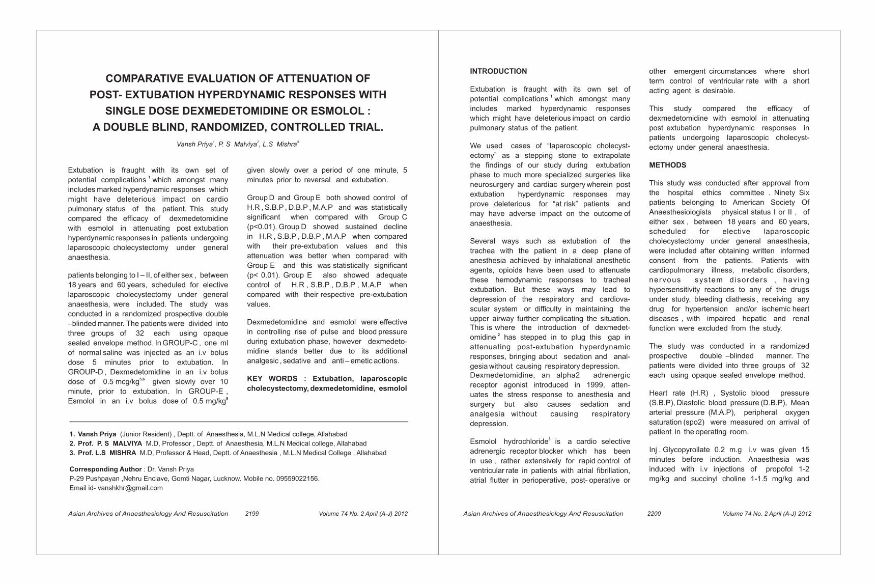
Group D and Group E both showed control of
H.R , S.B.P , D.B.P , M.A.P and was statistically
significant when compared with Group C
(p<0.01). Group D showed sustained decline
in H.R , S.B.P , D.B.P , M.A.P when compared
with their pre-extubation values and this
attenuation was better when compared with
Group E and this was statistically significant
(p< 0.01). Group E also showed adequate
control of H.R , S.B.P , D.B.P , M.A.P when
compared with their respective pre-extubation
values.
Dexmedetomidine and esmolol were effective
in controlling rise of pulse and blood pressure
during extubation phase, however dexmedeto-
midine stands better due to its additional
analgesic , sedative and anti – emetic actions.
KEY WORDS : Extubation, laparoscopic
cholecystectomy, dexmedetomidine, esmolol
Extubation is fraught with its own set of 1
potential complications which amongst many
includes marked hyperdynamic responses which
might have deleterious impact on cardio
pulmonary status of the patient. This study
compared the efficacy of dexmedetomidine
with esmolol in attenuating post extubation
hyperdynamic responses in patients undergoing
laparoscopic cholecystectomy under general
anaesthesia.
patients belonging to I – II, of either sex , between
18 years and 60 years, scheduled for elective
laparoscopic cholecystectomy under general
anaesthesia, were included. The study was
conducted in a randomized prospective double
–blinded manner. The patients were divided into
three groups of 32 each using opaque
sealed envelope method. In GROUP-C , one ml
of normal saline was injected as an i.v bolus
dose 5 minutes prior to extubation. In
GROUP-D , Dexmedetomidine in an i.v bolus 5,6dose of 0.5 mcg/kg given slowly over 10
minute, prior to extubation. In GROUP-E , 9
Esmolol in an i.v bolus dose of 0.5 mg/kg
other emergent circumstances where short
term control of ventricular rate with a short
acting agent is desirable.
This study compared the efficacy of
dexmedetomidine with esmolol in attenuating
post extubation hyperdynamic responses in
patients undergoing laparoscopic cholecyst-
ectomy under general anaesthesia.
METHODS
This study was conducted after approval from
the hospital ethics committee . Ninety Six
patients belonging to American Society Of
Anaesthesiologists physical status I or II , of
either sex , between 18 years and 60 years,
scheduled for elective laparoscopic
cholecystectomy under general anaesthesia,
were included after obtaining written informed
consent from the patients. Patients with
cardiopulmonary illness, metabolic disorders,
nervous system d isorders , hav ing
hypersensitivity reactions to any of the drugs
under study, bleeding diathesis , receiving any
drug for hypertension and/or ischemic heart
diseases , with impaired hepatic and renal
function were excluded from the study.
The study was conducted in a randomized
prospective double –blinded manner. The
patients were divided into three groups of 32
each using opaque sealed envelope method.
Heart rate (H.R) , Systolic blood pressure
(S.B.P), Diastolic blood pressure (D.B.P), Mean
arterial pressure (M.A.P), peripheral oxygen
saturation (spo2) were measured on arrival of
patient in the operating room.
Inj . Glycopyrollate 0.2 m.g i.v was given 15
minutes before induction. Anaesthesia was
induced with i.v injections of propofol 1-2
mg/kg and succinyl choline 1-1.5 mg/kg and
INTRODUCTION
Extubation is fraught with its own set of 1potential complications which amongst many
includes marked hyperdynamic responses
which might have deleterious impact on cardio
pulmonary status of the patient.
We used cases of “laparoscopic cholecyst-
ectomy” as a stepping stone to extrapolate
the findings of our study during extubation
phase to much more specialized surgeries like
neurosurgery and cardiac surgery wherein post
extubation hyperdynamic responses may
prove deleterious for “at risk” patients and
may have adverse impact on the outcome of
anaesthesia.
Several ways such as extubation of the
trachea with the patient in a deep plane of
anesthesia achieved by inhalational anesthetic
agents, opioids have been used to attenuate
these hemodynamic responses to tracheal
extubation. But these ways may lead to
depression of the respiratory and cardiova-
scular system or difficulty in maintaining the
upper airway further complicating the situation.This is where the introduction of dexmedet-
2omidine has stepped in to plug this gap in
attenuating post-extubation hyperdynamic
responses, bringing about sedation and anal-
gesia without causing respiratory depression.Dexmedetomidine, an alpha2 adrenergic
receptor agonist introduced in 1999, atten-
uates the stress response to anesthesia and
surgery but also causes sedation and
analgesia without causing respiratory
depression.
3Esmolol hydrochloride is a cardio selective
adrenergic receptor blocker which has been
in use , rather extensively for rapid control of
ventricular rate in patients with atrial fibrillation,
atrial flutter in perioperative, post- operative or
Asian Archives of Anaesthesiology And Resuscitation 2200Asian Archives of Anaesthesiology And Resuscitation 2199 Volume 74 No. 2 April (A-J) 2012Volume 74 No. 2 April (A-J) 2012
1. Vansh Priya (Junior Resident) , Deptt. of Anaesthesia, M.L.N Medical college, Allahabad
2. Prof. P. S MALVIYA M.D, Professor , Deptt. of Anaesthesia, M.L.N Medical college, Allahabad
3. Prof. L.S MISHRA M.D, Professor & Head, Deptt. of Anaesthesia , M.L.N Medical College , Allahabad
Corresponding Author : Dr. Vansh Priya
P-29 Pushpayan ,Nehru Enclave, Gomti Nagar, Lucknow. Mobile no. 09559022156.
Email id- [email protected]
1 2 3 Vansh Priya , P. S Malviya , L.S Mishra
COMPARATIVE EVALUATION OF ATTENUATION OF
POST- EXTUBATION HYPERDYNAMIC RESPONSES WITH
SINGLE DOSE DEXMEDETOMIDINE OR ESMOLOL :
A DOUBLE BLIND, RANDOMIZED, CONTROLLED TRIAL.

calculation website . Chi-square test , Tukey 11HDS and student t- test ( unpaired and one way
ANOVA) were used where appropriate, to test
the significance of data. Data are being
presented as mean ±S.D. A 'p ́ value of <0.05
was considered significant.
RESULTS
In total , 96 patients were initially randomized
into three study who eventually completed the
study successfully. These patients were man-
aged under conventional general anaesthesia.
The demographics , baseline haemodynamics
and pulmonary functions were comparable
between the study groups. There was no
significant difference in the duration of surgery
, I.V fluid infused and estimated blood loss
between the groups.
The control group ( Group C) showed post -
extubation elevation in H.R , S.B.P , D.B.P ,
M.A.P when compared with their respective
pre-extubation values at various time intervals
under observation. The control group showed
a control in the parameters under study at 15
minutes post-extubation.
Group D and Group E both showed control of
H.R , S.B.P , D.B.P , M.A.P and was statistically
significant when compared with Group C
(p<0.01).
Group D showed sustained decline in H.R ,
S.B.P, D.B.P, M.A.P when compared with their
pre-extubation values and this attenuation
was better when compared with Group E and
this was statistically significant (p< 0.01).
Group E also showed adequate control of
H.R , S.B.P , D.B.P , M.A.P when compared
with their respective pre-extubation values.
In Group D , 5 patients (15.64% ) had episode
of vomiting , 4 patients (12.5 %) complained of
tracheal intubation was performed . Patient
was maintained on soda lime breathing circuit.
Anaesthesia was maintained with 66:33
(N2O:O2) ventilation & isoflurane ( 0.5% -1%).
Adequate skeletal muscle relaxation was
maintained with loading dose of vecuronium
(0.04-0.06 mg/kg) followed by intermittent i.v
boluses of 0.01 mg/kg as and when required.
If hemodynamic values changed by more
than 15% from baseline, anaesthetic concentr-
ation of isoflurane was readjusted.
Isoflurane was stopped 10 minutes prior to
the end of surgery . In GROUP-C , one ml of
normal saline was injected as an i.v bolus
dose 5 minutes prior to extubation. In
GROUP-D , Dexmedetomidine in an i.v bolus 5,6dose of 0.5 mcg/kg given slowly over 10
minute, prior to extubation. In GROUP-E , 9
Esmolol in an i.v bolus dose of 0.5 mg/kg
given slowly over a period of one minute, 5
minutes prior to reversal and extubation.
Residual neuromuscular block was antago-
nized with neostigmine 0.05 mg/kg and
glycopyrolate 0.2 mg. S.B.P , D.B.P, M.A.P,
H.R, SPO2 were measured prior to
administration of drugs under study and then
at 1, 5, 10, 15, 20 minutes after extubation.
10Patients were observed at 30 minutes after
extubation for post operative nausea, vomiting,
analgesia and sedation. Degree of post operative pain was recorded
on visual analog scale ( VAS ). Sedation score
was evaluated post operatively and maximum
at any time was recorded using Ramsay
sedation scale.
Requirement of rescue analgesic and anti-
emetics was noted for the first 30 minutes
after extubation.
Statistical analysis was performed using
Microsoft excel 2007 and online statistical
system (CNS) , which causes decreased
neuronal activity and augmentation of the
vagal activity. The role of alpha-2 agonists in
regulating the autonomic and cardiovascular
responses whereby they inhibit release of
catecholamines ( norepinephrine) from the
sympathetic nerve terminals by augmentation
of a vasoconstrictive effect.
Esmolol , a cardio selective beta blocker with
distribution half-life of 2 minutes and a short
duration of action due to rapid clearance
(clearance half-life = 9 minutes). The rapid
metabolism of esmolol by RBC'S esterases
allows its beta-blocking activity to be lowered
rapidly by changing the rate of infusion and
obtaining rapid reversibility of effect in the
minutes following interruption of the infusion.
The esmolol dose is therefore manageable
and individual adjustments can be made in
function of a patient's clinical status. Such
properties mean that esmolol is indicated for
short-term treatment of hypertension and
tachycardia during the perioperative period
and in clinical situations that require easy
unblocking of beta receptors.
In the present study rather than using
dexmedetomidine in conventional loading dose
of 1 mcg/kg over a period of ten minutes we
have used it in a dose of 0.5 mcg/kg over a
period of ten minutes prior to extubation and
noted obtundation of post extubation
haemodynamic responses. As far as esmolol is
concerned, in our study we have restricted
ourselves in using it in a bolus dose of 0.5
mg/kg which was not followed by its infusion.Various studies have used dexmedetomidine
in doses ranging from 0.1 to 10 mcg /kg/hr with
not so much conclusive data but assosciated
with a significant incidence of bradycardia
and hypotension in higher doses. We used
dexmedetomidine in a dose of 0.5 mcg/kg
pain and 28 patients (87.5%) were sedated
when observed at 30 minutes in post
operative period.
In comparison to Group D , Group E and
Group C showed increased incidence of
vomiting and pain and reduced incidence of
patients being sedated and this was
statistically significant (p<0.01)
No statistically significant differences were
found in the incidence of pain , vomiting and
sedation between Group C and Group E (p
>0.05).
DISCUSSION
We have studied the effects of
Dexmedetomidine & Esmolol, agents with
known sympatholytic effects. We therefore,
before embarking upon our study had
anticipated a certain degree of control over
post-extubation rise in blood pressure and
heart rate. To our expectation both the drugs
produced results along the expected line
although in varying fashion. Since
Dexmedetomidine has more of central effects
than esmolol, it produced other beneficial
effects like sedation and analgesia.
Numerous drugs and their combinations have
been tried in the past and studies have
highlighted the use of these drugs in varying
doses for suppression of stress responses but
not without significant incidence of quite a few
side-effects , especially with higher doses of
opioids.
The analgesic , sedation , anxiolytic ,
sympatholytic and blunting of exaggerated
haemodynamic responses by administration
of dexmedetomidine are being extensively
studied and are mainly mediated by the
activation of alpha-2 receptors located in the
post-synaptic terminals in the central nervous
Asian Archives of Anaesthesiology And Resuscitation 2202Asian Archives of Anaesthesiology And Resuscitation 2201 Volume 74 No. 2 April (A-J) 2012Volume 74 No. 2 April (A-J) 2012
direct respiratory depressant effect. On the
contrary it is demonstrated that a lower dose
of dexmedetomidine decreases the risk of
apnoea and is considered a better alternative
in critical patients in whom narcotics can
cause severe respiratory depression.
Dexmedetomidine enables a smooth transition
from the time of administration of reversal to
the post-extubation phase by suppressing the
CNS sympathetic activity , leading to a high
quality of extubation , as was observed in
the majority of our patients in Group D.
There were certain limitations as the present
study was carried out in a surgical procedure
of short duration and on a smaller number of
patients. More studies are required to
establish the effects of a single dose of
dexmedetomidine in surgeries of longer
duration. Morever, the effect was seen in
ASA I/II patients, but the usefulness will be
of immense help in high –risk cardiac and
neuro-surgical patients who we could not
study because we did not have an advanced
cardiac and neurosurgery set-up at our
institute. The use of bi-spectral index system
would have been ideal in drawing the
conclusions.
over a period of 10 minutes prior to
extubation and observed obtundation of post
extubation hyperdynamic responses as
evident by sustained decline in H.R , S.B.P ,
D.B.P , M.A.P when compared with their
respective pre-extubation values. Studies
using dexmedetomidine have commonly
reported cardiac side effects like bradycardia ,
sinus pause , which is mainly due to
sympatholytic effect as well as preservation
of baroreflex mechanisms. But none of the
patients in our study had such an incidence
, which could have warranted the use of
atropine possibly due to the usage of low
dose of dexmedetomidine given in the form
of slow i.v infusion over a period of ten
minutes.
Esmolol on the other hand attenuated post-
extubation hyperdynamic responses if not
obtunding them altogether.
However , to substantiate the cardiovascular
safety of such drugs , such a small study of
ours is not sufficient and larger meta-analytical
studies are required . The rapid speed of
infusion also determines, to a large extent ,
the higher incidence of side effects such as
apnoea and irregular ventilation , and occurs
due to increased central sedation rather than
Asian Archives of Anaesthesiology And Resuscitation 2204Asian Archives of Anaesthesiology And Resuscitation 2203 Volume 74 No. 2 April (A-J) 2012Volume 74 No. 2 April (A-J) 2012
TABLE I : COMPARISON OF H. RATE AT TIME (T1) - one minute post- extubation
N= 32 GROUP -I GROUP-II GROUP -III
MEAN± SD
RANGE
132±9. 6
98-148
84. 6± 10. 1
68-115
95 ± 15. 9
52-122
HR = heart rate , SD = standard deviation
N= 32 GROUP -I GROUP-II GROUP -III
MEAN± SD
RANGE
114±5. 31
107-126
90. 3±9. 51
66-110
101±11. 6
78-132
MAP= mean arterial pressure , SD = standard deviation

Key words: Fibreoptic intubation, propofol,
ketamine
INTRODUCTION
Fibreoptic intubation is an integral part of caring
for patients in whom airway access is expected to
be difficult and direct laryngoscopy is deemed
difficult or unsafe. The techniques vary,
depending on whether nasal or oral approach is
used and patient is awake or anaesthetized. The
goals of procedural sedation are to provide an
adequate level of sedation to make the procedure
tolerable, while minimizing pain and anxiety,
maximizing amnesia, minimizing adverse drug
related events and maintaining stable
cardiovascular and respiratory status.
METHOD
The study population included 70 premedicated
adult patients aged 18-60 years, ASA І-ІІ
undergoing fibreoptic intubation. Exclusion
criteria included patient`s refusal, hypersensitivity
to any study medication, patient having any
cardio-respiratory illness or coagulopathy.
Hospital Ethical Committee approval and
informed written consent was obtained. A large
ABSTRACT
Fibreoptic intubation is an integral part of caring
for patients in whom airway access is expected to
be difficult. The purpose of our study was to
compare the intubating conditions and
hemodynamic response during fibreoptic
intubation with two different concentrations of
propofol and ketamine, ratio (1:1) in group 1 and
(1:2) in group 2, used for optimization of fibreoptic
intubation. Intubating conditions were scored on
1-3 grade based on coughing, swallowing, patient
movement, laryngospasm, and ease of
intubation. The level of sedation was determined
by Ramsay Sedation Scale, grading from 1-6, with
a target to achieve a score of 5 (patient exhibits
sluggish response to light glabellar tap or loud
auditory stimulus). Significantly, intubating
conditions were better in group 1 and
hemodynamic variation were less. The group 2
patients showed significant increase in pulse rate,
blood pressure, excessive secretions and
intubating conditions were inferior as compared
to other group. We conclude acceptable
intubating conditions with use of propofol and
ketamine (1:1) and it may be used for fibreoptic
intubation in difficult airway situations .
Asian Archives of Anaesthesiology And Resuscitation 2206Asian Archives of Anaesthesiology And Resuscitation 2205 Volume 74 No. 2 April (A-J) 2012Volume 74 No. 2 April (A-J) 2012
1 2 3 4Rajiv Gautam , B.K. Raw , L.S. Mishra , Gaurav Misra
1.MD, Assistant Professor, Dept of Anaesthesiology, M.L.N. Medical College, Allahabad, India,
2.MD, Assistant Professor, Dept of Anaesthesiology, M.L.N. Medical College, Allahabad, India, 3.MD, Professor,
M.L.N. Medical College, Allahabad, India, 4.Post graduate Student, M.L.N. Medical College, Allahabad, India.
AFFILIATION: Department of Anaesthesiology and Critical Care, M.L.N. Medical College, Allahabad, U.P, India .
Corresponding Author: Dr .Rajiv Gautam, 106 B/4A Galla Bazar Teliyarganj, Allahabad
Mob. No. 08756043432. [email protected]
EASE OF FIBREOPTIC INTUBATION WITH TWO DIFFERENT
CONCENTRATION OF PROPOFOL AND KETAMINE
8. Aksu R, Akin A, Biçer C, Esmaoğlu A, MD,
ZeynepTosun, MD,AdemBoyaci, MD.
Comparison of the effects of dexmedetomidine
versus fentanyl on airway reflexes and
hemodynamic responses to tracheal
extubation during rhinoplasty: A double-blind,
randomized, controlled study. Current
therapeutic research. 2009; 70:209-220.
9. Fuhrman TM, Ewell CL, Pippin WD, Weaver
JM. Comparison of the efficacy of esmolol and
alfentanil to attenuate the hemodynamic
responses to emergence and extubation. J
ClinAnesth. 1992; 4(6):444-7
10.Bilotta F, Lam AM, Doronzio A, Cuzzone V,
Delfini R, Rosa G. Esmolol blunts postop-
erative hemodynamic changes after propofol-
remifentanil total intravenous fast-track
neuroanesthesia for intracranial surgery. J Clin
Anesth. 2008; 20(6):426-30.
11.Wang YQ, Guo QL, Xie D. Effect of different
doses of esmolol on cardiovascular
responses to tracheal XueBao. 2003;
28(3):259-62.
CONCLUSION
Both the drugs, dexmedetomidine and esmolol
were effective in controlling rise of pulse and
blood pressure during extubation phase,
however dexmedetomidine stands better due
to its additional analgesic , sedative and anti –
emetic actions.
REFERENCES
1. Rosenblatt William H, Sukhupragarm W.
Airway management. In :Barash Paul G In thClinical Anesthesia 6 ed. WK/LW&W 2009;
6:769.
2. Gertler R, Brown HC, Mitchell DH, Silvius EN.
Dexmedetomidine : a novel sedative -
analgesic agent. Proc (Bayluniv. Med centr. )
2001; 14:13-21
3. Quon CY, Gorczynski RJ. Pharmacodynamics
and onset of action of esmolol in anesthetized
dogs. JPharmacol ExpTher. 1986; 237;:912-8.
4. Wiest DB, Haney JS. Clinical pharma-
cokinetics and therapeutic efficacy of esmolol.
Clin Pharmacokinet. 2012; 51:347-56.
5. Turan, C., Turan, G., Ozgultekin, A., Dýncer,
E., Yuksel, G. The effects of two different doses
of dexmedetomidine on extubation: 9AP3-5.
European Journal of Anaesthesiology:2007 -
Volume 24 - Issue - p 114
6. Turan G, Ozgultekin A, Turan C, Dincer E,
Yukse lG. Advan tageous e f fec ts o f
dexmedetomidine on haemodynamic and
recovery responses duringextubation for
intracranial surgery. Eur J Anaesthesiol. 2008;
25:816-20.
7. D. Jain, R. Khan, M. Maroof. The effect of
dexmedetomidine onstress response to
extubation. IJA. 2009 VOL. 21

oxygen nasal prongs with 0xygen flow at 5
l/minute. All the patients were monitored with
NIBP, electrocardiography (ECG), pulse oximetry
(SpO ), heart rate (HR), and end tidal carbon 2
dioxide (EtCO ), measured every 3 minutes. The 2
measurement started before commencement of
the intravenous line (IV) line and continued 5
minutes after successful intubation. The patients
were also assessed for apnoea, which was
defined as the loss of respiratory efforts for more
than 20 seconds or fall of SpO below 95%. 2
Complaints of pain/discomfort were treated by an
incremental increase in the study drug dose.
Other adverse events including necessity for
airway intervention, hypotension, hypoxia,
myoclonus, seizure, rash, dysphoric emergence
phenomenon (agitation,hallucination), vomiting
were also noted.
The study drug was discontinued after successful
intubation with reinforced endotracheal tube of
appropriate size, and the total drug requirements
were noted. The fiberoptic intubation was
attempted by an experienced anaesthesiologist.
Once intubation was complete and the
endotracheal tube secured, general anaesthesia
was administered and surgery was allowed to
proceed.
Statistical analysis was performed using Microsoft
Excel 2007 and statistical software plug in. Chi-
square test and student t- test were used where
appropriate to test the significance of data. A 'p
´value of <0.05 was considered significant.
bore intravenous canula was inserted for drug and
continuous fluid administration. All the patients
were premedicated with inj. glycopyrrolate 0.2 mg
intramuscularly, 30 min prior to induction of
anaesthesia. All the patients received inj.
midazolam 1 mg intravenously (IV), alongwith
v a s o c o n s t r i c t o r n o s e d r o p s b e f o r e
commencement of the procedure and 10%
xylocaine spray in oral cavity. The patients were
randomly assigned to one of the two groups, using
a "slips of paper in a box" technique.
Group 1: A propofol/ketamine admixture was
prepared by an assistant who was not involved in
clinical management of study patients. According
to a prestudy randomization schedule of study
group assignment, a ketofol (1:1): propofol 8.33
mg/ml, ketamine 8.33 mg/ml by mixing 5 ml
propofol 1% (10 mg/ml) with 1 ml ketamine (50
mg/ml) in Group 1.
Group 2: In group 2, Ketofol (1:2) : propofol 7.14
mg/ml, ketamine 14.28 mg/ml by mixing 5 ml
propofol 1% (10mg/ml) with 2 ml ketamine (50
mg/ml) was given for sedation.
A bolus dose was given as 2mg/kg followed by a
maintainence dose. The level of sedation was
assessed at 1-3 minutes intervals , and the
maintainence dose was adjusted to achieve
Ramsay Sedation Scale of 5 before starting the
procedure.
During the procedure patients were kept on
intubation was easy in 34 of the patients in
group 1, with difficulty only in 1 patient, whereas
intubation was difficult in 7 patients in group 2 due
to coughing, patient movement and excess
secretion as compared to group 1.
Haemodynamic variables were comparable
initially in both groups. There was a significant rise
in blood pressure and pulse rate in group 2 after
the start of the procedure, but no significant
alteration in group1. The SpO values showed no 2
significant difference between both the groups,
during the procedure.
Discussion: Awake fibreoptic intubation is a
technique used in patients with difficult airway.
Controlled sedation and analgesia are paramount
RESULTS
The two groups were similar regarding
demographic profile (Table 1). There were no
intergroup difference between baseline
hemodynamic measurement and SpO value. 2
Intubation was successsfull in all the patients in
group 1and also in group 2.
Score obtained for each variable of intubating
conditions alongwith the number of patients is
shown in table 2. In group 1 the overall incidence
of coughing, swallowing, patient movement was
remarkably less as compared to group 2. There
was no incidence of laaryngospasm in any of the
group during the procedure. The overall ease of
intubation was judged on a 3 point scale. The
Asian Archives of Anaesthesiology And Resuscitation 2208Asian Archives of Anaesthesiology And Resuscitation 2207 Volume 74 No. 2 April (A-J) 2012Volume 74 No. 2 April (A-J) 2012
1: Demographic profile
Group 1 Group 2 P value
Number of patients (n)
Male: Female
Age in years
Weight (kg)
35
14:21
38±9.35
54.85±7.90
35
15:20
38.17±7.60
55.71±5.95
>0.05
>0.05
>0.05
>0.05
Table 2: Scoring criteria for intubating conditions and score of patients
Coughing Swallowing Movement LaryngospasmEase of
intubationGrades
Group 1
No
1 1 1 1 12 2 2
2
8
2 23 3 3 3 3
No
1%
1%
Group 2
33
94.3
29 28
80 2082.9
2 0 0 33 35
35
0 000
0 0 0 000100
100
0 0 0 000
0 0 0 000
5.7 5.7 2.8
6 7
17.1
34 341 1
2.9
8.6
97.1 97.194.3
27
77.1 22.9
32 3
91.4
Coughing1-nil,2-mild,3-severe
Swallowing 1-nil, 2-mild, 3-severe
Movement 1-nil, 2-mild, 3-severe
Laryngospasm 1-nil, 2-mild, 3-severe
Ease of intubation 1-easy, 2-difficult, 3-impossible

Asian Archives of Anaesthesiology And Resuscitation 2209 Volume 74 No. 2 April (A-J) 2012
Lippincott-Raven, 1996.
2. Donnelly RF, Willman E, Andolfatto G.
Stability of ketamine-propofol mixture for
procedural sedation and analgesia in the
Emergency Department. Can J Hosp Pharm
2008;61(6):426-430.
3. Hui TW, Short TG, Hong W, Suen T, Gin T,
Plummer J. additive interactions between
propofol and ketamine when used for
anesthesia induction in female patients.
Anaesthesiology 1995;82:641-48.
4. Akin A, Esmaoglu A, Guler G, et al. propofol
and propofol-ketamine in pediatric patients
undergoing cardiac catheterization. Pediatr
Cardiol.2005;26:553-557.
5. Goh PK, Chiu CL, Wang CY et al.
Randomized double-blind comparison of
ketamine-propofol, fentanyl-propofol and
propofol saline on hemodynamics and
laryngeal mask airway insertion conditions.
Anaesth Intensive Care. 2005;33:223-8
6. Hwang J, Jeon Y, Park H, et al. Comparison of
alfentanil and ketamine in combination with
propofol for patient-controlled sedation during
fiberoptic bronchoscopy. Acta Anaesth
Scand.2005;49:1334-1338.
7. Tosun Z, Aksu R, Guler G, et al. propofol-
ketamine Vs propofol-fentanyl for sedation
during pediatric upper gastrointestinal
endoscopy. Paediatr Anaesth.2007;17:983-8.
8. William EV, Andolfatto G. A prospective
evaluation of ketofol for procedural sedation
and analgesia in the emergency department.
Ann Emerg Med. 2007;49:23-30.
to this technique, but deep sedation can result in
loss of airway with severe consequences. 1Ovassapian concluded that conscious sedation
is desirable to make this procedure tolerable.
Our primary outcome measures were the
endoscopy and intubation scores which quantified
the patient tolerance of endoscopy and intubation:
both were significantly improved with propofol and
ketamine in the ratio 1:1.
The stability and safety of intravenous
propofol/ketamine combination (ketofol) in a
single syringe is already established and found to 2
be physically compatible and chemically stable .The sedative effects of propofol and ketamine
have been found to be additive for anaesthesia 3
induction . The addition of low dose ketamine to
propofol have been found to preserve MAP
without prolonging recovery or incidence of 4.
adverse events The coadministration of propofol
and low dose ketamine provides better LMA
insertion conditions and during fibreoptic
bronchoscopy as compared to fentanyl-5,6propofol/propofol/alfentanil-propofol . This
combination also provides effective level of
sedation with stable hemodynamics in pediatric
patients undergoing upper gastrointestinal 7 8
endoscopy . William EV, Andolfatto G had similar
results showing ketofol (1:1), to be safe and
effective for procedural sedation and analgesia,
for painful procedures in the emergency
department.
In conclusion our study has shown that addition of
ketamine to propofol (1:1) provides better
hemodynamics, acceptable intubating conditions,
patients were more cooperative and comfortable
when compared to the combination of propofol
and ketamine(1:2)
REFERENCES
1. Ovasssapian A. Fibreoptic Endoscopy and ndthe Difficult Airway, 2 Edn. New York:
Asian Archives of Anaesthesiology And Resuscitation 2210 Volume 74 No. 2 April (A-J) 2012
CO-AMOXICLAV PROPHYLAXIS IN LSCS CAUSING
ANAPHYLAXIS AND INTRAUTERINE FETAL DEATH
1. Prasoon Gupta, M.B.B.S, M.D, Senior Resident
2. Ranvinder Kaur, M.B.B.S, M.D, Associate Professor
3. Lalita Chaudhary, M.B.B.S, M.D, Professor
4. Aruna Jain, M.B.B.S, M.D, Dir Professor & Head
Department-department of Anaesthesiology & Critical Care
Institute-lady Hardinge Medical College & Smt. Sucheta Kriplani Hospital, New Delhi-110001, India
Corrosponding Author- Dr. Prasoon Gupta
A-119, Raj Park, S.p Road, Nangloi, New Delhi-110086
Mob-9891571699, E Mail- [email protected]
1 2 3 4Prasoon Gupta , Ranvinder Kaur , Lalita Chaudhary , Aruna Jain
collapse without antecedent respiratory difficulty”.
This quotation from Austen's, describes exactly 1
the presentation of our case. We carried out the
causality, severity and preventability of ADR as 2 3
per Naranjo scale , Hartwig scale and Modified 4
Schumock and Thornton scales (Figure 1).
CASE HISTORY
A 32 year primigravida with precious pregnancy was prepared for elective LSCS. In ward patient was tested for sensitivity to co-amoxiclav by intra-dermal skin testing, yielded negative result. Her pre-anaesthetic examination was insignificant. While the patient was prepared to be shifted to OT she was administered prophylactic dose of co-amoxiclav. Around 10-15 minutes after giving antibiotic, she started complaining of nausea and shivering with rashes and flushing. Her pulse was 125/min and BP was 80/60 mm of Hg. She was administered Inj adrenaline (1:10000)1ml i.v, Inj hydrocortisone 100 mg i.v & Inj chlorpheniramine 25mg i.v. Cardiotocograph revealed drop in fetal heart rate. She was immediately rushed to the emergency OT.Patient was preoxygenated; inj ketamine 75mg and inj succinylcholine 75mg was given for
ABSTRACT
Anaphylaxis is an uncommon event during pregnancy but if it does arise, it has a serious implication for both the mother and fetus. We are reporting a case of level 5/level 6 anaphylactic reaction to Co-amoxiclav (Amoxicillin and Clavulinic acid) which occurred in a term parturient in spite of a negative intra-dermal skin testing which ultimately led to intrauterine fetal death. In the post operative period, the mother required hemodynamic support and progressed to DIC. Aggressive and timely management spared the mother from serious morbidity. This case highlights that life threatening anaphylactic reaction can occur at any time during pregnancy and that all staff in a maternal unit should be prepared to promptly recognize and treat this condition.
Key-words: Anaphylaxis, ADR (adverse drug reaction), Pregnancy, Co-amoxiclav
INTRODUCTION
Anaphylaxis is life threatening systemic allergic reaction which may present as “primary vascular

Asian Archives of Anaesthesiology And Resuscitation 2211 Volume 74 No. 2 April (A-J) 2012 Asian Archives of Anaesthesiology And Resuscitation 2212 Volume 74 No. 2 April (A-J) 2012
cases per 100,000 deliveries (0.002 to 0.004%) in
women near or at the time of delivery in hospitals 6in Texas.
Anaphylaxis is an uncommon event during
pregnancy that can have serious implications for
both mother and fetus. In obstetric population,
hypotension with anaphylaxis has been
associated with poor fetal outcome because
asphyxia from maternal hypotension. Immune
mediators are unlikely to harm the fetus, because
IgE does not cross the placenta and also the
maternal decidua catalyze deamination of
histamine and related mediators.
One of the strategies employed to determine the
predisposing factor for developing drug induced
anaphylaxis is administration of test dose of the
drug. In our patient although the test dose of co-
amoxiclav was negative, patient developed
anaphylaxis. A similar report has been cited 7
earlier.
The management of anaphylaxis in the pregnancy is essentially same as non pregnant
8 state. It consists of maternal resuscitation and close monitoring of the fetal status; with preparation for immediate delivery of fetus, if compromised. Treatment depends on the severity of the reaction and consists of fluid resuscitation,
oxygen, epinephrine, H and H blockers, 1 2
bronchodilators and corticosteroids.
The use of epinephrine in the obstetric population h a s r a i s e d c o n c e r n s o f d e c r e a s i n g utero-placental perfusion and worsening of fetal
9distress. It has been suggested that vasoconstriction caused by epinephrine further compromise uterine blood flow during anaphylaxis and that ephedrine (25-50 mg) may be preferable because it improves cardiac output while sparing uterine blood flow. Furthermore, patients with anaphylaxis are prone to cardiac arrhythmias if intravenous epinephrine is used. Despite this fact, epinephrine has greater efficacy and efficiency in this situation and remains the
intubation. Anaesthesia was maintained with
O +N O (50:50), intermittent isoflurane and 2 2
rocuronium. Inj fentanyl 50 µg given after delivery.
Baby revealed absent heart sounds and
respiration & finally declared dead. At the end of
the surgery patient was reversed but soon she
developed rising pulse rate, 150 beats /min and
BP dropped to 80/50mm of Hg. Therefore, i.v
fluids were made fast and Inj dopamine @5
µg/kg/min was started. She was shifted to the
I.C.U where BP showed a falling trend. Dopamine
infusion was slowly increased to 10µg/kg/min and
dobutamine was added @5µg/kg/min. Blood
samples for tryptase levels at 2, 4 and 24hours
after I.C.U admission, were found to be raised
(2hours - 140nmol/lt, at 4hours - 200nmol/lt and at
24hours - 15nmol/ l t , normal value is
<13.5nmol/lt). On day 2, the patient's BP began to
stabilize and dopamine infusion was gradually
reduced and dobutamine stopped. Clinically
patient started to ooze from operated wound. Her
biochemical investigations showed Hb-6.4gm%, 3 3TLC-30000/mm , Platelet count 40000/mm , LFT
deranged, Blood urea-79 mg/dl, Serum
creatinine-2.0 and INR-1.9. D-dimer was positive
in 1:2 dilutions.
The temporal association of onset of symptoms to
administration of co-amoxiclav and increased
levels of serum tryptase confirmed that it was
anaphylactic reaction and triggering agent was
co-amoxiclav. Provisional diagnosis of
anaphylactic shock with DIC was made. She was
administered 6 units whole blood, 16 units FFP
and 14 unit platelets over 96 hours. Patient's
clinical, haematological and biochemical
parameters began to improve. She was th
extubated on 5 day and subsequently th
discharged on 7 day.
DISCUSSION
The lifetime prevalence of anaphylaxis in the 5
general population is estimated as 0.05 to 2%. Data regarding the prevalence among pregnant women is limited. Prevalence is estimated as 2.7
In our case, the causality assessment revealed
the ADR to be “probable” attributed to co-
amoxiclav. The severity of ADR was found to be
“Level 5/ Level 6” as the patient suffered
intrauterine fetal death and required intensive
medical care and the ADR was found to be “not
preventable”.
Amniotic fluid embolism may have same
presentation with raised tryptase level. But it
generally develops during delivery or immediate
post partum period, diagnosed by finding fetal
tissue in maternal blood at autopsy and mostly a
diagnosis of exclusion.
gold standard for treatment of anaphylaxis in the
pregnancy. Epinephrine is used as 5-10 µg iv
bolus (0.2µg/kg of 1:10000 solution) for
hypotension and at 0.1-0.5mg iv doses in
presence of cardiovascular collapse. Airway
support with 100%O increase oxygen delivery. 2
Fluid replacement will compensate for peripheral
vasodilatation. H blocker (diphenhydramine), H 1 2
blockers (ranitidine), bronchodilators (albuterol
and ipratropium nebulizers) and corticosteroid
(hydrocortisone) should be given. Corticosteroids
can decrease the airway swelling and prompt
recurrence of symptoms. Epinephrine infusion
(0.05-0.1 µg/kg/min in 1:10000) may be 10necessary to maintain blood pressure.
FIGURE-1
NARANJO ADR PROBABILITY SCALE
To the best of our knowledge, this is first report of
anaphylaxis to co-amoxiclav in pregnancy
despite a negative skin testing. Since anaphylaxis
can occur with any drug, all members in maternal
unit should be trained to identify and manage this
emergency.

Asian Archives of Anaesthesiology And Resuscitation 2213 Volume 74 No. 2 April (A-J) 2012 Asian Archives of Anaesthesiology And Resuscitation 2214 Volume 74 No. 2 April (A-J) 2012
Hartwig Severity Assessment Scale
Modified Schumock and Thornton scales
3 Hartwig SC, Siegel J, Schneider PJ.
Preventability and severity assessment in
reporting adverse drug reactions. Am J Hosp
Pharm 1992; 49:2229-2232.
4 Schumock GT, Thornton JP. Focussing on the
preventability of adverse drug reactions.
Hosp Pharm 1992; 27:538.
5 Clark S, Camargo CA Jr. Epidemiology of
anaphylaxis. Immunol Allergy Clin North Am
2007;27:145
REFERENCES
1 Austen KF. Systemic anaphylaxis in the
human being. N Engl J Med 1974;291:661-
664.
2 Naranjo CA, Busto U, Sellers EM, Sandor P,
Ruiz I, Roberts EA et al. A method for
estimating the probability of adverse drug
reactions. Clin Pharmacol Ther 1981;30:239-
245.
9 Chaudhuri K, Gonzales J, Jesurun CA, Ambat
MT, Mandal- Chaudhari S. Anaphylactic
shock in pregnancy : a case study and review
of the literature. Int J Obstet Anesth 2008
Oct;17(4):350-357
10 Gei Alfredo F, Pacheco Luis D, Vanhook
James, Hankins Gary D. The use of
continuous infusion of epinephrine for
anaphylactic shock during labour. Obst
Gynaecol 2003;102:1332-1335
6 Mulla ZD, Ebrahim MS, Gonzalez JL.
Anaphylaxis in the obstetric patient: analysis
of a statewide hospital discharge database.
Ann Allergy Asthma Immunol 2010;104:55
7 Palaian S, Kishore PV, Mishra P. Co-
Amoxiclav: A common antibiotic with an
uncommon presentation of anaphylaxis - A
rare case report. Pharmacologyonline1:6-
10(2007)
8 Heinly TH, Lieberman P. Anaphylaxis in
Pregnancy. Immunol Aller Clin N Am 2000;
Vol 20 No 4:831-838

Asian Archives of Anaesthesiology And Resuscitation 2215 Volume 74 No. 2 April (A-J) 2012 Asian Archives of Anaesthesiology And Resuscitation 2216 Volume 74 No. 2 April (A-J) 2012
A STUDY TO EVALUATE THE EFFICACY OF PROSEAL
LARYNGEAL MASK AIRWAY AND LARYNGEAL TUBE
SUCTION IN PATIENTS FOR SURGERY UNDER GENERAL
ANAESTHESIA WITH CONTROLLED VENTILATION
rd1. MANINDER KAUR : Junior Resident 3 year, Department of Anaesthesia and Critical Care, Sri Guru Ram
Das Institute of Medical Sciences and Research, Amritsar
2. RUCHI GUPTA : MD, DNB, Professor and Head, Department of Anaesthesia and Critical Care, Sri Guru Ram
Das Institute of Medical Sciences and Research, Amritsar
3. KULJIT SINGH AUJLA : MD, Professor, Department of Anaesthesia and Critical Care, Sri Guru Ram Das
Institute of Medical Sciences and Research, Amritsar
4. CHITESHWAR WALIA : MD, Associate Professor, Department of Anaesthesia and Critical Care, Sri Guru
Ram Das Institute of Medical Sciences and Research, Amritsar
5. LAKSHMI MAHAJAN : MD, Assistant Professor, Department of Anaesthesia and Critical Care, Sri Guru Ram
Das Institute of Medical Sciences and Research, Amritsarrd6. NAYYAAMAT K SANDHU : Junior Resident 3 year, Department of Anaesthesia and Critical Care, Sri Guru
Ram Das Institute of Medical Sciences and Research, Amritsar
Correspondence Address – DR. MANINDER KAUR, 2, Roop Nagar, Garha Road, Jalandhar-144022.
Punjab. Telephone No.: 09464632003. Email- [email protected]
1 2 3 4 5 6Maninder Kaur , Ruchi Gupta , Kuljit Singh Aujla , Chiteshwar Walia , Lakshmi Mahajan , Nayyaamat K Sandhu
no airway manipulations and better hands-free
anaesthesia in group A (PLMA) versus group B
(LTS) (p<0.05). The secondary outcome criteriaof
hemodynamic parameters were comparable
between groups. The ventilatory variables (TV,
EtCO , peak airway pressures) were better in 2
group A patients versus group B (p<0.05). There
were increased cuff pressures (p<0.05), higher
incidence of sore throat (p<0.05) and trauma
(p>0.05) in group B. No patient aspirated in either
group. Conclusion: The PLMA & LTS were
comparable devices for airway management but
PLMA usage provided superior ventilatory
parameters and hands-free anaesthesia with no
incidence of sore throat in patients versus LTS.Keywords: proseal laryngeal mask airway,
laryngeal tube, airway manipulations, ventilatory
parameters.
ABSTRACT
Introduction: Proseal Laryngeal Mask Airway
(PLMA) and Laryngeal Tube Suction (LTS) are
two supraglottic airway devices with provision of
gastric drainage. We compared their efficacy
under general anaesthesia with controlled
ventilation. Material & Methods: 30 patients in
each group A & B had either PLMA or LTS
inserted.Primary outcome variables were
insertion time, hands free anaesthesia and airway
manipulations. Secondary outcome variables
were assessment for hemodynamic & ventilatory
parameters and incidence of complications.
Results: Both groups were comparable for
demographic data, ease of insertion and number
of attempts. Evaluation of primary outcome
parameters showed shorter mean insertion time,
the following protocol: Premedication was given
with intramuscular inj. glycopyrrolate 5-10mcg/kg
& Inj. pentazocine 0.5mg/kg 45 min prior to
surgery. After baseline vital sign monitoring in the
operating room, the patient was preoxygenated
for 3 minutes with 100% oxygen. Anaesthesia was
induced with Inj. Propofol (2-3mg/kg) IV and Inj.
succinylcholine (1-2mg/kg) IV.
Such device was inserted as per manufacturer's
recommendations. Additionally, the following
procedures were performed.
After deflation of the LTS cuff, a water-soluble
lubricant was applied. Placing the patient in
sniffing position, jaw thrust was used to assist
airway placement in all cases. Following
placement of the tip of LTS against the hard palate
behind the upper incisors, the device was
advanced in the centre of the mouth until
resistance was felt. If no resistance was felt, the
LTS was positioned with the second bold line on
the tube between the upper and lower incisors.
Using a cuff inflator (VBM, Germany), the cuff was
inflated to an intracuff pressure of ≤ 60 cm H O. 2
The size was decided as per patient's height, “i.e. 7<155 cm - size 3; 155–180 cm - size 4.”
The PLMA was inserted using the introducer
technique in all cases by applying water soluble
lubricant and jaw thrust. Size 3 PLMA was used for 8
females and size 4 for males. The cuff was
inflated using the same VBM cuff inflator as LTS 5
upto intracuff pressure of ≤ 60 cm H2O.
Patient evaluation was done on basis of primary
and secondary outcome variables.Insertion time
(time from jaw relaxation upto connection with
anaesthetic circuit & checking of adequate
ventilation) & number of insertion attempts were
recorded. Assessment of ease of insertion was
graded – easy, moderately difficult, difficult and
impossible.
Adequacy of ventilation was assessed by
INTRODUCTION
Proseal Laryngeal Mask Airway (PLMA) and
Laryngeal Tube-S (LTS) are two supraglottic
devices which have become standard of care in
cannot-intubate cannot-ventilate situations and
advanced cardiac life support. Because of the
gastric drainage tube, they are gaining popularity
in a variety of surgeries, even when regurgitation
can be a problem. Various studies have been
conducted to compare these devices. While
several studies have found the two devices 1-4comparable others found one superior to the
5,6.other
We conducted a prospective randomized study to
evaluate the efficacy of the two devices. Primary
outcome variable was hands-free anaesthesia.
Secondary outcomes included assessment for
insert ion t ime, requirement of a irway
manipulations, hemodynamic & ventilatory
parameters and the incidence of complications.
MATERIAL & METHODS
Sixty ASA I and II patients aged 18-60 yrs of either
sex scheduled for elective surgery under general
anaesthesia with controlled ventilation were
included in the study.They were randomly
assigned (by computerized random number
generation) to have either PLMA (group A; n=30)
or LTS (group B; n=30) inserted. The sample size
chosen was sufficient for this study based on
previous literature taking into consideration drop-
out rates. Ethical committee approval and written
informed consent was obtained. Exclusion criteria
were: morbid obesity with BMI ≥ 40, pregnant
patients, patients with active gastro-esophageal
reflux, esophageal pathology, pulmonary
pathology, ENT procedures, intraperitoneal
surgical procedures, glottic or subglottic airway
obstruction, mouth opening < 20 mm and ASA
physical status III, IV & V.
Anaesthetic management was standardized by

coincided, it was presumed that aspiration had
occurred, patient observed for at least 72 hours
postoperatively. Management on the lines of
aspiration was done; taking a chest X-Ray,
administration of intravenous antibiotics,
s te ro ids& bronchod i la to rs . Any o ther
complications occurring during insertion,
maintenance, emergence or immediately
postoperatively were recorded & treated. Before
leaving the recovery room, any damage to oral
structures was noted and patient observed for at
least 24 hrs postoperatively.
An Airway Assessment of device was classified -Excellent - Easy insertion, no coughing/bucking,
no additional airway manipulations required, st
device passed at 1 attempt.
Good - Moderately di ff icul t insert ion,
coughing/bucking, for <30sec, slight airway
manipulation required, up to 2 attempts required.
Fai r – D i f f i cu l t i nse r t i on , excess ive
cough ing /buck ing , s i gn i f i can t a i rway
manipulation, >2 attempts required.
Poor – Inability to insert the device, presence of
laryngospasm or coughing, airway obstruction.
For judging the efficacy, if ≥3 criteria's were met,
patient was considered in that particular grade; if
<3 criteria's were met- patient was considered in a
lower grade of classification.
The power of 0.99 and α error of 0.05 was
calculated with primary analysis of power being
done for hands free anaesthesia. Statistical
analysis was done with student t-test for
parametric data and Chi-square test for non-
parametric data. Intragroup comparison was
performed with paired t-test and intergroup with
student t-test.
RESULTS
Both groups were comparable with respect to
observing the movements of chest wall, minimum
air leak in the neck, oxygen saturation, carbon
dioxide concentration (square wave EtCO graph) 2
and ventilatory parameters (TV, EtCO , peak 2
airway pressures, cuff pressures). Above
parameters along with pulse rate, blood pressure
and respiratory rate (hand ventilation) were
recorded. A well lubricated gastric tube (14 Fr G or
16Fr G) was passed in all patients. Anaesthesia
was maintained using O :N O = 40:60, halothane-2 2
1 MAC and vecuronium- 0.08 mg/kg bolus & 0.02
mg/kg top-up as required by TOF monitoring.
Positive low pressure ventilation was done.
Intraoperatively, any airway obstruction,
inadequate seal with large gas leak was managed
by increasing the volume of air in the cuff or
manipulation of patient's airway “i.e. chin lift, jaw
thrust, turning the head, repositioning the airway
device.” These manipulations were graded as:
Excellent - Hands free anaesthesia-if no
manipulation required.
Good-If these manipulations were required only
initially.
Fair-If subsequent intraoperative manipulation
required.
Poor–Manipulation required throughout the
surgical procedure.
An inability to insert the device in 3 attempts,
maintain oxygen saturation or raised carbon
dioxide levels even after manipulation at any time
during surgery and failure to pass a gastric tube
was termed as failed insertion. This was followed
by endotracheal intubation with an appropriate
size tube. Reversal was given using inj.
neos t igmine (0 .05mg/kg) IV and in j .
glycopyrrolate (5-10mcg/kg) IV after completion
of surgery. The device was removed when patient
started obeying commands. After device removal,
any blood was noted. Device tip & gastric pH was
determined using pH paper & compared. If they
higher peak airway pressures upto 75 minutes
and higher EtCO levels upto 45 minutes of device 2
insertion with group B versus group A (p<0.05)
(Figures 1, 2 & 3). There was higher incidence of
sore throat (p<0.05) and trauma (p>0.05) in group
B versus group A (Figure 5). The higher incidence
of these complications correlated to the
significantly higher cuff pressures seen with LTS
as compared to PLMA (Figure 4). The device tip
and gastric fluid pH were never same, thus
indicating that there was no incidence of possible
aspiration in any patient with either device.
demographic data (age, sex, weight, type and
duration of surgery) (Table 1). Evaluation of
primary outcome showed significantly better
hands free anaesthesia in group A (PLMA), as
compared to group B (LTS) (p<0.05).
The secondary outcome criteria for efficacy in
terms of hemodynamic parameters were
comparable between the groups. Group A (PLMA)
showed significantly shorter mean insertion time
and no airway manipulations as compared to
group B (LTS) (p<0.05) (Table 2). There were
significantly lower expiratory tidal volumes and
Asian Archives of Anaesthesiology And Resuscitation 2217 Volume 74 No. 2 April (A-J) 2012 Asian Archives of Anaesthesiology And Resuscitation 2218 Volume 74 No. 2 April (A-J) 2012
Table 1
Group A(PLMA); Group B(LTS)

Asian Archives of Anaesthesiology And Resuscitation 2219 Volume 74 No. 2 April (A-J) 2012 Asian Archives of Anaesthesiology And Resuscitation 2220 Volume 74 No. 2 April (A-J) 2012
Table 2
Ex-excellent, G-good, F-fair, P-poor;
E- easy, MoD-moderately difficult, D-difficult, I-impossible
Figure 1: End tidal carbon dioxide in percentage
Group A- PLMA, Group B- LTS; T- time intervals in minutes
Figure 2: Expiratory tidal volume in ml
Group A- PLMA, Group B- LTS; T- time intervals in minutes
Figure 3: Peak airway pressures in cm of H2O
Group A- PLMA, Group B- LTS; T- time intervals in minutes
Figure 4: Cuff pressures in cm of H2O
Group A- PLMA, Group B- LTS; T- time intervals in minutes
Group A(PLMA); Group B(LTS)

We found that the PLMA insertion was superior to
LTS as it provided a shorter insertion time
(p=0.000). The shorter insertion time can depend 1,9upon various factors like use of introducer and
4user experience .Insertion of the PLMA with an
introducer was easier and faster since it provided
a preformed shape for negotiation of soft tissue,
occupied less space than the finger, directed the
DISCUSSION
The primary outcome variable of our study was
hands free anaesthesia which was significantly
better in group A (PLMA). The secondary outcome
variables of our study were the assessment for
inser t ion t ime, a i rway manipu la t ions,
hemodynamic & ventilatory parameters and the
incidence of complications.
pressures with LTS versus PLMA (PLMA- 19.4 ± 6 1cm H O, LTS- 27.5 ± 3 cm H O). Cook et al had 2 2
quoted that the initial success with the devices did
not guarantee optimal ventilation under surgical
conditions and the reliability of the PLMA was
greater than the LTS.2
There was a highly significant rise in cuff pressure
with time in both groups from baseline, but LTS
always had significantly higher cuff pressures
versus PLMA. Various complications had been
quoted in the literature with PLMA and LTS usage. 1,2,4,12-16 In our study, the major side effect we
encountered was sore throat in group B, which
correlated well to significantly increased cuff 3,11,14pressures with the group. The correlation
between cuff pressures and sore throat have been
reported by various studies 7-,19] and our study
substantiated their results. They attributed it to be
due to diffusion of nitrous oxide into the cuff and
higher volumes required to inflate its two cuffs of
LTS which matched the anatomical region where
the cuff pressure of LTS exerted its effect (pharynx
and hypopharynx). They have also reported poor
relationship between intracuff pressure and
pressure exerted on pharynx in case of PLMA
versus LTS. The incidence of trauma was lower
with group A versus group B (p>0.05). The other
complication being increased chances of
aspiration, was not seen in our cases but Gatini et
al reported one such case due to malposition of
PLMA.
LIMITATIONS
Our study had a few limitations. Firstly, we did not
confirm the position of the devices with use of a
fiberscope and hence were unable to comment
whether the difference in the insertion conditions
and ventilatory parameters were due to
malposition or any other reason. Secondly, we did
not measure and compare the neck size of the
patients, and thus, could not rule out a difference
in the same to be the reason for variation in
insertion and ventilation with the two devices in
cuff around the oropharyngeal inlet and facilitated 9full depth of insertion. Whereas with LTS, this
resistance had to be negotiated solely by the
advancing tip and the large oropharyngeal cuff
could cause airway obstruction and delay in 10
insertion.
Similarly, airway manipulations and hands-free
anaesthesia which go hand in, hand depend upon
final position of device in situ, height of the patient
and length of the neck. Our study showed
significantly less insertion time and manipulations
in group A compared to group B (p<0.05). This
could be because of the fact that the PLMA rested
over the laryngeal inlet, whereas the distal end of
LTS plugged the esophageal opening. Use of
fibreoptic laryngoscope to confirm the position 1,5,6
helps in reducing manipulations , although not
performed in this study.
Ease of insertion and number of attempts were
comparable in both groups. There was one failure
in the group A (PLMA) where endotracheal tube
was inserted leading to exclusion of the case from
statistical analysis. In this case, intubation was
also possible only after two attempts by an expert,
possibly signifying altered airway anatomy.
The better hemodynamic stability being one of the
considerations for use of LMA vis-à-vis 5
endotracheal tube , but when we compared
PLMA in relation to LTS, our results were similar.
As far as the ventilatory parameters such as
expiratory tidal volumes, EtCO levels & peak 2
airway pressures were concerned; in contrast to 11
previous observations by Cartensen et al 4 6(2003), Klaver et al (2007) and Cattano et al
(2012), we found PLMA performed better than
LTS (p<0.05); probably due to ventilatory
inadequacy and smaller ventilation ports of LTS.
However, the oxygen saturation and respiratory
rate were comparable in the two groups.
Consistent with our finding, Gaitini L A et al (2004)
also found statistically significantly higher P peak
Asian Archives of Anaesthesiology And Resuscitation 2221 Volume 74 No. 2 April (A-J) 2012 Asian Archives of Anaesthesiology And Resuscitation 2222 Volume 74 No. 2 April (A-J) 2012
Figure 5: Complications
Group A- PLMA, Group B- LTS

5. , Carstensen S, Gleim M, Claus L,
Tonner P H, Steinfath M et al. A comparison of
the proseal laryngeal mask airway, the
laryngeal tube S and the oesophageal-
tracheal combitube during routine s u r g i c a l
procedures.European Journal of Anaesthe-
siology. 2005;22(5):341-6.
6. Cattano D, Ferrario L, Patel C, Normand K,
Seitan C, Hagberg C A. Laryngeal tube suction-
D, combitube and proseal laryngeal mask
airway: randomized clinical trial. Journal of
Anesthesiology and Clinical Science [Internet].
2012 [cited 2012 Aug 8];1:1-8. Available from:
ht tp : / /www.hoajon l ine.com/ jacs/2049-
9752/1/8.
7. VBM GmBH .Laryngeal tube sonda
instructions. Sulz. Germany; 2000
8. Intavent Ltd. Laryngeal mask airway proseal
instruction manual. Maidenhead, UK; 2002.
9. BrimacombeJ, Keller C. The proseal laryngeal
mask airway. A randomized crossover study
with the standard laryngeal mask airway in
p a r a l y z e d , a n e s t h e t i z e d p a t i e n t s .
Anesthesiology. 2000 Jul;93(1):104-9.
10.Khan SA, Khan RM, Siddiqui MMH. A better
method of laryngeal tube insert ion.
Anaesthesia. 2003;58:614-5.
11.Carstensen S, Bein B, Claus L, Steinfath M,
Dorges V. Proseal laryngeal mask airway and
laryngeal tube S - modified airway devices for
lung ventilation and gastric drainage.
Anesthesiology. 2003;99:1257-61.
12. Gaitini LA., Vaida SJ, Somri M, Ben-David B,
Hagberg C A. A randomized controlled trial
comparing the proseal laryngeal mask airway
with the sonda laryngeal tube in mechanically
vent i la ted pat ients . Anesthes io logy.
2002;96:1319-22.
Bein Bour study. Thirdly, N O was used in our study; in 2
case air/oxygen was used, probably the cuff
pressures would have been lower.
CONCLUSION
We conclude that both PLMA and LTS proved
similar in airway management& hemodynamic
parameters. However, PLMA was superior to LTS
with respect to insertion time, airway
manipulations and ventilatory parameters. Also,
the incidence of complications was significantly
lower with PLMA usage.
CONFLICTS OF INTEREST
The author(s) declare(s) that there is no conflict of
interests regarding the publication of this article.
REFERENCES
1. Gaitini LA, Vaida SJ, Somri M, Yanovski B, Ben-
David B, Hagberg CA. A randomized controlled
trial comparing the proseal laryngeal mask
airway with the laryngeal tube suction in
m e c h a n i c a l l y v e n t i l a t e d p a t i e n t s .
Anesthesiology. 2004;101(2):316-20.
2. Cook T M, Cranshaw J. Randomized crossover
comparison of proseal laryngeal mask airway
with laryngeal tube sonda during anaesthesia
with controlled ventilation. British Journal of
Anaesthesia. 2005;95(2):261–6.
3. Esa K, Azarinah I, Muhammad M, Helmi M A,
Jaafar M Z. A comparison between laryngeal
tube suction II airway and proseal laryngeal
mask airway in laparoscopic surgery. Medical
Journal of Malaysia. 2011;66(3):182-6.
4. Klaver N S, Kuizenga K, Ballast A, Fidler V. A
comparison of clinical use of the laryngeal tube
S and the proseal laryngeal mask airway by
f irst-month anaesthesia residents in
anaesthetized patients. Anaesthesia.
2007;62:723-7.
patients. Masui. 2008 Sep;57(9):1131
5.[Japanese].
17.Keller C, Brimacombe J, Benzer A. Calculated
vs. measured pharyngeal mucosal pressures
with the laryngeal mask airway during cuff
inflation: assessment of four locations. British
Journal of Anaesthesia. 1999;82(3):399–401.
18.Brimacombe J, Keller C, Roth W, Loeckinger
A. Large cuff volumes impede posterior
pharyngeal mucosal perfusion with the
laryngeal tube airway. Canadian Journal of
Anesthesia. 2002;49(10):1084–7.
19.Brimacombe J, Keller C, Puhringer F.
Pharyngeal mucosal pressure and perfusion. A
fiber optic evaluation of the posterior pharynx in
anesthetized adult patients with a modified
cuffed oropharyngeal airway. Anesthesiology.
1999;91:1661–5.
13. , Amini A, Sadeghi SE, Gureishi M,
Chohedri A. A comparison of the laryngeal
tube-S and proseal laryngeal mask during
outpatient surgical procedures. European
Journal of Anaesthesiology. 2007;24(10):847-
51.
14.Roth H, Genzwuerker H V, Rothhaas A, Finteis
T, Schmeck J. The proseal laryngeal mask
airway and the laryngeal tube suction for
venti lat ion in gynecological patients
undergoing laparoscopic surgery. European
Journal of Anaesthesiology. 2005;22(2):117-
22.
15.Genzwuerker HV, Altmayer S, Hinkelbein J,
Gernoth C, Viergutz T, Ocker H. Prospective
randomized comparison of the new laryngeal
tube suction LTS II and the LMA-proseal for
e l e c t i v e s u r g i c a l i n t e r v e n t i o n s .
Ac taAnaes thes io log icaScand inav ica .
2007;51(10):1373-7.
16.Ueno M, Suzuki A, Takahata O, Iwasaki H.
Comparison of laryngeal tube suction and the
proseal laryngeal mask airway in anesthetized
Zand F
Asian Archives of Anaesthesiology And Resuscitation 2223 Volume 74 No. 2 April (A-J) 2012 Asian Archives of Anaesthesiology And Resuscitation 2224 Volume 74 No. 2 April (A-J) 2012

Asian Archives of Anaesthesiology And Resuscitation 2225 Volume 74 No. 2 April (A-J) 2012 Asian Archives of Anaesthesiology And Resuscitation 2226 Volume 74 No. 2 April (A-J) 2012
a) Highest academic qualification and institutional affiliation
1. DNB, Assistant Professor
2. MD , Associate Professor
3. MD, Professor
b) Department
1. Department of Anaesthesia, SRMS IMS
2. Department of Pulmonary Medicine, SRMS IMS
3. Department of Anaesthesia, JN Medical College, AMU
c) Corresponding Author
Name : Dr. Geeta Karki
Address : Flat no A-21, Doctor's Residence, SRMS IMS, Bhojipura, Bareilly, UP-243202
A COMPARATIVE STUDY OF THE EFFECTS OF EPIDURAL
BUPIVACAINE AND BUPIVACAINE – FENTANYL MIXTURE IN
LOWER ABDOMINAL SURGERY
1 2 3Geeta Karki , Lalit Singh , S Moied Ahmed
ABSTRACT
Combination of epidural opioids with local
anaesthetics to improve the quality of epidural
anaesthesia and duration of analgesia in the
immediate postoperative period is a subject of
extensive study. This method is very useful
because addition of opioids does not cause an
increase in side effects while improving the quality
of analgesia. We evaluated in this prospective
randomized double blind study the effect of 100 µg
of fentanyl added to 20 ml of 0.5% bupivacaine
plain with respect to the onset of sensory block,
hemodynamic stability, quality of analgesia, mean
duration of postoperative analgesia and incidence
of side effects, if any. Sixty ASA I-II patients aged
between 18 – 65 yrs scheduled for elective
surgery under epidural anaesthesia were divided
into two groups. Patients were randomly allocated
to receive either 100µg fentanyl ( Group I, n = 30 )
or 2 ml of normal saline ( Group II, n= 30 )
combined with 20 ml of 0.5% bupivacaine plain.
Epidural anaesthesia was given in sitting position
in L space. Time of onset and highest level of 4-5
sensory block was assessed using pin prick test,
motor block was assessed by modified Bromage
scale and quality of analgesia by VAS ( Visual
analogue scale ). In group I there was faster onset
of sensory block, better hemodynamic stability
and quality of analgesia and longer mean duration
of analgesia than group II. The incidence of side
effects was higher in group I but not clinically
significant.
Key Words : Epidural , Fentanyl , Bupivacaine ,
Analgesia INTRODUCTION
John Milton complained in 'Paradise lost', "Pain is
perfect misery, the worst of all evils". The same
feeling is being uttered by human beings since
ages, for pain is as old as mankind, and can be
even older. There are ample reasons to believe
that it is inherent to life. Apart from its
humanitarian aspect, there are therapeutic values
of pain relief specially in post-operative period.
International Association for study of pain has
defined pain as an "unpleasant sensory and
emotional experience associated with actual or
potential damage or described in terms of such
damage".
Epidural anaesthesia is a central neuraxial block
technique with many applications. Improvements
in equipment, drugs and technique have made it a
popular and versatile anaesthetic technique, with
applications in surgery, obstetrics and pain
control. Both single injection and catheter
techniques can be used. Single shot epidural,
without the use of a catheter, is still widely used in
various settings, and is effective in providing
intraoperative anaesthesia and analgesia in the
immediate post-operative period. The major
disadvantage of single shot epidurals is that the
duration of post-operative analgesia is limited to
the duration of action of the drug given and cannot
be topped up. The most commonly used local
anaesthetics are lignocaine 2% or bupivacaine
0.5%. The former has a faster onset but shorter
duration and the latter has a slower onset and
longer duration.
In clinical practice the ideal local anaesthetic
should combine rapid onset and a long duration of 1action . At present, no anaesthetic agent has such
characteristics, but there have been numerous
studies of the effects of mixtures of different
anaesthetic solutions in an attempt to combine the 2
two properties . The effects of the association of
an opioid with a local anaesthetic in extradural
have been investigated little, or without control 3
studies .The addition of opioids to local
anaesthetic solutions has gained popularity; as
the opioids have a synergistic effect by acting 4directly on opioid receptors in the spinal cord .
Relief of pain during surgery is the primary aim
and most important component of balanced
anaesthesia. Adequate pain relief in post-
operative period has always been a problem. Pain
is severe on the first day after surgery and
diminished over the next 24 hours and is minimal
afterwards. The main aim is to ensure that the 5patient gets relief at the appropriate time .
More recently, the role of extradural opioids has
been investigated in enhancing the analgesic
effect of local anaesthetics. Because of its rapid
onset of action and lipid solubility, which
theoretically reduces rostral spread in
cerebrospinal fluid, fentanyl is considered to be a
suitable opioid for extradural administration.
In this study, our aim was to compare epidural
bupivacaine alone and bupivacaine-fentanyl
mixture with respect to onset of anaesthesia,
segmental spread, hemodynamic changes,
duration of analgesia and incidence of side
effects.
METHODS
After approval of ethics committee a written
informed consent was taken. Sixty patients of ASA
grade I & II , 18 to 65 yrs of age ,of either sex,
undergoing elective lower abdominal surgery
under epidural anaesthesia were included in the
study. The patients were randomly divided into 2
groups :Group I ( n = 30 ) received 100 µg fentanyl
with 20 ml 0.5% bupivacaine plain and Group II ( n
= 30 ) received 2 ml normal saline with 20 ml 0.5 %
bupivacaine plain .Patients under 18 years and
above 65 years , ASA physical status 3 or higher ,
with known hypersensitivity to opioids,
contraindication for epidural anaesthesia ,any
bleeding disorder ,uncontrolled diabetes,
hypertension, ischemic heart disease and
significant respiratory disease and those who
refused for epidural were excluded from the study.After the standard monitors were placed and
intravenous access was established patients
were preloaded with 10 ml/kg lactated Ringer's
solution. Following all aseptic precautions, 2%
lignocaine was infiltrated into the space in which
the anaesthetic was to be given. Epidural
anaesthesia was given in sitting position in L 3-4

Asian Archives of Anaesthesiology And Resuscitation 2227 Volume 74 No. 2 April (A-J) 2012 Asian Archives of Anaesthesiology And Resuscitation 2228 Volume 74 No. 2 April (A-J) 2012
shows the mean time for demand of analgesia in
postoperative period. The mean duration of
analgesia in the postoperative period in the
fentanyl group was 8.07±1.66 hours which was
longer than the mean duration of post-operative
analgesia in the control group which was
3.82±1.07 hours. Table 6 shows the incidence of
side effects. The incidence of side effects in the
study group was higher than in the control group
but not clinically significant (p > 0.05 ).
DISCUSSION
Use of epidural opioids with local anaesthetics to
improve the quality of anaesthesia and to prolong
the duration of analgesia in the immediate
postoperative period is a subject of extensive
study. This method is of great use because the
addition of opioids does not cause an increase in
the incidence of side effects while improving the
quality of analgesia. The present study was
undertaken to evaluate the efficacy of adding
fentanyl to epidural bupivacaine plain 0.5% for
epidural anaesthesia in lower abdominal surgery.The desired level of sensory blockade for surgical
anaesthesia was T-6. The mean time taken for
sensory blockade upto T-6 in control group was
20.33±1.87 min and in study group was
12.71±2.29 min which is comparable to results of
other studies conducted in past. Young IK Jang et 6al. observed that mean time for sensory loss upto
T-6 in control group was 17.6±1.6 min and in
fentanyl group was 8.1±1.1 min.
7Christelis N et al. observed that time taken for
sensory loss upto T-6 in fentanyl group was
14.7±9.1 min.
Heart rate, blood pressure and SpO was 2
observed every 5 min for 20 minutes, every 15
minutes for upto 1 hour and then every 30 minutes
till end of surgery. At all times, the mean heart rate
was higher in the study group than in control group
but the difference was not statistically significant
(p > 0.05). In the study group, the systolic blood
pressure was maintained near to the baseline
value, whereas in the control group there was a
significant fall in systolic blood pressure at 10
minutes post induction till 45 minutes after
induction (p<0.05). However, at the end of study
(90 min) there was no significant difference
between two groups (p>0.05). In the study
group, the diastolic blood pressure was
maintained near to the baseline value throughout
the study period, whereas in the control group
there was significant fall in diastolic blood
pressure at 15 minutes post induction till 45
minutes (p<0.05).The above observations show
that the hemodynamic stability was better
maintained in the study group than in the control
group which is comparable to the results of 7 3
Christelis N et al , Rucci FS et al. and Prakash S 8 7et al, Christelis N et al observed that the patients
were hemodynamically more stable in the study 3
group than in control group. Rucci FS et al .
observed that the patients in fentanyl group
showed fewer instances of acute hypotension 8
than the control group. Prakash S et al. observed
that pulse rate and blood pressure remained
stable in all patients given epidural bupivacaine
with fentanyl for labour analgesia.
Post operative pain is a dynamic event which is
dependent on multiple factors like the type of
surgery, the psychological state of patient, sex of
patient, etc. Visual Analogue Score (VAS) is an
effective measure of post-operative pain. In the
present study, the quality of analgesia in the
postoperative period was assessed by using the
Visual Analogue Scale (VAS) in the two groups.
The mean VAS at 120 minutes in the control group
was 5.60±1.54 and in the study group was
2.13±1.11. From 30 minutes to 120 minutes
postoperatively, the VAS was lower in the study
group than in the control group (p<0.001) which is
suggestive of a better quality of analgesia in the
study group than the control group. The
observation in the present study of a better quality
space through midline approach with a 18 G
Tuohy epidural needle. Epidural space was
identified by "loss of resistance" technique.
Aspiration test for CSF and blood was done. After
giving a test dose of 3 ml of lignocaine with
adrenaline and looking for the effect, total dose of
20 ml of Bupivacaine 0.5% was slowly given with
intermittent aspiration. After adequate level of
blockade was achieved, the surgery was
performed under epidural anaesthesia with
sedation (Injection Midazolam 1 mg i.v.). heart
rate , blood pressure , Spo2 respiratory rate were
monitored continuously. Sensory level was
assessed with pinprick and motor block with
Bromage scale. Duration of analgesia was
assessed by the time of appearance of pain (VAS
= 4 or more) and demand for analgesic in the
postoperative period. Incidence of side effects if
any, like – nausea, vomiting, voiding difficulty,
pruritis, backache, hypoxia etc were also
recorded .
Data are presented as mean and standard
deviation. The statistical analysis was performed
using SPSS version 20. Analysis of demographic
data was done by Chi-square test and for
quantitative data paired t-test was used. A “ p “
value of less than 0.05 was considered
statistically significant.
RESULTS
As shown in Figure 1 and 2, there was no
significant difference between the study groups
regarding mean age , height , weight, sex and type
of surgery (p>0.05).
As shown in Figure 3, in both the groups heart
rate, systolic blood pressure (SBP), diastolic
blood pressure (DBP) and oxygen saturation
were comparable preoperatively (p > 0.05).
Figure 4 shows the time taken for sensory
blockade at different dermatomal levels. The
mean time taken for attaining sensory blockade to
T6 level in the fentanyl group was 12.71±2.29
minutes which was faster than the mean time
taken for onset of sensory blockade in the control
group which was 20.33±1.87 minutes. Table 1
shows the changes in heart rate. It was seen that
upto 45 minutes post-induction, mean heart rate
in study group was significantly higher as
compared to control group. Although the mean
value of HR in study group was higher as
compared to control group at 60 and 90 minutes
too, but the difference was not significant
statistically(Figure 4) . Table 2 (Figure 5) shows
the changes in systolic blood pressure (SBP) in
the two groups. Statistically significant differences
between two groups was seen from 10 minutes
post induction till 45 minutes post induction with
study group showing significantly higher SBP as
compared to control group (p<0.05). However, at
1 min and 5 min post induction and at 60 minutes
till the end of study no significant difference
between two groups was seen (p>0.05). Table
3(Figure 6) shows changes in diastolic blood
pressure (DBP). Statistically no significant
difference between two groups was seen at 1 min,
5 min, 10 min, 45 min and 60 minutes post-
induction respectively though the mean value of
study group was higher at all the times. At 15 min,
20 min, 30 min and 90 min post induction, the
mean value of study group was significantly
higher as compared to control group (p<0.05).
Hemodynamic stability was thus better
maintained in the fentanyl group than the control
group in which there was a fall in the heart rate,
systolic and diastolic blood pressure. Table 4
shows the VAS score. At 0 minutes, there was no
significant difference in pain score (VAS) in two
groups (p=0.161), however from 30 minutes
onwards, control group had significantly higher
VAS score as compared to study group (p<0.001).
The quality of analgesia as evident from the VAS
score was better in the fentanyl group than in the
control group. The mean VAS at 2 hrs
postoperatively in the fentanyl group was
2.13±1.11 which was lower than the mean VAS in
the control group which was 5.60±1.54. Table 5

Asian Archives of Anaesthesiology And Resuscitation 2229 Volume 74 No. 2 April (A-J) 2012 Asian Archives of Anaesthesiology And Resuscitation 2230 Volume 74 No. 2 April (A-J) 2012
of analgesia in the study group than in the control
group is comparable to that of various studies 9conducted in the past. Tekin S et al. observed that
in the postoperative period after abdominal
hysterectomy, the Visual Analogue Scale (VAS)
scores were lower in the fentanyl group than in the 10
control group (p<0.05). Helbo Hansen et al.
observed that quality of analgesia was improved 11
in the fentanyl group (p<0.05). Halonen PM et al.
observed that patients in fentanyl group had
significantly less pain than the control group
(p=0.0256).
The mean time taken for analgesic demand in the
control group was 3.82±1.07 hours and that in the
study group was 8.07±1.66 hours, which is 10comparable to the results of Helbo Hansen et al ,
11 12Halonen PM et al. and Cohen SE et al. Helbo
10Hansen et al. observed that the time to first
sdupplemental analgesic request was 6 hour in
the fentanyl group which was significantly
prolonged than the control group (p<0.05). 11
Halonen PM et al. observed that the
postoperative time until treatment for pain
requested by the patients was longer in fentanyl
group than in control group (p<0.01). Cohen SE 12et al. observed that duration of analgesia was
significantly prolonged after epidural fentanyl with
bupivacaine (p<0.05).
Figure 1. Comparison of demographic profile of patients
Figure 2. Comparison of the type of surgery in the two groups.
Figure 3. Baseline vitals.
Figure 4. Time taken for sensory blockade
Asian Archives of Anaesthesiology And Resuscitation 2231 Volume 74 No. 2 April (A-J) 2012 Asian Archives of Anaesthesiology And Resuscitation 2232 Volume 74 No. 2 April (A-J) 2012
Figure 5. Comparison of SBP
Figure 6. Comparison of DBP
Table 1: Comparison of Heart Rate in two groups at different time intervals
Mean±SD
Table 2: Comparison of Systolic blood pressure (SBP) in two groups at
different time intervals
Mean±SD

Asian Archives of Anaesthesiology And Resuscitation 2233 Volume 74 No. 2 April (A-J) 2012 Asian Archives of Anaesthesiology And Resuscitation 2234 Volume 74 No. 2 April (A-J) 2012
Table 3: Comparison of Diastolic blood pressure (DBP) in two groups at different time intervals
Table 4: Pain Score (VAS) in two groups
Mean±SD
Mean±SD
Table 5: Time taken for Demand of Analgesia (hr)
t=11.787; p<0.001
Table 6 : Side Effects
The incidence of side effects was higher in the
study group as compared to the control group, but
not statistically significant (p > 0.05).All the seven
patients showing any side effect in control group
had hypotension.In the study group, 2 (6.7%)
patients had nausea/vomiting and 3 (10%)
patients each had pruritus and urinary retention. .
These observations are comparable to the results 14
of various authors. Milon D et al. reported that
fentanyl never induced any significant
hemodynamic variations. Pruritus and nausea
occurred but were not statistically significant. P.M. 11Halonen observed that mild pruritus not requiring
treatment was more common in fentanyl group
than the control group (p=0.0187) but not 14 statistically significant. Torda TA et al. observed
that pruritus and nausea/vomiting was observed
in the fentanyl group but there was no statistical
difference from the control group.
CONCLUSION
Single shot epidural block is a widely used
technique for lower abdominal and lower limb
surgeries. Its advantage is that it provides
intraoperative anaesthesia and analgesia in the
immediate postoperative period. The use of local
anaesthetics alone for epidural block is
characterized by its limited duration of action.
Adding adjuvants to local anaesthetics is a widely
used method to prolong the duration of analgesia.It can be concluded from the present study that
addition of 100 µg fentanyl to 0.5% bupivacaine
for epidural block is beneficial in terms of faster
onset, hemodynamic stability, improved quality of
analgesia and prolonged duration of analgesia in
the immediate postoperative period without any
increase in side effects.
REFERENCES
1. Cunnigham NL and Kaplan JA . A rapid onset,
long acting regional anesthetic technique.
Anesthesiology,1974; 41(5): 509-11.
2. Brodsky JB and Brock-Utne JG. Mixing local
anaesthetics. Br J Anaesth. 1978;50( 12),
:1269.
3. Rucci FS, Cardamone M and Migliori P .
Fentanyl and bupivacaine mixtures for
extradural blockade. Br J Anaesth.
1985;57(3): 275-84.
4. Cousins MJ, Mather LE. Intrathecal and
epidural adminis t rat ion of opio ids.
Anesthesiology 1984; 61(3): 276-310.
5. Jaishri Bogra, Nikhil Kothari, Chaudhary AK.
Comparison between extradural bupivacaine
and bupivacaine-fentanyl mixture for post-
operative pain relief in abdominal surgery.
Indian J Pain 2008; 22: 171- 4.
6. Young IK Jang, You Taek Shim, Young Jin
Han, Huhn Choe: The influence of fentanyl on
the action of epidural bupivacaine. Korean
Journal of Anesthesiology 1989; 3: 431-6.
7. Christelis N, Harrad J, Howell PR. A
comparison of epidural ropivacaine 0.75%
and bupivacaine 0.5% with fentanyl for
elective caesarean section. Int J Obstet
Anesth. 2005; 14: 212-18.
8. Prakash S, Rastogi V,Mishra LD, Jain A.
Comparison of epidural bupivacaine with
butorphanol or fentanyl in obstetric analgesia.
Indian J Pain 2008; 22(3): 152-6.
9. Tekin S, Topan I, Ekici NZ, Caglar H, Erincler T.
Comparison of analgesic activity of the
addition of neostigmine and fentanyl to
bupivacaine in post-operative epidural
analgesia. Saudi Med J 2006; 27(8): 1199-
203.
10. Helbo-Hansen HS, Bang U, Lindholm P,
Klitgaard NA. Maternal effects of adding
epidural fentanyl to 0.5% bupivacaine for
caesarean section. Int. J Obstet Anesth. 1993;
2(1): 21-6.

Asian Archives of Anaesthesiology And Resuscitation 2235 Volume 74 No. 2 April (A-J) 2012 Asian Archives of Anaesthesiology And Resuscitation 2236 Volume 74 No. 2 April (A-J) 2012
Asian Archives of Anaesthesiology and Resuscitation (AAAR) was started in 1971 by initiative of late Prof. W.E. Spoeral of University of Western Ontario, London. He visited JIPMER, Pondicherry in 1970-71 and helped in starting this journal .Since then, AAAR was published under able guidance of (late) Prof. N.P. Singh continuously till date.
EDITORIAL POLICY
AAAR publishes original articles, review articles, special, articles, medical intelligence articles, case reports, technical communications editorials, book reviews and letters to the editor.All papers, after editorial scrutiny are peer reviewed by at least two referees. Acceptance is based on significance, originality and validity of the material presented.
SUMMARY OF REQUIREMENTS
Type the manuscript double spaced, including title page, summary (abstract) and key words, text, acknowledgements, references, tables (each table complete with title and foot notes on a separate page) and legends for illustrations. Each of the above mentioned component of the manuscript should begin with a new page, maintaining the sequence. Illustrations must be of good quality, usually 1227 x 173 mm (5 x 7 in) but not larger than203 x 254 mm (8 x 10 in). Manuscript should be submitted articles may kindly be sent only on such requests. Authors should keep out the manuscript on white bond paper preferably ISO A4 size with margins of at least 25 mm(1 in). Type or print on only one side of the paper using double spacing throughout. Number the pages consecutively in the upper right hand corner of each page beginning with the title page.
Format, Style and Grammar
The article is expected to be written in simple and small sentences. Due care need to be exercised by all the authors towards spelling, grammar and
style of writing. The article needs to be written in 'past-participle passive voice' format.
Title page
The title page should carry: A) The Title of the article which must be concise,
functional and informative. It must be accurate and not be misleading. Very short and cryptic titles are to be avoided as the words in the title may be used by electronic search engines to identify and categorise the paper.
b) Name of each author typed in capitals across the title page immediately beneath the title of the article. A line should be drawn across the title page below the name(s) of author(s) in capitals. Each author's a) highest academic qualification, institutional affiliation; b) name of department (s) and institution(s) to which the work should be attributed ; (c) name, address No. and email ID of author responsible for correspondence should be indicated.
Authorship
All persons designated as authors should qualify for authorship. The order of authorship should be a joint decision of the co-authors. Each author should have participated sufficiently in the work to take public responsibility for the content. Authorship credit should be based only on substantial contributions to(a) conception and design or analysis and
interpretation of data; and to (b) drafting the article or revising it critically for
important intellectual content; and on (c) final approval of the version to be published.
Conditions (a), (b) and (c) must all be met.
Any part of an article critical to its main conclusions must be the responsibility of at l e a s t one author.
Editor may ask the authors to justify the assignment of authorship.
Summary and Key words
The second page should carry the summary
GUIDELINES TO CONTRIBUTORSAlso can be accessed form website:www.aaarnaccm.com and
you can send your manuscript on email: [email protected]
11. Halonen PM, Paatero H, Hovorka J, Haasio J.
Comparison of two fentanyl doses to improve
epidural anaesthesia with 0.5% bupivacaine
for caesarean section. Acta Anaesthesiol
Scand. 1993, 37: 774-9.
12. Cohen SE, Tan S, Albright GA, Halpern J.
Epidural fentanyl/bupivacaine mixtures for
obstetric analgesia. Anesthesiology 1987,
67(3): 403-7.
13. Milon D, Bentue-Ferrer D, Noury D,et al.
Epidural anesthesia using the bupivacaine-
fentanyl combination for caesarean section.
Ann Fr Anesth Reanim 1983; 2(4): 273-9.
14. Torda TA, Hann P, Mills G, Delson G and
Penman D. Comparison of extradural
fentanyl, bupivacaine and two fentanyl
bupivacaine mixtures for pain relief after
abdominal surgery. BJA 1995; 74(1): 35-40.

(abstract) preferably of not more than 350 words, summarizing the work systematically by disclosing context, objectives, design, setting, participants, interventions, main outcome measures, results and conclusions .The abstract should reflect the paper and describe the message succinctly and accurately. The format of the abstract may be based on the standard IMRAD structure (Introduction, Methods, Results And Discussion) of the paper below the summary, provide and identify as such, 3 to 5 key words that will assist indexers in cross indexing. Use terms from the medical subject headings (MeSH) list of Medline.
Text
The text of observational and experimental articles is usually but not necessarily divided into sections with headings viz., Introduction, Methods, Results and Discussion (IMRAD). Other types of articles such as case reports, reviews, editorials are likely to need other formats. Nevertheless, a fundamental structure is the basis of all scientific papers.
Introduction
Start on a new page stating clearly the question being answered in the study. To lead the reader to this point it is essential to review the relevant literature briefly. Do not include data or conclusions from the work being reported.
Material and methods
Over all the Material and Methods should answer three fundamental questions viz:How the study was designed? How the study was carried out? How the data were analysed? Though brevity is desirable, describe the selection of the observational or experimental subjects (patients of laboratory animals, including controls) clearly justify/ explain the sample size. Identify the methods, apparatus (manufacturer's name and address in parenthesis) and procedures in sufficient detail to enable other workers to reproduce the results. Give references to established methods, including statistical methods; provide references and brief descriptions for methods that have been published but are not well-known; describe new or substantially modified methods, give reasons for
using them and evaluate their limitations. Identify precisely all drugs or chemicals used, including generic name(s), dose(s), and route(s) of administration.
Ethics
When reporting experiments on human subjects, indicate whether the procedures followed were in accordance with ethical standards of the responsible committee on human experimentation (institutional or regional) and with the Helsinki Declaration of 1975, as revised in 2002. Indicate whether institutions or the Indian Council of Medical Research's guidelines were followed. No manuscript can be sent for publication in two journals at same time and it will be considered as ethical misconduct .The copyrights will be provided only to that journal where it is published first.
Legal Considerations
Authors should avoid the use of names, initials and hospital numbers which might lead to recognition of a patient. A patient must not be recognizable in photographs unless written consent of the subject has been obtained. A table or illustration that has been publ ished e lsewhere should be accompanied by a statement that permission for reproduction has been obtained from the publishers.
Statistics
Input from a statistician should be sought at the planning stage of the study. The statistical methods with enough details to enable a knowledgeable reader with access to the original data to verify the reported results, should be incorporated. Give a brief note of how you arrived at the chosen sample size of your study. Give the exact tests used to analyse the data statistically and include an appropriate reference if the test is not well known. If computer software was used, give the type and version of the software. When possible, quantify findings and present them with appropriate indicators or easurement error or uncertainty (such as 95% Confidence Intervals). Avoid sole reliance on statistical hypothesis testing such as the use of p values, which fails to convey important quantitative information.
Asian Archives of Anaesthesiology And Resuscitation 2237 Volume 74 No. 2 April (A-J) 2012 Asian Archives of Anaesthesiology And Resuscitation 2238 Volume 74 No. 2 April (A-J) 2012
Results
This section has to have two essential features: there should be an overall description of the major findings of the study; and the data should be presented clearly and concisely. Present your results in logical sequence in the text, tables and illustrations. Do not repeat in the text all the data in the table or illustrations; emphasise or summarise only important observations. It is worthwhile stating briefly what you did not find, as this may stop other workers in the area undertaking unnecessary studies.
Discussion
It is difficult not to write a long and detailed analysis of the literature that you know so well. A rough guide to the length of 'Discussion', however is that it should not be more than one third of the total length of the manuscript (IMRAD) Emphasise and summarise the new and important findings of the study and the inferences that follow from them. Discuss possible problems with the methods used. Compare your results with previous work or relate your observations to other relevant studies. Discuss the scientific and clinical implications of your findings. Do not repeat in detail data or other material given in the 'introduction' or the 'Results' section. Discuss and analyse the limitations of your study, including suggestion for future work.
Conclusions
Link the conclusions with the goals of the study but avoid unqualified statements and conclusions not completely supported by your data.
Acknowledgements
They should be brief and should include reference to the source of technical help, material support and financial assistance. Individuals named must approve their inclusion in the acknowledgements, before the paper is submitted.References
The references of the article are the foundation on which the work of the study is built. They provide the scientific background that justifies your study, including the methods used. AAAR follows
'Vancouver style' of quoting the references as superscripts in which references are numbered consecutively in the order in which they are first mentioned in the text. Identify references in text, tables, and legends by Arabic numerals in parentheses. References cited only in tables or in legends to figure should be numbered in accordance with a sequence established by the first identification in the text of the particular table or figure. Use the style of the examples below, which are based with slight modifications on the formats used by the U S National Library of Medicine in Medline database. The titles of journals should be abbreviated according to the style used in Medline. The references must be verified by the authors(s) against the original documents. Restrict references to those that have a direct bearing on the work described, preferably less than 25 for general articles and 6 for short communications.Examples of correct forms of references are given below.
A. Journals:
1. Standard journal article List all authors, but if number exceeds six, list only first three and add et al. Fery AM, Haynes AR, Owen KJ, Farrall M, Jack LA, Lai LY, et al. Predisposing locus for Alzheimer's disease on chromosome 21, Lancet 1989; 1: 352-5.
2. Organisation as author : The Royal Marsden Hospital Bonemarrow Transplantation Team. Failure of syngeneic bonemarrow graft without preconditioning in post- hepatitis marrow aplasia. Lancet 1977; 2: 742 4.
3. No author given : Coffee drinking and cancer of the pancreas (editorial). BMJ 1981; 283:628.
B. Books and other Monographs
1. Personal author(s): Colson JH, Armour WJ. Sports injuries and their treatment, 2nd rev. ed. London: S. Paul, 1986.
2. Editor(s), compiler as authors : Diener HC, Wilkinson M, editors. Drug-induced headache.
New York: Springer Verlag, 1988.
3. Chapters in a book: Weinstein L, Swartz MN. P a t h o l o g i c p r o p e r t i e s o f i n v a d i n g

microorganisms. In:Sodeman WA Jr, Sodeman WA, ed i t o r s . Pa tho log i c physiology: m e c h a n i s m s o f d i s e a s e . Philadelphia: Saunders, 1974: 457-72.
C. Other published Material
Newspaper article: Rensberger B, Specter B, CFCs may be destroyed by natural process. The Washington Post 1989 Aug. 7; Sect. A:2 (Col.5).
D. Unpublished Material
Lillywhite HD, Donald JA. Pulmonary blood flow regulation in an aquatic snake. Science. In press or Personal Communication
E. Internet References
Complete Website address and the location to be mentioned.
Tables
Do not include tables in the text. Type each table, double-spaced on a separate sheet.Number tables consecutively in the order of their first citation in the text and put a brief title for each. Give each table a short abbreviated heading, Mention explanatory matter as well as explanations of all non-standard abbreviations used in the table, in footnotes and not in the heading. Identify statistical measures of variations such as standard deviation and standard error of the mean. Indicate approximate position of each table in relation to the subject matter of the text right hand margin of the appropriate page of the manuscript. If you use data from another published or unpublished source, obtain permission and acknowledge fully. Maximum tables allowed in any manuscript is as follows:Maximum tables allowance
General Article (excluding abstract) 62Case Report
Brief Report 4Technical Communication 5Review Article 10Medical Intelligence Article 6Special Article 6Editorial 1Letter to the Editor 2
Illustrations (Figures)
Submit Figures Letters, numbers, and symbols should be clear and even throughout and of sufficient size that when reduced for publication each item will still be legible. Each figure should have a label pasted on its If a figure has been published, acknowledge the original source and submit written permission from the copyright holder to reproduce the material.
Units of measurement
All measurements length, height, weight and volume,etc. should be reported in metric units (metre, kilogram, or litre) or their decimal multiples. Temperatures should be given in degree Celsius. Blood pressure should be given in millimetres of mercury. All haematologic and clinical chemistry measurements should be reported in the metric system in terms of the International System of Units (SI).
Abbreviations and Symbols
Use only standard abbreviations. Avoid abbreviations in the title and abstract. The full term for which an abbreviation stands, for should precede its first use in the text unless it is a standard unit of measurement.
Correspondence
A. Letters to the editor include brief constructive comments concerning previously published articles or brief notations of general interest. The manuscripts must be double-spaced, and a title and two copies must be provided. Letters may be submitted at [email protected] .
B. The editor may change, delete or modify in any way all items of correspondence.Maximum Word Allowance: When submitting your manuscript, please observe the maximum word count allowed for each type of submission; and the maximum allowance for figures, tables, and references (word count should reflect text only and must be listed in the cover letter):
Maximum word allowance
General Article (excluding abstract) 3000 words
Asian Archives of Anaesthesiology And Resuscitation 2239 Volume 74 No. 2 April (A-J) 2012 Asian Archives of Anaesthesiology And Resuscitation 2240 Volume 74 No. 2 April (A-J) 2012
Case Report 800 wordsBrief Report 1000 wordsTechnical Communication 1500 wordsReview Article 4000 wordsMedical Intelligence Article 3000 wordsSpecial Article 2000 wordsEditorial 1500 wordsBook Review 750 wordsLetter to the Editor 200 wordsAbstract 350 wordsImplications 50 words
Non-textual Material Maximum Allowance
Figure and Tables No more than 3 each or a combination of 6 total. Do not duplicate data in tables and figures. References No more than 25 references per article, up to 40 references are acceptable.
Submission of manuscripts
Manuscr ip ts ( inc lud ing tab les, f igures, photographs,etc). accompanied by a covering letter should be signed by all the authors. The covering letter must provide an undertaking to the effect that (a) the article has not been published or submitted to or accepted for publication in any form in any other journal,(b) the authors vouch safe that the authorship of this article will not be contested by any one whose name (s) is/are not listed, (c) on acceptance the article will become copyright of AAAR (d) the sequence of the names of co-authors (e) the manuscript has been read and approved by all the authors,(f) name, address and the email ID of the corresponding author (responsible for communication).On final preparation, A letter of acceptance or otherwise, will normally be sent to the author within 3 (three) months. Articles which are not accepted cannot be sent to the author unless accompanied by adequate postage stamps.
A Completed checklist must accompany each manuscript submitted to Asian Archives of Anaesthesiology and Resuscitation.
Checklist for submitting the manuscript
General
1. Two complete sets of the manuscripts (including tables) are submitted.
2. A floppy disk or CD is submitted with two files :the complete manuscript and a separate file containing only the title page, abstract, and references.
3. Manuscript is typed double-spaced, with ample, left ,justified, margins.
4. Pages are numbered consecutively, starting with the title page.
Title Page
1. On the first page are typed the title, author name(s) and major degree(s), and affiliation(s).
2. The name, address, telephone and FAX numbers, and E-mai l address of the corresponding author are to be given.
3. The manuscript title is no longer than 100 characters (letters and spaces) and does not contain any abbreviations.
4. A short title (no more than 30 characters) is provided at the bottom of the page for use as a running foot.
Summary
* An abstract is provided. For all kind of articles, this abstract is limited to 200-250 words.
References
1. References correspond to the specifications of the Uniform Requirements for Manuscripts S u b m i t t e d t o B i o m e d i c a l J o u r n a l s ” promulgated by the International Committee of Medical Journal Editors.
2. References are identified in the text bysuperscript figures, eg., Miller.
3. Each reference is cited in the text. Those appearing in tables and figures should be cited in the text where the table or figure is mentioned.
4. References are numbered consecutively in the order in which they appear in the text. (Vancouver Style)
5. Unpublished data, personal communications, submitted manuscripts, statistical programs,

papers presented at meetings, and nonpeerreview publications are not listed in the bibliography.
6. The bibliography is typed doublespaced.
7. Abbreviations of Journal titles conform to those used in Index Medicus, National Library of Medicine.
Tables
1. Each table is typed on a separate sheet of paper with its title.
2. Tables are numbered with Arabic numerals.
3. Each table contains all necessary information in order that it may stand alone, independent ofthe text.
4. No table contains data that could be included in the text in several sentences.
5. Vertical lines are not used.
6. Irrelevant and extra tables must not be included Figures
1. Each figure is cited in the text.
2. Figures have been prepared with the journal column size in mind.
3. Letters and identifying marks are clear and sharp, and the critical areas of radiographs and photomicrographs are identified.
4. Legends and explanatory material appear in the accompanying caption and not no the figure itself.
5. Legends are typed together on one page. Legends for photomicrographs include information regarding stain and magnification.
Asian Archives of Anaesthesiology And Resuscitation 2241 Volume 74 No. 2 April (A-J) 2012
BOOK REVIEW
ANAESTHESIA AND ALLIED SCIENCES FOR PARAMEDICS,2013,first edition
Editor-Dr Manpreet Singh, MD, FCCP, FIMSA, FACEE,MAMS
Publisher : Mr Rahul Jain - SR health Sciences, (CBS Publishers,India)
Darya Ganj, Delhi, India
Phone of publisher: 09810825524
[email protected],[email protected]
The editor of this book, Dr Manpreet Singh is involved in teaching the students of BSc Medical Technology
and Operation Theatre, Trauma Technician courses, MBBS and MD (Anaesthesiology and Intensive
care) at Government Medical College, Chandigarh ,India.
'ANAESTHESIA AND ALLIED SCIENCES FOR PARAMEDICS' is first book of its kind and comprises of six
sections. All sections are colour coded for easy identification.
Section one consists of anatomy, physiology and clinical biochemistry for paramedics. Details of all
muscles, bones and joints along with their actions, nerves and vessels are compiled in a tabular form so
that it can be easily learnt and recapitulated by students. Essential physiology and clinical biochemistry are
concised subsections of this section.
Second section provides every detail about anaesthesia and its various sub-specialities. This section has
40 chapters i.e from history of anaesthesia till modular operation theatre suit details. Apart from basics of
anaesthesia and sub-specialities of anaesthesia, it highlights operation theatre suit, air-conditioning of
Operation theatre and ICU, sterilization, pain management, dialysis room management and transportation
of patients and anaesthesiologists.
Third section, 'Pharmacology in Anaesthesia' describes intricacies of all anaesthetic drugs and emergency
drugs. These drugs are described in tabular forms in easy language. This section will help the students to
explain the drugs that are asked in table viva during examination.
Asian Archives of Anaesthesiology And Resuscitation 2242 Volume 74 No. 2 April (A-J) 2012

Section four covers all the anaesthesia instruments. These includes anaesthesia machine , automated
external defibrillator, sutures, vaporizers and all airway management equipments. The details of
instruments will be very beneficial for the students during training periods, examination, table viva and day-
to-day practice.
Fifth section provides knowledge of 32 unique topics of modern anaesthetic practices that requires utmost
attention. It highlights brief knowledge about clinical audit, hospital waste management, ECG, EMG,
cardiopulmonary resuscitation 2010 guidelines, intensive care topics and physics in anaesthesia.
The final section 6, highlights all the scoring systems, algorithms and grading in anaesthesia. The students
will be elated to read this section as they will feel comfortable to find all gradings at one place.
This book will be extremely useful to all residents of anaesthesiology and paramedics i.e MSc. Operation
Theatre, BSc Medical Technology students, operation theatre technicians nurses, physiotherapists and
trauma technicians. I assure that the student will not move away from this comprehensive book that will be
useful in all types of examinations, skill development and knowledge augmentation.
The book is a sincere tribute to my father who had this dream for me. I am fortunate enough to have
blessings from Almighty, my teachers and parents. All the contributors of this book have provided me a
great support and deserve my heartfelt gratitude.
Dr Manpreet Singh (Editor)
India
Ph:09646121503
[email protected],[email protected]
Asian Archives of Anaesthesiology And Resuscitation 2243 Volume 74 No. 2 April (A-J) 2012