aspects on prostanoid and cholinergic effects …798901/fulltext01.pdfbarrier and the iop...
TRANSCRIPT
UMEÅ UNIVERSITY MEDICAL DISSERTATIONS New Series No 519 - ISSN 0346-6612 - ISBN 91-7191-354-8
From the Department o f Ophthalmology, Umeå University, Umeå, Sweden
ASPECTS ON PROSTANOID AND CHOLINERGIC EFFECTS ON AQUEOUS HUMOUR DYNAMICS IN
HUMAN EYES
Christina Lindén
Umeå University Umeå 1997

Copyright 1997 © Christina Lindén
ISBN 91-7191-354-8
Printed in Sweden by Solfjädern Printing Office
Umeå 1997
UMEÅ UNIVERSITY MEDICAL DISSERTATIONS New Series No 519 - ISSN 0346-6612 - ISBN 91-7191-354-8
From the Department of Ophthalmology, Umeå University, Umeå, Sweden
ASPECTS ON PROSTANOID AND CHOLINERGIC EFFECTS ON AQUEOUS HUMOUR DYNAMICS IN
HUMAN EYES
AKADEMISK AVHANDLING som med vederbörligt tillstånd av Rektorsämbetet vid Umeå universitet för avläggande av
medicine doktorsexamen kommer att offentligt försvaras i föreläsningssal B, 9 tr., Tandläkarhögskolan, fredagen den 21 november, kl 09.00
av
Christina Lindén
Umeå 1997
ABSTRACTAspects on prostanoid and cholinergic effects on aqueous humour dynamics in
human eyes
Christina LindénDepartment of Ophthalmology, Umeå University, S-901 85 Umeå, Sweden
The discovery of the ocular hypotensive effect of topically applied prostaglandins (PGs) has raised a number of questions about the mechanisms of action involved. The aim of the present thesis was to answer some of these questions.
PGs reduce the intraocular pressure (IOP) by increasing uveoscleral flow through the ciliary muscle, but the exact mechanism is not known. Morphological changes may be involved. PGs are also involved in the inflammatory response. In the first study the aim was to investigate the effect of latanoprost, a prostaglandin F2a-analogue, on the blood-aqueous barrier and the IOP restoration after long-term treatment. 26 glaucoma patients were treated with latanoprost (50 pg/ml) once daily for 6-12 months. Aqueous protein concentration was followed with a laser flare meter in 16 patients throughout this period. No change was observed. IOP increased slowly after withdrawal of treatment. It was concluded that latanoprost has no clinically significant effect on the permeability of the blood-aqueous barrier and that the IOP will return to pretreatment levels within a few weeks, indicating that any changes in the ciliary muscle morphology are reversible.
In 20 healthy volunteers it was attempted to prevent the ocular hypotensive effect of latanoprost by inhibiting uveoscleral flow by a pronounced ciliary muscle contraction. For this purpose a high dose of the cholinergic agonist, physostigmine (1 drop 8 mg/ml alternate hours) was used. However, the effects on IOP of the two drugs were mainly additive most likely due to a short-lasting effect of physostigmine on the ciliary muscle.
The progressive IOP reduction by physostigmine in the second study raised the question as to whether the drug reduces aqueous flow apart from enhancing outflow. On the contrary, in the third study repeated administrations of physostigmine, in 20 normal subjects, increased aqueous flow, measured with fluorophotometry, by about 25%.
From studies of patients it is known that latanoprost twice daily has less ocular hypotensive effect than once daily. This was the subject of the two remaining studies. The possibility that latanoprost causes a short-lasting increase in aqueous flow was examined in 18 healthy volunteers. Application of a second drop in the morning would blunt some of the early IOP lowering effect of latanoprost. Once or twice daily applications had similar effect on aqueous flow, a tendency to an increase without any difference between the dose regimens. The next study confirmed the difference in effect on IOP between once and twice daily applications in 40 normal subjects. The difference remained even when one of the two applications was omitted after two weeks’ treatment. The results indicate that applying latanoprost twice daily induces a modest receptor desensitisation.
Key words: Aqueous flow, blood-aqueous barrier, flare, human, intraocular pressure (IOP),latanoprost, physostigmine, prostaglandins, uveoscleral flow.

Vi börjar ana att vår vilsegång är ännu djupare än fö rs t vi trott
Harry Martinson Aniara

ABSTRACTAspects on prostanoid and cholinergic effects on aqueous humour dynamics in
human eyes
Christina LindénDepartment of Ophthalmology, Umeå University, S-901 85 Umeå, Sweden
New Series No 519 - ISSN 0346-6612, ISBN 91-7191-354-8
The discovery of the ocular hypotensive effect of topically applied prostaglandins (PGs) has raised a number of questions about the mechanisms of action involved. The aim of the present thesis was to answer some of these questions.
PGs reduce the intraocular pressure (IOP) by increasing uveoscleral flow through the ciliary muscle, but the exact mechanism is not known. Morphological changes may be involved. PGs are also involved in the inflammatory response. In the first study the aim was to investigate the effect of latanoprost, a prostaglandin F2a-analogue, on the blood-aqueous barrier and the IOP restoration after long-term treatment. 26 glaucoma patients were treated with latanoprost (50 pg/ml) once daily for 6-12 months. Aqueous protein concentration was followed with a laser flare meter in 16 patients throughout this period. No change was observed. IOP increased slowly after withdrawal of treatment. It was concluded that latanoprost has no clinically significant effect on the permeability of the blood-aqueous barrier and that the IOP will return to pretreatment levels within a few weeks, indicating that any changes in the ciliary muscle morphology are reversible.
In 20 healthy volunteers it was attempted to prevent the ocular hypotensive effect of latanoprost by inhibiting uveoscleral flow by a pronounced ciliary muscle contraction. For this purpose a high dose of the cholinergic agonist, physostigmine (1 drop 8 mg/ml alternate hours) was used. However, the effects on IOP of the two drugs were mainly additive most likely due to a short-lasting effect of physostigmine on the ciliary muscle.
The progressive IOP reduction by physostigmine in the second study raised the question as to whether the drug reduces aqueous flow apart from enhancing outflow. On the contrary, in the third study repeated administrations of physostigmine, in 20 normal subjects, increased aqueous flow, measured with fluorophotometry, by about 25%.
From studies of patients it is known that latanoprost twice daily has less ocular hypotensive effect than once daily. This was the subject of the two remaining studies. The possibility that latanoprost causes a short-lasting increase in aqueous flow was examined in 18 healthy volunteers. Application of a second drop in the morning would blunt some of the early IOP lowering effect of latanoprost. Once or twice daily applications had similar effect on aqueous flow, a tendency to an increase without any difference between the dose regimens. The next study confirmed the difference in effect on IOP between once and twice daily applications in 40 normal subjects. The difference remained even when one of the two applications was omitted after two weeks’ treatment. The results indicate that applying latanoprost twice daily induces a modest receptor desensitisation.
Key words: Aqueous flow, blood-aqueous barrier, flare, human, intraocular pressure (IOP),latanoprost, physostigmine, prostaglandins, uveoscleral flow.

CONTENTS
0. ORIGINAL PAPERS
1. INTRODUCTION1.1. Glaucoma1.2. Aqueous humour dynamics1.3. Treatment of glaucoma1.4. Prostaglandins
1.4.1. History1.4.2. Pharmacology1.4.3. Prostaglandin receptors1.4.4. Early animal experiments1.4.5. Early human studies1.4.6. Latanoprost1.4.7. Dose1.4.8. Mechanism of action1.4.9. Effects on blood-aqueous barrier1.4.10. Combination with cholinergics
1.5. Cholinergics1.5.1. History1.5.2. Pharmacology1.5.3. Acetylcholine receptors1.5.4. Mechanism of action
2. AIMS OF THE STUDY
3. MATERIALS AND METHODS3.1. Subjects
3.1.1. Patients3.1.2. Healthy volunteers
3.2. Drugs3.3. Methods and technical equipment
3.3.1. Determination of intraocular pressure3.3.2. Determination of anterior chamber flare3.3.3. Determination of aqueous flow and corneal endothelial permeability3.3.4. Determination of outflow facility3.3.5. Determination of anterior chamber depth and volume
3.4. Procedures3.5. Statistical methods and experimental design
4. RESULTS4.1. IOP restoration and BAB changes after long term latanoprost treatment4.2. Combination of latanoprost and physostigmine4.3. The effect of physostigmine on aqueous humour dynamics
4.4. The effect of latanoprost on aqueous humour dynamics4.5. Does latanoprost induce desensitisation?4.6. Side effects and withdrawals
5. DISCUSSION5.1. Mechanism of action for latanoprost5.2. The effect of latanoprost on the blood-aqueous barrier5.3. The effect of physostigmine on aqueous humour dynamics5.4. Does latanoprost induce desensitisation?
6. CONCLUSIONS
7 ACKNOWLEDGEMENTS
8 REFERENCES
PAPERS I-V
8
0. ORIGINAL PAPERS
This thesis is based on the following papers, which will be referred to by their Romannumerals:
I. Lindén C, Nuija E, Aim A. Effects on IOP restoration and blood-aqueous barrier after long term treatment with latanoprost in open angle glaucoma and ocular hypertension. Br J Ophthalmol. 1997;81:370-372.
II. Lindén C, Aim A. Latanoprost and physostigmine have mostly additive ocular hypotensive effects in human eyes. Arch Ophthalmol. 1997; 115: 857-861.
III.Lindén C, Aim A. Physostigmine increases aqueous humor production in human eyes. Curr Eye Res. 1997; in press.
IV.Lindén C, Aim A. Effects on intraocular pressure and aqueous flow of various dose regimens of latanoprost in human eyes. Acta Ophthalmol Scand. 1997; 75: 412-415.
V. Lindén C, Aim A. Latanoprost twice daily is less effective than once daily. Indication of receptor desensitization? Submitted for publication.
Reprints were made with permission from the publishers.
9

10

1. INTRODUCTION
Physostigmine, an indirect-acting cholinergic agent, was the first available medical treatment of glaucoma. Latanoprost ( 13,14-dihydro- 17-phenyl-18,19,20-trinor- PGF2a-isopropyl ester), a prostaglandin F2a (PGF2a) analogue, is the most recent contribution to the treatment of the same disease. The purpose of this thesis was to gain more information about the effect of these agents on aqueous humour dynamics in human eyes.
1.1. GlaucomaGlaucoma is a disease that may be defined as a progressive optic neuropathy with characteristic changes of the optic nerve head and the visual field. The pathogenesis is still under debate. However, increased intraocular pressure (IOP) is a common finding in glaucoma. The elevated IOP was once considered to be sufficient to explain the damage of the optic nerve, but it does not account for all types of the disease, e.g. normal tension glaucoma. Furthermore, all subjects with elevated IOP do not develop glaucomatous changes. The most common opinion is that elevated IOP is an important risk factor that aggravates the course of the disease, but other mechanisms such as impaired blood circulation may contribute to the damage.
Figure 1. Aqueous humour circulation.
Trabecular route
Uveoscleral route
Anteriorchamber
Lens
Ciliary body
11

1.2. Aqueous humour dynamicsThe IOP is maintained by the balance between inflow and outflow of aqueous humour in the eye (Fig. 1). The aqueous humour is produced by the non-pigmented ciliary epithelium in the ciliary body. Daytime formation is between 2 and 3 pi per minute (Brubaker 1991), nocturnal production is lower (Reiss et al. 1984). The fluid is mainly secreted into the posterior chamber. A small part probably enters the vitreous body, but the main portion leaves the posterior chamber and enters the anterior chamber through the pupil. The passage between the anterior surface of the lens and the posterior surface of the iris is propagated by the posterior chamber pressure. The newly formed aqueous humour then dilutes the anterior chamber fluid close to the pupillary border (Holm 1968, McLaren and Brubaker 1985). This will have special implications for the determination of the concentration of e.g. fluorescein in the anterior chamber due to uneven distribution. In the anterior chamber the cooling of the fluid close to the cornea induces a circulation within the chamber. Eventually the aqueous humour escapes from the eye at the irido-comeal angle. A major part leaves the eye via the trabecular or conventional route, i.e. through the trabecular meshwork, into Schlemm’s canal, collector channels, aqueous veins, and finally the general venous circulation. Another part uses the uveoscleral or unconventional route (Bill 1965), i.e. across the iris root and the ciliary muscle, through the connective tissue between the muscle bundles, into the suprachoroidal space, and out through the sclera. Diffusion through cornea and into iris vessels is negligible (Bill 1977). The flow through the trabecular meshwork is affected by the IOP as well as the episcleral venous pressure, while the uveoscleral flow is much less sensitive to changes in the IOP (Bill 1989).
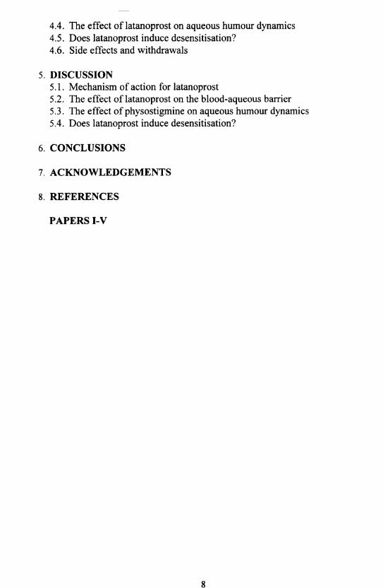
1.3. Treatment of glaucomaUntil now all glaucoma therapy has been aimed at reducing IOP. Although the elevated IOP is considered to be the result of an impaired outflow of aqueous humour, treatment aimed at inflow as well as outflow is used, whether it is surgical or medical. Surgical treatments affecting inflow include all cyclodestructive procedures. All filtering procedures as well as laser trabeculoplasty aim at increasing outflow. Medical therapy of chronic open angle glaucoma includes cholinergic agonists, adrenergic agonists and antagonists, carbonic anhydrase inhibitors, and the recently acquired prostaglandins. In Table 1 an overview of drugs for glaucoma treatment is presented.
12
Table 1. Drugs fo r treatment o f glaucoma and their main mechanisms o f action
Group Generic name Mechanism of action
Cholinergic agonistsIndirect acting (inhibitors o f acetylcholinesterase)
Physostigmine (= eserine) Trabecular outflow tPhospholineiodide Trabecular outflow t
Direct acting (imitates acetylcholine)Pilocarpine Trabecular outflow tCarbachol Trabecular outflow t
Adrenergic agonistscc2-agonists
Apraclonidine Aqueous humour Iß 2 -agonists
Epinephrine Trabecular anduveoscleral outflow t
Dipivefrin Trabecular anduveoscleral outflow t
Adrenergic antagonistsß-blockers, non-selective
Timolol Aqueous humour ILevobunolol Aqueous humour IKarteolol Aqueous humour I
ß-blockers, relatively ßi-selectiveBetaxolol Aqueous humour I
Carbonic anhydrase inhibitorsAcetazolamide (systemic administration) Aqueous humour iDichlorphenamide (systemic administration) Aqueous humour IDorzolamide Aqueous humour I
ProstaglandinsLatanoprost Uveoscleral outflowt
1.4. Prostaglandins1.4.1. HistoryIn 1930 two American gynaecologists, Kurzrok and Lieb (1930), observed that the human uterus muscle contracted when it was exposed to seminal fluid. Goldblatt (1933, 1935) in England and von Euler (1934) in Sweden independently verified that seminal fluid contracted smooth muscle, von Euler (1934) identified two substances
13
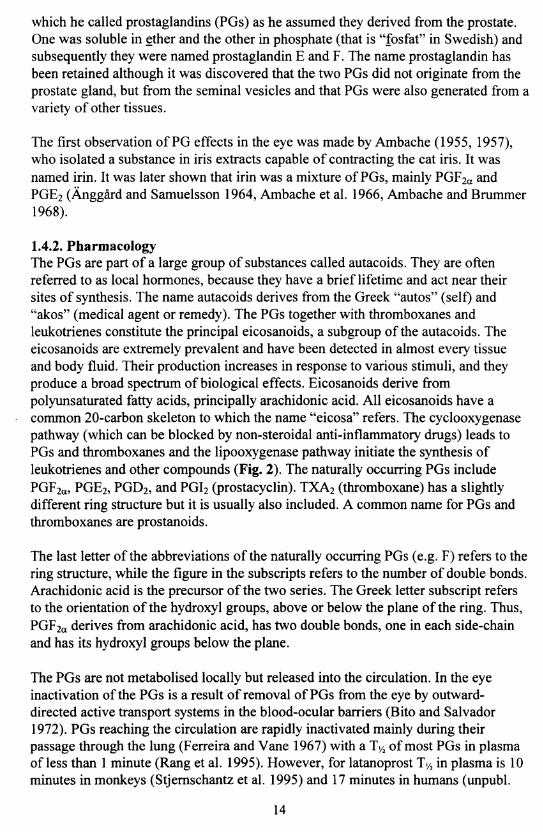
which he called prostaglandins (PGs) as he assumed they derived from the prostate. One was soluble in ether and the other in phosphate (that is “fosfat” in Swedish) and subsequently they were named prostaglandin E and F. The name prostaglandin has been retained although it was discovered that the two PGs did not originate from the prostate gland, but from the seminal vesicles and that PGs were also generated from a variety of other tissues.
The first observation of PG effects in the eye was made by Ambache (1955, 1957), who isolated a substance in iris extracts capable of contracting the cat iris. It was named irin. It was later shown that irin was a mixture of PGs, mainly PGF2ct and PGE2 (Änggård and Samuelsson 1964, Ambache et al. 1966, Ambache and Brummer 1968).
1.4.2. PharmacologyThe PGs are part of a large group of substances called autacoids. They are often referred to as local hormones, because they have a brief lifetime and act near their sites of synthesis. The name autacoids derives from the Greek “autos” (self) and “akos” (medical agent or remedy). The PGs together with thromboxanes and leukotrienes constitute the principal eicosanoids, a subgroup of the autacoids. The eicosanoids are extremely prevalent and have been detected in almost every tissue and body fluid. Their production increases in response to various stimuli, and they produce a broad spectrum of biological effects. Eicosanoids derive from polyunsaturated fatty acids, principally arachidonic acid. All eicosanoids have a common 20-carbon skeleton to which the name “eicosa” refers. The cyclooxygenase pathway (which can be blocked by non-steroidal anti-inflammatory drugs) leads to PGs and thromboxanes and the lipooxygenase pathway initiate the synthesis of leukotrienes and other compounds (Fig. 2). The naturally occurring PGs include PGF2a, PGE2, PGD2, and PGI2 (prostacyclin). TXA2 (thromboxane) has a slightly different ring structure but it is usually also included. A common name for PGs and thromboxanes are prostanoids.
The last letter of the abbreviations of the naturally occurring PGs (e.g. F) refers to the ring structure, while the figure in the subscripts refers to the number of double bonds. Arachidonic acid is the precursor of the two series. The Greek letter subscript refers to the orientation of the hydroxyl groups, above or below the plane of the ring. Thus, PGF2a derives from arachidonic acid, has two double bonds, one in each side-chain and has its hydroxyl groups below the plane.
The PGs are not metabolised locally but released into the circulation. In the eye inactivation of the PGs is a result o f removal of PGs from the eye by outward- directed active transport systems in the blood-ocular barriers (Bito and Salvador 1972). PGs reaching the circulation are rapidly inactivated mainly during their passage through the lung (Ferreira and Vane 1967) with a TVi of most PGs in plasma of less than 1 minute (Rang et al. 1995). However, for latanoprost T./, in plasma is 10 minutes in monkeys (Stjemschantz et al. 1995) and 17 minutes in humans (unpubl.
14

obs.). It is mainly inactivated in the liver. Most of the metabolites are excreted into the urine, but a minor part is eliminated via the biliary system into the faeces (Stjemschantz et al. 1995).
ESSENTIAL FATTY ACID IN DIET
ARACHIDONIC ACID
Lipooxygenase pathway
Leukotrienes
^ Cyclooxygenase pathway
oAnti-inflammatory drugs, e.g. aspirin, indomethacin
Prostacyklin Thromboxanesynthetasesynthetase
P G I2(prostacyclin)
TX A 2(thromboxane)
Figure 2. Biosynthesis o f the products o f arachidonic acid.
1.4.3. Prostaglandin receptorsThe diversity of the effects of PGs is explained by the existence of a number of different prostanoid receptors. The receptors are named DP-, EP- (4 subtypes), FP-, IP-, and TP-receptors for the natural prostaglandin, PGD2, PGE2, PGF2ot, PGI2, or TXA2, for which they have the greatest affinity (Coleman et al. 1994). All natural PGs have affinity for more than one receptor resulting in a mixed pharmacological response. Thus, selective agonists or antagonists can be expected to have more specific effects than naturally occurring PGs.
The FP receptor has a widespread distribution within the tissues of the eye including the ciliary muscle and the ciliary processes (Ocklind et al. 1996).
1.4.4. Early animal experimentsThere is a marked variation in the effect of different PGs between different species. PGs are involved in the inflammatory process and, from an ophthalmologic point of
15

view, they have generally been associated with inflammation. This is particularly true for rabbits, the most common experimental animal for studies of the effect of PGs on IOP in the late 1960s and 1970s. The rabbit eye responds to injuries with disruption of the blood-aqueous barrier, which at least in part is PG-mediated.
In an early attempt to administer PGs topically, PGEi was applied to rabbit eyes in doses from 0.5 to 50 pg and caused a dose-dependent increase in IOP, hyperaemia and break-down of the blood-aqueous barrier (BAB) (Kass et al. 1972). Later it was reported that a topically applied low dose, 5 pg, of another PG, PGF2a, gave a reduction in IOP in the rabbit eye lasting for several hours (Camras et al. 1977). Subsequent studies have confirmed that PGF2a (Lee et al. 1984) as well as PGE2, PGD2 (Woodward et al. 1989), PGE3, and PGD3 (Kulkami and Sririvasan 1985) reduce IOP in the rabbit eye.
Persistent IOP-reduction was shown also for PGF2a and PGE2 in normo- or hypertensive eyes of several species including cats, dogs, and monkeys (Camras and Bito 1981, Stem and Bito 1982, Lee et al. 1984, Camras et al. 1987, Crawford et al. 1987, Groeneboer et al. 1989, Wang et al. 1990, Gum et al. 1991).
1.4.5. Early human studiesThe first report on the ocular effects of topically applied PGF2„ (Fig. 3) in humans was published in 1985 (Giuffrè 1985). 200 pg of the tromethamine salt reduced IOP significantly for 4-24 hours in normotensive eyes, but caused marked hyperaemia, ocular pain and headache.
With the purpose of reducing the side-effects and to increase the bioavailability, PGF2a was made more lipid-soluble by estérification of the carboxyl group (Bito and Baroody 1987). PGF2ct-isopropyl ester (PGF2a-IE) (Fig. 3) crosses the comeal epithelium more readily and can be used in much lower doses than the tromethamine salt. This ester prodrug is hydrolysed to PGF2a during its passage through the cornea by two enzymes, butyryl-cholinesterase and carboxylesterase (Camber et al. 1986, Cheng-Bennet et al. 1994).
In the first study performed on normal volunteers 1-10 pg of PGF2a-IE was found to reduce IOP in a dose-dependent manner, but the side-effects were still a problem (Villumsen and Aim 1989). A diester was also tried, but it did not improve the therapeutic index (Villumsen and Aim 1990).
16

HO HO
COOH
PGFor PGR -IE
COOCH(CH3)2
HO
COOCH(CK)
Latanoprost
a - chain
© - chain
Figure 3. Structural formulas ofPGF2a, PGF2a-IE and latanoprost.
1.4.6. LatanoprostIn the attempts to find a more selective FP-agonist a part of the omega-chain was substituted with a phenyl-ring. The result was PhXA34, the mixture of the 15-R and 15-S epimers of the main drug studied in this thesis; latanoprost ( 13,14-dihydro- 17- phenyl- 18,19,20-trinor- PGF2a-IE, code name PhXA41) (Fig. 3). Latanoprost is the pure 15-R epimer, which is about 10 times more biologically active than the S epimer (Stjemschantz and Resul 1992). To reach a similar effect, the dose needs to be approximately doubled for PhXA34 compared with latanoprost.
Latanoprost is a biologically inactive prodrug. It is hydrolysed to its active form in cornea or in plasma. About 1% of topically applied latanoprost enters the monkey eye (Stjemschantz et al. 1995). In human aqueous humour a peak concentration is reached approximately 214 hours after application (Sjöquist et al. 1997).
Latanoprost is a more selective FP-receptor agonist than PGF2a (Stjemschantz et al. 1995) and this has resulted in an improved therapeutic profile with less hyperaemia, negligible irritation, and a retained IOP-lowering effect (Aim et al. 1995b, Watson et al. 1996, Camras et al. 1996a).
The drug concentration in a vial of latanoprost (50 pg/ml) is about IO'4 M, whereas in aqueous humour and in plasma the peak concentrations reach about IO'7 M and 10'10 M, respectively (unpubl. obs.). Thus the concentration in aqueous humour is within the range for FP-receptor stimulation (Table 2) but below the range for stimulation of other prostanoid receptors. The peak concentration in plasma is below the stimulation of FP-receptors, which may explain the favourable systemic side effect profile of latanoprost.
17

Table 2. Receptor profile o f PGp2 a and latanoprost. EC so values in moles/l.
1.4.7. DoseThe optimal concentration for reducing IOP in humans has been found to be about 50 pg/ml of latanoprost, i.e. 0.005% (Villumsen and Aim 1992, Hotehama and Mishima 1993, Aim et al. 1993/ The maximum IOP reduction occurs 6 to 12 hours after administration of a single dose (Aim and Villumsen 1991, Hotehama and Mishima 1993).
Once daily administration has been found to be superior to twice daily with regard to the ocular hypotensive effect during the day (Nagasubramanian et al. 1993, Aim et al. 1995a). This observation was addressed in two of the studies in this thesis (IV, V). A true reduction of the efficacy of latanoprost by e.g. a receptor adaptation at twice daily, but not once daily, applications is a possible explanation for the difference in effect. Another possibility is that latanoprost has a dual effect on aqueous humour dynamics. A small increase in aqueous flow lasting e.g. 6-8 hours would blunt some of the early effect of latanoprost on IOP and explain why applying a drop also in the morning would cause a less effective pressure reduction during the day.
1.4.8. Mechanism of actionThe main mechanism of action of latanoprost in the reduction of IOP in monkeys is increased uveoscleral outflow (Stjemschantz et al. 1995). An increase of 60 % in uveoscleral outflow was found after 3 pg daily for 5 days, whereas trabecular outflow was unchanged (Stjemschantz et al. 1995). This confirms the previous results from studies of PGF2a (Crawford and Kaufman 1987, Crawford et al. 1987) and PGF2a-IE (Nilsson et al. 1989, Gabelt and Kaufman 1989, Gabelt and Kaufman 1990) in monkeys. There is also indirect evidence to support the same mode of action of latanoprost in humans (Toris et al. 1993). In study II an attempt to support this theory was made. The exact mechanism behind the increase in uveoscleral outflow is not known, but relaxation (Poyer et al. 1995) and/or structural changes (Lütjen-Drecoll
18
and Tamm 1988) of the ciliary muscle have been suggested. The effect on IOP of possible morphological changes in the ciliary muscle is investigated in study I.
A reduction in aqueous humour production cannot explain the ocular hypotensive effect of latanoprost. A tendency towards an increase in aqueous flow with PGF2a and PGF2ct-IE has been shown in monkeys (Crawford et al. 1987, Nilsson et al. 1989) as well as in humans for PhXA34 (Aim and Villumsen 1991) and latanoprost (Ziai et al. 1993, Toris et al. 1993).
1.4.9. Effects on BABBreak-down of the blood-aqueous barrier (BAB) has been observed in rabbits
(Camras et al. 1977, Lee et al. 1984) while the BAB in monkeys seems to be much more resistant (Camras et al. 1987, Crawford et al. 1987). No signs of BAB breakdown with respect to protein content have been noted in human eyes during shortterm studies (Ziai et al. 1993, Hotehama and Mishima 1993). In three six-month studies no increased frequency of cells or flare in the aqueous humour were observed (Aim et al. 1995b, Watson et al. 1996, Camras et al. 1996a), but clinical observations may well fail to detect small changes in BAB permeability. In study I, where a subgroup of patients were followed with laser flare measurements, this issue has been addressed.
1.4.10. Combination with cholinergicsCombinations of drugs are common in glaucoma treatment. When combining latanoprost and cholinergic agonists an antagonistic effect might theoretically be expected as the cholinergic agonists contract the ciliary muscle. In cynomolgus monkeys pretreatment with pilocarpine was found to prevent the ocular hypotension of PGF2ct (Crawford and Kaufman 1987). However, studies in the same species using pilocarpine after PGs are equivocal (Millar and Kaufman 1995, Nilsson et al. 1989). In humans latanoprost, 0.006% twice daily, and pilocarpine, three times a day, provided a small additive effect (Friström and Nilsson 1993). Study II was conducted to further investigate the clinically important combination of latanoprost and cholinergics and to acquire more knowledge on the mechanism of action of latanoprost in humans. We chose to study young volunteers in which contraction of the ciliary muscles is maximal and physostigmine was used since it causes an intense contraction of the muscle.
1.5. Cholinergics1.5.1. HistoryPhysostigmine (eserine)* comes from the ordeal bean of Calabar. It was brought from Nigeria, where it was used in trials for witchcraft**, to England by a medical missionary in about 1840. Its pharmacology and properties were studied by Thomas Richard Frazer (1841-1919) and Douglas Argyll Robertson (1837-1909). Albrecht von Graefe (1828-1870) utilised the miosis of the Calabar bean extract preoperatively to facilitate iridectomy for glaucoma, but it was one of his students, Ludwig Laquer (1838-1909), who introduced it as an effective treatment of glaucoma about 20 years
19

later, in 1876. Laquer himself suffered from glaucoma and had experienced the relief using physostigmine when his ocular tension was increased, which he recognised when he observed coloured rings around a lighted match. Later he had successful bilateral basal iridectomies. Unfortunately he developed photophobia due to the iris colobomas and he became a recluse.
Almost at the same time, 1877, Adolph Weber, another of von Graefe’s former students, described pilocarpine as a potent miotic. Pilocarpine*** derives from a shrub belonging to the rue family, having its origin in Central America. Pilocarpine has stood the test of time, but physostigmine is no longer in clinical use in Sweden.
*The woody vine from which Calabar bean originates was identified and in 1846 christened to Physostigma venenosum. When the active alkaloid was isolated by Jobst and Hesse in 1864 they called it physostigmine. In 1865 Véé and Leven independently identified the same alkaloid and named it ésérine after the native name of the vine, ésére.** It is known to be used as ordeal poison; death implied guilt!♦♦♦Derives from the plant Pilocarpus microphyllus.
1.5.2. PharmacologyAgents affecting the autonomic (involuntary, visceral or vegetative) nervous system are named autonomic drugs. Depending on which part of the system they act on, they can be divided into sympathetic and parasympathetic agents. The transmitter substance in the parasympathetic system is acetylcholine. It is also the neurotransmitter at preganglionic sympathetic synapses, at somatic motor nerve endings, and at certain central nervous system synapses. Acetylcholine is synthesised and stored within the cholinergic neurones and released from nerve endings. It activates a pre- or post-junctional receptor until it is metabolised by the enzyme acetylcholinesterase and its effect terminated.
Cholinergic drugs are either direct, acting by imitating acetylcholine, or indirect, acting by binding acetylcholinesterase, causing an accumulation of naturally occurring acetylcholine. Pilocarpine is an example of a direct acting cholinergic drug, whereas physostigmine belongs to the indirect acting drugs.
1.5.3. Acetylcholine receptorsThere are two major types of acetylcholine receptors; nicotinic and muscarinic. Nicotinic receptors are present in autonomic ganglia and skeletal muscles such as levator, orbicularis, and extraocular muscles. Muscarinic receptors are situated in postganglionic parasympathetic smooth muscle and glands, for example in the iris and ciliary body. It is the muscarinic effects of the cholinergic drugs that are used for glaucoma treatment.
Five different subtypes of muscarinic receptors, ml-m5, coupled to G-proteins, have so far been characterised pharmacologically and/or by molecular cloning (Erickson- Lamy 1994). The exact distribution and effects of the muscarinic receptor subtypes
20

are not known. M3 is the predominating receptor in bovine iris sphincter and ciliary processes (Honkanen et al. 1990), and in human nonpigmented ciliary epithelial cells (Wax and Coca-Prados 1989). In rabbits the ciliary epithelium contains muscarinic receptors coupled to an inhibitory G-protein (Jumblatt et al. 1990).
1.5.4. Mechanism of actionThe main mechanism by which cholinergic drugs lower IOP is enhanced aqueous outflow (Båråny 1962). The contraction of the longitudinal fibres of the ciliary muscle exerts a tractional force on the scleral spur, which separates the trabecular sheets and leads to increased trabecular outflow facility (Kaufman and Båråny 1976). Uveoscleral outflow is reduced (Bill and Wålinder 1966). Studies on the effect on aqueous flow of pilocarpine, the most commonly used cholinergic agonist today in glaucoma treatment, have yielded different results (Berggren 1965, Bill and Wålinder 1966, Wålinder and Bill 1969, Nagataki and Brubaker 1982, Miichi and Nagataki 1983), but the present opinion is that this is of minor clinical significance. However, due to the progressive IOP reduction by physostigmine, in the second study, the question was raised if physostigmine also reduces aqueous flow apart from enhancing outflow. Thus, in study III the effect of a large dose of physostigmine on aqueous humour flow was studied in healthy volunteers.
21
2. AIMS OF THE STUDY
The discovery of the ocular hypotensive effect of prostaglandins and the subsequent development of latanoprost for glaucoma treatment has raised a number of questions about the mechanisms of action involved. This provides the basis of the present study. In this context, the effects on aqueous humour dynamics in human eyes have been studied in relation to a well established compound, physostigmine. Thus, prostanoid and cholinergic effects were investigated according to the following specific aims:
• To evaluate whether long term treatment with the prostaglandin analogue latanoprost has a deleterious effect on the blood-aqueous barrier or ciliary muscle in humans (I).
• To investigate if a pronounced ciliary muscle contraction, induced by physostigmine, can abolish the ocular hypotensive effect of latanoprost via inhibition of uveoscleral outflow (II).
• To investigate if part of the progressive reduction of IOP, when applying physostigmine at alternate hours, is due to a reduced aqueous flow (III).
• To determine if a difference in daytime IOP in normal eyes between different dose regimens of latanoprost exists and to establish whether any difference could be attributed to a change in aqueous flow (IV).
• To evaluate whether the less effective IOP-reduction of twice daily applications of latanoprost can be compatible with a modest desensitisation at the level of the FP- receptor or its associated intracellular signalling pathways (V).
22
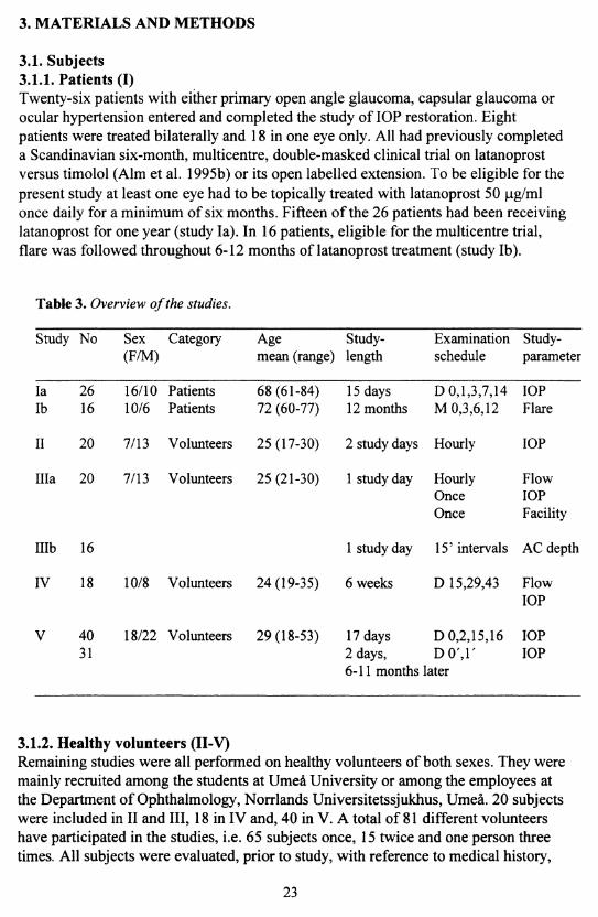
3. MATERIALS AND METHODS
3.1. Subjects3.1.1. Patients (I)Twenty-six patients with either primary open angle glaucoma, capsular glaucoma or ocular hypertension entered and completed the study of IOP restoration. Eight patients were treated bilaterally and 18 in one eye only. All had previously completed a Scandinavian six-month, multicentre, double-masked clinical trial on latanoprost versus timolol (Aim et al. 1995b) or its open labelled extension. To be eligible for the present study at least one eye had to be topically treated with latanoprost 50 pg/ml once daily for a minimum of six months. Fifteen of the 26 patients had been receiving latanoprost for one year (study la). In 16 patients, eligible for the multicentre trial, flare was followed throughout 6-12 months of latanoprost treatment (study lb).
Table 3. Overview o f the studies.
Study No Sex(F/M)
Category Agemean (range)
Study-length
Examinationschedule
Study-parameter
lalb
2616
16/1010/6
PatientsPatients
68 (61-84) 72 (60-77)
15 days 12 months
D 0,1,3,7,14 M 0,3,6,12
IOPFlare
II 20 7/13 Volunteers 25 (17-30) 2 study days Hourly IOP
Ilia 20 7/13 Volunteers 25 (21-30) 1 study day HourlyOnceOnce
FlowIOPFacility
Illb 16 1 study day 15’ intervals AC depth
IV 18 10/8 Volunteers 24(19-35) 6 weeks D 15,29,43 FlowIOP
V 4031
18/22 Volunteers 29(18-53) 17 days D 0,2,15,16 2 days, D 0 ',1 '6-11 months later
IOPIOP
3.1.2. Healthy volunteers (II-V)Remaining studies were all performed on healthy volunteers of both sexes. They were mainly recruited among the students at Umeå University or among the employees at the Department of Ophthalmology, Norrlands Universitetssjukhus, Umeå. 20 subjects were included in II and III, 18 in IV and, 40 in V. A total of 81 different volunteers have participated in the studies, i.e. 65 subjects once, 15 twice and one person three times. All subjects were evaluated, prior to study, with reference to medical history,
23

determination of visual acuity, refraction, IOP and slit-lamp biomicroscopic examination of anterior and posterior segments. Gonioscopy was performed in II, III and V. No drugs on a regular basis, except for oral contraceptives, were permitted. Contact lens wearers were not allowed to use their lenses within one week prior to study start and during the study.
More detailed information is given in the papers I-V. An overview of the studies is presented in Table 3.
In all studies the protocols followed the tenets of the Declaration of Helsinki and they were reviewed and approved by the Medical Products Agency and by the Ethics Committee of the Medical faculty of Umeå University. Informed consent was obtained from each participant.
3.2. DrugsLatanoprost 50 pg/ml for topical administration was used in I, II, IV, and V. Latanoprost eye drops contain 0.2 mg/ml benzalkonium chloride as preservative.They were supplied by the manufacturer (Pharmacia AB / Pharmacia & Upjohn AB, Sweden). Study II and V were double-masked. Coded, randomly assigned bottles of latanoprost and placebo (its vehicle) were provided by the same manufacturer.
Physostigmine salicylate, 8 mg/ml, an ex tempore preparation without preservative was prepared by the hospital pharmacy at Umeå University Hospital and used in II and III.
For applanation tonometry a combination of anaesthetic and fluorescein was used (Ofitan Flurekain®, Leiras, Finland) (I-V).
For determination of aqueous flow a 2 per cent sodium fluorescein solution without preservative (Smith & Nephew, UK) was instilled (III, IV).
For tonography an anaesthetic without preservative (Tetrakain, 10 mg/ml, Chauvin Pharmaceuticals Ltd, England) was applied (IV).
3.3. Methods and technical equipment3.3.1. Determination of intraocular pressure (la, II, Illa, IV, V)Goldmann applanation tonometry was used for IOP determinations in all studies. Within each centre the same daily calibrated Goldmann applanation tonometer (Haag- Streit™, Bern, Switzerland) was utilised. In order to minimise errors all tonometry on a given subject was performed by the same person throughout each study (Thorbum 1978). The median of three readings in each eye was used for calculations. If both eyes were treated in study la the mean IOP of the two eyes was used in the analyses.
24
3.3.2. Determination of anterior chamber flare (Ib)Break-down of the BAB with the subsequent increase of protein and/or cells can be observed as flare and/or floaters in the anterior chamber. For quantitative measurements of protein concentration in study lb a commercially available laser flare meter (FM-500, Kowa) was used. The instrument measures the scattered light intensity produced by proteins as photon counts per millisecond (Sawa et al. 1988). At each time point 10 determinations were performed and the mean was used for calculations. If both eyes were treated, the average of the two eyes was used. Normal values for young healthy volunteers and for adult cataract patients have been reported to be 4.1 (standard deviation (SD): 1.0) and 6.2 (SD: 2.5) photon counts/ms, respectively (Sawa et al. 1988).
3.3.3. Determination of aqueous flow and corneal endothelial permeability (Ilia, IV)Aqueous flow was studied with fluorophotometry by clearance of topically applied fluorescein, i.e. the corneal depot method, in study Ilia and IV. Basically the same technique, utilising a commercial instrument (Fluorotron Master™, Coherent, USA) for determinations of corneal and aqueous fluorescein concentrations, has been used in our department since 1984.
The cornea was loaded with a 2 per cent sodium fluorescein solution without preservative (Smith & Nephew, UK). This was done six hours before start of measurements to allow an even distribution within the corneal stroma. In order to reach an adequate concentration of fluorescein in cornea at the start of measurements, the number of instillations varied between five (IV) and 10-15 (Ilia).
The fluorescein penetrates the corneal epithelium, enters the stroma, forming a depot within the stroma, from where it leaks into the anterior chamber. The losses through the epithelium and the limbus are very small and almost all fluorescein, trapped within the stroma, will enter the anterior chamber, from where it is cleared by diffusion and flow. In normal eyes the diffusional loss into the vasculature of the iris is small, about 25 pl/min (Brubaker 1989), and the principal route for the fluorescent aqueous to leave the eye is through the iridocorneal angle. The rate of flow can be deduced by knowing the rate of loss of fluorescein in proportion to its concentration in the cornea and the anterior chamber, i.e. the clearance.
Measurements of the fluorescein concentrations were made every hour (Illa, IV). When physostigmine was studied (III), each eye was measured 3-6 times hourly and the mean value was used for calculations to minimise errors of uneven distribution within the aqueous due to the miotic pupil. Aqueous flow (Illa, IV) was calculated according to the mathematical model described by Brubaker (1989, 1995) including calculations of the corneal endothelial permeability (IV).
The actual volume of the anterior chamber is not critical for the calculation of changes in aqueous flow when the subjects act as their own control. When studying
25

healthy volunteers, a standard value of 200 p.1 is often used. However, this may introduce an error if the volume of the anterior chamber varies during the course of the investigation, as can be expected when physostigmine (III) is applied. In order to estimate the magnitude of this problem we determined the volume of the anterior chamber at different time points after application of physostigmine in a separate series of experiments (Illb). The flow was then calculated according to three different protocols; assuming 1) a standard volume of 200 pi or 2) an anterior chamber volume corresponding to the minimum or 3) maximum anterior chamber volume observed in Illb.
3.3.4. Determination of outflow facility (Ilia)Outflow facility was determined by tonography, which is based on the observation that external pressure on the eye causes a lowering in IOP with time. Tonography quantitates this effect by measuring IOP continuously and following the decrease in IOP, due to loading of the eye, with the weight of the tonometer. In study III a 4- minute tonography with the Pneumatonograph™ (Digilab Inc., USA), using the 10 g weight, was performed at the end of the study day. Outflow facility (C-value) was calculated from the pressure decay curves and standard tables presented in the tonographer manual based on the tables from Eisenlohr et al. (1962). This method measures the total outflow facility, which is the sum of the true facility (flow of aqueous humour through the outflow channels, i.e. trabecular outflow and uveoscleral outflow) and pseudofacility (the apparent flow secondary to a reduced rate of aqueous humour formation). C-values in normal eyes have been reported to range between 0.15 to 0.34 pl/min/mm Hg with a mean of 0.24 (Grant 1950). Tonography remains an accepted research tool, but its clinical usefulness is limited mainly due to lack of precision.
3.3.5. Determination of anterior chamber depth and volume (Illb)In the second part of study III (Illb) the depth of the anterior chamber was determined by using the pachymeter attachment to the Haag-Streit™ slit-lamp. Three measurements of the depth in each eye were performed in a masked fashion and the median was used for calculations. The corneal diameter was measured with callipers and the anterior chamber volume was calculated according to Brubaker (1995).
3.4. Procedures (I-V)In study la, IOP was followed for 14 days (Day 0, 1, 3, 7 and 14) after withdrawal of latanoprost. In lb, aqueous flare was measured 2-4 times with a laser flare meter during 6-12 months of latanoprost treatment.
In study II, one drop of physostigmine salicylate was instilled in one randomly assigned eye every other hour between 7 AM and 7 PM on the first study day. At 8 a m
both eyes received one drop of either latanoprost or placebo. This protocol was repeated on the second study day with latanoprost administered to previously placebo-treated eyes and vice versa. IOP was measured hourly throughout the day in both eyes.
26

In study Ilia, the drops of physostigmine salicylate were instilled in one eye in a similar way as in study II. The fellow eye remained untreated. Fluorophotometry of the anterior segment was performed hourly between 7 AM and 8 PM and the aqueous flow was calculated. Subsequently the subjects underwent tonography and tonometry. The change in anterior chamber depth and volume induced by physostigmine was assessed separately (Illb). After a baseline depth measurement at 7 am one drop of 8 mg/ml physostigmine salicylate was instilled in the same eye which had received physostigmine in the study of aqueous humour dynamics (Ilia). This instillation was repeated 2 hours later to mimic the same conditions as in Illb. Anterior chamber depth was followed every 15 minutes for 4 hours.
In study IV, latanoprost was instilled in one eye. Three dose regimens (one/three drops once daily or one drop twice daily) were evaluated. IOP was measured at the end of each 14-day treatment period. Aqueous flow and endothelial permeability were assessed by fluorophotometry.
In study V, latanoprost was instilled once daily (8 PM) in one eye of 40 healthy volunteers and twice daily (8 AM and 8 PM) in the other eye in a randomised, masked fashion for 2 weeks with the last drop on the evening of Day 15. IOP was determined at 8 AM, noon and 4 PM on Day 0,2, 15 and 16. Also, the effect of a single dose of latanoprost was measured 6-11 months after cessation of treatment. Nine subjects did not participate in this part of the study since they had since moved from Umeå.
3.5. Statistical methods and experimental design (I-V)The response variables chosen in each study are described in papers I-V. The experimental design with each patient or subject being his own control, permitted use of paired analysis (I, III-V). The two-tailed Student’s paired f-test was used for the statistical inference. Values of p<0.05 were considered statistically significant. In paper II, the design of the study was that of a 22 factorial experiment and the statistical analysis was performed by an analysis of variance (ANOVA). The F-test was used to assess statistical significance. In all studies, the 95% confidence interval of the mean (95% Cl) was calculated as the measure of the uncertainty of the estimates.
27

4. RESULTS
4.1. IOP restoration and BAB changes after long term latanoprost treatment (I)After cessation of latanoprost, the mean difference (pretreatment IOP - IOP during follow up) in treated eyes from Day 0 (end of treatment) to Day 14 was significantly lower than pretreatment IOP at all time points. However, the difference gradually declined from 7.2 mm Hg on Day 1 (p<0.001) to 1.3 mm Hg on Day 14 (p<0.05) (Fig. 4).
O)
O -10
Time (days)
Figure 4. Mean difference (pre-treatment IOP- IOP during follow-up) versus time after cessation of latanoprost treatment. Error bars denote 95% confidence interval.
No statistically significant differences in flare were seen throughout the 6-12 months of latanoprost treatment. Neither was there a marked increase in flare in any single individual. The maximal flare values before treatment were 13.0 in treated eyes and 9.4 photon counts/ms in untreated eyes. The corresponding figures during treatment were 10.5 and 10.3.
4.2. Combination of latanoprost and physostigmine (II)The mean pretreatment IOPs at 7 AM were 13.5 (95% Cl: 12.2-14.8), 13.8 (12.4- 15.2), 13.2 (11.7-14.6) and 13.4 (12.0-14.7) mm Hg in eyes randomised to placebo, latanoprost, physostigmine or the combination, respectively. The mean IOP over the day, in the vehicle-treated eye, ranged between 12.8 and 14.6 mm Hg. Latanoprost reduced IOP significantly (pO.Ol) 3-12 hours after application with a maximal effect of 1.9 mm Hg observed 8 hours post treatment. The reduction obtained with physostigmine administered every second hour, and observed from 1 h after the dose, was even more pronounced (p<0.01). The effect increased throughout the day and finally reached a maximum of 3.3 mm Hg. No significant interaction was seen until 7 to 10 hours after application of latanoprost.
28

□ — latanoprost ■ — ■ physostigmine* - - combination• — theoreticalO)I
EE
CLOcCDOc2
7 AM 1 PM
Time
Figure 5. The estimated IOP reduction versus time, fo r latanoprost, physostigmine and the combination compared with placebo. The "theoretical” IOP reduction for the combination if there had been full additivity is also shown. ■ denotes the times fo r instillation o f physostigmine. □ denotes the time fo r instillation o f latanoprost.
Fig. 5 illustrates the effect of the drugs and their interaction versus time. It shows the IOP reduction hour by hour for latanoprost and physostigmine compared with placebo and for the combination of physostigmine and latanoprost. It also displays how large the IOP reduction would have been for the combination if there had been full additivity between physostigmine and latanoprost. The figure demonstrates that for the first 6 h after application of latanoprost, the effect of the combination and the "theoretical" combined effect are almost identical.
4.3. The effect of physostigmine on aqueous humour dynamics (III)Anterior chamber volume (Illb)Prior to treatment (7.00 AM), the mean volume of the anterior chamber in the physostigmine treated eyes was 210 pi (95% Cl: 199-221). The minimum volume was found 30 minutes after drop administration. The mean reduction at that time,7.30 AM, was 13% (95% CI: 11-16%, range: 2-20%). The chamber depth then slowly increased until the next drop was given. The control eye was not affected.
Aqueous humour dynamics (Ilia)Aqueous flowThe mean hourly flow in the eyes treated with physostigmine and control eyes is presented in Table 4. As can be seen, the variation of the anterior chamber volume had only a minor effect on calculated flow values. Aqueous humour production increased during the day in both eyes. This was more pronounced in the
29

physostigmine treated eye. There was a statistically significant difference between the two eyes from 9 AM, when the maximum anterior chamber volumes were used for calculations, and from 10 AM for the minimum volumes (p<0.05-p<0.001). The mean flow during the day was 3.47 (95% Cl: 3.15-3.79) or 3.39 (3.07-3.72) pl/min, respectively, in the physostigmine treated eye and 2.72 (2.51-2.93) pl/min in the control eye. This increase (28 or 25%) is statistically significant (p<0.001) irrespective of anterior chamber volume.
IOPMean IOP at 8 PM was 9.8 (95% Cl: 8.8-10.8) mm Hg in eyes treated with physostigmine and 13.0 (11.9-14.0) mm Hg in control eyes (pO.001), i.e. a pressure difference of 3.2 (2.3-4.0) mm Hg (p<0.001) between treated and untreated eyes.
Outflow facilityMean tonographic outflow facility was 0.48 (95% Cl: 0.43-0.54) in physostigmine treated eyes and 0.35 (0.30-0.39) pl/min/mm Hg in control eyes (p<0.001) at the end o f the study day, 8 PM. This is an increase in mean outflow facility with 0.14 (0.09- 0.18) pl/min/mm Hg (p<0.001).
Table 4. Mean hourly aqueous flow and its 95% Cl in eyes treated with physostigmine and control eyes. For physostigmine treated eyes, the values obtained from flow calculations, using both maximum and minimum anterior chamber volumes, are shown. n=20.
Time Physostigmine Maximum volume pl/min
Physostigmine Minimum volume (il/min
Control eye
pl/min
7 AM 2.28 (2.01-2.54) 2.20 (1.93-2.46) 2.22 (1.91-2.52)8 AM 2.37 (2.14-2.60) 2.29 (2.06-2.53) 2.33 (2.01-2.66)9 AM 2.64 (2.37-2.91) 2.57 (2.29-2.84) 2.34 (2.07-2.62)10 AM 3.04 (2.74-3.34) 2.97 (2.67-3.26) 2.57 (2.28-2.85)11 AM 3.14 (2.88-3.40) 3.07 (2.81-3.32) 2.69 (2.39-2.98)noon 3.44 (3.10-3.78) 3.36 (3.02-3.70) 2.76 (2.51-3.01)1 PM 3.42 (3.05-3.78) 3.34 (2.98-3.71) 2.72 (2.49-2.95)2 PM 3.74 (3.31-4.17) 3.66 (3.23-4.09) 2.79 (2.56-3.02)3 PM 3.77 (3.42-4.12) 3.69 (3.35-4.04) 2.81 (2.61-3.01)4 PM 3.92 (3.51-4.32) 3.84 (3.43-4.25) 2.81 (2.64-2.99)5 PM 4.11 (3.58-4.63) 4.03 (3.50-4.56) 2.94 (2.73-3.15)6 PM 4.22 (3.69-4.75) 4.14 (3.61-4.67) 3.02 (2.80-3.24)7 PM 4.12 (3.59-4.66) 4.04 (3.51-4.58) 3.03 (2.79-3.27)8 PM 4.41 (3.63-5.19) 4.34 (3.56-5.12) 3.15 (2.86-3.45)
30

4.4. The effect of latanoprost on aqueous humour dynamics (IV)IOPThe differences in IOP between treated and untreated eyes as well as the differences between the three different dose regimens are presented in Table 5 a-b. All three tested dose regimens lowered IOP significantly. Both once daily treatments, irrespective of whether one or three drops were given, lowered the IOP significantly more than the twice daily regimen. Between the two once daily treatments there was no statistically significant difference.
Table 5a. IOP at noon with the three different dose regimens and the differences (IOP in treated eye less IOP in control eye). Mean and 95% Cl.
Treatment Treated eye Control eye Difference nmm Hg mm Hg mm Hg
Once daily 11.3(10.5-12.1) 14.5(13.5-15.5) -3.2 (-3.9-2.5) 18Twice daily 12.4(11.4-13.3) 14.3(13.6-15.0) -2 . 0 (-2 .8 - 1 .1 ) 17Once daily Gdroos) ili no.i-i2.n 15.0 fl4.0-15.9i . 3 . 9 M.6--3.2) 17
Table 5b. Difference in IOP reductions between the three different dose regimens. The IOP reduction is calculated as IOP in treated eye less IOP in control eye. Mean and 95% Cl.
Treatments Difference in IOP mm Hg
p-value
Once-twice daily -1.2 (-2.0-0.4) < 0 . 0 1
Once daily (3 drops)-twice daily -1.9 (-2.7-1.1) < 0 . 0 0 1
Once daily (3 drops)-once daily 0.7 (-0.1-1.5Ì 0.088
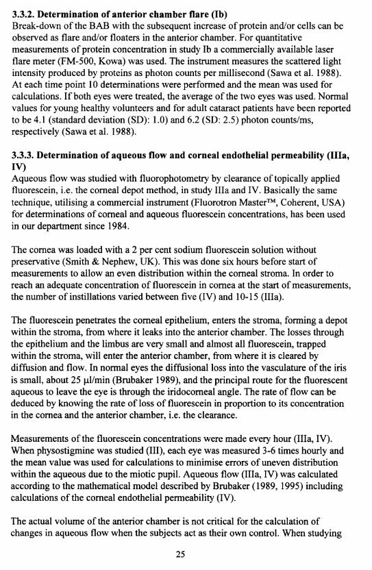
Aqueous humour flowTable 6 presents aqueous flow in both eyes and the differences in flow between treated and control eyes. All three dose regimens had a tendency to increase aqueous flow, varying between 6-13%, but a statistically significant difference was only detected for the one drop daily dose regimen (p<0.05). None of the pairwise differences between the three different dose regimens was statistically significant. This was true whether aqueous flow in the treated eye or the difference in flow between treated and control eyes was used as the response variable.
31
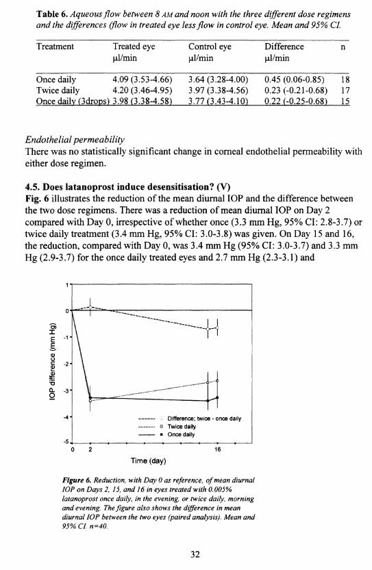
Table 6. Aqueous flow between 8 AM and noon with the three different dose regimens and the differences (flow in treated eye less flow in control eye. Mean and 95% CL
Treatment Treated eye Control eye Difference npl/min pl/min pl/min
Once daily 4.09 (3.53-4.66) 3.64 (3.28-4.00) 0.45 (0.06-0.85) 18Twice daily 4.20 (3.46-4.95) 3.97 (3.38-4.56) 0.23 (-0.21-0.68) 17Once daily (3drons) 3.98 (3.38-4.58) 3.77 (3.43-4.10) 0.22 (-0.25-0.68) 15
Endothelial permeabilityThere was no statistically significant change in corneal endothelial permeability with either dose regimen.
4.5. Does latanoprost induce desensitisation? (V)Fig. 6 illustrates the reduction of the mean diurnal IOP and the difference between the two dose regimens. There was a reduction of mean diurnal IOP on Day 2 compared with Day 0, irrespective of whether once (3.3 mm Hg, 95% Cl: 2.8-3.7) or twice daily treatment (3.4 mm Hg, 95% Cl: 3.0-3.8) was given. On Day 15 and 16, the reduction, compared with Day 0, was 3.4 mm Hg (95% Cl: 3.0-3.7) and 3.3 mm Hg (2.9-3.7) for the once daily treated eyes and 2.7 mm Hg (2.3-3.1) and
O)IEE<DOÇ - 2 -
ë
è -3-
■4 - : Difference; twice - once daily □ Twice daily ■ O nce daily
160 2
Time (day)
Figure 6. Reduction, with Day 0 as reference, o f mean diurnal IOP on Days 2, 15, and 16 in eyes treated with 0.005% latanoprost once daily, in the evening, or twice daily, morning and evening. The figure also shows the difference in mean diurnal IOP between the two eyes (paired analysis). Mean and 95% Cl. n=40.
32
2.6 mm Hg (2.3-3.0) for those treated twice daily. The IOP reduction was statistically significant at all time points (p<0.001) but the difference between the once or twice daily regimes was only statistically significant on Day 15 and 16. One daily dose resulted in a significantly lower IOP compared with two doses (p<0.001, paired analysis) on Day 15 (0.7 mm Hg with 95% CI: 0.3-1.0). The difference between the two dose regimens remained (pO.OOl) on Day 16 (0.6 mm Hg with 95% CI: 0.3-1.0) when no morning dose had been given.
Re-exposure to a single dose of latanoprost 6-11 months later did not reveal any remaining difference (p>0.05) in effect between the eyes that were previously treated once or twice daily. Thus the partial loss of effect observed on Day 16 was no longer seen 6-11 months later.
4.6. Side effects and withdrawals (I-V)In the long-term study of flare in patients (lb), side effects were reported elsewhere (Aim et al. 1995b, Camras et al. 1996b) and in the study of IOP restoration there were no side effects. The most frequent side effect of latanoprost in the studies of healthy volunteers was hyperaemia, mostly mild and transient, but, in a few isolated cases, more pronounced. All subjects treated with physostigmine complained of various degrees of blurred vision, more than 90% had ocular pain or headache and about 80% experienced muscular twitches. No serious adverse events occurred.
All included patients and volunteers completed the studies without protocol violation with one exception. One of the healthy volunteers became pregnant during study II and she was immediately withdrawn. She has later given birth to a healthy baby.
33

5. DISCUSSION
5.1. Mechanism of action for latanoprost (I, II, IV)The mechanism of action of latanoprost in primates appears to be enhanced unconventional aqueous outflow (Crawford and Kaufman 1987, Crawford et al. 1987, Nilsson et al. 1989) and there is indirect evidence suggesting that increased uveoscleral outflow is the main mechanism of action in humans (Toris et al. 1993).
(II) The objective of study II was to investigate whether an intense ciliary muscle contraction would abolish the hypotensive effect of latanoprost, by blocking the uveoscleral outflow route. For this reason we applied a supramaximal dose of physostigmine salicylate, 8 mg/ml, alternate hours to the eyes of young, healthy volunteers, presumed to have a vigorous ciliary muscle.
The IOP-lowering effect of physostigmine increased with repeated doses throughout the study day. The peak effect of latanoprost was found 8 hours post-dose. There was no significant interaction between the two drugs until 6 hours after application of latanoprost and its initial effect was fully additive to the almost maximal effect of physostigmine.
Summarising the results of study II and previous studies concerning the combination of latanoprost (or PGF2a and analogues) and cholinergics, the following conclusions can be drawn
• The possibility that pretreatment with a cholinergic agonist should prevent latanoprost from reaching the receptor, as has been suggested for monkeys (Millar and Kaufman 1995) does not appear to be true for human eyes.
• Alternate hourly administration of physostigmine is still not sufficient to maintain a sustained contraction of the ciliary muscle in the young human eye. This was confirmed in study III, which showed that the physostigmine effect on anterior chamber depth (Illb) was as short-lasting as previously reported for the effect of pilocarpine on accommodation (Berggren 1985). It cannot be excluded that more frequent administrations of physostigmine could have abolished the ocular hypotensive effect of latanoprost.
• In monkeys the IOP-lowering effect of PGF2a was successfully reduced with topical application of either a high (Crawford and Kaufman 1987) or a low (Nilsson et al. 1989) dose of pilocarpine. A species difference may well exist, but, from a clinical point of view, the major conclusion is that the additive effect of latanoprost and pilocarpine, 20 mg/ml t.i.d., previously observed (Villumsen 1992, Friström and Nilsson 1993) is also seen with more frequent administrations of a potent cholinergic agonist.
34

A significant interaction between the two drugs was seen only between 7 and 10 hours after application of latanoprost. There may be several explanations for this finding.
• One hypothesis is that repeated administration of physostigmine induces several, perhaps even prolonged, periods of mechanical obstruction of the uveoscleral outflow. The increasing interaction in conjunction with the increasing effect of physostigmine during the day supports this explanation.
• A second hypothesis may be that the interaction observed might also be a "pseudophenomenon", caused by the fact that it is more difficult to reach the same reduction of the IOP, in absolute values, when IOP is already reduced. In this study we investigated healthy volunteers. Their normal IOP was further reduced by two potent agents, which means that we obtained an IOP that may have approached its lower limit. This would be the case if the lower limit was set by the episcleral venous pressure.
• The third hypothesis might be that latanoprost may reduce IOP below the episcleral venous pressure by acting on uveoscleral flow. A possible speculation might be that backflow through the trabecular meshwork does occur (Ethier et al. 1995) and physostigmine may keep the meshwork wide open and prevent the IOP to reach a value beneath that of the episcleral veins. In this situation the lower limit of the IOP may then be set by the episcleral venous pressure in physostigmine treated eyes.
• The final hypothesis is that latanoprost has a dual mechanism of action on aqueous humour dynamics, e.g. an early effect on the conventional outflow through the trabecular meshwork independent of physostigmine, and a late effect that involves the uveoscleral route and is blocked by physostigmine. As the mechanism of action for latanoprost in human eyes is not known in detail, this possibility cannot be ruled out. However, studies in monkeys found no effect on conventional outflow which made this possibility less likely (Crawford et al. 1987).
The fact that latanoprost and physostigmine were found to have a mainly additive ocular hypotensive effect does not abolish the hypothesis that the main mechanism of action of latanoprost is also increased uveoscleral flow in humans. A prolonged ciliary muscle contraction may be needed to inhibit uveoscleral outflow sufficiently to prevent the effect of latanoprost.
(I) Morphological changes have been found in the ciliary muscle in monkeys after topical application of PGF2a (Lütjen-Drecoll and Tamm 1988, Tamm et al. 1990).The possibility of similar changes in the human ciliary muscle is particularly important since latanoprost is intended for long-term treatment. Irreversible changes could result in a persisting ocular hypotensive effect. In study I the time for
35
restoration of IOP to pretreatment levels after withdrawal of latanoprost was studied. It can be concluded that the IOP reduction is transient even after treatment for up to 12 months. It declined slowly but some reduction remained for 14 days after discontinuation of treatment. If the morphological changes in monkeys and humans and for PGF2a and latanoprost are similar, the slow restoration of IOP after long-term treatment suggests a correspondingly slow change in extracellular material. However, morphological examinations of relevant tissue specimens are needed to clarify this issue. As latanoprost is a much more specific FP-receptor agonist than PGF2a (Stjemschantz et al. 1995), it is possible that the effect on the ciliary muscle morphology differs between PGF2a and latanoprost. A study by Svedbergh and Forsberg (1993) on monkeys supports this hypothesis. Different doses and different species can also contribute to different results. However, the effect on IOP of possible morphological changes in the human ciliary muscle is reversible and the IOP will be restored to pretreatment levels within a few weeks after discontinuation of treatment.
(IV) Study IV confirms the results of previous investigators (Aim and Villumsen 1991, Ziai et al. 1993, Toris et al. 1993) who found that reduction in aqueous humour production cannot explain the eye pressure lowering effect of latanoprost. In fact, there was a tendency to increased flow with all three dose regimens used in the study. Primarily, the study was designed to detect a difference between one and two daily drops. Nagasubramanian et al. (1993) found a reduction in IOP of 2.1 mm Hg, between once and twice daily application, in ocular hypertensive eyes. Assuming an outflow facility of the same order as reported by Ziai et al. (1993) for ocular hypertensive eyes, 0.20 - 0.26 pl/min/mm Hg, we calculated that a difference in aqueous flow between once and twice daily applications of about 15% could explain the difference in IOP observed by Nagasubramanian et al. (1993). No such difference in aqueous flow between the two dose regimens was observed, despite the fact that we did find a difference in IOP reduction in favour of once daily treatment. It is noteworthy, that the mean increase in aqueous flow was even smaller in the eyes treated with two or three daily drops compared with the one-drop eyes, although this difference was not statistically significant. Our hypothesis was that application of latanoprost in the morning would cause an increase in aqueous flow during the early part of the day which would blunt the early IOP-lowering effect of latanoprost. Whereas for latanoprost applied in the evening, only the effect on outflow would remain explaining the difference in effect on IOP between the two dose regimens.The results of the present study make this hypothesis highly improbable.
5.2. The effect of latanoprost on the blood-aqueous barrier (I)Break-down of the BAB due to PG-administration is common in rabbits (Camras et al. 1977, Lee et al. 1984) while the BAB in monkeys seems to be much more resistant (Camras et al. 1987, Crawford et al. 1987). Several human short-term studies with different techniques, e.g. iris fluorescein angiography (Giufffè 1985), fluorophotometry (Aim and Villumsen 1991, Ziai et al. 1993, Villumsen and Aim 1989) and flare meter measurements (Hotehama and Mishima 1993, Ziai et al. 1993)
36

have failed to detect any significant effect on the BAB caused by topical treatment with PGF2a and latanoprost. In two of the fluorophotometry studies there was a short- lasting (up to 4 hours) increase of about 10% in aqueous fluorescein concentration after oral administration of fluorescein (Aim and Villumsen 1991, Villumsen and Alm 1989). However, fluorescein is a much smaller molecule than proteins and, subsequently, fluorophotometry may be too sensitive for clinical evaluation of the BAB permeability. Also, the clinical significance of this small effect and its relation to BAB permeability is not clear. The concentration of proteins in the anterior chamber is likely to be a more relevant measure of the permeability of the BAB. In the present investigation, where 16 patients treated with latanoprost once daily were followed with laser flare measurements for 6-12 months, there was no statistically significant difference between the means of the treated and the non-treated eyes at any time point or between pre- and post-treatment measurements in treated eyes. Nor did any treated eye show a larger change in flare values than could be seen in nontreated eyes. It may then be concluded that latanoprost has no effect on the permeability of the BAB to proteins in patients with normal BABs. However, it should be kept in mind that clinical studies are performed on a selected group of patients, and separate studies or long-term clinical experience may be necessary to detect a possible latanoprost-induced activation of an inflammatory response in eyes pre-disposed to anterior segment inflammation or in aphakic eyes.The lack of effect on the BAB may reflect the receptor profile of latanoprost. In rabbits EP2 receptors, for which latanoprost has little affinity (Stjemschantz et al. 1995), are involved in break-down of the BAB (Protzman and Woodward 1990).
5.3. The effect of physostigmine on aqueous humour dynamics (III)The primary objective of study III was to investigate whether physostigmine, a potent acetylcholinesterase inhibitor, reduced aqueous humour production, beside its main mechanism of action on outflow facility. A second IOP-reducing effect seemed reasonable considering the large effect on IOP in study II. Several studies have shown that pilocarpine reduces aqueous flow in monkeys (Bill and Wålinder 1966, Wålinder and Bill 1969, Miichi and Nagataki 1983). However, we found an increase of 25-28% in aqueous humour production in this study on healthy volunteers. This is in accordance with Nagataki and Brubaker (1982) who found a 14% increase in flow with pilocarpine after one drop of 5 mg/ml applied to normal human eyes.
Fluorophotometry presumes an adequate mixing of fluorescein in the anterior chamber. When using a miotic, an uneven distribution of fluorescein can be expected (Anseimi et al. 1968, Holm 1968). In order to minimise this error, we made frequent measurements of fluorescence. This precaution seemed to take care of any variation in fluorescein concentration in the physostigmine treated eyes as there was an equally good fit of the aqueous concentrations to an exponential decay in both eyes.
A second problem with physostigmine is its influence on anterior chamber volume by the forward movement of the iris-lens diaphragm as a consequence of the ciliary muscle contraction. In Illb, we estimated the influence of this effect on the calculated
37

aqueous flow by frequent measurements of anterior chamber depth. In Ilia, attempts were made to estimate the magnitude of that error by calculating flow for maximal and minimal anterior chamber volumes as well as for a fixed standard volume of 200 pi. The results were very similar irrespective of the method used. The greatest change in anterior chamber volume (20%) measured in any treated eye in this study resulted in a calculated maximum difference in aqueous flow of only 4.2%, whether maximum or minimum volume was used in the calculation.
It can be concluded that species differences are likely to exist for the effect of cholinergic agents on aqueous flow. In rabbits a muscarinic receptor coupled to an inhibitory G-protein in the ciliary epithelium (probably the nonpigmented) has been found (Jumblatt et al. 1990) and a study on rabbit ciliary processes in vitro suggests that pilocarpine does reduce aqueous flow in this species (Berggren 1965).
In summarising the data from study III, a modest increase in outflow facility was found, from 0.35 to 0.48 pl/min/mm Hg (37%), but this was not large enough to explain the substantial reduction in IOP, 3.2 mm Hg (25%), observed at the end of the study day. There was also a progressive increase in aqueous humour flow during the day such that, at the end of the study day, the difference between the two eyes was more than 1 pl/min. Thus, the data do not fit the Goldmann equation (modified to include uveoscleral flow) used to describe aqueous humour dynamics
IOP = ((Fjn - Fuv)/C)+Pev
where Fj„ denotes aqueous flow, Fuv uveoscleral outflow, C facility and, Pev episcleral venous pressure. Considering the effects on C and Fin in the present study, one would have expected little effect on IOP, instead of the marked reduction observed. We did not measure either Fuv or Pev- The early effect of cholinergic agonists is a reduced Fuv (Bill 1967) and an increased Pev (Wilke 1974). However, there are several possible explanations for the apparent lack of fit of the modified Goldmann equation to our data. As pointed out by Kaufman (1989) and others, the equation is a too simplistic model for describing the true aqueous humour dynamics. Also, the techniques for determining the variables do not give an account of the aqueous humour dynamics for a specific time point. Flow is determined over a period of time (at least one hour).IOP is an instant measure but it is influenced by the immediate previous history of aqueous dynamics, while outflow is a more or less instant measurement but during an artificial situation. When the value obtained for outflow facility is used for calculation one assumes that the whole trabecular meshwork is open and available. This is probably the case during tonography but may not be true throughout the whole day. The general conclusion that can be drawn from the data is that the Goldmann equation should be used with caution for calculating variables that have not been measured.
In conclusion, physostigmine was found to increase aqueous flow. Repeated administration enhanced this effect. Physostigmine also increased outflow facility.
38
Although it was not confirmed by the determination of outflow facility the increase in outflow must have exceeded the increase in aqueous flow since IOP was markedly reduced.
5.4. Does latanoprost induce desensitisation*? (V)Study V confirms previous reports in humans that latanoprost applied twice daily loses some of its effect during continued treatment (Aim et al. 1993) and that administration of latanoprost eye drops once daily has a better IOP reducing effect than twice daily applications after 1-2 weeks of treatment (Nagasubramanian et al. 1993, Aim et al. 1995, IV). In summarising the results of this and previous studies concerning the partial loss of effect with twice daily applications of latanoprost the following can be concluded:
• The phenomenon is highly reproducible. In several studies, about one third of the effect has been lost after treatment with twice daily applications for one week or more (Camras et al. 1992, Aim et al. 1993, Nagasubramanian et al. 1993, Aim et al. 1995, IV).
• It is not induced by the morning drop as can be concluded from the present study(V) and from a previous study (IV), examining if a slight stimulation of aqueous flow could explain the difference between once and twice daily applications.
• To some extent it is dose-dependent. Thus, Camras et al. (1992) found that part of the effect on IOP was lost after application of 0.01% of the epimeric mixture PhXA34 (corresponding to about 0.005% latanoprost) twice daily for 6 days, but not with 0.003%. For higher concentrations no clear relation to dose has been found. Three concentrations of latanoprost, 0.0035%, 0.006%, and 0.0115%, applied twice daily resulted in a similar loss of effect after 4 weeks’ treatment (Aim et al. 1993).
• It seems to be more dependent on the dose regimen than the dose. Thus 3 drops of 0.005% latanoprost applied in the evening induced no loss of effect while one drop applied twice daily did (IV).
• The difference between the two dose regimens is not seen on the first day of treatment. The amount of time needed to induce this effect is not fully known but it is seen after 6-7 days’ treatment (Camras et al. 1992, Aim et al. 1993, Nagasubramanian et al. 1993) and it is maintained for at least three months without any tendency to further loss of effect (Aim et al. 1995a).
* The word is used as a global term to describe the loss of cellular sensitivity, subsequent to agonist treatment, regardless of mechanism (Triggle 1981).
39
• The partial loss of effect with twice daily treatments is reversible. Application of latanoprost 6-11 months later, had the same effect in both eyes.
From the above it seems clear that a true partial loss of effect of latanoprost on IOP develops after the first days of treatment if it is applied twice daily. Some adaptation of one or more of the tissues involved in the outflow of aqueous cannot be excluded by these studies, but is less likely. An adaptation at the level of the FP-receptor or its associated intracellular signalling pathways seems a more likely explanation. A loss of the effect is a common phenomenon for many receptor agonists. The FP-receptor is a G protein-coupled receptor (Coleman 1994) and for many such receptors the binding of an agonist to the receptor leads to desensitisation (Chuang et al. 1996). Desensitisation of FP-receptors has been demonstrated in vitro in the bovine iris sphincter after exposure to 25 pM PGF2a for 45 minutes (Yousufzai et al. 1989, Tachado et al. 1993), and in cultured rat astrocytes exposed to 10 pM PGF2oc for 4 hours (Gotoh et al. 1994). As the half-life of latanoprost in the anterior chamber is 3-4 hours (unpubl. obs.), it might be expected that the FP-receptors will be exposed to latanoprost for a sufficiently long period to cause desensitisation when administered twice daily. Many different mechanisms can induce desensitisation and a gradual loss of the efficiency of the drug. Exhaustion of an intermediate mediator or increased metabolic degradation of the drug is unlikely to be involved in the partial loss of efficiency of latanoprost since no known intermediate mediator is involved and prostaglandins are not metabolised in the eye (Eakins 1976). Other mechanisms include a loss of receptors, i.e. down-regulation (Triggle 1981), a conformational change of the receptor (Chuang et al. 1996) or changes in signalling pathways e.g. the second messenger level. While a modest down-regulation of FP receptors at twice daily, but not once daily, applications of latanoprost could explain the results, it is not possible to unequivocally determine the actual mechanism from the present study.
40
6. CONCLUSIONS
• Long-term treatment with latanoprost has no clinically important effects on the permeability of the BAB and IOP will return to pretreatment levels within a few weeks after discontinuation of treatment. The results indicate that latanoprost is safe for long-term treatment with respect to the BAB and that the effect on IOP of possible morphological changes in the ciliary muscle are reversible. (I)
• Latanoprost and physostigmine have a mainly additive ocular hypotensive effect. Even frequent administrations of a potent cholinergic agonist failed to abolish the ocular hypotensive effect of latanoprost in humans. Although a short-lasting mechanical interference causing some interaction seems reasonable it can be concluded that latanoprost and cholinergic agonists can be combined. (II)
• Contrary to the hypothesis it was found that physostigmine increased aqueous flow. Repeated administration enhanced this effect. Physostigmine also increased outflow facility. Although it was not confirmed by the determination of outflow facility the increase in outflow must have exceeded the increase in aqueous flow since IOP was markedly reduced. (Ill)
• Twice daily applications of latanoprost results in less IOP reduction than once daily applications. Latanoprost showed a tendency to increase aqueous flow, compared with untreated eyes but, not of the order that would be needed to explain the reduced effect on IOP. Other causes have to be considered. (IV)
• It seems reasonable that twice daily dosing of latanoprost, but not once daily, induce a modest desensitisation at the level of the FP-receptor or its associated intracellular signalling pathways. This may explain the smaller effect on IOP of twice daily administration, compared with once daily. (V)
41

7. ACKNOWLEDGEMENTSI wish to express my sincere gratitude to:
Professor Albert Aim, my inspiring tutor, for teaching me glaucoma, for introducing me to the scientific field, for sharing his vast knowledge and for good friendship during the years.
Professor Lillemor Wachtmeister, head of the Department of Ophthalmology, for support and for providing excellent research conditions.
Dr. Ola Sandgren for never failing encouragement and support.
Michael Hansson, outstanding research nurse, a key person in the carrying out of these studies by organising, assisting and keeping me and the volunteers in a good mood.
Eva Nuija for nice and stimulating collaboration and co-authorship.
Birgit Johansson for skilful assistance with all administrative problems.
Ulla Aim for correcting my English, but most of all for generous hospitality.
George Wilson for rescuing a lady in distress.
Colleagues and staff at the Department of Ophthalmology in Umeå for support and interest, sometimes so much that they volunteered as participants.
Colleagues and friends at the department of Ophthalmology in Uppsala for nice chats and for copeing with my visits.
Professor Lennart Berggren and professor Johan Stjemschantz for valuable advice.
Staff at Pharmacia & Upjohn for taking interest in the studies, assistance when needed and for providing latanoprost and placebo eye drops.
Björn, Hanna and Emil for always being there.
42

8. REFERENCES
Alm A, Villumsen J. PhXA34, a new potent ocular hypotensive drug. A study on dose-response relationship and on aqueous humor dynamics in healthy volunteers. Arch Ophthalmol. 1991; 109: 1564-1568.
Aim A, Villumsen J, Tömquist P, Mandahl A, Airaksinen J, Tuulonen A, Marsk A, Resul B, Stjemschantz J. Intraocular pressure-reducing effect of PhXA41 in patients with increased eye pressure. A one-month study. Ophthalmology. 1993; 100:1312-1317.
Alm A, Widengård I, Kjellgren D, Söderström M, Friström B, Heijl A, Stjemschantz J. Latanoprost administered once daily caused a maintained reduction of intraocular pressure in glaucoma patients treated concomitantly with timolol. Br J Ophthalmol. 1995a; 79: 12-16.
Aim A, Stjemschantz J, the Scandinavian Latanoprost Study Group. Effects on intraocular pressure and side effects of 0.005% latanoprost applied once daily, evening or morning. A comparison with timolol. Ophthalmology. 1995b; 102: 1743-1752.
Ambache N. Irin, a smooth-muslce contracting substance present in rabbit iris. J Physiol (London). 1955; 129: 65P-66P.
Ambache N. Properties of irin, a physiological constituent of the rabbit’s iris. J Physiol (London). 1957; 135: 114-132.
Ambache N, Kavanagh L, Whiting J. Some differences in uveal reactions between cats and rabbits. J Physiol (London). 1966; 182: 110-130.
Ambache N, Brummer HC. A simple chemical procedure for distinguishing E from F prostaglandins, with application to tissue extracts. Br J Pharmacol. 1968; 33: 162-170.
Änggård E, Samuelsson B. Smooth muscle stimulating lipids in sheep iris. The identification of prostaglandin F2a- Prostaglandins and related factors 21 .Biochem Pharmacol. 1964; 13: 281-283.
Anselmi P, Bron AJ, Maurice DM. Action of drugs on the aqueous flow in man measured by fluorophotometry. Exp Eye Res. 1968; 7: 487-496.
Båråny EH. The mode of action of pilocarpine on outflow resistance in the eye of a primate (Cercopithecus ethiops). Invest Ophthalmol. 1962; 1 :112-121.
43

Berggren L. Effect of parasympathomimetic and sympathomimetic drugs on secretion in vitro by the ciliary processes of the rabbit eye. Invest Ophthalmol. 1965;4:91-97.
Berggren L. Individual responsiveness to topical pilocarpine and the effects of variations in application technique. Acta Ophthalmol. 1985; 63: 642-646.
Bill A. The aqueous humor drainage mechanism in the cynomolgus monkey (Macaca irus) with evidence for unconventional routes. Invest Ophthalmol. 1965; 4:911-919.
Bill A, Wålinder P-E. The effects of pilocarpine on the dynamics of aqueous humor in a primate (Macaca irus). Invest Ophthalmol. 1966; 5: 170-175.
Bill A. Effects of atropine and pilocarpine on aqueous humour dynamics in cynomolgus monkeys (Macaca irus). Exp Eye Res. 1967; 6: 120-125.
Bill A. Basic physiology of the drainage of aqueous humor. Exp Eye Res. 1977; suppl. 25:291-304.
Bill A. Uveoscleral drainage of aqueous humor: Physiology and pharmacology. In The ocular effects o f prostaglandins and other eicosanoids (Eds. Bito LZ, Stjemschantz J). Pp. 417-427. Alan R. Liss Inc. New York, USA. 1989.
Bito LZ, Salvador EV. Intraocular fluid dynamics. III. The site and mechanism of prostaglandin transfer across the blood intraocular fluid barriers. Exp Eye Res. 1972; 14:233-241.
Bito LZ, Baroody RA. The ocular pharmacokinetics of eicosanoids and their derivatives. 1. Comparison of ocular eicosanoid penetration and distribution following the topical application of PGF2a, PGF2a-l-methyl ester, and PGF2a-l- isopropyl ester. Exp Eye Res. 1987; 44: 217-226.
Brubaker RF. Measurement of aqueous flow by fluorophotometry. In The glaucomas (Eds. Ritch R, Shields MB, Krupin T). Pp. 337-344. The CV Mosby Company. St. Louis, USA. 1989.
Brubaker RF. Flow of aqueous humor in humans. The Friedenwald lecture. Invest Ophthalmol Vis Sci. 1991; 32: 3145-3166.
Brubaker RF. Clinical evaluation of the circulation of aqueous humor. In Duane ’s clinical ophthalmology, Vol. 3, 46th chap. (Eds. Tasman W, Jaeger EA). Pp. 1-11. Lippincott-Raven Publishers. Philadelphia, USA. 1995.
44

Camber O, Edman P, Olsson L-I. Permeability of prostaglandin F2a and prostaglandin F2a esters across cornea in vitro. Int J Pharm. 1986; 29: 259-266.
Camras CB, Bito LZ, Eakins KE. Reduction of intraocular pressure by prostaglandins applied topically to the eyes of conscious rabbits. Invest Ophthalmol Vis Sci. 1977; 16: 1125-1134.
Camras CB, Bito LZ. Reduction of intraocular pressure in normal and glaucomatous primate (Aotus trivirgatus) eyes by topically applied prostaglandin F2ot. Curr Eye Res. 1981; 1: 205-209.
Camras CB, Bhuyan KC, Podos SM, Bhuyan DK, Master RWP. Multiple dosing of prostaglandin F2a or epinephrine on cynomolgus monkey eyes. II. Slit-lamp biomicroscopy, aqueous humor analysis, and fluorescein angiography. Invest Ophthalmol Vis Sci. 1987; 28: 921-926.
Camras CB, Schumer RA, Marsk A, Lustgarten JS, Serie JB, Stjemschantz J, Bito LZ, Podos SM. Intraocular pressure reduction with PhXA34, a new prostaglandin analogue, in patients with ocular hypertension. Arch Ophthalmol. 1992; 110: 1733- 1738.
Camras CB, the United States Latanoprost Study Group. Comparison of latanoprost and timolol in patients with ocular hypertension and glaucoma. A six- month, masked, multicenter trial in the United States. Ophthalmology. 1996a; 103: 138-147.
Camras CB, Aim A, Watson P, Stjemschantz J, the Latanoprost Study Groups. Latanoprost, a prostaglandin analog, for glaucoma therapy. Efficacy and safety after 1 year of treatment in 198 patients. Ophthalmology. 1996b; 103: 1916-1924.
Chuang TT, Iacovelli L, Sailese M, De Blasi A. G protein-coupled receptors: heterologous regulation of homologous desensitization and its implications. Trends Pharmacol Sci. 1996; 17:416-421.
Cheng-Bennett A, Chan MF, Chen G, Gac T, Garst ME, Gluchowski C, Kaplan LJ, Protzman CE, Roof MB, Sachs G, Wheeler LA, Williams LS, Woodward DF. Studies on a novel series of acyl ester prodrugs of prostaglandin F2a. Br J Ophthalmol. 1994; 78: 560-567.
Coleman RA, Smith WL, Narumiya S. International union of pharmacology classification of prostanoid receptors; properties, distribution, and structure of the receptors and their subtypes. Pharmacol Rev. 1994; 46: 205-229.
45
Crawford K, Kaufman PL. Pilocarpine antagonizes prostaglandin F2a-induced ocular hypotension in monkeys. Evidence for enhancement of uveoscleral outflow by prostaglandin F2a. Arch Ophthalmol. 1987; 105: 1112-1116.
Crawford K, Kaufman PL, Gabelt BT. Effects of topical PGF2a on aqueous humor dynamics in cynomolgus monkeys. Curr Eye Res. 1987; 6: 1035-1044.
Eakins KE. Prostaglandins and the eye. In Advances in Prostaglandin Research. Prostaglandins: Physiological, Pharmacological and Pathological Aspects. (Ed. Karim SMM). Pp. 63-81. University Park Press. Baltimore, USA. 1976.
Eisenlohr JE, Langham ME, Maumenee AE. Manometric studies of the pressure- volume relationship in living and enucleated eyes of individual human subjects. Br J Ophthalmol 1962; 46: 536-548.
Erickson-Lamy K. Muscarinic receptors in the ciliary muscle and outflow pathways. In Encounters in glaucoma research 1: Receptor biology and glaucoma. (Eds. Anderson DR, Drance SM). Pp. 1-32. Fogliazza editore. Milano, Italy. 1994.
Ethier CR, Coloma FM, de Kater AW, Allingham RR. Retroperfusion studies of the aqueous outflow system. Part 2: Studies in human eyes. Invest Ophthalmol Vis Sci. 1995; 36: 2466-2475.
von Euler US. Zur Kenntnis der Pharmakologischen Wirkungen von Nativsekreten und Extrakten männlicher accessorischer Geschlechtsdrüsen. Nauyn Schmiedeberg's Arch Exp Pathol Pharmacol. 1934; 175:78-84.
Ferreira SH, Vane JR. Prostaglandines: their disappearance from and release into the circulation. Nature. 1967; 216: 868-873.
Friström B, Nilsson SEG. Interaction of PhXA41, a new prostaglandin analogue, with pilocarpine. A study on patients with elevated intraocular pressure. Arch Ophthalmol. 1993; 111: 662-665.
Gabelt BT, Kaufman PL. Prostaglandin F2a increases uveoscleral outflow in the cynomolgus monkey. Exp Eye Res. 1989; 49: 389-402.
Gabelt BT, Kaufman PL. The effect of prostaglandin F2a on trabecular outflow facility in cynomolgus monkeys. Exp Eye Res. 1990; 51: 87-91.
Giuffrè G. The effects of prostaglandin F2ce in the human eye. Graefe 's Arch Clin Exp Ophthalmol. 1985; 222: 139-141.
46
Goldblatt MW. A depressor substance in seminal fluid. JSoc Chem and Industry. 1933; 5: 1056-1057.
Goldblatt MW. Properties of human seminal plasma. JPhysiol (London). 1935; 84: 208-218.
Gotoh M, Kitanaka J, Hirasawa Y, Kondo K, Baba A. Desensitization of prostaglandin F2a receptor-mediated phosphoinositide hydrolysis in cultured rat astrocytes. Neurochem Res. 1994; 19:679-685.
Grant VM. Tonographic method for measuring the facility and rate of aqueous flow in human eyes. Arch Ophthalmol. 1950; 44: 204-214.
Groeneboer MC, Hoyng PFJ, Kuizenga A. Prostaglandin F2a isopropyl ester versus Iloprost phenacyl ester in rabbit and beagle eyes. Curr Eye Res. 1989; 8: 131-138.
Gum GG, Kingsbury S, Whitley RD, Garcia A, Gelatt KN. Effect of topical PGA2, PGA2 isopropylester, and PGF2a isopropylester on intraocular pressure in normotensive and glaucomatous canine eyes. JOcul Pharmacol. 1991; 7: 107-116.
Holm O. A photogrammetric method for estimation of the pupillary aqueous flow in the living human eye, I. Acta Ophthalmol. 1968; 46: 254-277.
Honkanen RE, Howard EF, Abdel-Latif AA. M3-muscarinic receptor subtype predominates in the bovine iris sphincter smooth muscle and ciliary processes. Inv Ophthalmol Vis Sci. 1990; 31: 590-593.
Hotehama Y, Mishima HK. Clinical efficacy of PhXA34 and PhXA41, two novel prostaglandin F2a-isopropyl ester analogues for glaucoma treatment. Jpn J Ophthalmol. 1993; 37: 259-269.
Jumblatt JE, North GT, Hackmiller RC. Muscarinic cholinergic inhibition of adenylate cyclase in the rabbit iris-ciliary body and ciliary epithelium. Invest Ophthalmol Vis Sci. 1990; 31: 1103-1108.
Kass MA, Podos SM, Moses RA, Becker B. Prostaglandin Ei and aqueous humor dynamics. Invest Ophthalmol. 1972; 11: 1022-1027.
Kaufman PL, Bärany EH. Loss of acute pilocarpine effect on outflow facility following surgical disinsertion and retrodisplacement of the ciliary muscle from the scleral spur in the cynomolgus monkey. Invest Ophthalmol Vis Sci. 1976; 15: 793-807.
47

Kaufman PL. Pressure-dependent outflow. In The glaucomas (Eds. Ritch R,Shields MB, Krupin T). Pp. 219-240. The CV Mosby Company. St. Louis, USA. 1989.
Kulkami PS, Srinivasan BD. Prostaglandins E3 and D3 lower intraocular pressure. Invest Ophthalmol Vis Sci. 1985; 26: 1178-1182.
Kurzrok C, Lieb C. Biochemical studies on human semen. II The action of semen on the human uterus. Proc Soc Exp Biol Med. 1930; 28: 268-272.
Lee P, Podos SM, Severin C. Effect of prostaglandin F2a on aqueous humor dynamics of rabbit, cat and monkey. Invest Ophthalmol Vis Sci. 1984; 25: 1087-1093.
Lütjen-Drecoll E, Tamm E. Morphological study of the anterior segment of cynomolgus monkey eyes following treatment with prostaglandin F2a. Exp Eye Res. 1988;47:761-769.
McLaren JW, Brubaker RF. A two-dimensional scanning ocular fluorophotometer. Invest Ophthalmol Vis Sci. 1985; 26: 144-152.
Miichi H, Nagataki S. Effects of pilocarpine, salbutamol, and timolol on aqueous humor formation in cynomolgus monkeys. Invest Ophthalmol Vis Sci. 1983; 24: 1269-1275.
Millar JC, Kaufman PL. PGF2a/pilocarpine interactions on IOP and accomodation in monkeys. Exp Eye Res. 1995; 54: 677-683.
Nagasubramanian S, Sheth GP, Hitchings RA, Stjemschantz J. Intraocular pressure-reducing effect of PhXA41 in ocular hypertension. Comparison of dose regimens .Ophthalmology. 1993; 100: 1305-1311.
Nagataki S, Brubaker RF. Effect of pilocarpine on aqueous humor formation in human beings. Arch Ophthalmol. 1982; 100: 818-821.
Nilsson SFE, Samuelsson M, Bill A, Stjemschantz J. Increased uveoscleral outflow as a possible mechanism of ocular hypotension caused by prostaglandin F2a-l-isopropylester in the cynomolgus monkey. Exp Eye Res. 1989; 48: 707-716.
Ocklind A, Lake S, Wentzel P, Nistér M, Stjemschantz J. Localization of the prostaglandin F2a receptor messenger RNA and protein in the cynomolgus monkey eye. Invest Ophthalmol Vis Sci. 1996; 37: 716-726.
Poyer JF, Millar C, Kaufman PL. Prostaglandin F2a effects on isolated rhesus monkey ciliary muscle. Invest Ophthalmol Vis Sci. 1995; 36: 2461-2465.
48

Protzman CE, Woodward DF. Prostanoid-induced blood-aqueous barrier breakdown in rabbits involves the EP2 receptor subtype. Invest Ophthalmol Vis Sci. 1990;31:2463-2466.
Rang HP, Dale MM, Ritter JM. Local hormones, inflammation and allergy. In Pharmacology, 3rd edition, 11th chapter. Pp. 214-245. Churchill Livingstone. New York, USA. 1995.
Reiss GR, Lee DA, Topper JE, Brubaker RF. Aqueous humor flow during sleep. Invest Ophthalmol Vis Sci. 1984; 25: 776-778.
Sawa M, Tsurimaki Y, Tsuru T, Shimizu H. New quantitative method to determine protein concentration and cell number in aqueous in vivo. Jpn J Ophthalmol. 1988; 32: 132-142.
Sjöquist B, Almegård B, Khalifeh V, Aim A. The bioavailability of Xalatan® in the human eye. Invest Ophthalmol Vis Sci. 1997; 38: 248. Abstract.
Stem FA, Bito LZ. Comparison of the hypotensive and other ocular effects of prostaglandins E2 and F2a on cat and rhesus monkey eyes. Invest Ophthalmol Vis Sci. 1982; 22: 588-598.
Stjemschantz J, Resul B. Phenyl substituted prostaglandin analogs for glaucoma treatment. Drugs Fut. 1992; 17: 691-704.
Stjemschantz J, Selén G, Sjöquist B, Resul B. Preclinical pharmacology of latanoprost, a phenylsubstituted PGF2a analogue. In Advances in prostaglandin, thromboxane, and leukotriene research. (Eds. Samuelsson B et al.). Pp. 513-518. Raven press. New York, USA. 1995.
Svedbergh B, Forsberg I. A morphological study on the effects of chronic administration of latanoprost (LP) on the ciliary muscle and trabecular meshwork in monkeys. Invest Ophthalmol Vis Sci. 1993; 34: 932. Abstract.
Tachado SD, Zhang Y, Abdel-Latif AA. Protein kinase C is involved in cyclic adenosine monophosphate formation due to PGF2a desensitization in bovine iris sphincter. Invest Ophthalmol Vis Sci. 1993; 34: 2023-2032.
Tamm E, Rittig M, Liitjen-Drecoll E. Elektronenmikroskopische und immunhistochemische Untersuchungen zur augendrucksenkenden Wirkung von Prostaglandin F2a. Fortschr Ophthalmol. 1990; 87: 623-629.
Thorbum W. The accuracy of clinical applanation tonometiy. Acta Ophthalmol. 1978; 56:1-5.
49
Toris CB, Camras CB, Yablonski ME. Effects of PhXA41, a new prostaglandin F201 analog, on aqueous humor dynamics in human eyes. Ophthalmology. 1993; 100: 1297-1304.
Triggle DJ. Desensitization. In Towards Understanding Receptors. Current Reviews in Biomedicine 1. (Ed. Lamble JW). Pp. 28-33. Elsevier/North-Holland Biomedical Press BV. Amsterdam, The Netherlands. 1981.
Villumsen J, Aim A. Prostaglandin F2a-isoproylester eye drops: effects in normal human eyes. Br J Ophthalmol. 1989; 73: 419-426.
Villumsen J, Aim A. Ocular effects of two different prostaglandin F2a esters. A doublemasked cross-over study on normotensive eyes. Acta Ophthalmol. 1990; 68: 341-343.
Villumsen J, Aim A. PhXA34 - a prostaglandin F2a analogue. Effect on intraocular pressure in patients with ocular hypertension. Br J Ophthalmol. 1992; 76: 214-217.
Villumsen J. Effect of the prostaglandin F2a analogue PhXA41 in human eyes treated with timolol and pilocarpine. In Effects in human eyes o f topical prostaglandin F2 a-isopropylester and 17-phenyl substituted analogues o f prostaglandin F2 a-isopropylester. Section 6. Umeå University Medical Dissertations. Umeå, Sweden. New Series No 336 - ISBN 91-7174-652-8. 1992.
Wang RF, Camras CB, Lee PY, Podos SM, Bito LZ. Effects of prostaglandin F2a, A2, and their esters in glaucomatous monkey eyes. Invest Ophthalmol Vis Sci. 1990;31:2466-2470.
Watson P, Stjemschantz J, the Latanoprost Study Group. A six-month, randomized, double-masked study comparing latanoprost with timolol in open- angle glaucoma and ocular hypertension. Ophthalmology. 1996; 103: 126-137.
Wax MB, Coca-Prados M. Receptor-mediated phosphoinositide hydrolysis in human ocular ciliary epithelial cells. Invest Ophthalmol Vis Sci. 1989; 30: 1675- 1679.
Wilke K. Early effects of epinephrine and pilocarpine on the intraocular pressure and the episcleral venous pressure in the normal human eye. Acta Ophthalmol. 1974; 52:231-241.
Woodward DF, Williams LS, Chen J. Marked differential effects of prostanoid metabolites on rabbit intraocular pressure. Ophthalmic Res. 1989; 21: 428-435.
50
Wålinder PE, Bill A. Influence of intraocular pressure and some drugs on aqueous flow and entry of cycloleucine into the aqueous humor of vervet monkeys (Cercopithecus ethiops). Invest Ophthalmol. 1969; 8: 446-458.
Yousufzai SYK, Tachado SD, Carter KD, Abdel-Latif AA. Short-term desensitization of prostaglandin F2„ receptors increases cyclic AMP formation and reduces inositol phosphates accumulation and contraction in the bovine iris sphincter. Curr Eye Res. 1989; 8: 1211-1220.
Ziai N, Dolan JW, Kacere RD, Brubaker RF. The effects on aqueous dynamics of PhXA41, a new prostaglandin F2a analogue, after topical application in normal and ocular hypertensive human eyes. Arch Ophthalmol. 1993; 111: 1351-1358.
51