aspergillus · louis frank, m.d., and o. m. alton, b.s. louisville, ky. ... "naïodine"...
TRANSCRIPT
ASPERGILLOSIS: A CASE OF POSTOPERATIVESKIN INFECTION
Louis Frank, M.D., and O. M. Alton, B.S.Louisville, Ky.
Since our student days we have been familiar with the factthat molds may at times be pathogenic in their action. Atten-tion had been called to this pathogenicity even before Virchow'swork in 1856 describing the infectious process and identifyingthe organism. Baumgarten in his "Mykologie," published in1890, describes the lesions resulting from experimental injec-tions of Aspergillus niger and discusses very fully the patho-logic changes and the microscopic observations.Myers and Dunn 1 reported a case of aspergillosis in which
the infection was located on the dorsal aspect of the hand ofa farmer. The lesion was in the nature of a granuloma.Most often this infection is seen in the auditory canal and
in the lungs. In the lung it may run a very chronic course
and defy accurate diagnosis for a long time. In the auditorycanal it is a question whether the mold acts as a true patho-genic organism or is present as an epiphyte. In the lung,however, it is truly parasitic or pathogenic in character.Recently our attention, has been called to a postoperative
dermatosis due to Aspergillus and, having seen two previouscases, we have thought the matter of sufficient interest torecord ; also, we have noted no similar postoperative skininfections reported in the literature.A woman, aged 40, was operated on for abdominal tumor,
the operation being devoid of any feature worthy of comment.The usual preoperative local preparations were made and theusual dressings applied. At the end of about sixteen days,the dressings were removed and revealed what has beenobserved on two other occasions ; namely, the dressing hadthe appearance of being studded with black powder, and atone end of the wound, near the site of one of the lower staysutures, there was a spot that had the appearance of a largeulcer, about \y2 inches (3.7 cm.) in diameter, with sharplydefined edges, slightly elevated and filled with a yellowish-black material resembling pus. In addition, at various pointsin the skin under the gauze dressings covering the woundwere pustules varying in size from one-sixth to one-fourth or
three-eighths inch in diameter. They were elevated, containeda purulent looking material varying in color from yellowish,in the smaller, to almost black in the larger pustules. Theywere quite superficial and did not penetrate through the trueskin except for the large ulcer-like area at the stay suture,which showed the black discoloration not only over its entirebase but also in the depths of the suture tract. The edges ofall were sharply defined with the slightest margin, possiblya line in thickness, of redness circumscribing the pustule. Asstated, the infection did not extend beyond the gauze pad usedas dressing and was definitely confined to this area. As a
result of this observation, it was at once recognized that wehad encountered an implantation of a species of mold fungus,most likely Aspergillus niger. There was no postoperativeelevation of temperature nor any other untoward symptoms.Following exposure of the wound field to air and washingwith iodine and alcohol, the apparent ulcers and pustules hadentirely disappeared by the next morning, leaving dry, slightlyscaly, areas outlined by a faint red line to show where theyhad been.The history and the appearance is characteristic of three
cases observed. The first was seen thirty years ago and thesecond about twelve years ago, each of them exhibiting similarpustulation confined absolutely to the area of the gauze dress-ing and healing rapidly after exposure and painting with 3 percent tincture of iodine followed by alcohol and dressing appliedso as not to exclude air. A photograph of the second case,which followed an emergency appendectomy, failed to showthe details sufficiently to warrant publication.
From the Department of Surgery, Norton Memorial Infirmary.1. Myers, J. T., and Dunn, A. D.: Aspergillus Infection of the
Hand, J. A. M. A. 95:794 (Sept. 13) 1930.
At first the inclination was to construe the aspergillus foundin these cases as truly pathogenic in character, but after con-sideration one may come to the conclusion that this is not trueand that the condition can be called an infection onl> in thebroadest use. of the term. The explanation may be that fol-lowing the preparation of the patient's skin there is in theoperating room atmospheric contamination by this organism.It is sealed up by the dressing, skin reaction ensues, blebsare formed under the adhesive plaster or any other dressingthat may be used, and the serum in these blebs furnishes a
most desirable pabulum for the growth, or there may be abit of oozing from a stay suture stitch hole or from the lipsof the wound, and such would act as an excellent culturemedium.Culture of other packages of dressings put up at the same
time as those used in this case proved absolutely sterile, norwas there any evidence of any skin infection in any otherclean case 'in which operation was performed the same dayor on previous or subsequent days.Of the 375 species of Aspergillus, 57 are pathogenic, and 40
are pathogenic for man. The majority of pathogenic specieshave been found in the ear. These organisms have also beenfound in the lungs, bronchi and throat, about the nails, in anulcerated cornea, and in the feces. The organisms were iso-lated from the beards of two Africans by Castellani.
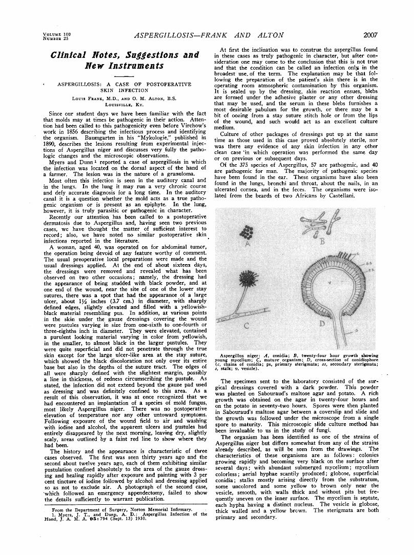
Aspergillus niger: A, conidia; B, twenty-four hour growth showingyoung mycelium; C, mature organism; D, cross-section of conidiophore(c, chains of conidia; ps, primary sterigmata; ss, secondary sterigmata;s, stalk; v. vesicle).
The specimen sent to the laboratory consisted of the sur-gical dressings covered with a dark powder. This powderwas planted on Sabouraud's maltose agar and potato. A richgrowth was obtained on the agar in twenty-four hours andon the potato in seventy-two hours. Spores were then plantedin Sabouraud's maltose agar between a coverslip and slide andthe growth was followed under the microscope from a singlespore to maturity. This microscopic slide culture method hasbeen invaluable to us in the study of fungi.The organism has been identified as one of the strains of
Aspergillus niger but differs somewhat from any of the strainsalready described, as will be seen from the drawings. Thecharacteristics of these organisms are as follows : coloniesgrowing rapidly and becoming very black on the surface afterseveral days ; with abundant submerged mycelium ; myceliumcolorless ; aerial hyphae scantily produced ; globose, superficialconidia ; stalks mostly arising directly from the substratum,some uncolored and some yellow to brown only near thevesicle, smooth, with walls thick and without pits but fre-quently uneven on the inner surface. The mycelium is septate,each hypha having a distinct nucleus. The vesicle is globose,thick walled and a yellow brown. The sterigmata are bothprimary and secondary.

Large numbers of calcium oxalate crystals were found inSabouraud's maltose agar near the growing organisms. Thedisaccharide maltose is hydrolyzed by the enzyme maltaseproduced, by this aspergillus and is broken down into twomolecules of the monosaccharide dextrose :
CiiHbOu + H20 i= 2C„Hi»06Citric acid is then fermented from the dextrose as an inter-
mediate product :
C«H]2Oo C„H807 + 2H20This reaction is not fully understood. Buchanan thinks that
possibly the acid is synthesized from some of the decomposi-tion products of the sugar.Oxalic acid is formed from the citric acid. When the acid
formed is neutralized by the addition of chalk, one half ofthe calculated theoretical yield is obtained.This is an interesting phenomenon. The finding of large
numbers of calcium oxalate crystals in the mediums may giveone an immediate lead as to the type of organisms present.614 Heyburn Building.
REPORT OF CASES OF CARBON MONOXIDE POISONINGTREATED BY METHYLENE BLUE INJECTION
A. W. Christopherson, M.D., Hermiston, Ore.
I wish to report further confirmative evidence of the valueof methylene blue (methylthionine chloride U. S. P.) in thetreatment of carbon monoxide poisoning.Recently five men were brought to me for emergency treat-
ment following a "mysterious" poisoning. They had beenworking with a road construction crew and were blastingrock in a tunnel measuring 6 by 10 by 115 feet. The blastwas set at the depth of the tunnel and ten minutes later two ofthe men entered the tunnel to commence work. After a fewmoments one of them sensed the danger and warned the otherto run. The latter collapsed almost immediately and theformer ran but collapsed at the mouth of the tunnel, wherehe was dragged to safety. One man ran into the tunnel andrescued the fallen man, but he also collapsed when safelywithin reach of others. These three were rushed to the hospital,where I attended them.Within thirty minutes two more men were brought to the
hospital. One had entered the tunnel with an air hose toblow the tunnel clean and in a few minutes he collapsed.Another witness rushed in and placed the victim on a beltconveyor and both were brought to safety.I immediately diagnosed the cases as carbon monoxide poi-
soning and put all to bed with high elevation of the foot ofthe bed, external heat, and pure oxygen inhalation. Threerequired caffeine sodiobenzoate and two, atropine sulphate forcardiac and respiratory stimulation.Two of the men were profoundly comatose and required
artificial respiration, as their voluntary respirations varied fromthree to five per minute. Their pulses were weak, rapid andirregular. Their color varied ; the younger man, about 28years of age and robust, was flushed pink from head to foot;the other, a man of 55 who had dissipated heavily, was cyanoticand in convulsions.An attempt was made immediately to obtain ready prepared
méthylène blue, 1 per cent, for intravenous injection, but asit was not available, a solution was prepared locally. An hourhad elapsed when the solution was ready and as the twoseverely affected patients appeared unimproved, they were bothgiven 50 cc. of the solution. Before the injection was com-
pleted, they became conscious and talked rationally. Bothmade rapid, uneventful recoveries and, while more cautiouslyhandled, seemed to have recuperated sooner than the threeremaining patients.The remaining cases responded promptly to the oxygen and
the dye was not used. One of these patients later had thestormiest recovery of the lot and is the only one remainingunder my care at this time, having a local systolic murmurover the heart apex, which may or may not be organic.It would appear that the méthylène blue had a remarkably
specific action in these cases of carbon monoxide poisoning.
Council on Pharmacy and ChemistryREPORTS OF THE COUNCIL
The Council has authorized publication of the followingïports. Paul Nicholas Leech, Secretary.
NAÏODINE NOT ACCEPTABLEFOR N. N. R.
"Naïodine" is a product of the Emile Logeais Laboratoriesof Boulogne sur Seine, France, distributed in the United Statesby E. Fougera and Company. It is proposed for subcutaneousintramuscular or intravenous injection for the relief of painfrom whatever cause.
The advertising material for Naïodine, widely promoted tothe medical profession, is pernicious. Rarely today can oneread claims of therapeutic usefulness more imaginative in con-
ception or less supported by fact.Naïodine is claimed to be a "one per cent solution of hyper-
active sodium iodide (Nal) . .
.
stabilized by a specialprocess, exclusive of any other active principle, toxic or other-wise."There is no chemical method known to the Council whereby
a simple, highly ionizable salt like sodium iodide can be rendered"hyperactive," whatever that may mean. The implication inthe advertising circulars that this iodide preparation is of a
special order of therapeutic effectiveness is highly improbable.It is well known that the addition of small amounts of alkali,preservation in hard glass, and protection from light may tendto prevent the decomposition of iodide solutions.The following are some of the claims made for this prepara-
tion :
"NAÏODINE is indicated for the relief of pain, and distress, whateverthe organ affected, whatever the site of pain, and whatever the nature ofthe pain or distress present, whether it be due to neuralgia, neuritis,spasm, inflammation, sympathetic pain, anguish or anxiety [Italics ours]."NAÏODINE is as powerfully effective against the anguish accom-
panying the withdrawal treatment in cases of toxicomanía as it is againstthe so called 'essential' sciatica, the crises of asthma, sinusalgia, thetarsalgia affecting adolescents, uterine or ovarian pain in dysmenorrhealpatients, pain present in advanced cancer, myalgia, rheumatic or rheuma-toid pain and the like."In brief, NAÏODINE constitutes a type of sedative preparation, a
faithful antispasmodic, and an antalgic. It may be added that theinjection of NAÏODINE is painless.""The sodium iodide present in NAÏODINE. when introduced paren-
terally, possesses a veritable nenronotropic effect. When absorbed bythe capillaries, it progressively impregnates the nerve centers, somewhatas bromides impregnate them, diminishes and suppresses promptly theirsensitiveness to pain
.
.
.
large doses must be tmployed from thevery first, and must be repeated without hesitation. There is no reasonwhatever for hesitating, since Naïodine is atoxic and since it producesno modification whatever of the respiratory or circulatory centers
.
. .""Injectable NAÏODINE permits atoxic treatment of all algic states.
It replaced morphin and other aiuilticsiis, all of which are toxic.N'aiodine is Atoxic and Painless. Any dosage may be injected, without
the least difficulty. It has no contra-indications.""Use and dosage: The Treatment of Attack, by massive doses: 20 to
30 cc. daily, given in one or several injections. Maintaining Treatment,with diminishing dosage: 10 to 5 cc. daily." "Injections ... inany muscular mass, deep subcutaneous, or intravenous."
Naïodine is further recommended for "sciaticas, lumbagos,shingles, trigeminal neuralgias, syphilitic and rheumatismaliritis . . . dyspnea, angina pectoris, asthma, pulmonaryemphysema, toxicomanías . .
.
phlegmons, burns, acutearthritis, painful chronic arthritis, acute otitis, etc."A brochure also is distributed, composed about a photograph
of a woman's head in marble reproduction ; at the bottomappears "Naïodine—cure atoxique" ; in one margin, "ne plussouffrir," repeated several times in type of diminishing size.Naïodine is claimed to be absolutely atoxic ; nowhere is there
mention of the possibility of iodism, of anaphylactoid reactionsfrom intravenous injection, or of the possibility of inducingserious or even fatal pulmonary edema in susceptible individuals,particularly in the pathologic pulmonary conditions for whichNaïodine is recommended. The routine use of injections ofsodium iodide to replace "morphine and other analgesics" as