assembly of dna architectures in a non-aqueous solution · assembly of dna architectures in a...
TRANSCRIPT
Assembly of DNA Architectures in a Non-aqueous Solution
by Amethist S. Finch, Christopher M. Anton Christina M. Jacob
Thomas J. Proctor, and Dimitra N. Stratis-Cullum
ARL-RP-0405 September 2012
A reprint from Advanced Materials
Approved for public release; distribution unlimited.
NOTICES
Disclaimers
The findings in this report are not to be construed as an official Department of the Army position
unless so designated by other authorized documents.
Citation of manufacturer’s or trade names does not constitute an official endorsement or
approval of the use thereof.
Destroy this report when it is no longer needed. Do not return it to the originator.

Army Research Laboratory Adelphi, MD 20783-1197
ARL-RP-0405 September 2012
Assembly of DNA Architectures in a Non-aqueous Solution
Amethist S. Finch, Christina M. Jacob, Thomas J. Proctor,
and Dimitra N. Stratis-Cullum Sensors and Electron Devices Directorate, ARL
Christopher M. Anton Episensors, Inc.,
590 Territorial Drive
Bolingbrook, IL 60440
A reprint from Advanced Materials
Approved for public release; distribution unlimited.
ii
REPORT DOCUMENTATION PAGE Form Approved OMB No. 0704-0188
Public reporting burden for this collection of information is estimated to average 1 hour per response, including the time for reviewing instructions, searching existing data sources, gathering and maintaining the
data needed, and completing and reviewing the collection information. Send comments regarding this burden estimate or any other aspect of this collection of information, including suggestions for reducing the
burden, to Department of Defense, Washington Headquarters Services, Directorate for Information Operations and Reports (0704-0188), 1215 Jefferson Davis Highway, Suite 1204, Arlington, VA 22202-4302.
Respondents should be aware that notwithstanding any other provision of law, no person shall be subject to any penalty for failing to comply with a collection of information if it does not display a currently valid
OMB control number.
PLEASE DO NOT RETURN YOUR FORM TO THE ABOVE ADDRESS.
1. REPORT DATE (DD-MM-YYYY)
September 2012
2. REPORT TYPE
3. DATES COVERED (From - To)
4. TITLE AND SUBTITLE
Assembly of DNA Architectures in a Non-aqueous Solution
5a. CONTRACT NUMBER
5b. GRANT NUMBER
5c. PROGRAM ELEMENT NUMBER
6. AUTHOR(S)
Amethist S. Finch, Christopher M. Anton, Christina M. Jacob, Thomas J. Proctor
and Dimitra N. Stratis-Cullum
5d. PROJECT NUMBER
5e. TASK NUMBER
5f. WORK UNIT NUMBER
7. PERFORMING ORGANIZATION NAME(S) AND ADDRESS(ES)
U.S. Army Research Laboratory
ATTN: RDRL-SEE-E
2800 Powder Mill Road
Adelphi, MD 20783-1197
8. PERFORMING ORGANIZATION
REPORT NUMBER
ARL-RP-0405
9. SPONSORING/MONITORING AGENCY NAME(S) AND ADDRESS(ES)
10. SPONSOR/MONITOR’S ACRONYM(S)
11. SPONSOR/MONITOR'S REPORT
NUMBER(S)
12. DISTRIBUTION/AVAILABILITY STATEMENT
Approved for public release; distribution unlimited.
13. SUPPLEMENTARY NOTES
E-Mail: [email protected]
14. ABSTRACT
In the present work, the procedures for the creation of self-assembled DNA nanostructures in aqueous and non-aqueous media
are described. DNA-Surfactant complex formation renders the DNA soluble in organic solvents offering an exciting way to
bridge the transition of DNA origami materials electronics applications. The DNA retains its structural features, and these
unique geometries provide an interesting candidate for future electronics and nanofabrication applications with potential for
new properties. The DNA architectures were first assembled under aqueous conditions, and then characterized in solution
(using circular dichroism (CD) spectroscopy) and on the surface (using atomic force microscopy (AFM)). Following aqueous
assembly, the DNA nanostructures were transitioned to a non-aqueous environment, where butanol was chosen for optical
compatibility and thermal properties. The retention of DNA hierarchical structure and thermal stability in non-aqueous
conditions were confirmed via CD spectroscopy. The formation and characterization of these higher order DNA-surfactant
complexes is described in this paper. 15. SUBJECT TERMS
DNA nanostructures; bioelectronics; biodirected assembly; CTAC; DNA
16. SECURITY CLASSIFICATION OF: 17. LIMITATION
OF ABSTRACT
UU
18. NUMBER
OF PAGES
16
19a. NAME OF RESPONSIBLE PERSON
Amethist S. Finch a. REPORT
UNCLASSIFIED
b. ABSTRACT
UNCLASSIFIED
c. THIS PAGE
UNCLASSIFIED 19b. TELEPHONE NUMBER (Include area code)
(301) 394-0326
Standard Form 298 (Rev. 8/98)
Prescribed by ANSI Std. Z39.18
Nanomaterials 2012, 2, 275-285; doi:10.3390/nano2030275
nanomaterials ISSN 2079-4991
www.mdpi.com/journal/nanomaterials
Communication
Assembly of DNA Architectures in a Non-Aqueous Solution
Amethist S. Finch 1,*, Christopher M. Anton 2, Christina M. Jacob 1, Thomas J. Proctor 1 and
Dimitra N. Stratis-Cullum 1
1 RDRL-SEE-B, Adelphi, MD 20783, USA; E-Mails: [email protected] (C.M.J.);
[email protected] (T.J.P.); [email protected] (D.N.S.-C.) 2 Episensors, Inc., 590 Territorial Drive, Bolingbrook, IL 60440, USA;
E-Mail: [email protected]
* Author to whom correspondence should be addressed; E-Mail: [email protected];
Tel.: +1-301-394-0326.
Received: 18 June 2012; in revised form: 8 August 2012 / Accepted: 20 August 2012 /
Published: 31 August 2012
Abstract: In the present work, the procedures for the creation of self-assembled DNA
nanostructures in aqueous and non-aqueous media are described. DNA-Surfactant complex
formation renders the DNA soluble in organic solvents offering an exciting way to bridge
the transition of DNA origami materials electronics applications. The DNA retains its
structural features, and these unique geometries provide an interesting candidate for future
electronics and nanofabrication applications with potential for new properties. The DNA
architectures were first assembled under aqueous conditions, and then characterized in
solution (using circular dichroism (CD) spectroscopy) and on the surface (using atomic
force microscopy (AFM)). Following aqueous assembly, the DNA nanostructures were
transitioned to a non-aqueous environment, where butanol was chosen for optical
compatibility and thermal properties. The retention of DNA hierarchical structure and
thermal stability in non-aqueous conditions were confirmed via CD spectroscopy. The
formation and characterization of these higher order DNA-surfactant complexes is
described in this paper.
Keywords: DNA nanostructures; bioelectronics; biodirected assembly; CTAC; DNA
OPEN ACCESS

Nanomaterials 2012, 2 276
1. Introduction
Rapid development in electronic materials manufacturing has led to the development of standard
devices with sub-100 nm features. These advances in science and engineering are remarkable but have
reached a fundamental limit using a top-down approach, leading researchers to look towards nature to
realize the next level of technological innovation [1,2]. Structural DNA nanotechnology has seen the
development of a rich toolbox of building blocks over the last two decades [3,4]. These tools allow for
the creation of a variety of structural architectures with distinct mechanical properties, and allow
the assembly of a variety of 2D and 3D structures with exquisite control of periodicity and
complexity [5–11].
Specifically, DNA has been a focus of many studies because it is well-characterized, relatively
inexpensive, easy to synthesize, self-sorting, and readily modified with a variety of functional groups.
Additionally, DNA is programmable at the molecular level with the ability to pre-organize into
well-defined secondary and tertiary structures, and its size is to scale with traditional nanomaterial
building blocks [2]. DNA assemblies range from simple geometric shapes to more complex higher
order structures, all varied by use of the genetic code. These characteristics make DNA an ideal target
for use as templates for materials applications, and for biologically enabled electronics and devices
such as capacitors [12], sensors [13], wires [14–16], electro-optic modulators [17,18], waveguides [19],
semi-conductor materials [20], electromagnetic interference (EMI) shielding materials [21], field
effect transistors (FET) [22], energy storage materials [23], and organic light emitting diodes
(OLED) [24,25].
Despite the promise of DNA assembly, a major hurdle in the use of assembled DNA architectures
lies in fact that DNA architectures rely on aqueous phase assembly and are, thereby, not amenable to
traditional manufacturing processes [26]. Development of methodologies that enable non-aqueous
assembly of biological materials would address this key barrier to successful coupling of biological
materials and their use in manufacturing of electronic devices. In addition, the ability to transition these
DNA architectures into organic solvents should allow these materials to be used as interesting
alternatives to traditional dielectrics since the interface and surface morphology seems to play a crucial
role in device performance [27]. In this paper, we will address a critical issue of the biological
interface by complexing the DNA with a surfactant, which allows it to be soluble in a variety of
non-aqueous solvents and greatly increases its thermal stability. This is the first report of higher order
DNA nanoarchitectures assembled in non-aqueous conditions in literature; these procedures are
necessary in avoiding unwanted oxidation of certain electronic substrates and for providing added
mechanical stability to the biological materials.
2. Results and Discussion
For these experiments, a DNA-cationic surfactant complex was created using either
hexadecyltrimethylammonium-chloride (CTAC) or bromide (CTAB). This DNA-surfactant complex
(DNA-CTMA) rendered the DNA water insoluble, causing it to precipitate out of its aqueous solution,
and also had the added benefit of making the DNA more mechanically and thermally stable [22]. The
DNA-CTMA complex was soluble in a variety of non-aqueous solvents, including isopropanol,
Nanomaterials 2012, 2 277
ethanol, methanol, acetonitrile, and butanol [28]. Previous CD studies have shown that when using
genomic DNA, the DNA-CTMA compound retains its double-helical structure in these solvents at
temperatures over 100 °C [19].
Many different research laboratories have published sequences and protocols for creating a large
variety of shapes and structures using differing numbers of DNA strands and different strand
lengths [3]. For this work, the wagon wheel DNA (wwDNA) was selected as the initial template,
primarily due to its overall size [29]. Specifically, wagon wheel templates have a void size of 10 nm or
less and have a relatively large feature size (>15 nm), facilitating visualization of the samples by AFM.
Furthermore, this template only consists of two DNA strands that combine to form a higher order
structure using a T-junction motif, which greatly simplifies the protocol for creating the initial
template. To demonstrate the versatility of this approach, several other T-junction motif-based DNA
architectures were used, illustrating compatibility with structural diversity and ease of assembly.
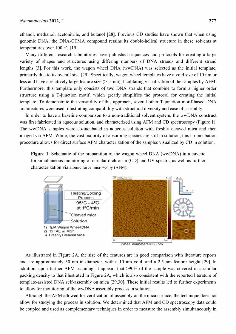
In order to have a baseline comparison to a non-traditional solvent system, the wwDNA construct
was first fabricated in aqueous solution, and characterized using AFM and CD spectroscopy (Figure 1).
The wwDNA samples were co-incubated in aqueous solution with freshly cleaved mica and then
imaged via AFM. While, the vast majority of absorbing species are still in solution, this co-incubation
procedure allows for direct surface AFM characterization of the samples visualized by CD in solution.
Figure 1. Schematic of the preparation of the wagon wheel DNA (wwDNA) in a cuvette
for simultaneous monitoring of circular dichroism (CD) and UV spectra, as well as further
characterization via atomic force microscopy (AFM).
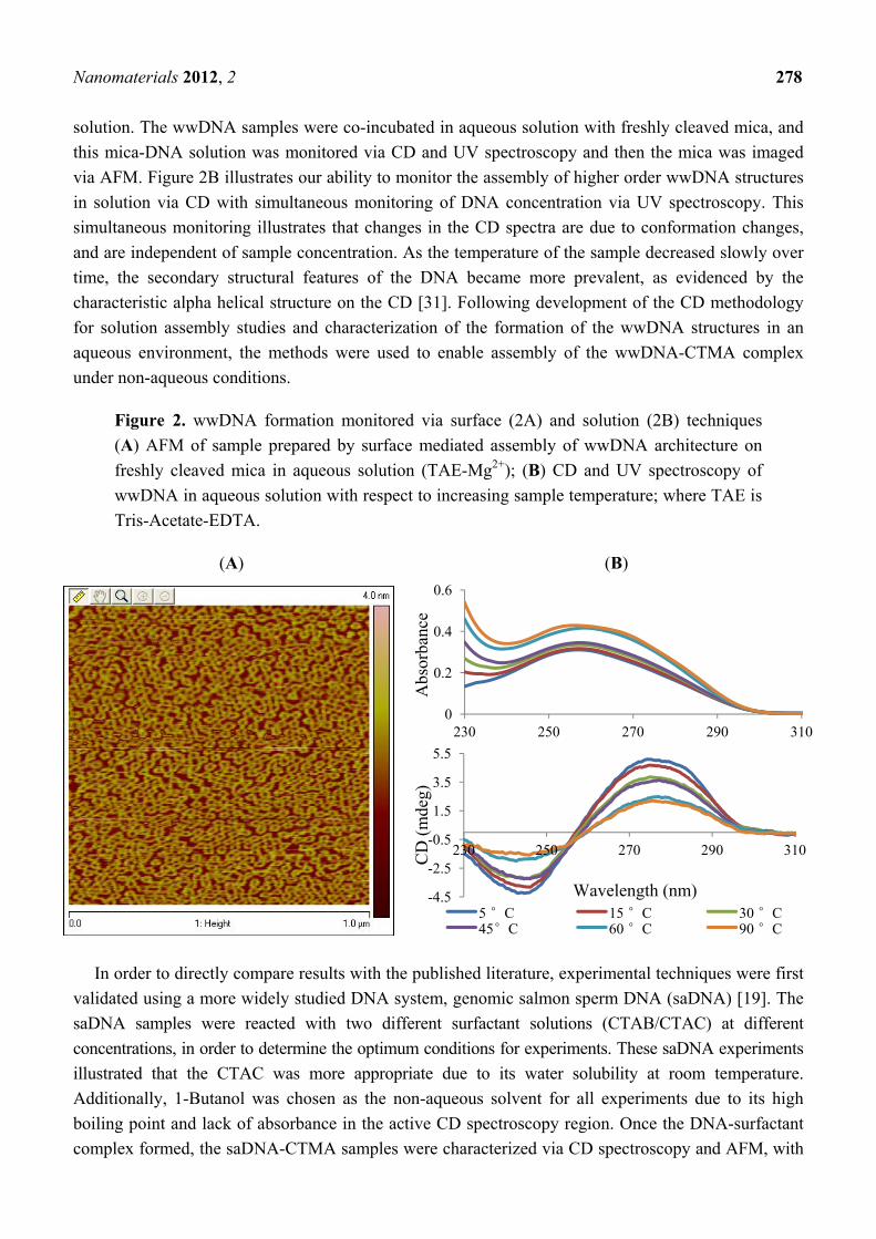
As illustrated in Figure 2A, the size of the features are in good comparison with literature reports
and are approximately 30 nm in diameter, with a 10 nm void, and a 2.5 nm feature height [29]. In
addition, upon further AFM scanning, it appears that >90% of the sample was covered in a similar
packing density to that illustrated in Figure 2A, which is also consistent with the reported literature of
template-assisted DNA self-assembly on mica [29,30]. These initial results led to further experiments
to allow for monitoring of the wwDNA assembly process in solution.
Although the AFM allowed for verification of assembly on the mica surface, the technique does not
allow for studying the process in solution. We determined that AFM and CD spectroscopy data could
be coupled and used as complementary techniques in order to measure the assembly simultaneously in
Nanomaterials 2012, 2 278
solution. The wwDNA samples were co-incubated in aqueous solution with freshly cleaved mica, and
this mica-DNA solution was monitored via CD and UV spectroscopy and then the mica was imaged
via AFM. Figure 2B illustrates our ability to monitor the assembly of higher order wwDNA structures
in solution via CD with simultaneous monitoring of DNA concentration via UV spectroscopy. This
simultaneous monitoring illustrates that changes in the CD spectra are due to conformation changes,
and are independent of sample concentration. As the temperature of the sample decreased slowly over
time, the secondary structural features of the DNA became more prevalent, as evidenced by the
characteristic alpha helical structure on the CD [31]. Following development of the CD methodology
for solution assembly studies and characterization of the formation of the wwDNA structures in an
aqueous environment, the methods were used to enable assembly of the wwDNA-CTMA complex
under non-aqueous conditions.
Figure 2. wwDNA formation monitored via surface (2A) and solution (2B) techniques
(A) AFM of sample prepared by surface mediated assembly of wwDNA architecture on
freshly cleaved mica in aqueous solution (TAE-Mg2+); (B) CD and UV spectroscopy of
wwDNA in aqueous solution with respect to increasing sample temperature; where TAE is
Tris-Acetate-EDTA.
(A) (B)
In order to directly compare results with the published literature, experimental techniques were first
validated using a more widely studied DNA system, genomic salmon sperm DNA (saDNA) [19]. The
saDNA samples were reacted with two different surfactant solutions (CTAB/CTAC) at different
concentrations, in order to determine the optimum conditions for experiments. These saDNA experiments
illustrated that the CTAC was more appropriate due to its water solubility at room temperature.
Additionally, 1-Butanol was chosen as the non-aqueous solvent for all experiments due to its high
boiling point and lack of absorbance in the active CD spectroscopy region. Once the DNA-surfactant
complex formed, the saDNA-CTMA samples were characterized via CD spectroscopy and AFM, with
0
0.2
0.4
0.6
230 250 270 290 310
Abs
orba
nce
-4.5
-2.5
-0.5
1.5
3.5
5.5
230 250 270 290 310
CD
(m
deg)
5 °C 15 °C 30 °C45°C 60 °C 90 °C
Wavelength (nm)
Nanomaterials 2012, 2 279
1-butanol as the solvent (Figure S1 in supporting information). The CD confirmed literature reports and
illustrates that the structure of the saDNA does not change upon creation of the saDNA-CTMA complex,
as shown by the characteristic dsDNA CD spectra [31,32]. These results and experimental controls
conducted with the saDNA provided a standard sample for comparison with the wwDNA.
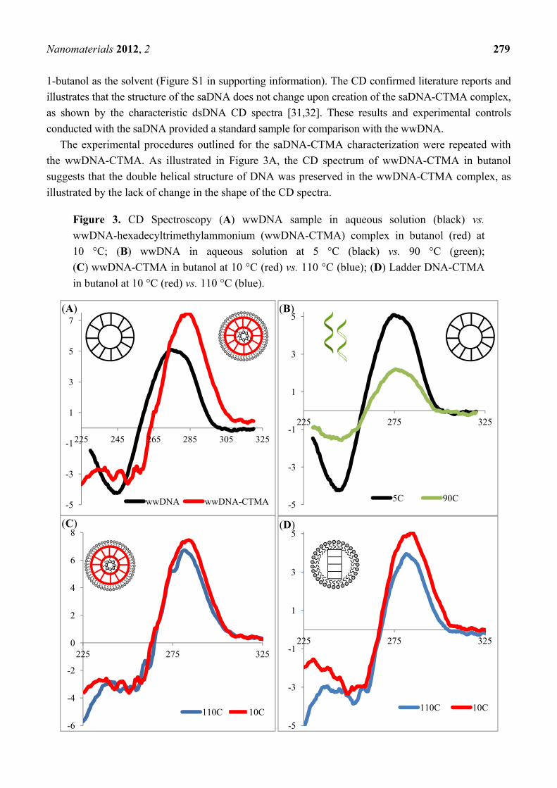
The experimental procedures outlined for the saDNA-CTMA characterization were repeated with
the wwDNA-CTMA. As illustrated in Figure 3A, the CD spectrum of wwDNA-CTMA in butanol
suggests that the double helical structure of DNA was preserved in the wwDNA-CTMA complex, as
illustrated by the lack of change in the shape of the CD spectra.
Figure 3. CD Spectroscopy (A) wwDNA sample in aqueous solution (black) vs.
wwDNA-hexadecyltrimethylammonium (wwDNA-CTMA) complex in butanol (red) at
10 °C; (B) wwDNA in aqueous solution at 5 °C (black) vs. 90 °C (green);
(C) wwDNA-CTMA in butanol at 10 °C (red) vs. 110 °C (blue); (D) Ladder DNA-CTMA
in butanol at 10 °C (red) vs. 110 °C (blue).
-5
-3
-1
1
3
5
7
225 245 265 285 305 325
wwDNA wwDNA-CTMA -5
-3
-1
1
3
5
225 275 325
5C 90C
-6
-4
-2
0
2
4
6
8
225 275 325
110C 10C
-5
-3
-1
1
3
5
225 275 325
110C 10C
(A) (B)
(C) (D)
Nanomaterials 2012, 2 280
Although the spectrum from aqueous to butanol is slightly shifted, this shift is in agreement with
literature reports that the overall shape of the spectra remains the same, suggesting retention of overall
structure [31,32]. For comparison, Figure 3B illustrates the differences between the high and low
temperature CD spectra of the wwDNA in aqueous solution. The wwDNA-CTMA in butanol was then
subject to the same thermal melting conditions as the aqueous wwDNA sample. As illustrated in
Figure 3C, no loss of features or loss of helical structure is detected with increased temperature up to
the limits of the instrument (110 °C). This thermal melting experiment was repeated with the ladder
DNA-CTMA sequence in butanol (Figure 3D), illustrating the ubiquity of this technique for
transitioning DNA architectures to non-aqueous solvents.
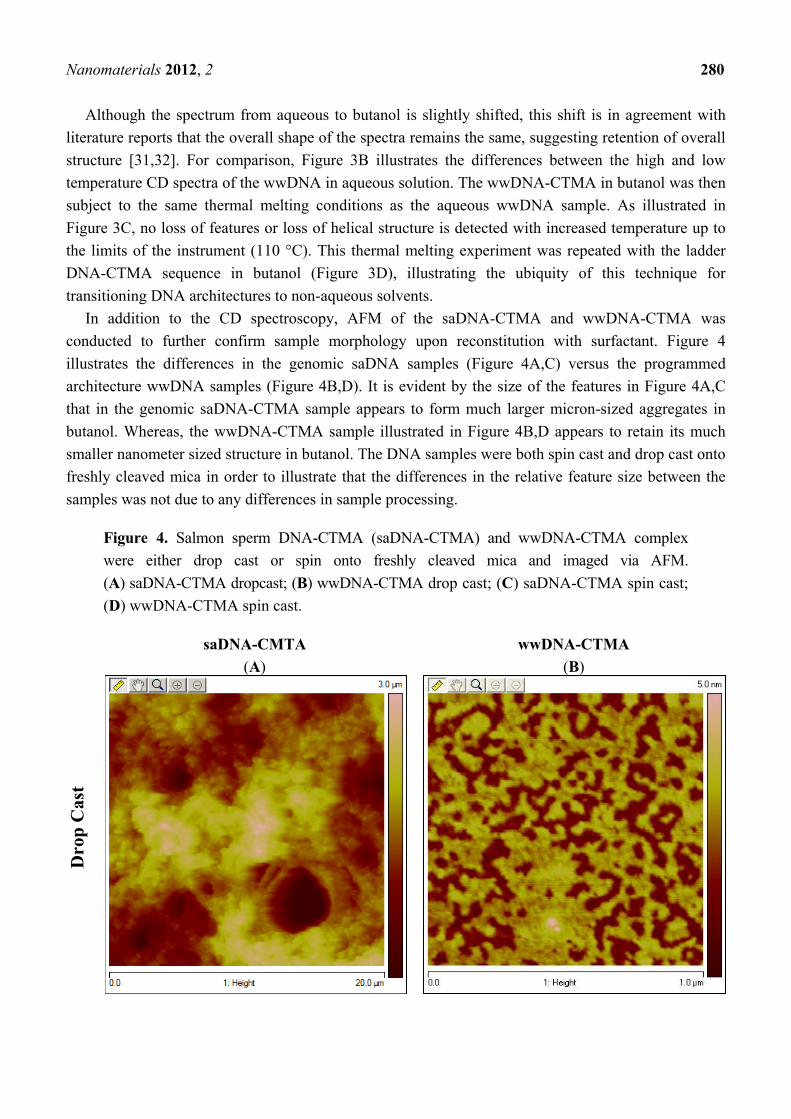
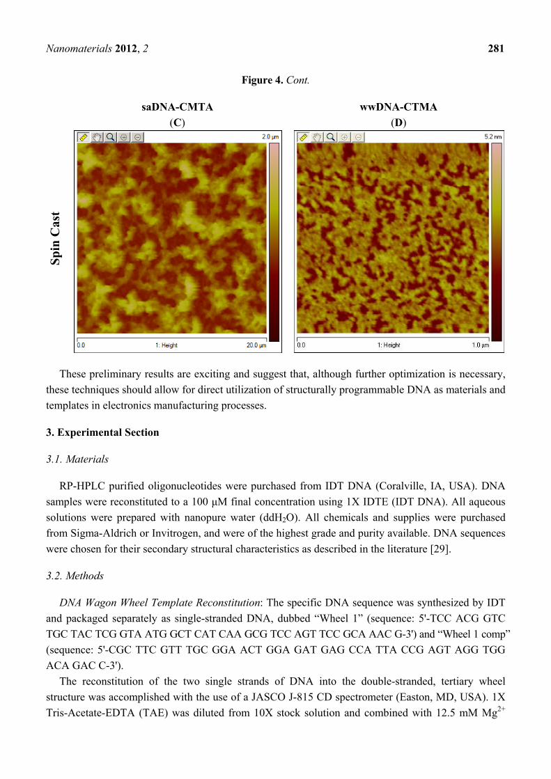
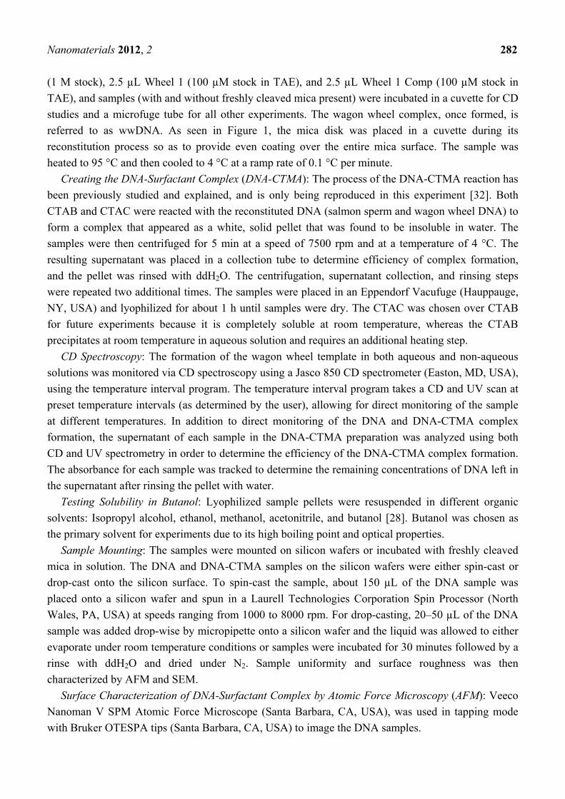
In addition to the CD spectroscopy, AFM of the saDNA-CTMA and wwDNA-CTMA was
conducted to further confirm sample morphology upon reconstitution with surfactant. Figure 4
illustrates the differences in the genomic saDNA samples (Figure 4A,C) versus the programmed
architecture wwDNA samples (Figure 4B,D). It is evident by the size of the features in Figure 4A,C
that in the genomic saDNA-CTMA sample appears to form much larger micron-sized aggregates in
butanol. Whereas, the wwDNA-CTMA sample illustrated in Figure 4B,D appears to retain its much
smaller nanometer sized structure in butanol. The DNA samples were both spin cast and drop cast onto
freshly cleaved mica in order to illustrate that the differences in the relative feature size between the
samples was not due to any differences in sample processing.
Figure 4. Salmon sperm DNA-CTMA (saDNA-CTMA) and wwDNA-CTMA complex
were either drop cast or spin onto freshly cleaved mica and imaged via AFM.
(A) saDNA-CTMA dropcast; (B) wwDNA-CTMA drop cast; (C) saDNA-CTMA spin cast;
(D) wwDNA-CTMA spin cast.
saDNA-CMTA wwDNA-CTMA
Dro
p C
ast
(A) (B)
Nanomaterials 2012, 2 281
Figure 4. Cont.
saDNA-CMTA wwDNA-CTMA
Sp
in C
ast
(C) (D)
These preliminary results are exciting and suggest that, although further optimization is necessary,
these techniques should allow for direct utilization of structurally programmable DNA as materials and
templates in electronics manufacturing processes.
3. Experimental Section
3.1. Materials
RP-HPLC purified oligonucleotides were purchased from IDT DNA (Coralville, IA, USA). DNA
samples were reconstituted to a 100 μM final concentration using 1X IDTE (IDT DNA). All aqueous
solutions were prepared with nanopure water (ddH2O). All chemicals and supplies were purchased
from Sigma-Aldrich or Invitrogen, and were of the highest grade and purity available. DNA sequences
were chosen for their secondary structural characteristics as described in the literature [29].
3.2. Methods
DNA Wagon Wheel Template Reconstitution: The specific DNA sequence was synthesized by IDT
and packaged separately as single-stranded DNA, dubbed “Wheel 1” (sequence: 5'-TCC ACG GTC
TGC TAC TCG GTA ATG GCT CAT CAA GCG TCC AGT TCC GCA AAC G-3') and “Wheel 1 comp”
(sequence: 5'-CGC TTC GTT TGC GGA ACT GGA GAT GAG CCA TTA CCG AGT AGG TGG
ACA GAC C-3').
The reconstitution of the two single strands of DNA into the double-stranded, tertiary wheel
structure was accomplished with the use of a JASCO J-815 CD spectrometer (Easton, MD, USA). 1X
Tris-Acetate-EDTA (TAE) was diluted from 10X stock solution and combined with 12.5 mM Mg2+
(C) (D)
Nanomaterials 2012, 2 282
(1 M stock), 2.5 µL Wheel 1 (100 µM stock in TAE), and 2.5 µL Wheel 1 Comp (100 µM stock in
TAE), and samples (with and without freshly cleaved mica present) were incubated in a cuvette for CD
studies and a microfuge tube for all other experiments. The wagon wheel complex, once formed, is
referred to as wwDNA. As seen in Figure 1, the mica disk was placed in a cuvette during its
reconstitution process so as to provide even coating over the entire mica surface. The sample was
heated to 95 °C and then cooled to 4 °C at a ramp rate of 0.1 °C per minute.
Creating the DNA-Surfactant Complex (DNA-CTMA): The process of the DNA-CTMA reaction has
been previously studied and explained, and is only being reproduced in this experiment [32]. Both
CTAB and CTAC were reacted with the reconstituted DNA (salmon sperm and wagon wheel DNA) to
form a complex that appeared as a white, solid pellet that was found to be insoluble in water. The
samples were then centrifuged for 5 min at a speed of 7500 rpm and at a temperature of 4 °C. The
resulting supernatant was placed in a collection tube to determine efficiency of complex formation,
and the pellet was rinsed with ddH2O. The centrifugation, supernatant collection, and rinsing steps
were repeated two additional times. The samples were placed in an Eppendorf Vacufuge (Hauppauge,
NY, USA) and lyophilized for about 1 h until samples were dry. The CTAC was chosen over CTAB
for future experiments because it is completely soluble at room temperature, whereas the CTAB
precipitates at room temperature in aqueous solution and requires an additional heating step.
CD Spectroscopy: The formation of the wagon wheel template in both aqueous and non-aqueous
solutions was monitored via CD spectroscopy using a Jasco 850 CD spectrometer (Easton, MD, USA),
using the temperature interval program. The temperature interval program takes a CD and UV scan at
preset temperature intervals (as determined by the user), allowing for direct monitoring of the sample
at different temperatures. In addition to direct monitoring of the DNA and DNA-CTMA complex
formation, the supernatant of each sample in the DNA-CTMA preparation was analyzed using both
CD and UV spectrometry in order to determine the efficiency of the DNA-CTMA complex formation.
The absorbance for each sample was tracked to determine the remaining concentrations of DNA left in
the supernatant after rinsing the pellet with water.
Testing Solubility in Butanol: Lyophilized sample pellets were resuspended in different organic
solvents: Isopropyl alcohol, ethanol, methanol, acetonitrile, and butanol [28]. Butanol was chosen as
the primary solvent for experiments due to its high boiling point and optical properties.
Sample Mounting: The samples were mounted on silicon wafers or incubated with freshly cleaved
mica in solution. The DNA and DNA-CTMA samples on the silicon wafers were either spin-cast or
drop-cast onto the silicon surface. To spin-cast the sample, about 150 µL of the DNA sample was
placed onto a silicon wafer and spun in a Laurell Technologies Corporation Spin Processor (North
Wales, PA, USA) at speeds ranging from 1000 to 8000 rpm. For drop-casting, 20–50 µL of the DNA
sample was added drop-wise by micropipette onto a silicon wafer and the liquid was allowed to either
evaporate under room temperature conditions or samples were incubated for 30 minutes followed by a
rinse with ddH2O and dried under N2. Sample uniformity and surface roughness was then
characterized by AFM and SEM.
Surface Characterization of DNA-Surfactant Complex by Atomic Force Microscopy (AFM): Veeco
Nanoman V SPM Atomic Force Microscope (Santa Barbara, CA, USA), was used in tapping mode
with Bruker OTESPA tips (Santa Barbara, CA, USA) to image the DNA samples.
Nanomaterials 2012, 2 283
4. Conclusions
To summarize, these results show for the first time, formation of higher order DNA structures, both
in aqueous and non-aqueous solutions. Characterization was performed via AFM (surface) and CD
spectroscopy (solution). The aqueous wwDNA samples were combined with surfactant CTAC to form
DNA-CTMA complexes, which were soluble in a variety of organic solvents. Future work will include
optimization of the material surface for single layer deposition of the templated DNA-CTMA complex,
as well as transitioning to a wide range of structures. These nanostructured materials have great
promise in creating bottom-up features smaller than those created by current top-down
lithographic techniques.
Acknowledgements
The authors would like to thank Amy Manocchi for her assistance with the AFM and James Sumner
for initial research discussions. The authors would also like to thank the Sensors and Electron Devices
Directorate of the U.S. Army Research Laboratory for funding these efforts.
References
1. Galatsis, K.; Wang, K.L.; Ozkan, M.; Ozkan, C.S.; Huang, Y.; Chang, J.P.; Monbouquette, H.G.;
Chen, Y.; Nealey, P.; Botros, Y. Patterning and templating for nanoelectronics. Adv. Mater. 2010,
22, 769–778.
2. Noy, A. Bionanoelectronics. Adv. Mater. 2011, 23, 807–820.
3. Sacca, B.; Niemeyer, C.M. DNA origami: The art of folding DNA. Angew. Chem. Int. Ed. 2012,
51, 58–66.
4. Seeman, N.C., Nanomaterials based on DNA. Annu. Rev. Biochem. 2010, 79, 65–87.
5. Andersen, E.S.; Dong, M.; Nielsen, M.M.; Jahn, K.; Subramani, R.; Mamdouh, W.; Golas, M.M.;
Sander, B.; Stark, H.; Oliveira, C.L.P.; et al. Self-assembly of a nanoscale DNA box with a
controllable lid. Nature 2009, 459, 73–76.
6. Douglas, S.M.; Dietz, H.; Liedl, T.; Hogberg, B.; Graf, F.; Shih, W.M. Self-assembly of DNA
into nanoscale three-dimensional shapes. Nature 2009, 459, 414–418.
7. Ke, Y.; Douglas, S.M.; Liu, M.; Sharma, J.; Cheng, A.; Leung, A.; Liu, Y.; Shih, W.M.; Yan, H.
Multilayer DNA origami packed on a square lattice. J. Am. Chem. Soc. 2009, 131, 15903–15908.
8. Ke, Y.; Sharma, J.; Liu, M.; Jahn, K.; Liu, Y.; Yan, H. Scaffolded DNA origami of a DNA
tetrahedron molecular container. Nano Lett. 2009, 9, 2445–2447.
9. Kuzuya, A.; Komiyama, M. Design and construction of a box-shaped 3D-DNA origami. Chem.
Commun. 2009, 28, 4182–4184.
10. Kuzuya, A.; Komiyama, M. DNA origami: Fold, stick, and beyond. Nanoscale 2010, 2, 310–322.
11. Rothemund, P.W. Folding DNA to create nanoscale shapes and patterns. Nature 2006, 440,
297–302.
12. Tang, H.; Chen, L.; Xing, C.; Guo, Y.-G.; Wang, S. DNA-templated synthesis of cationic
poly(3,4-ethylenedioxythiophene) derivative for supercapacitor electrodes. Macromol. Rapid
Commun. 2010, 31, 1892–1896.
Nanomaterials 2012, 2 284
13. Lin, Y.; Tao, Y.; Ren, J.; Pu, F.; Qu, X. Highly sensitive and selective detection of
thiol-containing biomolecules using DNA-templated silver deposition. Biosens. Bioelectron. 2011,
28, 339–343.
14. Shukla, S.; Sastry, M. Probing differential Ag+-nucleobase interactions with isothermal titration
calorimetry (ITC): Towards patterned DNA metallization. Nanoscale 2009, 1, 122–127.
15. Boeneman, K.; Prasuhn, D.E.; Blanco-Canosa, J.B.; Dawson, P.E.; Melinger, J.S.; Ancona, M.;
Stewart, M.H.; Susumu, K.; Huston, A.; Medintz, I.L. Self-assembled quantum dot-sensitized
multivalent DNA photonic wires. J. Am. Chem. Soc. 2010, 132, 18177–18190.
16. Kim, H.J.; Roh, Y.; Hong, B. Selective formation of a latticed nanostructure with the precise
alignment of DNA-templated gold nanowires. Langmuir 2010, 26, 18315–18319.
17. Heckman, E.M.; Aga, R.S.; Rossbach, A.T.; Telek, B.A.; Bartsch, C.M.; Grote, J.G. DNA
biopolymer conductive cladding for polymer electro-optic waveguide modulators. Appl. Phys.
Lett. 2011, 98, 103304:1–103304:3.
18. Popescu, R.; Moldoveanu, M.; Rau, I. Biopolymer thin films for photonics applications. Key Eng.
Mater. 2009, 415, 33–36.
19. Heckman, E.; Bartsch, C.; Yaney, P.; Subramanyam, G.; Ouchen, F.; Grote, J.G. DNA-Surfactant
Thin-Film Processing and Characterization. In Materials Science of DNA; CRC Press: Boca
Raton, FL, USA, 2011; pp. 179–230.
20. Gao, L.; Ma, N. DNA-templated semiconductor nanocrystal growth for controlled DNA packing
and gene delivery. ACS Nano 2012, 6, 689–695.
21. Zang, D.Y.; Grote, J. DNA-based nanoparticle composite materials for EMI shielding. Proc. SPIE
2012, doi: 10.1117/12.905284.
22. Singh, T.B.; Sariciftci, N.S.; Grote, J.G. Bio-Organic Optoelectronic Devices Using DNA.
In Organic Electronics; Meller, G., Grasser, T., Eds.; Springer: New York, NY, USA, 2010;
Volume 223, pp. 189–212.
23. Séverac, F.; Alphonse, P.; Estève, A.; Bancaud, A.; Rossi, C. High-energy Al/Cuo
nanocomposites obtained by DNA-directed assembly. Adv. Funct. Mater. 2012, 22, 323–329.
24. Kobayashi, N. Bioled with DNA/conducting polymer complex as active layer. Nonlinear Opt.
Quantum Opt. 2011, 43, 233–251.
25. Sun, Q.; Chang, D.W.; Dai, L.; Grote, J.; Naik, R. Multilayer white polymer light-emitting diodes
with deoxyribonucleic acid-cetyltrimetylammonium complex as a hole-transporting/electron-blocking
layer. Appl. Phys. Lett. 2008, 92, 251108:1–251108:3.
26. Singh, T.B.; Sariciftci, N.S.; Grote, J.G. Bio-organic optoelectronic devices using DNA. Adv.
Polym. Sci. 2010, 223, 189–212.
27. Horowitz, G. Interfaces in Organic Field-Effect Transistors. In Organic Electronics; Meller, G.,
Grasser, T., Eds.; Springer: New York, NY, USA, 2010; Volume 223, pp. 113–153.
28. Finch, A.S.; Jacob, C.M.; Sumner, J.J. DNA architectures for templated material growth.
Proc. SPIE 2011, doi:10.1117/12.899397.
29. Hamada, S.; Murata, S. Substrate-assisted assembly of interconnected single-duplex DNA
nanostructures. Angew. Chem. Int. Ed. 2009, 48, 6820–6823.
30. Sun, X.; Hyeon Ko, S.; Zhang, C.; Ribbe, A.E.; Mao, C. Surface-mediated DNA self-assembly.
J. Am. Chem. Soc. 2009, 131, 13248–13249.
Nanomaterials 2012, 2 285
31. Kypr, J.; Kejnovska, I.; Renciuk, D.; Vorlickova, M. Circular dichroism and conformational
polymorphism of DNA. Nucleic Acids Res. 2009, 37, 1713–1725.
32. Tanaka, K.; Okahata, Y. A DNA-lipid complex in organic media and formation of an aligned cast
film. J. Am. Chem. Soc. 1996, 118, 10679–10683.
© 2012 by the authors; licensee MDPI, Basel, Switzerland. This article is an open access article
distributed under the terms and conditions of the Creative Commons Attribution license
(http://creativecommons.org/licenses/by/3.0/).
DEFENSE TECH INFO CTR
ATTN DTIC OCA (PDF)
8725 JOHN J KINGMAN RD STE 0944
FT BELVOIR VA 22060-6218
US ARMY RSRCH LAB
ATTN IMAL HRA MAIL & RECORDS MGMT
ATTN RDRL CIO LL TECHL LIB
ATTN RDRL SEE B D STRATIS-CULLUM
ATTN RDRL SEE E A FINCH (2 COPIES)
ATTN RDRL SEE L BLISS
ADELPHI MD 20783-1197