assessment by doppler ultrasound of entheseal …£o sandra td 2014.pdf · asas assessment of...
TRANSCRIPT
ASSESSMENT BY DOPPLER ULTRASOUND OF ENTHESEAL
LESIONS IN SPONDYLOARTHRITIS:
A LONGITUDINAL STUDY TO DETERMINE STRUCTURAL DAMAGE
AND DISEASE ACTIVITY LESIONS
SANDRA ISABEL SALVADOR FALCÃO Tese para obtenção do grau de Doutor em Medicina na Especialidade em Investigação Clínica na Faculdade de Ciências Médicas
Outubro, 2014

ASSESSMENT BY DOPPLER ULTRASOUND OF ENTHESEAL
LESIONS IN SPONDYLOARTHRITIS:
A LONGITUDINAL STUDY TO DETERMINE STRUCTURAL DAMAGE
AND DISEASE ACTIVITY LESIONS
Sandra Isabel Salvador Falcão Orientadores: Professor Doutor Jaime da Cunha Branco e
Professor Doutor Eugenio de Miguel
Tese para obtenção do grau de Doutor em Medicina na Especialidade em
Investigação Clínica
Outubro, 2014

Identidade
Preciso ser um outro
para ser eu mesmo
Sou grão de rocha
Sou o vento que a desgasta
Sou pólen sem insecto
Sou areia sustentando
o sexo das árvores
Existo onde me desconheço
aguardando pelo meu passado
ansiando a esperança do futuro
No mundo que combato morro
no mundo por que luto nasço
Mia Couto, in "Raiz de Orvalho e Outros Poemas"

“…for some unexplained reason, ultrasonography applied to disorders of
tendons, musculature, soft tissues, and even bones have been largely
ignored by many physicians…”
Donald Resnick

v
TABLE OF CONTENTS
ACKNOWLEDGEMENTS .................................................................................................... 8
ABBREVIATIONS ............................................................................................................. 10
SUMMARY ...................................................................................................................... 12
INTRODUCTION .............................................................................................................. 15
“Once upon a time in Rheumatology land...” ............................................................. 15
Enthesis: “The synovial-entheseal complex” .............................................................. 17
Spondyloarthritis: the entheseal disease ................................................................... 20
Ultrasound of enthesis – enthesopathy and enthesitis features and scores ............. 27
DETAIL DESCRIPTION ..................................................................................................... 40
AIMS ............................................................................................................................... 43
RESULTS .......................................................................................................................... 44
PART I .......................................................................................................................... 45
PART II ......................................................................................................................... 52
PART III ........................................................................................................................ 56
PART IV ........................................................................................................................ 63
DISCUSSION .................................................................................................................... 70
CONCLUSION .................................................................................................................. 76
REFERENCES ................................................................................................................... 78

vi
FIGURES INDEX
Figure 1. Diagrammatic representation of the two types of enthesis: fibrocartilaginous
and fibrous. ..................................................................................................................... 18
Figure 2. Diagrammatic representation of the synovio-entheseal complex, using the
Achilles tendon enthesis organ to illustrate the concept. ............................................. 19
Figure 3. A view on the relationship between inflammation and ankylosis in SpA. ...... 22
Figure 4. Roles of BMPs and WNTs in endochondral bone formation. ......................... 24
Figure 5. The “TNF brake” hypothesis. ........................................................................... 25
Figure 6. Madrid sonography enthesitis index. .............................................................. 34

vii
TABLES INDEX
Table 1. Principal scores of enthesis ultrasound examination in spondyloarthritis
patients ........................................................................................................................... 29
Table 2. Validity aspects of enthesis ultrasound studies ............................................... 36

viii
ACKNOWLEDGEMENTS
Uma tese de doutoramento pode ser comparada a uma longa viagem. No início não
sabemos muito bem o que vamos encontrar e sentimentos como a ansiedade ou o
receio podem entrar em conflito com a curiosidade de descoberta pelo desconhecido.
Tal como numa viagem, é feita de momentos, uns bons outros menos bons, uns mais
enriquecedores do que outros, mas no final o que se sente é um sentimento de
preenchimento. Por nós, mas também pelos que nos acompanharam nesta viagem.
Estes muitas das vezes são aqueles que nos mantêm no caminho certo, impedindo que
um qualquer descarrilamento tenha um desfecho fatal. A estas pessoas quero
simplesmente agradecer a sua presença.
Durante a elaboração dos trabalhos que levaram a esta dissertação não posso deixar
de começar por agradecer aos meus dois orientadores. Obrigada professor Jaime
Branco por, para além de me oferecer “um trampolim”, me ter deixado manter as
minhas asas. Recordo-me do dia em que o conheci. No final de uma longa conversa
sincera disse-lhe: “Vai correr bem”. Ao qual me respondeu: “Claro que sim.” Obrigada
professor Eugenio de Miguel por partilhar comigo a sua paixão pela ecografia, por ser
um mentor exemplar e por me fazer acreditar. Neste caso a viagem passou muito além
de uma simples ida a Madrid. Nunca me esquecerei da forma como fui recebida,
integrada e, até, diria adotada. Obrigada por me ter integrado na sua linha de
investigação, pela planificação da rota, pelas orientações, discussões, contínuo
estímulo, conselhos inestimáveis, constante disponibilidade e atenção, mas acima de
tudo, pela amizade. Aos restantes membros da unidade de investigação do
departamento de Reumatologia do Hospital Universitário La Paz, o meu Muito
Obrigada. E a ti, Diana, um especial “Gracias” por me teres feito sentir em casa, pelos
almoços de domingo e por seres uma boa amiga.
No serviço de reumatologia do Hospital de Egas Moniz muito aconteceu desde que eu
entrei na especialidade. Muitas pessoas passaram, algumas ficaram e poucas sairam,
mas todas me marcaram. Obrigada Dr. Bravo Pimentão por me apresentar à
reumatologia. Dr. Alves de Matos não posso deixar de lhe agradecer as suas duras

ix
palavras que tanto me ajudaram a crescer. Obrigada a todos os colegas, enfermeira
Maria José, Ana e Sandra por tornarem o dia a dia mais leve.
A ti Filipa, a minha companheira dos “dias” duros de roer, dos dias de sol, dos dias
“assim assim”, enfim de todos os dias. Obrigada pela cumplicidade, pela tua força
contagiante de viver.
A todos os colegas e amigos da ESPER que partilham a paixão pela ecografia um muito
obrigada.
A todos os amigos que acompanharam esta minha viagem muito obrigada. Prometo
nos próximos meses não falar da tese ou dos artigos por terminar!
Não posso ainda deixar de agradecer à família – a todos os presentes a aos que já
partiram. Obrigada pai e mãe, tios, primos, por me ajudarem a ser quem sou. À minha
sis, meu farol e super-mulher, Zé e picolinos – abominável homem das neves e
principessa – muito, muito obrigada por existirem, por me fazerem rir mesmo quando
me apetece chorar. E per te...

x
ABBREVIATIONS
ASAS Assessment of SpondyloArthritis international Society
ASDAS Ankylosing Spondylitis Disease Activity Score
BASDAI Ankylosing Spondylitis Disease Activity Index
BASFI Bath Ankylosing Spondylitis Functional Index
BASRI Bath Ankylosing Spondylitis Radiology Index
BMI Body Mass Index
BMP Bone Morphogenetic Proteins
CASPAR ClASsification criteria for Psoriatic Arthritis
CRP C-reactive protein
DAS Disease Activity Score
DKK-1 Dickkopf-related protein 1
DISH Diffuse Idiopathic Skeletal Hyperostosis
ERAP1 Endoplasmic reticulum aminopeptidase 1
ESR Erythrocyte sedimentation rate
ESSG European Spondylarthropathy Study Group
ESU Early Spondyloarthritis Unit
FOP Fibrodysplasia Ossificans Progressiva
GUESS Glasgow Ultrasound Enthesitis Scoring System
HLA Human leucocyte antigen
IBD Inflammatory bowel disease
IL Interleukin
IL23R Interleukin-23 receptor
MASEI Madrid Sonography Enthesitis Index
MASES Maastricht Ankylosing Spondylitis Enthesitis Score
MEI Mander Enthesis Index
MRI Magnetic resonance imaging
OMERACT Outcome Measures in Rheumatology Clinical Trials
PG Proteoglycan-rich matrix
PGE2 Prostaglandin E2

xi
PsA Psoriatic Arthritis
RA Rheumatoid Arthritis
SEI Sonographic enthesitic index
SpA Spondyloarthritis
TNF Tumor necrosis factor
VAS Visual analogic scale
WNT Wingless type like signaling

12
SUMMARY
Enthesitis is the hallmark of spondyloarthritis (SpA), and is observed in all subtypes.
Wide information on SpA abnormalities, including synovitis, tendinitis and enthesitis,
can be efficiently perceived by Doppler ultrasound. Furthermore, several studies on
imaging of enthesis showed that imaging techniques are better than clinical
examination to detect enthesis alterations; and vascularized enthesitis detected by
Doppler ultrasound appears to be a valuable diagnostic tool to confirm SpA diagnosis.
However, data published until now concerning entheseal elementary alterations that
characterize SpA enthesitis (enthesis inflammatory activity) or enthesopathy
(permanent structural changes) reflect rather the authors’ empiric opinion than a
methodological validation process. In this sense it seems crucial to identify elementary
entheseal lesions associated with activity or damage, in order to improve monitoring
and treatment response in SpA patients. The development of better assessment tools
is today a challenge and a need in SpA.
The first study of this thesis focused on the analysis of the reliability of inter-lector and
inter-ultrasonography equipment of Madrid sonography enthesitis index (MASEI).
Fundamental data for the remaining unrolling project validity.
In the second and third studies we concerned about two entheseal elemental lesions:
erosions and bursa. In literature erosions represent a permanent structural damage,
being useful for monitoring joint injury, disease activity and therapeutic response in
many rheumatic diseases; and to date, this concept has been mostly applied in
rheumatoid arthritis (RA). Unquestionably, erosion is a tissue-related damage and a
structural change. However, the hypothesis that we decided to test was if erosions
represent a permanent structural change that can only grow and worsen over time, as
occurs in RA, or a transitory alteration. A longitudinal study of early SpA patients was
undertaken, and the Achilles enthesis was used as a model. Our results strongly
suggested that previously detected erosions could disappear during the course of the
disease, being consistent with the dynamic behavior of erosion over time. Based on
these striking results it seems reasonable to suggest that the new-bone formation

13
process in SpA could be associated with the resolution of cortical entheseal erosion
over time. These results could also be in agreement with the apparent failure of anti-
tumor necrosis factor (TNF) therapies to control bone proliferation in SpA; and with
the relation of TNF-α, Dickkopf-related protein 1 (Dkk-1) and the regulatory molecule
of the Wnt signaling pathway in the bone proliferation in SpA. In the same model, we
then proceeded to study the enthesis bursa. Interestingly, the Outcome Measures in
Rheumatology Clinical Trials (OMERACT) enthesopathy definition does not include
bursa as an elementary entheseal lesion. Nonetheless, bursa was included in 46% of
the enthesis studies in a recently systematic literature review, being in agreement with
the concept of “synovio-entheseal complex” that includes the link between enthesitis
and osteitis in SpA. It has been clarified in recent data that there is not only a close
functional integration of the enthesis with the neighboring bone, but also a connection
between enthesitis and synovitis. Therefore, we tried to assess the prevalence and
relevance of the bursa-synovial lesion in SpA. Our findings showed a significant
increase of Achilles bursa presence and thickness in SpA patients compared to controls
(healthy/mechanical controls and RA controls). These results raise awareness to the
need to improve the enthesopathy ultrasonographic definition.
In the final work of this thesis, we have explored new perspectives, not previously
reported, about construct validity of enthesis ultrasound as a possible activity outcome
in SpA. We performed a longitudinal Achilles enthesis ultrasound study in patients with
early SpA. Achilles ultrasound examinations were performed at baseline, six- and
twelve-month time periods and compared with clinical outcome measures collected at
basal visit. Our results showed that basal erythrocyte sedimentation rate (ESR) and C-
reactive protein (CRP) are higher in patients with Doppler signal in enthesis, and even
that higher basal ESR, CRP and Ankylosing Spondylitis Disease Activity Score (ASDAS)
predicted a higher Doppler signal (an ultrasound alteration accepted as representative
of inflammation) six months later. Patients with very high disease activity assessed by
ASDAS (>3.5) at baseline had significantly higher Achilles total ultrasound score verified
at the same time; and ASDAS <1.3 predicted no Doppler signal at six and twelve
months. This seems to represent a connection between classical biomarkers and
clinical outcomes associated with SpA activity and Doppler signal, not only at the same

14
time, but also for the following months. Remarkably, patients with inactive disease
(ASDAS < 1.3) at baseline had no Doppler signal at six and twelve months. These
findings reinforce the potential use of ultrasound related techniques for disease
progression assessment and prognosis purposes. Intriguingly, Ankylosing Spondylitis
Disease Activity Index (BASDAI) didn’t show significant differences between different
cut-offs concerning ultrasound lesions or Doppler signal, while verified with ASDAS.
These results seem to indicate that ASDAS reflects better than BASDAI what happens
in the enthesis.
The work herein discussed clearly shows the potential utility of ultrasound in enthesis
assessment in SpA patients, and can be important for the development of ultrasound
activity and structural damage scores for diagnosis and monitoring purposes.
Therefore, local promotion of this technique constitutes a medical intervention that is
worth being tested in SpA patients for diagnosis, monitoring and prognosis purposes.

15
INTRODUCTION
“Once upon a time in Rheumatology land...”
Ultrasonography is a well-known and widely used method within several medical
specialties, such as cardiology and gynecology, but not in rheumatology. Possibly this is
related with the clinical expertise of senior rheumatologists – accustomed to “old
methods”, not feeling any interest in this revolutionary technique – and the relatively
slow learning process of this imaging method. Initial developments in the field were
led by radiologists. In the ‘70s, ultrasound B-scanning was used in the differentiation of
Baker’s cyst and thrombophlebitis, and in a relatively short period of time was
considered the technique of choice for the detection and assessment of popliteal
cysts.1,2 In the early ‘80s Tiliakos and colleagues3 used ultrasound to identify
tophaceous versus rheumatoid nodules, and Aisen and colleagues4 provided new
insights about ultrasound use for measuring the articular cartilage thickness in
humans, as well as to detect changes in its surface and internal characteristics. During
that decade several studies were published supporting the role of ultrasound in the
detection of soft tissues changes, enlargement of joint cavity, effusion and synovial
reaction; and in measuring disease activity in RA.5-8 This research was mainly focused
on large joints because the low frequency transducers that were available at that time
did not allow a careful assessment of small joints. Even so, these data strongly
contributed to the progress of knowledge and to promote a widespread interest in
ultrasound. In 1988 De Flaviis and colleagues9 published the first description of
ultrasound detection of bone erosion in rheumatoid arthritis. In the ‘90s, the dramatic
improvement of spatial resolution, due to the new generation high frequency probes,
opened up new possibilities for the exploration of otherwise undetectable anatomical
details. Ultrasound research during this period was enhanced by the growing use of
color Doppler and power Doppler and by the first prototypes of three dimension
ultrasound. In 1993, Martinoli and colleagues elegantly demonstrated that the internal
network of fine parallel and linear echoes that characterizes tendinous echotexture is
caused by specular reflections at the interface between collagen bundles and
endotendineum septa.10 In the same year, Grassi and colleagues published the first

16
study of the metacarpophalangeal joints in patients with RA with a 13 MHz probe.11
Ultrasound was able to detect a wide spectrum of abnormalities including joint cavity
widening, effusion, synovial thickening, bone erosions, loss of definition of the
metacarpal articular cartilage, widening of the flexor tendon sheath, irregularities of
flexor and extensor tendons and tendon rupture. In the following year, Lehtinen and
colleagues clearly demonstrated the potential of ultrasound to provide morphological
information of enthesis, which is unobtainable by a clinical assessment of patients with
SpA.12 Ultrasound demonstrated its pivotal role in giving more detailed information
about the causes of pain at the insertions of tendons; being described a wide range of
sonographic changes, such as edema at the insertion of the tendon, bursitis, focal
intra-tendinous changes and periosteal changes. The late ‘90s and early ‘2000s were
characterized by a constant increase of ultrasound studies focused on its application in
several clinical conditions, such as diagnosis of monarticular symptoms, psoriatic
arthritis, seronegative spondyloarthritis, juvenile rheumatoid arthritis, polymyalgia
rheumatic, osteoarthritis, crystal deposition diseases, enthesitis, preoperative
evaluation of tendons, intra-articular steroid injections, synovial biopsy and therapy
monitoring. Ultrasound was also compared with other well-known imaging techniques.
Wakefield and colleagues in 2000 verified that ultrasound was capable to detect more
erosions that conventional radiography, especially in early RA;13 Szkudlarek and
colleagues showed that power Doppler ultrasound is a reliable technique for assessing
inflammatory activity in metacarpophalangeal joints of patients with rheumatoid
arthritis, using dynamic magnetic resonance imaging (MRI) as the standard;14 and
Terslev and colleagues published that estimates of synovial inflammatory activity by
Doppler ultrasound and post-contrast magnetic resonance were comparable.15
Ultrasound has also demonstrated its value in therapy monitoring. In 2002, Hau and
colleagues verified that ultrasound was able to detect a decrease in synovial
vascularization of small fingers joints in RA patients one month after treatment with
TNF blocker,16 while Terslev and colleagues described a significant decrease in synovial
vascularization after intra-articular treatment with glucocorticosteroids in patients
with RA.17 Power Doppler ultrasound with an echo contrast agent has also proven to
be a useful tool in distinguishing between inflammatory and non-inflammatory
pannus.18 Several papers have reported that ultrasound is more sensitive than clinical

17
examination in the detection of entheseal abnormalities of lower limbs in SpA,19 and
synovitis in RA.20 In the field of guided procedures ultrasound also represented an
enormous progress concerning intra-lesional injections.21 The constant progress in
ultrasound technology allowed amazing improvements in its images, and in the quality
of relevant information that can be achieved. Thus, it is not surprising that ultrasound
has revealed the potential to make a clinically substantial impact in the assessment of
the extra-articular involvement of rheumatic diseases (salivary glands, skin, lung, and
blood vessels).22-26 Despite the growing evidence of the clinical value of ultrasound in
daily practice, the dissemination of this imaging technique is still limited. It is
predictable that ultrasound has a long way to go, but it will certainly be more relevant
in the near future.
Enthesis: “The synovial-entheseal complex”
The conceptual understanding of enthesis has changed in recent years, with new
developments coming from the integration of anatomical, histological and imaging
data.27 The term enthesis was firstly defined as the site of attachment of tendon,
ligament, joint capsule or fascia to bone, with the functions of anchorage and stress
dissipation.28 Nowadays it has become clear that enthesis is often more than a focal
attachment, and can form part of an elaborate “enthesis organ” or “synovial-entheseal
complex” that may include functional integration with a synovial membrane.29
Furthermore, there are two types of enthesis, one purely fibrous and the other
containing fibrocartilage in the insertional zone (Figure 1).
18
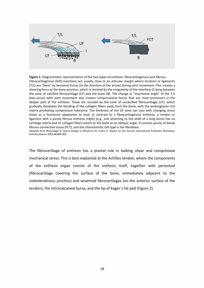
Figure 1. Diagrammatic representation of the two types of enthesis: fibrocartilaginous and fibrous. Fibrocartilaginous (left) insertions are usually close to an articular margin where tendons or ligaments (T/L) are “bent” by tensional forces (in the direction of the arrow) during joint movement. This creates a shearing force at the bone junction, which is resisted by the irregularity of the interface (I) lying between the zone of calcified fibrocartilage (CF) and the bone (B). The change in “insertional angle” of the T/L that occurs with joint movement also creates compressional forces that are most prominent in the deeper part of the enthesis. These are resisted by the zone of uncalcified fibrocartilage (UF), which gradually dissipates the bending of the collagen fibers away from the bone, with the proteoglycan-rich matrix promoting compression tolerance. The thickness of the UF zone can vary with changing stress levels as a functional adaptation to load. In contrast to a fibrocartilaginous enthesis, a tendon or ligament with a purely fibrous enthesis (right) (e.g., one attaching to the shaft of a long bone) has no cartilage matrix and its collagen fibers attach to the bone at an oblique angle. It consists purely of dense fibrous connective tissue (FCT), and the characteristic cell type is the fibroblast. Adapted from McGonagle D, Marzo-Ortega H, Benjamin M, Emery P. Report on the Second international Enthesitis Workshop. Arthritis Rheum 2003;48:896-905.
The fibrocartilage of enthesis has a pivotal role in balking shear and compressive
mechanical stress. This is best explained at the Achilles tendon, where the components
of the enthesis organ consist of the enthesis itself, together with periosteal
(fibrocartilage covering the surface of the bone, immediately adjacent to the
osteotendinous junction) and sesamoid fibrocartilages (on the anterior surface of the
tendon), the retrocalcaneal bursa, and the tip of Kager´s fat pad (Figure 2).

19
Figure 2. Diagrammatic representation of the synovio-entheseal complex, using the Achilles tendon enthesis organ to illustrate the concept. The synovial membrane (SM), which is intimately related to the enthesis itself, lines much of the retrocalcaneal bursa (RCB), except in the region where the sesamoid fibrocartilage (SF) in the deep part of the tendon presses against the periosteal fibrocartilage (PF) covering the superior tuberosity. Macrophages (M) are an integral part of the synovium, and their anatomic proximity to fibrocartilage adjacent to insertions could contribute to an inflammatory response in relation to degenerative changes (DC) in the walls of the bursa or at the enthesis itself. Although a young healthy enthesis is probably avascular, blood vessel invasion (VI) of the enthesis is common in older individuals. The blood vessels may come from the underlying bone at sites of focal absence of the subchondral bone plate, as depicted, or they may invade from tissue on the surface of the tendon, including synovium. Adapted from McGonagle D, Lories RJ, Tan AL, Benjamin M. The concept of a "synovio-entheseal complex" and its implications for understanding joint inflammation and damage in psoriatic arthritis and beyond. Arthritis Rheum 2007;56:2482-91.
Together, these structures help to dissipate load over a wide area. Thus, although
there is no neighboring synovial joint lined by articular cartilage at this location, the
enthesis is still intimately related with the synovial membrane that covers the tip of
the protruding fat pad. In addition, it should also be remembered that the enthesis
organ is also present at numerous other sites in a close anatomic relationship to
synovial joints, and can play a non-negligible role in pathophysiology damage process
in inflammatory arthritis. For instance, traditional defined concepts, such as the “bare
area” and “cartilage-pannus junction”30 may be neither essential nor necessary for the
erosive process in RA. Histologic studies have demonstrated that periarticular erosion
formation in RA has a propensity to occur adjacent to ligaments in which bone
microdamage is common,31,32 suggesting that inflammation drives the inherent
propensity for damage to occur at characteristically predisposed sites.

20
Given the extent and complexity of enthesis, it is likely that the presence of synovium
and synovial fluid in the retrocalcaneal bursa and bursae, associated with other
attachment site, reflect a physiologic role identical to that in synovial joints. Type A
and type B bursae synoviocytes are likely to be involved in maintaining the rheological
properties of synovial fluid, lubricating and nourishing periosteal and sesamoid
fibrocartilages.27 Although “synovio-entheseal complex” seems to be advantageous in
health, the very fact that a tissue prone to microdamage is closely related with
synovium means that, in fact, the “synovio-entheseal complex” is an enabling
immunologic environment and may be a region that is particularly prone to
inflammation.
Spondyloarthritis: the entheseal disease
Spondyloarthritis describes a group of interrelated rheumatic conditions comprising
ankylosing spondylitis (AS), psoriatic arthritis (PsA), arthritis/spondylitis with
inflammatory bowel disease (IBD) and reactive arthritis. One of the major clinical
problems is, and always has been, the search for proof of sacroiliitis in patients who
present with the typical clinical picture of AS and normal X-ray films of sacroiliac joints.
These patients were classified for years as having undifferentiated SpA or diagnosed
with an alternative rheumatic disorder or even a non-rheumatic condition, such as
mechanical back pain. The observation that many patients with inflammatory back
pain but without sacroiliitis on X-ray films represent the earliest phase of AS and will
develop radiographic changes diagnostic for AS within a period of time, usually
measured in years, has created a platform for the development of the new disease
concept.33 It has been demonstrated that the presence of HLA-B27 and/or MRI findings
of active sacroiliac joints inflammation in these patients could, with a significant
degree of accuracy, help to predict which patients will subsequently develop the
classical picture of AS.34 This led to the development of the concept of pre-
radiographic AS, which, together with classical AS, formed the basis for the new entity
of axial SpA.35 Other SpA with predominant spinal involvement, such as related to
psoriasis or inflammatory bowel disease, and sharing many clinical and imaging

21
features with AS can also be classified in the group of axial SpA. Therefore, it has been
suggested that patients with SpA should be distinguished according to their clinical
presentation as patients with predominantly axial SpA or with predominantly
peripheral SpA.36,37 These new concepts not only unifies patients with a similar disease
pattern (an entity that shares clinical, pathological and genetic characteristics),
permitting advanced research, but also provides the opportunity for earlier diagnosis
and better management of patients with pre-radiographic AS and AS-like psoriatic and
inflammatory bowel disease-related arthritis.
The main pathological feature in SpA is the chronic inflammatory involvement of the
enthesis and the adjacent bone,38 which may sometimes be present several years as an
isolated clinical manifestation.39 As previously described, the typical clinical articular
affectation are the sacroiliac joints and spinal inflammation, as well as peripheral
arthritis and enthesitis, often with a nonsymmetrical distribution. Although features of
joint destruction can be dramatic, in particular in some forms of PsA, skeletal damage
in SpA is only partially due to the loss of articular cartilage and bone erosion. In
contrast, new cartilage and bone formation, presenting as ankylosing enthesopathy
and leading to bony spurs, syndesmophytes, enthesophytes and eventually joint or
spine ankylosis, are hallmark signs of this disease. However, the relationship between
enthesitis, new-bone formation and bone erosion in SpA remains poorly understood.
The introduction of targeted therapies, in particular anti-TNF drugs, has met
unprecedented success in the treatment of signs and symptoms of SpA. Some studies
reported the histological and immunohistochemical features of the bone adjacent to
entheseal sites and the actual point of enthesis contact with the bone, where
fibrocartilage is abundant, particularly in the early stages of SpA. In early and
established SpA, human studies showed that the predominant infiltrating cell at the
entheseal fibrocartilage is the macrophages,40 while in the underlying bone is
lymphocytic infiltration.41 These facts are in accordance with two pathophysiologic
processes well recognized in SpA: the importance of innate immunity where the
macrophages have a crucial role, and that autoimmunity in SpA might be primarily
directed against a bone antigen.40,42 As macrophages are the main source of TNF, it is
not surprising the apparent good response of enthesitis to biologic blockade with anti-

22
TNF.43 Nonetheless, current radiographic relatively short follow-up data suggest that
these drugs do not affect the process of ankylosis.44-46 This apparent lack of structural
effect is in sharp contrast to what is seen in the erosive destruction of joints in RA.47
Intriguingly, continuous treatment with nonsteroidal anti-inflammatory drugs, as
compared with on-demand treatment, does appear to influence ankylosis in AS.48 It
seems that prostaglandin E2 (PGE2) inhibition may act differently than TNF blockade,
and PGE2 modulation has shown to delay new bone formation in AS. However, one
can remain doubtful whether this effect of PGE2 inhibition truly proofs the link
between inflammation and new bone formation, as PGE2 is an important mediator for
osteoblast differentiation and function, and inhibition of new bone formation may
merely reflect the anti-anabolic effect of PGE2 inhibition, rather than its anti-
inflammatory effect.49 Nevertheless, the pathophysiological SpA process can be seen
as a complex slow waltz between pro-inflammatory molecules and new tissue
formation, with restoration of tissue integrity or tissue remodeling as a final
outcome.50 It seems that the development of SpA is dependent on a multi-step process
that leads to chronic or recurrent inflammation, but also to the triggering of new tissue
formation, completely or partially independent of inflammation (Figure 3).51
Figure 3. A view on the relationship between inflammation and ankylosis in SpA. The primary event is considered “entheseal stress”. Biomechanical factors and microdamage are likely to play roles in this. Entheseal stress leads to triggering of an acute inflammatory reaction and of progenitor cells. In most instances, the acute events go unnoticed and homeostasis is restored. Under specific circumstances, the acute events can turn into a chronic situation in which inflammation and/or

23
ankylosis are prominent. Different pathways regulate chronic inflammation and new tissue formation, but these pathways are likely to influence each other. Genetic factors are likely to steer chronic inflammation and new tissue formation. For the latter aspects, clues may be found in other bone-forming diseases. ERAP1, endoplasmic reticulum aminopeptidase 1; IBD, inflammatory bowel disease; IL23R, interleukin-23 receptor; BMP, bone morphogenetic proteins; WNT, wingless type like signaling; DISH, diffuse idiopathic skeletal hyperostosis; FOP, fibrodysplasia ossificans progressiva.
The degree to which inflammation and the new bone formation are linked remains
conjectural, but data from MRI studies of spinal inflammation support the concept of
such coupling; however, these studies also suggest a role for the involvement of no
inflammatory pathways, such as those involving bone morphogenetic proteins (BMPs),
wingless type like signaling (Wnt) proteins and dickkopf – related protein 1 (DKK-1), in
the formation of new bone (Figure 4).

24
Figure 4. Roles of BMPs and WNTs in endochondral bone formation. (a) Physiological endochondral bone formation is stimulated by BMPs. WNT signaling plays a supportive role in relation to BMPs. However, some WNTs have a negative effect on early chondrocyte differentiation. (b) In the presence of inflammation, TNF may stimulate BMP signaling but also the expression of DKK1, which acts a WNT antagonist. The balance between TNF, BMP and WNT signaling may determine the onset and progression of ankylosis. DKK, dickkopf. Adapted from Lories RJ, Luyten FP, de Vlam K. Progress in spondylarthritis. Mechanisms of new bone formation in spondyloarthritis. Arthritis Res Ther. 2009;11:221.
The “TNF brake” hypothesis was proposed to explain the sequence of events of new
syndesmophytes formation in an established inflammatory lesion (Figure 5).52

25
Figure 5. The “TNF brake” hypothesis.
In well‑established (mature) inflammatory lesions, repair pathways leading to new bone formation activated through BMPs, Wnts and other signaling proteins are held in check by inhibitors, such as
sclerostin and DKK‑1; DKK‑1 is up regulated by TNF. Although pro-inflammatory cytokines such as TNF
“trigger” the expression of BMP, TNF also up regulates DKK‑1; resolution of inflammation by anti‑TNF
therapy and the associated reduction in DKK‑1 would thereby allow new bone formation to proceed.
This hypothesis was put forward to explain the observation that new syndesmophytes
are more likely to develop at sites were inflammation has been resolved (low TNF, low
DKK-1, high Wnt) as opposed to sites of persistent inflammation (high TNF, high DKK-1,
low Wnt). In a more detailed elaboration of the hypothesis, it was proposed that early
inflammatory lesions are resolved without sequealae, such as new bone, if effective
therapy is instituted and inflammation resolves prior to activation of bone formation
pathways by triggers such as TNF.53 While complex inflammatory lesions, and the
majority having fat lesions, can also be resolved following anti-TNF therapy, they are
associated with the development of new syndesmophytes. Fat lesions, both
established and newly evolving, are also associated with new bone formation. These
data support a window-of-opportunity concept of disease modification for anti-
inflammatory therapy in SpA, especially if it is used early in the disease course; and a
model of new bone formation that is dependent on the activation of inflammatory
pathways followed by tissue metaplasia that includes fat.

26
The extent of inflammatory evolvement in SpA has been changed in the last years.
Currently, the relationship between enthesitis and osteitis is well known. They are
particularly characteristics of SpA, but they can also be a feature of degenerative and
mechanically enthesopathy. For example, in SpA associated plantar fasciitis, the HLA
B27 gene appears to determine the extent and severity of the condition, but not the
susceptibility to osteitis; and 50% of patients with plantar fasciitis have an associated
osteitis, as determined by MRI.54 It is reasonable to consider that osteitis is a feature of
mechanically induced enthesopathy, suggesting common mechanisms of osteitis in
SpA and mechanically induced disease. Furthermore, osteitis adjacent to functional
enthesis (i.e., where there is contact, but no attachment to bone) has been reported in
SpA and mechanically related foot and ankle pain, where MRI studies demonstrated
bone edema in wraparound regions of tendons.55,56 Like true insertions, these sites are
also prone to adjacent periostitis. These observations have important implications for
understanding pathogenesis of SpA, because they reinforce the role of mechanical
stress (shear and compression) as a distinct etiophatogenic factor inducing bone
abnormalities.
Bony spurs are other well recognized lesions in SpA. Although, they also can be a
feature in degenerative, metabolic enthesopathies (e.g., acromegaly), they are
widespread in DISH, in athletes, and occur in healthy individuals not necessarily as
indication of disease.57 Enthesophytes appear as irregular outgrowths of varying size
that extend from the bone into the tendon or ligament and often develop in parallel
with osteophytes at the periphery of articular cartilage.58 Studies at Achilles tendon
level have demonstrated that bony spurs can developed without the need for
preceding microtears or any inflammatory reaction, and they are formed mainly by
endochondral59 ossification of enthesis. Bony spur formation appears to be initiated by
vascular invasion into the tendon from the underlying bone marrow. The capillaries
migrate along the rows of fibrocartilage cells that have developed by metaplasia from
tendon fibroblasts. Each fibrocartilage cell within a row dies in turn, becomes
reabsorbed, and thus creates space for the invading capillary. Bone is subsequently
deposited along the walls of the tunnels and a spur is formed.60 Thus, the potential for
bony spur development continues beyond the growing period, and bone grows into

27
the tendons and not vice versa. However, this does not normally happen while the
avascularity of the enthesis fibrocartilage is maintained. Increased loading of tendon is
likely to lead to increased fibrocartilage formation and, simultaneously, to trigger
osteoblast activity at enthesis and bony spur formation.61
A number of known factors may contribute to structural damage and chronicity in SpA.
The cytokines such as TNF play a pivotal role, but other factors should not be
neglected, such as structural properties of HLA-B27; activation of the immune system
by the presence of inflammatory bowel disease or infection; polymorphisms in
cytokines and cytokine processing molecules, that lead to either more severe
inflammation or delayed clearance of inflammation; biomechanical factors that lead to
stress responses or microdamage in the enthesis; and specific genetic factors, not yet
identified and different from those that determine disease susceptibility.
Ultrasound of enthesis – enthesopathy and enthesitis features and
scores
The term "enthesopathy" is usually used to designate entheseal lesions related to any
pathology, including degenerative changes; while the concept of "enthesitis" is used
when entheseal inflammation is prevalent, both occurring in the course of SpA.
The prevalence of enthesitis in SpA is not easy to determine. Firstly, related to the
expected subclinical enthesis involvement in SpA; and secondly connected with the
apparent lack of diagnostic accuracy of the clinical examination, related to the absence
of enthesis visible signs of inflammation. Nonetheless, there are three validated scores
to clinically assess enthesitis in patients with AS: Mander enthesis index (MEI),62
Maastricht Ankylosing Spondylitis enthesitis score (MASES)63 and Major;64 and two
validated indices for PsA (Gladman65 and Leeds66). The MEI was published in 1987 and
evaluates 66 enthesis. The high number of enthesis to be accessed per patient, and a
graduation score system established according to the intensity of pain by each enthesis
pressure, makes it particularly difficult to apply in clinical practice.62 Subsequently the

28
index MASES was published, as a simplification of the previous index, that explores 13
enthesis concerning the presence or absence of pain (I and VII costochondral joints, V
lumbar spinous process, Achilles tendon, anterior and superior iliac spine, iliac crest).
The Major index includes 12 enthesis assessments: iliac crests, trochanters, medial and
lateral epicondyles, Achilles and plantar fascia. The Gladman index assesses 8 enthesis:
rotator cuff, anterior tuberosity of the tibia, Achilles and plantar fascia; and the Leeds
index includes 6 enthesis in the evaluation: Achilles, medial femoral condyle and
lateral epicondyle. The exploration is done by exerting sustained pressure with the
fingertips on the enthesis (sufficient to blanch the finger nail of the examiner -
approximately 4 Kg), which makes you lose objectivity while considering the pain
threshold, as it is not the same for each patient.
The OMERACT defines enthesopathy as an “abnormally hypoechoic (loss of normal
fibrillar architecture) and/or thickened tendon or ligament at its bony attachment (may
occasionally contain hyperechoic foci consistent with calcification), seen in 2
perpendicular planes that may exhibit Doppler signal and/or bony changes including
enthesophytes, erosions, or irregularity”.67
This definition has the advantage of focusing on the main characteristic of entheseal
abnormalities, but has the great limitation of not clearly defining the difference
between enthesopathy and enthesitis. Despite previous data concerning the
usefulness of ultrasound in the evaluation of enthesis in the course of SpA, it seems
that the most affected peripheral enthesis are located in the lower limbs38 and the
combination of grey-scale with power Doppler increases diagnostic accuracy for SpA.68
In recent years, numerous scoring systems have been developed with great
heterogeneity regarding sites, abnormalities included and evaluation with power
Doppler (Table 1). Also, the prevalence of enthesitis in SpA is variable depending on
entheseal site and diagnostic ultrasound criteria (see Table 1).

29
Table 1. Principal scores of enthesis ultrasound examination in spondyloarthritis patients
Score B mode parameters
and settings
Doppler
parameters and
setting
Description of sites of
vascularization
Sites Percentage of abnormal
enthesis on ultrasound in
spondyloarthritis
Scoring system
Balint 2002
Glasgow
ultrasound
enthesitis
scoring system
(GUESS)1919
Thickness, bursitis,
enthesophytes and
erosions
7-15 MHz
NA NA Quadriceps tendon
enthesis, proximal and
distal patellar ligament,
Achilles tendon, plantar
aponeurosis
56% (22% on clinical
examination) – no controls
GUESS score (0 to 36): each item score one point, total possible
score on both lower limbs is 36.
Quadriceps tendon thickness ≥6.1mm, patellar ligament thickness
(proximal, distal) ≥4mm, Achilles tendon thickness ≥5.29mm,
plantar aponeurosis thickness ≥4.4mm
Sub score: soft tissue score (thickness and bursitis) and bone
score (erosions and enthesophytes)
D´Agostino
200368
Thickness, bursitis,
calcification or
periosteal changes,
hypoechogenicity
13 MHz
Binary (0 or 1)
PRF 0.75KHz, gain
50-53 DB
(constant
temperature of
the room)
Cortical bone
insertion, body of the
tendon, bursa,
junction
tendon/enthesis
Bilaterally: great
trochanter, pubis,
quadriceps tendon
enthesis, patellar tendon
(proximal insertion),
Achilles tendon, plantar
aponeurosis, tibialis
anterior tendon
insertion, medial and
lateral epicondyles
38% (14% on clinical
examination) versus 10% for
mechanical back pain and
14% for rheumatoid arthritis
patients
stage 1: Vascularization at the cortical junction without abnormal
findings in Grey-scale
stage 2a: Vascularization associated with swelling and/or
decreased echogenicity at the cortical junction in Grey-scale
stage 3a: Same as stage 2a, plus erosions of cortical bone and/or
calcification of enthesis, and optional surrounding bursitis
stage 2b: Abnormal findings in B mode as in stage 2a, but without
vascularization
stage 3b: Abnormal findings in B mode as in stage 3a, but without
vascularization
Kiris 200669 Bursitis, calcification,
hypoechogenicity,
cortical reactive
changes (cortical
reabsortive changes,
Semi-quantitative
(0-3) and a final
sum of each
tendon examined
PRF 0.5-1 KHz,
Tendon/enthesis: no
precision concerning
the exact location of
vascularization
I and VII costochondral
joints, V lumbar spinous
process, Achilles tendon,
anterior and superior
iliac spine, iliac crest
44.8% (51.5% on clinical
examination) – no controls
Cumulative power Doppler score:
grade 0: no flow signal
grade 1: mild flow signal refers to the presence of separate dot
signals or short linear signals
grade 2: moderate flow signal refers to the presence of clearly

30
new bone formation,
cortical irregularity)
7-14 MHz
gain 50-55, low
wall filter
(constant
temperature of
the room)
discernible vascularity with either many small vessels or several
long vessels with or without visible branching though involving
less than half of the enthesis
grade 3: severe flow signal refers to the presence of vessels
involving more than half of the enthesis
Alcalde 2007
Sonographic
enthesitic
index (SEI)70
Thickness/loss of
thickness, bursitis,
hypoechogenicity,
peritendinous edema,
calcifications, tendon
tears, erosions
7.5 MHz
NA NA Quadriceps tendon
enthesis, proximal and
distal patellar ligament,
Achilles tendon, plantar
aponeurosis
25% ( 8% on clinical
examination) versus 0%
healthy controls matched for
age
SEI = the total sum of SEI-A (acute injury) and SEI-C (chronic
injury). Maximum SEI scoring is 76 points
SEI-A (0 to 36): each variable is scored as 0 (absence) or 1
(presence): thickening of tendon/aponeurosis, hypoechogenicity
of tendon/aponeurosis, peritendinous/periaponeurotic edema,
bursitis (where applicable)
SEI-C (0 to 40): each variable is scored as 0 (absence) or 1
(presence): tendon tear, loss of thickness, tendon calcification,
bone erosion
De Miguel 2009
Madrid
sonography
enthesitis index
(MASEI)71
Thickness, bursitis,
structure/hypoechoge
nicity, calcification,
erosions
7-12 MHz
Binary (0 or 3)
PRF 0.4 KHz, gain
20 dB, low wall
filter
Enthesis, tendon,
bursitis
Quadriceps tendon
enthesis, proximal and
distal patellar ligament,
Achilles tendon, plantar
aponeurosis, distal
brachial triceps tendon
NA MASEI score (0 to 136 on both sides):
Calcifications were scored on a semi-quantitative score of 0 to 3
Doppler and erosions were scored as 0 or 3 points
Scores for tendon structure, tendon thickness and bursa were
either 0 or 1.
Calcifications were examined at the area of the enthesis insertion,
and scored as 0 if absent, or 1 if a small calcification or ossification
with an irregularity of enthesis cortical bone profile was seen.
Calcifications were given a score of 2 if there was clear presence
of enthesophytes or if medium sized calcifications or ossification
were observed. Lastly, they were classified as a 3 if large
calcifications or ossifications were present. To simplify things,
ossifications and enthesophytes at the enthesis were also
included as calcifications.

31
D´Agostino
200972
Thickness and/or
hypoechogenicity,
calcifications and/or
enthesophytes,
erosions
13MHz
Binary (0-1) and
semi-quantitative
(0-3)
PRF 0.5 KHz, gain
113 dB, medium
wall filter
Enthesis insertion into
the cortical bone
Quadriceps tendon
enthesis, proximal
patellar ligament,
Achilles tendon, plantar
aponeurosis, lateral
epicondyle
NA Grey-scale: hypoechogenicity/thickness: 0 to 1,
Calcification/enthesophyte: 0 to 1, erosion: 0 to 1
Doppler : (0 to 3): 0: no signal, 1: minimal (1 spot), 2: moderate (2
spot), 3: severe (> = 3 spots) or Doppler scored as 0 to 1
(absent/present)
Fillippucci
200973
Tendon
hypoechogenicity and
thickening, entheseal
hypoechogenicity,
bursal effusion,
calcifications (tendon,
enthesis),
enthesophytes,
erosions and bone
irregularities
6-18 MHz
Binary (0-1)
and semi-
quantitative (0-2)
PRF 0.75 KHz, low
wall filter
Enthesis, tendon,
bursitis
Achilles tendon NA Soft tissue inflammation (seven items): tendon hypoechogenicity,
Tendon thickening, Entheseal hypoechogenicity, Bursal effusion,
PDS signal at tendon level, PDS signal at entheseal level, PDS
signal at bursal level
Tissue damage (five items): Intratendineous calcifications,
Entheseal calcifications, Enthesophytes, Bone erosions, Bone
Irregularities (the last not used to calculate total score)
(1) a total score for soft tissue inflammation, which resulted from
the sum of the scores assigned to the 7 US findings indicative of
soft tissue inflammation, ranging from 0 to 7 with
presence/absence data and from 0 to 14 with semiquantitative
scores (from 0 to 2: 0:none; 1 mild-moderate; 2:severe);
(2) a total score for tissue damage, which resulted from the sum
of the scores assigned to the 4 US findings indicative of tissue
damage, ranging from 0 to 4 with presence/ absence data and
from 0 to 8 with semiquantitative scores.
Scoring system: 0: none, 1: mild-moderate, 2: severe
Tendon thickness: 0:<5.3mm, 1: between 5.3 and 6.3mm,
2:>6.3mm
Bursal size: 0:<2mm, 1:between 2 and 4mm, 2:>4mm
Bone erosions: 0: no bone erosion, 1: between 0.1 and 2mm,
2:>2mm
32
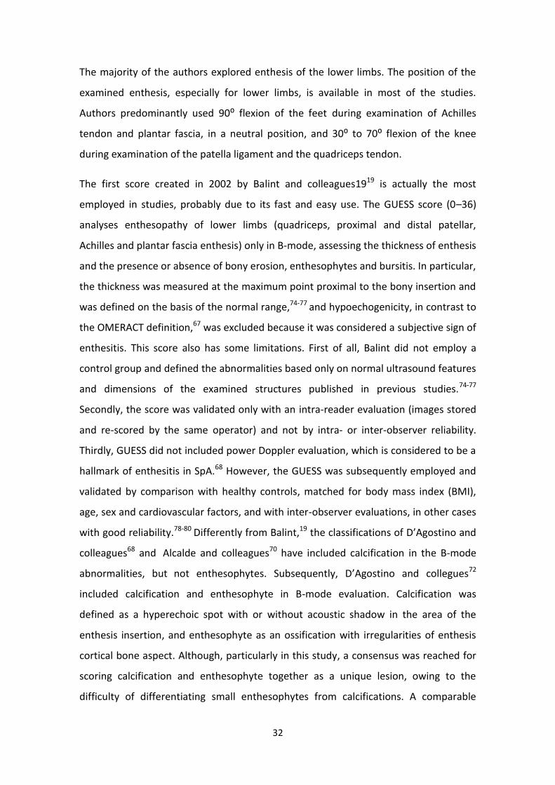
The majority of the authors explored enthesis of the lower limbs. The position of the
examined enthesis, especially for lower limbs, is available in most of the studies.
Authors predominantly used 90⁰ flexion of the feet during examination of Achilles
tendon and plantar fascia, in a neutral position, and 30⁰ to 70⁰ flexion of the knee
during examination of the patella ligament and the quadriceps tendon.
The first score created in 2002 by Balint and colleagues1919 is actually the most
employed in studies, probably due to its fast and easy use. The GUESS score (0–36)
analyses enthesopathy of lower limbs (quadriceps, proximal and distal patellar,
Achilles and plantar fascia enthesis) only in B-mode, assessing the thickness of enthesis
and the presence or absence of bony erosion, enthesophytes and bursitis. In particular,
the thickness was measured at the maximum point proximal to the bony insertion and
was defined on the basis of the normal range,74-77 and hypoechogenicity, in contrast to
the OMERACT definition,67 was excluded because it was considered a subjective sign of
enthesitis. This score also has some limitations. First of all, Balint did not employ a
control group and defined the abnormalities based only on normal ultrasound features
and dimensions of the examined structures published in previous studies.74-77
Secondly, the score was validated only with an intra-reader evaluation (images stored
and re-scored by the same operator) and not by intra- or inter-observer reliability.
Thirdly, GUESS did not included power Doppler evaluation, which is considered to be a
hallmark of enthesitis in SpA.68 However, the GUESS was subsequently employed and
validated by comparison with healthy controls, matched for body mass index (BMI),
age, sex and cardiovascular factors, and with inter-observer evaluations, in other cases
with good reliability.78-80 Differently from Balint,19 the classifications of D’Agostino and
colleagues68 and Alcalde and colleagues70 have included calcification in the B-mode
abnormalities, but not enthesophytes. Subsequently, D’Agostino and collegues72
included calcification and enthesophyte in B-mode evaluation. Calcification was
defined as a hyperechoic spot with or without acoustic shadow in the area of the
enthesis insertion, and enthesophyte as an ossification with irregularities of enthesis
cortical bone aspect. Although, particularly in this study, a consensus was reached for
scoring calcification and enthesophyte together as a unique lesion, owing to the
difficulty of differentiating small enthesophytes from calcifications. A comparable

33
consensus was reached for scoring increased thickness and hypoechogenicity of
enthesis insertion as a unique feature, arguing that both are signs of acute
inflammation. Meanwhile, De Miguel and colleagues71 and Filippucci and colleagues73
used judgment in their scores. A semi-quantitative score was used regarding the
calcification dimension, but it is not clear or well defined how to distinguish between
the different sizes. Additionally, while Balint19 separated abnormalities into soft tissue
(thickness and bursitis) and bone (enthesophytes and erosions), Fillippucci73 grouped
the entheseal lesions in soft tissue inflammation (tendon hypoechogenicity, entheseal
hypoechogenicity, bursal effusion, power Doppler signal at tendon level, at entheseal
level and at bursal level) or tissue damage (intratendineous calcifications, entheseal
calcifications, enthesophytes, bone erosions and bone irregularities), and Sonographic
Enthesitic Index (SEI)70 described acute (thickening, hypoechogenicity, edema of
surrounding structures and bursitis) versus chronic (tear, loss of thickness, calcification
and erosions) alterations. In the same study, Alcalde70 demonstrated that edema, tears
and loss of thickness do not have great frequency or relevance in SpA and are probably
difficult to define because they are not well described and quantified in other studies.
Similarly to GUESS, SEI do not include power Doppler evaluation, with the limitations
that may come. The first score including power Doppler was proposed by D´Agostino.68
This score presents some difficulties in application because there are too many sites
evaluated. It is not clear if the mix of abnormalities with B-mode and power Doppler is
only descriptive, or if there is a rationale for classification linked to different degrees of
severity. In this study the intra- and inter-observer variability was excellent but it was
calculated on the final score (not for a single defect), and it was not validated in
subsequent studies. Furthermore, power Doppler was considered only as a binary
system (present/absent). The power Doppler signal (similar to B-mode lesions such as
thickness or bursitis) might be important for follow-up purposes, together with other
clinical and serological indices, to guide the choice of drugs and to monitor the efficacy
of treatments. For this reason, semi-quantitative systems of power Doppler scoring
were proposed.69,72,73 Kiris and collegues69 made a descriptive and topographic
classification (presence of separate linear signals - grade 1; discernible vessels involving
less than half enthesis - grade 2; and vessels involving more than half enthesis - grade
3), followed by D’Agostino and colleagues72 with a simplified version, describing only

34
the number of power Doppler spots (one - score 1; two - score 2; and ≥3 signals - score
3). The most attractive part of these methods is to evaluate a total power Doppler
score, calculated by summing the flow signal grades on enthesis, which might be
particularly useful for future clinical trials.
The systematic ultrasound exploration of MASEI is represented in Figure 6.
Figure 6. Madrid sonography enthesitis index.
Despite these promising results, the use of power Doppler for the management of SpA
has remained less frequent than other innovative imaging techniques such as MRI. This
discrepancy is probably due to the perception that ultrasound remains an unreliable
imaging technique. The operator dependence on ultrasound performance, artefacts of
power Doppler on image acquisition, the optimization of power Doppler dependence
on the type of used device and on settings are some of the given reasons for this fact.

35
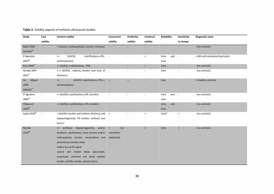
The validity of enthesis ultrasound involves various aspects, not always clearly defined
in the studies. The discrepancies in methods, the lack of comparison with a gold
standard, such as biopsy, or the lack of evaluation of a real prognostic value of
entheseal lesions detected by ultrasound makes it difficult to compare several studies
efficiently (see table 2).
36
Table 2. Validity aspects of enthesis ultrasound studies
Study Face
validity
Content validity Concurrent
validity
Predictive
validity
Construct
validity
Reliability Sensitivity
to change
Diagnostic value
Balint 2002
(GUESS)19
- + erosions, enthesophytes, bursitis, thickness - - - - - - (no controls)
D´Agostino
200368
- ++ (GUESS, +calcifications,+PD,-
enthesophytes)
- - + Intra and
inter
- + (RA and mechanical back pain)
Kiris 200669 - ++ (GUESS, +calcifications, +PD) - - + Intra - - (no controls)
Alcalde 2007
(SEI)70
- +++ (GUESS, +edema,+tendon tear,+loss of
thickness)
- - - Inter - - (no controls)
De Miguel
2009
(MASEI)71
- ++ (GUESS,+calcifications,+PD,+,-
enthesophytes)
+ + - Inter - + (healthy controls)
D´Agostino
200972
- ++ (GUESS,+calcifications,+PD,-bursitis) - - - Intra and
inter
- - (no controls)
Fillippucci
200973
- ++ (GUESS,+calcifications,+PD,+tendon) - - - Intra and
inter
- - (no controls)
Aydin 201081 - + (Achilles tendon and enthesis thickness and
hypoechogenicity, PD tendon, enthesis and
bursa )
+ - + Intra* + - (no controls)
Naredo
201082
- ++ (enthesis hypoechogenicity and/or
thickness, calcifications, bone erosion and/or
enthesophyte, bursitis, intraenthesis and
perienthesis (tendon body
and/or bursa) PD signal
Lateral and medial elbow epicondyle,
quadriceps, proximal and distal patellar
tendon, Achilles tendon, plantar fascia
+ (no
correlation
stablished)
- + Intra + - (no controls)

37
De Miguel
201183
- ++ (GUESS,+calcifications,+PD,+,-
enthesophytes)
MASEI
+ + - - - + (inflammatory and non-inflammatory
controls#)
D´Agostino
201184
- ++ (GUESS,+calcifications,+PD,-bursitis)
Quadriceps tendon enthesis, proximal
patellar ligament, Achilles tendon, plantar
aponeurosis, lateral and medial epicondyle,
gluteus medius tendon
+ + - - - + (no controls)
* The inter-observer agreement for US evaluation of the Achilles tendon was tested in a previous study. # Non-inflammatory controls: healthy persons, non-inflammatory lumbar pain and posterior uveitis unrelated to SpA; inflammatory controls: patients from ESPERANZA project that did not meet SpA diagnostic criteria. Face = credibility for measuring what it is supposed to; content = comprehensiveness of all aspects of the attribute to be measured; concurrent = degree to which a measure reflects a gold standard applied at the same time; predictive = degree to which a measure predicts a future gold standard outcome; construct = consistency with theoretical concepts; reliability = intra- and inter-observer variation to allow reliable detection of this change; sensitivity to change = variation of the measure over time (e.g., follow-up after treatment); diagnostic value = ability to distinguish between different diseases. GUESS = Glasgow Ultrasound Enthesitis Scoring System; MASEI = Madrid Sonographic Enthesis Index; PD = Power Doppler; SEI = Sonographic Enthesitic Index; RA = rheumatoid arthritis
38
The capability of ultrasound to evaluate enthesitis earlier and better than radiography
is well demonstrated but, until now, there only have been a few studies that have
compared ultrasound with more sensitive imaging techniques, such as MRI63,85 and
scintigraphy,86 but did not clearly investigate the correlation between B-mode and
power Doppler findings.
The enthesis ultrasound sensitivity to change has been evaluated in SpA patients
treated with anti-TNF drugs. These studies have the limitation for a relative short
period of follow-up, but they showed a reduction of B-mode and power Doppler
enthesis abnormalities, such as morphologic abnormalities (tendon hypoechogenicity
and/or thickening), power Doppler signal, and bursitis.81,82
On the other hand, the diagnostic value of ultrasound (ability to distinguish between
different diseases) was verified not only by D’Agostino and colleagues,68 who
compared SpA to rheumatoid arthritis and mechanical back pain, but also by De
Miguel and colleagues (MASEI total score ≥ 18 points was the best cut-off point for
differentiation between cases and controls (healthy persons), and demonstrated a
sensitivity, specificity, positive and negative likelihood ratios of 83.3%, 82.8%, 4.8%,
and 0.2%, respectively, for the diagnosis of SpA regardless of the presence of other
clinical manifestations).71 Similar results have been established by the same authors in
early SpA. In a cross sectional, blinded and controlled study with 113 early SpA patients
De Miguel and colleagues achieved for a MASEI total score ≥ 20 points a likelihood
ratio of 5.3, with a specificity of 89.47% and a sensitivity of 55.75% for SpA diagnosis.83
Additionally, in a two years prospective cohort study with 118 early SpA patients
D’Agostino and colleagues84 demonstrated that the power Doppler ultrasound
detection of at least one vascularized enthesis provided good predictive value for
diagnosing SpA (sensitivity 76.5%, specificity 81.3%, positive likelihood ratio 4.1, OR
14.1;p<0.0001).
As described, the various published ultrasound studies for SpA entheseal assessment,
meet both permanent structural damage and non-permanent entheseal injuries
related with inflammatory disease activity. This fact is in agreement with the increasing
knowledge about entheseal ultrasound study in SpA. However, for its use in daily

39
practice, a profound understanding on the behavior of entheseal structural alterations
should be developed. The OMERACT enthesopathy definition includes the main lesions
of the enthesis at bone and enthesis tendon insertion identified by ultrasonography,
and it is now widely cited and accepted in the ultrasound community. Nevertheless,
this definition does not comprehend bursitis as an elementary lesion, or the distinction
between injuries related with entheseal structural damage or inflammatory activity.
Until now published data concerning these entheseal alterations reflect rather the
authors’ empiric opinion than a methodological validation process. Progress in this
study area is one of the main objectives of a reduced number of ultrasound
investigation teams; and I am integrated in one of these groups.

40
DETAIL DESCRIPTION
In 2006, the integration into a research group in a reference center for inflammatory
rheumatic diseases - Rheumatology Department of Hospital Universitario La Paz in
Madrid - not only provided me with additional training in the field of musculoskeletal
ultrasound, but also my involvement with a research group in SpA, headed by Prof.
Eugenio de Miguel. Since then, and in partnership with this research group, several
studies have been developed in the musculoskeletal ultrasound area and have been
used to assess inflammatory rheumatic diseases.87-98
The integration into the research group occurred slowly and progressively in a two
steps pathway. Firstly, a structured learning process in what concerns structural lesions
in SpA and systematic entheseal exploration was developed; and secondly, the
validation process to guarantee the entrance in this research team was preceded by
head to head reliability studies with other members of the investigation group.99
Since 2006 longitudinal studies have been developed with more than five years of
follow-up of SpA patients; and several papers have been published.71,83,100-107 The
developed database has been used for different analysis. Clinical, analytical,
radiographic and 2D and 3D ultrasound data are integral part of the records.
The patient sample was selected from individuals attending the Early Spondyloarthritis
Unit (ESU), as part of the ESPERANZA program, a nation-wide health management
program designed to provide excellence in care for early SpA, promoted by the
Rheumatology Spanish Foundation. The referral criteria included: 1) age below 45; 2)
symptom duration between three and 24 months; and 3) at least one of the following:
a) inflammatory low back pain, defined as at least two of the following: insidious
onset, morning stiffness for more than 30 minutes, or clear improvement of the
symptoms with physical activity but not relieved by rest; b) asymmetric arthritis,
preferably of the lower limbs; or c) low back pain or arthralgia and at least one of the
following: psoriasis, inflammatory bowel disease, anterior uveitis, family history of
spondylitis, psoriasis, radiographic sacroiliitis or HLA-B27+ status. Patients will be
classified as SpA according to accepted classification criteria, as follows: 1) ankylosing

41
spondylitis if they fulfilled the modified New York criteria; 2) psoriatic arthritis if they
fulfilled the classification criteria for psoriatic arthritis (CASPAR) criteria; 3) SpA
without definitive radiographic sacroiliitis (at least bilateral grade II or unilateral grade
III) and undifferentiated SpA if the European Spondylarthropathy Study Group (ESSG)
preliminary criteria for classification of SpA were fulfilled without any other specific
diagnostic criteria; 4) reactive arthritis if the patient fulfilled ESSG criteria or had
arthritis, confirmed by a rheumatologist, with recent evidence of related infection; 5)
arthritis-associated inflammatory bowel disease if IBD was present in a patient with
the New York criteria or ESSG criteria; and 6) anterior uveitis if it had been diagnosed
by an ophthalmologist. The diagnosis of IBD required typical histological findings of
Crohn’s disease or ulcerative colitis. Exclusion criteria included previous history of
ankle surgery, peripheral neuropathy, or corticosteroid injection within the previous 6
weeks in the Achilles tendon. All patients completed the Spanish version of Bath
Ankylosing Spondylitis Disease Activity Index (BASDAI), Bath Ankylosing Spondylitis
Functional Index (BASFI) and Bath Ankylosing Spondylitis Radiology Index (BASRI).
Peripheral joint count, entheseal clinical evaluation and analytical data were also
registered on the same day of the visit. The subjects have been followed-up in a
regular scheme with systematic clinical, analytical and imaging records. The controls
were non SpA inflammatory patients and asymptomatic subjects, without any known
medical history of inflammatory or mechanical musculoskeletal disease. They were
selected among hospital workers and friends of patients, all of whom volunteered to
participate after receiving an explanation of the procedure.
The ultrasound protocol was performed using a Logiq 9 (General Electrics Medical
Systems, Milwaukee, WI) equipped with a linear probe at 9-14 MHz and a broadband
high-frequency (8-15 MHz) volumetric probe. Focus was positioned at the level of the
region of interest; grey-scale frequency was 15 MHz; Doppler settings were
standardized with a pulse repetition frequency of 400 Hz, wall filter of 48Hz and color-
mode frequency of 7.5 MHz. The color gain was 36-45 (increased to the highest value
not generating Doppler signals under the bony cortex). The sonographer was blinded
to patients´ clinical or therapeutic data and subjects were advised to withhold these

42
data from the ultrasound examiner. All acquired images were stored in a digital format
to be subsequently analyzed.
The study of enthesis was conducted according to local regulations and the Declaration
of Helsinki, and local approval was obtained from the ethical committee and
institutional review board of Hospital Universitario La Paz - Madrid. All patients and
controls signed an informed consent.
Nowadays, the development of new technologies that enable the recording of images
in digital format, and their further analysis after acquisition, allowed not only reliability
studies, but also established long distance working partnerships. This was the followed
methodology for 2D and 3D data processing and analysis.
Data collected in this project is expected to contribute for a better understanding of
the behavior of entheseal damage in SpA, identifying new assessment tools for
diagnosis and follow-up purposes, and hopefully providing physician with improved
tools for assessing disease prognosis and response to treatment.

43
AIMS
The main objective of this work is to improve the knowledge of SpA entheseal lesions.
Namely, understand the behavior of entheseal erosion and the importance of the
entheseal bursa that could be involved in futures scores of structural damage or
disease activity; analyze the validity of enthesis ultrasound in the quantification of SpA
disease activity and to contribute for enthesitis ultrasound definition, using the Achilles
tendon as a model.
Our specific objectives are:
I. To evaluate if Doppler ultrasound is a reliable method to assess entheseal
structural lesions in SpA in a well-trained observer;
II. To know whether erosion in SpA represents a persistent structural damage
that can be used for structural damage ultrasound scores, or as a non-
permanent lesion that should be included in future ultrasound disease
activity scores;
III. To assess the prevalence and the relevance of the bursa-synovial lesions in
SpA;
IV. To determine the predictive value of entheseal ultrasound lesions in SpA,
and its relationship with other well-established SpA activity or structural
damage outcome measures.
44
RESULTS
In agreement with the Decreto-Lei 388/70, art. 8º, the results presented and discussed
in this thesis were published in the following scientific peer-reviewed journals:
I. Falcão S, De Miguel E, Castillo C, Branco JC, Martín-Mola E. Doppler
ultrasound – a valid and reliable tool to assess spondyloarthritis. Acta
Reumatol Port 2012;37(3):212-7.
II. de Miguel E*, Falcao S*, Castillo C, Plasencia C, García M, Branco JC, Martín-
Mola E. Enthesis erosion in spondyloarthritis is not a persistent structural
lesion. Ann Rheum Dis 2011;70:2008-10.
(*de Miguel E and Falcao S contributed equally for this work)
III. Falcao S, de Miguel E, Castillo-Gallego C, Peiteado D, Branco J, Martín Mola
E. Achilles enthesis ultrasound: the importance of the bursa in
spondyloarthritis. Clin Exp Rheumatol 2013;3:422-7.
IV. Falcao S, Castillo-Gallego C, Peiteado D, Branco J, Martín Mola E, de Miguel
E. Can we use enthesis ultrasound as an outcome measure of disease
activity in Spondyloarthritis? A study at Achilles level. Rheumatology
2014;doi:10.1093/rheumatology/keu399.

45
PART I
Doppler ultrasound – a valid and reliable tool to assess spondyloarthritis

46

47
48
49

50

51

52
PART II
Enthesis erosion in spondyloarthritis is not a persistent structural lesion

53

54

55

56
PART III
Achilles enthesis ultrasound: the importance of the bursa in spondyloarthritis

57

58

59

60

61

62

63
PART IV
Can we use enthesis ultrasound as an outcome measure of disease activity in
Spondyloarthritis? A study at the Achilles level.

64

65

66

67

68

69
70
DISCUSSION
It is well established that enthesitis is a distinctive feature of SpA, is transversal to all
SpA subtypes, and may sometimes be present several years as an isolated clinical
manifestation. Despite the relevance of peripheral enthesitis assessment in the last
years – as corroborated by its inclusion in the recent developed Assessment of
SpondyloArthritis international Society (ASAS) new classification criteria for axial and
peripheral SpA,35,36 and in the last EULAR recommendations for the management of
PsA108 – it remains uncertain which is the best form to perform its diagnosis. Several
studies on imaging of enthesis showed that imaging techniques such as MRI or
ultrasound are superior to clinical examination for enthesitis diagnosis, and some
asymptomatic enthesitis might only be detected by imaging techniques. However, as
the enthesitis diagnosis can be assessed by ultrasound, it is fundamental to study and
define the elemental lesions that build the concept of enthesitis in SpA, and its
relationship with other well-established SpA outcome measures. The aim of this
dissertation thesis was to improve the knowledge of SpA entheseal lesions; namely,
understand the behavior of entheseal erosion and the importance of the entheseal
bursa that could be involved in futures scores of structural damage or disease activity;
analyze the validity of enthesis ultrasound in the quantification of SpA disease activity
and to contribute for enthesitis ultrasound definition.
The first study of this thesis (part I) focused on the analysis of the reliability of inter-
lector and inter-ultrasonography equipment of MASEI index. Fundamental data for the
remaining unrolling project validity. This work represented the validation process for
my incorporation in the research team. In addition, it has always been said that the
main problem of ultrasound is the interobserver variability. These types of studies are
fundamental to spread and generalize this technique. With the proper knowledge and
training ultrasound has proven, in many cases, to be more reproducible than other
oldest techniques used in clinical daily practice.
In part II we were concerned about the Achilles enthesis erosions behavior over time.
In literature erosions represent a permanent structural damage, being useful for

71
monitoring joint injury, disease activity and therapeutic response in many rheumatic
diseases; and to date, this concept has been mostly applied in RA.109 However, in this
sense, it is important to emphasize that RA erosion and SpA enthesis erosions likely
represent a different aetiophatogenic disease-response mechanism. Unquestionably,
erosion is a tissue-related damage and a structural change. However, the important
question is whether erosions represent a permanent structural change that can only
grow and worsen over time, as occurs in RA, or a transitory alteration. The initial
observation leading to the development of the hypothesis was based on the
identification of the dynamic nature of the entheseal erosions. Unfortunately, this was
just an observation in the daily practice that had to be tested. Initially, we also thought
that this could be related with the variability of ultrasound image readings or patient
entheseal ultrasound exploration; but in some cases we had collected pictures of
erosions that we were not able to reproduce in later ultrasound examinations. This
coupled with the fact that the vertebral erosions disappear in SpA patients made us
hypothesize that these lesions might disappear over time. To avoid bias related with
the lack of reliability of entheseal exploration or ultrasound image reading we used 3D
technology that allowed us to observe the scan time and review as many times as
necessary the images. Previous published data about enthesis ultrasound erosion in
SpA classified this elementary entheseal alteration as a structural damage.70,73
Nevertheless, our findings in the longitudinal study of Achilles enthesis in early SpA are
consistent with the dynamic behavior of erosion over time (part II). Our results
strongly suggest that previously detected erosions could disappear during the course
of the disease. Furthermore, at six and twelve months of follow-up, 25% and 50% of
basal erosions disappeared, respectively; and among the new erosions that appeared
at six months, 40% disappeared six months later.110 Based on these striking results it
seems reasonable to suggest that the new-bone formation process in SpA could be
associated with the resolution of cortical entheseal erosion over time. Consistent with
these findings, prior results on spinal MRI showed that new syndesmophytes in SpA
developed from inflammatory lesions in spinal vertebral corners into fat infiltration or
erosion, and progressed to bone sclerosis and syndesmophyte formation.111 This result
could also be in agreement with the apparent failure of anti-TNF therapies to control
bone proliferation in SpA; and with the relation of TNF, Dkk-1 and the regulatory

72
molecule of the Wnt pathway in the bone proliferation in SpA.51 This can be important
to the development of ultrasound activity and structural damage scores to improve
assessment, treatment response and prognosis in SpA patients.
Following the study of ultrasound elementary entheseal lesions in SpA, another trend
was to analyze the bursal entheseal area (part III). There are multiple studies that
added the bursa to the elementary entheseal lesions considered in the OMERACT
enthesopathy definition.19,68-73 In fact, bursa was not included in the OMERACT
enthesopathy definition,67 but was included in 46% of the enthesis studies in a recently
systematic literature review,103 being in agreement with the concept of “synovio-
entheseal complex” that includes the link between enthesitis and osteitis in SpA. It has
been clarified in recent data that there is not only a close functional integration of the
enthesis with the neighboring bone, but also a connection between enthesitis and
synovitis.29 Additionally, entheseal morphologic abnormalities, Doppler signal and
bursa were the only elementary lesions that were associated with anti-TNF therapies
response.81,82 Therefore, bursa may be important in quantifying the therapeutic
response in SpA patients, and may be related with disease activity. Consequently,
bursa was another of the injuries of interest in our ultrasound enthesis SpA third study
draft. Therefore, we tried to assess the prevalence and relevance of the bursa-synovial
lesion in SpA. Our findings showed a significant increase of Achilles bursa presence and
thickness in SpA patients compared to controls (healthy/mechanical controls and RA
controls). Furthermore, when bursa’s thickness was measured, our results showed an
increase in SpA patients with statistical significant differences. The ROC curve analysis
showed 60.4% sensitivity and 68.5% specificity, for SpA diagnosis, when bursa was >1
mm, and 34% sensitivity and 87% specificity when bursa was >1.5 mm. A cut-off of
bursa >2 mm showed a low sensitivity of 19.8% with a specificity of 97.8% in front of
the overall group, and a sensitivity of 19.8% and a specificity of 95.7% with a positive
likelihood ratio of 4.6 in front of healthy controls. A likelihood ratio between 2 and 5
generates small, but sometimes important changes in probability. In this study, a
striking finding was the relatively low prevalence and thickness of bursa in RA control
group (21.7% in RA control group versus 58.7% in healthy controls; p<0.01).112 One
possible explanation could be that this control population was composed by RA

73
patients all treated with disease modifying anti-rheumatic drugs without advanced
deformities, and low disease activity. On the other hand in the healthy control
population the higher bursa presence could be related with overuse. In agreement
with what has been shown by other authors, the presence of Doppler signal seems to
have a high significance in the correct classification of SpA patients.68,71,83,84 Doppler
signal was also associated with other clinical measures accepted for assessment of SpA
disease activity (CRP, heel pain, patient VAS for pain and global disease activity
evaluation, number of swollen joints and BASDAI question 3), but not with axial
question of BASDAI, it even had a negative association with spine pain (BASDAI
question 2). The association with the number of swollen joints, BASDAI question 3 and
CRP is in agreement with the idea that bursal-synovial specific factors could trigger
innate immune responses and may be pivotal players in the phenotypic expression of
SpA, as suggested by the synovio-entheseal complex concept proposed by McGonagle
and colleagues.29,32,39 In this sense, and supporting the idea of the importance of the
participation of the synovial bursal tissue in enthesis damage, previous reported data
have demonstrated that erosions typically occur in the bursal proximal portion of the
enthesis in SpA patients, possibly establishing a link between these lesions.110,113
Therefore, after analyzing the available data in our cohort of patients our study
effectively can conclude that the entheseal bursa can be seen in other pathologies
than in SpA. Although, bursa has some power to discriminate between SpA and other
diseases, mainly if is used in combination with other elementary lesions. Moreover, its
correlation with other activity parameters makes it a significant injury to be included in
future scores of disease activity; for monitoring response to treatment purposes, and
to be included in the definition of enthesitis.
In general, patient disease activity assessment is always difficult, particularly in SpA.
The concept of disease activity, a reflection of the underlying inflammation,
encompasses a wide range of measures and domains. To its assessment we can use
both the patient and the physician’s perspectives, single disease activity parameters
(e.g., ESR or CRP) or a composite index. Probably, a disease activity composite index
can capture multiple important aspects of disease activity and better represent the
truth. In general, and referring to the OMERACT initiative, such indices should be

74
truthful, discriminative and feasible.114 The BASDAI is an example of an expected based
index, composed by six domains (fatigue, back pain, peripheral joint pain and swelling,
enthesitis, and severity and duration of morning stiffness) with a high level of face
validity, but represents only the subjective perspective of the patient. Nonetheless,
BASDAI is probably the most commonly used score in clinical practice, and for
therapeutic guidance in SpA.115 In order to reduce the well-known limitations of
subjective components based in the patient perspective – or currently used indices,
such as BASDAI – ASAS has developed ASDAS (statistically derived in analogy with the
development of DAS in RA), focusing on the hypothesis that a better selection of
patient perspective components and an objective laboratory parameter could improve
the composite score.116 Based on feasibility, the ASAS membership selected the ASDAS
version, which included back pain, duration of morning stiffness, patient global
assessment, peripheral joint complaints and CRP as the preferred version. The
enthesis, one of the more important targets in the pathogenesis of SpA, are
undervalued in the assessment of disease activity. The inclusion of enthesis as an
outcome in SpA patients is represented in BASDAI as question 4, but not in ASDAS.
However, ASAS core set for clinical record keeping and for disease-controlling anti-
rheumatic treatments validated enthesitis score, such as MASES, San Francisco and
Berlin.117 Furthermore, it is consensual that clinical examination lacks sensitivity and
specificity for enthesitis detection; and that imaging technics, such as ultrasound, can
be efficiently used for this purpose. This is the reason why in recent years a large
number of studies have been published on ultrasound entheseal alterations in SpA
diseases.118-121 Activity in SpA patients is probably related with at least three aspects:
axial, synovial and enthesis involvement. Whatever composite score used as an
outcome in SpA should include these domains. The fourth study of this thesis (part IV)
explored new perspectives, not previously reported, about construct validity of
enthesis ultrasound as a possible activity outcome in SpA.122 The question remains of
how are ultrasound findings related with other well-known measures of disease
activity, and its relevance. In this sense, our results were exciting because they showed
that basal ESR and CRP are higher in patients with Doppler signal in enthesis, and even
that higher basal ESR, CRP and ASDAS predicted a higher Doppler signal (an ultrasound
alteration accepted as representative of inflammation) six months later. This seems to

75
represent a connection between classical biochemical or immunological aspects
associated with inflammation and Doppler signal, not only at the same time, but also
for the following months. Patients with higher values of ESR and CRP had also higher
total Achilles score at basal visit, six and twelve months examinations; this could be a
predictor of worst prognosis in these patients, as the score included also structural
damage lesions. The same correlation was also established at baseline in patients with
higher levels of ASDAS; and, remarkably, patients with inactive disease (ASDAS < 1.3)
at baseline had no Doppler signal at six and twelve months. Furthermore, Doppler
signal at basal visit predicted a higher total ultrasound score at six and twelve months.
These findings reinforce the potential use of ultrasound related techniques for disease
progression assessment and prognosis purposes. Nonetheless, BASDAI didn’t show
significant differences between different cut-offs concerning ultrasound lesions or
Doppler signal, while verified with ASDAS. These results seem to indicate that ASDAS
reflects better than BASDAI what happens in the enthesis. In conclusion, Doppler
seems to be a valid tool to assess entheseal inflammation in SpA patients, and has
significant correlation with other commonly used disease activity measures. As a status
measure, it seems that ASDAS better reflects the entheseal inflammatory disease
process in SpA than BASDAI. Our last study strengthens the construct validity of
enthesis ultrasound and provides further evidence that enthesis ultrasound could be a
useful tool for disease assessment in patients with SpA. Therefore local promotion of
this technique constitutes a medical intervention that is worth being tested in SpA
patients for diagnosis, monitoring and prognosis purposes.

76
CONCLUSION
In the last decade ultrasound has been shown to be remarkably attractive in the
evaluation of rheumatic diseases. Nevertheless, it is with some surprise that this
technique continues to be ignored by many physicians. The independence that it
provides in assisting diagnosis, as well as in the practice of ultrasound guided
procedures, makes it a unique technique. Perhaps as disadvantage, we can consider
the operator dependency and the relatively long learning process, when compared
with other techniques. Similarly to Fernando Pessoa Coke description, I can define it
as: "First you find it strange. Then you cannot get enough of it." or, in a good
Portuguese way, “Primeiro estranha-se. Depois, entranha-se”. This slogan led to the
banning of Coca-Cola by the Portuguese authorities, for about 50 years, allegedly for
being a product capable of creating addiction. The expression still in use today and it is
just a small slice of the masterpiece left by Fernando Pessoa.
The OMERACT definition of enthesopathy is a broad concept that includes a wide
range of structural lesions found in inflammatory and degenerative diseases. With this
work we intended to open new horizons in order to understand the importance of
other structural enthesis lesions not included in this definition, such as the entheseal
bursa; but also the behavior of lesions that were empirically considered as permanent
structural damage, namely the entheseal cortical erosions. In the latter case, with the
demonstration of the dynamic behavior of entheseal erosions in SpA patients, we not
only revolutionized the classical erosion concept, but also reinforced the importance of
the new bone formation in the pathophysiologic process in SpA. This work seeks to add
objective data for a better definition of enthesopathy in SpA.
As physicians researchers we try to awaken consciousness towards the use of Doppler
ultrasound in the assessment of patients with SpA. The SpA for many years have been
forgotten at the expense of other inflammatory diseases, such as RA. Perhaps this was
related with the relatively lack of therapeutic options to change the disease course, or
even to efficiently improve the patient’s quality of life. The increasing development of
new pharmacological tools for the treatment of this condition boosted the

77
development of new concepts for early diagnosis, as well as the creation of new
mechanisms for patients’ disease assessment. This is clearly evident in new developed
concepts, such as pre-radiographic AS or ASDAS. Despite the consistent data of this
work supporting the use of Doppler ultrasound in the assessment of disease activity,
the results obtained using just one enthesis as a model are remarkable. This fact is
certainly in straight connection with the wide entheseal involvement in SpA, and the
chosen enthesis. The Achilles is a complex and well-structured superficial enthesis,
with excellent acoustic window, that can be easily assessable by Doppler ultrasound.
As people with dreams we hope that the work herein discussed makes some helpful
contribution in the Doppler ultrasound history, underwriting towards the spread of this
technique in the daily practice of rheumatology. Fernando Pessoa’s Coke slogan could
be easily applied here. My personal experience has shown me that, as time-consuming
as it might be in the beginning, using Doppler ultrasound has become essential in my
daily practice to the point where I “cannot get enough of it.”

78
REFERENCES
1. McDonald DG, Leopold GR. Ultrasound B-scanning in the differentiation of Baker's
cyst and thrombophlebitis. Br J Radiol 1972;45:729-32.
2. Moore CP, Sarti DA, Louie JS. Ultrasonographic demonstration of popliteal cysts in
rheumatoid arthritis. A noninvasive technique. Arthritis Rheum 1975;18:577-80.
3. Tiliakos N, Morales AR, Wilson CH Jr. Use of ultrasound in identifying tophaceous versus
rheumatoid nodules. Arthritis Rheum 1982;25:478-9.
4. Aisen AM, McCune WJ, MacGuire A, et al. Sonographic evaluation of the cartilage of the
knee. Radiology 1984;153:781-4.
5. Spiegel TM, King W, Weiner SR, Paulus HE. Measuring disease activity: comparison of joint
tenderness, swelling, and ultrasonography in rheumatoid arthritis. Arthritis Rheum
1987;30:1283-8.
6. Baunin C, Moreno P, Clément JL, Railhac JJ, Cahuzac JP. Value of ultrasonics in painful hip in
children. Chir Pediatr 1986;27:75-8.
7. Koski JM. Axillar ultrasound of the glenohumeral joint. J Rheumatol 1989;16:664-7.
8. Koski JM. Ultrasonographic evidence of hip synovitis in patients with rheumatoid arthritis.
Scand J Rheumatol 1989;18:127-31.
9. De Flaviis L, Scaglione P, Nessi R, Ventura R, Calori G. Ultrasonography of the hand in
rheumatoid arthritis. Acta Radiol 1988;29:457-60.
10. Martinoli C, Derchi LE, Pastorino C, Bertolotto M, Silvestri E. Analysis of echotexture of
tendons with US. Radiology 1993;186:839-43.
11. Grassi W, Tittarelli E, Pirani O, Avaltroni D, Cervini C. Ultrasound examination of
metacarpophalangeal joints in rheumatoid arthritis. Scand J Rheumatol 1993;22:243-7.
12. Lehtinen A, Taavitsainen M, Leirisalo-Repo M. Sonographic analysis of enthesopathy in the
lower extremities of patients with spondylarthropathy. Clin Exp Rheumatol 1994;12:143-8.
13. Wakefield RJ, Gibbon WW, Conaghan PG, et al. The value of sonography in the detection of
bone erosions in patients with rheumatoid arthritis: a comparison with conventional
radiography. Arthritis Rheum 2000;43:2762-70.
14. Szkudlarek M, Court-Payen M, Strandberg C, Klarlund M, Klausen T, Ostergaard M. Power
Doppler ultrasonography for assessment of synovitis in the metacarpophalangeal joints of
patients with rheumatoid arthritis: a comparison with dynamic magnetic resonance imaging.
Arthritis Rheum 2001;44:2018-23.

79
15. Terslev L, Torp-Pedersen S, Savnik A, et al. Doppler ultrasound and magnetic resonance
imaging of synovial inflammation of the hand in rheumatoid arthritis: a comparative study.
Arthritis Rheum 2003;48:2434-41.
16. Hau M, Kneitz C, Tony HP, Keberle M, Jahns R, Jenett M. High resolution ultrasound detects
a decrease in pannus vascularisation of small finger joints in patients with rheumatoid arthritis
receiving treatment with soluble tumour necrosis factor alpha receptor (etanercept). Ann
Rheum Dis 2002;61:55-8.
17. Terslev L, Torp-Pedersen S, Qvistgaard E, Danneskiold-Samsoe B, Bliddal H. Estimation of
inflammation by Doppler ultrasound: quantitative changes after intra-articular treatment in
rheumatoid arthritis. Ann Rheum Dis 2003;62:1049-53.
18. Carotti M, Salaffi F, Manganelli P, Salera D, Simonetti B, Grassi W. Power Doppler
sonography in the assessment of synovial tissue of the knee joint in rheumatoid arthritis: a
preliminary experience. Ann Rheum Dis 2002;61:877-82.
19. Balint PV, Kane D, Wilson H, McInnes IB, Sturrock RD. Ultrasonography of entheseal
insertions in the lower limb in spondyloarthropathy. Ann Rheum Dis 2002;61:905-10.
20. Kane D, Balint PV, Sturrock RD. Ultrasonography is superior to clinical examination in the
detection and localization of knee joint effusion in rheumatoid arthritis. J Rheumatol
2003;30:966-71.
21. Grassi W, Farina A, Filippucci E, Cervini C. Sonographically guided procedures in
rheumatology. Semin Arthritis Rheum 2001;30:347-53.
22. Salaffi F, Carotti M, Iagnocco A, et al. Ultrasonography of salivary glands in primary
Sjögren’s syndrome: a comparison with contrast sialography and scintigraphy. Rheu¬matology
2008; 47: 1244-9.
23. Gargani L, Doveri M, D’Errico L et al. Ultrasound lung comets in systemic sclerosis: a chest
sonography hallmark of pulmo¬nary interstitial fibrosis. Rheumatology 2009; 48: 1382-7.
24. Di Geso L, Filippucci E, Girolimetti R et al. Reliability of ultrasound measurements of dermal
thickness at digits in systemic sclerosis: role of elastosonography. Clin Exp Rheumatol 2011; 29:
926-32.
25. Schmidt WA, Kraft HE, Vorpahlk K, Volker L, Gromnica-Ihle EJ. Color duplex
ultrasonography in the diagnosis of temporal arteritis. N Engl J Med 1997; 337: 1336-42.
26. Di Geso L, Zardi EM, Afeltra A et al. Comparison between conventional and au¬tomated
software-guided ultrasound assess¬ment of bilateral common carotids intima-media thickness
in patients with rheumatic diseases. Clin Rheumatol 2012; 31: 881-4.
27. McGonagle D, Marzo-Ortega H, Benjamin M, Emery P. Report on the Second international
Enthesitis Workshop. Arthritis Rheum 2003;48:896-905.

80
28. Benjamin M, Moriggl B, Brenner E, Emery P, McGonagle D, Redman S. The "enthesis
organ" concept: why enthesopathies may not present as focal insertional disorders. Arthritis
Rheum 2004;50:3306-13.
29. McGonagle D, Lories RJ, Tan AL, Benjamin M. The concept of a "synovio-entheseal
complex" and its implications for understanding joint inflammation and damage in psoriatic
arthritis and beyond. Arthritis Rheum 2007;56:2482-91.
30. Schett G. Joint remodelling in inflammatory disease. Ann Rheum Dis. 2007;66 Suppl 3:42-4.
31. McGonagle D, Tan AL, Møller Døhn U, Ostergaard M, Benjamin M. Microanatomic studies
to define predictive factors for the topography of periarticular erosion formation in
inflammatory arthritis. Arthritis Rheum 2009;60:1042-51.
32. Benjamin M, McGonagle D. Histopathologic changes at "synovio-entheseal complexes"
suggesting a novel mechanism for synovitis in osteoarthritis and spondylarthritis. Arthritis
Rheum 2007;56:3601-9.
33. Rudwaleit M, Haibel H, Baraliakos X, et al. The early disease stage in axial spondylarthritis:
results from the German Spondyloarthritis Inception Cohort. Arthritis Rheum 2009; 60: 717-27.
34. Bennett AN, McGonagle D, O’Connor P, et al. Severity of baseline magnetic resonance
imaging evident sacroiliitis and HLA-B27 status in early inflammatory back pain predict
radiographically evident ankylosing spondylitis at eight years. Arthritis Rheum 2008; 58: 3413-
18.
35. Rudwaleit M, van der Heijde D, Landewé R, et al. The development of Assessment of
SpondyloArthritis international Society classification criteria for axial spondyloarthritis (part II):
validation and final selection. Ann Rheum Dis 2009;68:777-83.
36. Rudwaleit M, van der Heijde D, Landewé R, et al. The Assessment of SpondyloArthritis
International Society classification criteria for peripheral spondyloarthritis and for
spondyloarthritis in general. Ann Rheum Dis 2011;70:25-31.
37. Zeidler H, Amor B. The Assessment in Spondyloarthritis International Society (ASAS)
classification criteria for peripheral spondyloarthritis and for spondyloarthritis in general: the
spondyloarthritis concept in progress. Ann Rheum Dis 2011;70:1-3
38. D´Agostino MA, Olivieri I. Enthesitis. Best Pract Res Clin Rheumatol 2006;20:473-86.
39. McGonagle D, Khan MA, Marzo-Ortega H, O´Connor P, Gibbon W, Emery P. Enthesitis in
spondyloarthropathy. Curr Opin Rheumatol 1999;11:244-250.
40. McGonagle D, Marzo-Ortega H, O'Connor P, et al. Histological assessment of the early
enthesitis lesion in spondyloarthropathy. Ann Rheum Dis 2002;61:534-7.
41. Bollow M, Fischer T, Reisshauer H, et al. Quantitative analyses of sacroiliac biopsies in
spondyloarthropathies: T cells and macrophages predominate in early and active sacroiliitis-
cellularity correlates with the degree of enhancement detected by magnetic resonance
imaging. Ann Rheum Dis 2000;59:135-40.

81
42. McGonagle D, Stockwin L, Isaacs J, Emery P. An enthesitis based model for the
pathogenesis of spondyloarthropathy. additive effects of microbial adjuvant and
biomechanical factors at disease sites. J Rheumatol. 2001;28:2155-9.
43. McGonagle D, Benjamin M, Marzo-Ortega H, Emery P. Advances in the understanding of
entheseal inflammation. Curr Rheumatol Rep 2002;4:500-6.
44. van der Heijde D, Landewe R, Einstein S, et al. Radiographic progression of ankylosing
spondylitis after up to two years of treatment with etanercept. Arthritis Rheum 2008, 58:1324-
1331.
45. van der Heijde D, Landewe R, Baraliakos X, et al. Radiographic findings following two years
of infliximab therapy in patients with ankylosing spondylitis. Arthritis Rheum 2008, 58:3063-
3070.
46. van der Heijde D, Landewe R, Maksymowych W, et al. Adalimumab (HUMIRA®) therapy for
Ankylosing spondylitisover 2 years does not demonstrate inhibition of radiographic
progression compared with a historical control group. Arthritis Rheum 2008, 58:S413.
47. van der Heijde D, Klareskog L, Rodriguez-Valverde V, et al. Comparison of etanercept and
methotrexate, alone and combined, in the treatment of rheumatoid arthritis: two-year clinical
and radiographic results from the TEMPO study, a double-blind, randomized trial. Arthritis
Rheum 2006, 54:1063-1074.
48. Wanders A, van der Heijde D, Landewe R, et al. Nonsteroidal anti-inflammatory drugs
reduce radiographic progression in patients with ankylosing spondylitis: a randomized clinical
trial. Arthritis Rheum 2005, 52:1756-1765.
49. Schett G. Independent development of inflammation and new bone formation in
ankylosing spondylitis. Arthritis Rheum 2012. doi: 10.1002/art.34441.
50. Sieper J, Appel H, Braun J, Rudwaleit M. Critical appraisal of assessment of structural
damage in ankylosing spondylitis: implications for treatment outcomes. Arthritis Rheum 2008,
56:649-656.
51. Lories RJ, Luyten FP, de Vlam K. Progress in spondylarthritis. Mechanisms of new bone
formation in spondyloarthritis. Arthritis Res Ther. 2009;11:221.
52. Maksymowych WP. Disease modification in ankylosing spondylitis. Nat Rev Rheumatol
2010;6:75-81.
53. Maksymowych WP, Morency N, Conner-Spady B, Lambert RG. Suppression of inflammation
and effects on new bone formation in ankylosing spondylitis: evidence for a window of
opportunity in disease modification. Ann Rheum Dis 2013;72:23-8.
54. McGonagle D, Marzo-Ortega H, O'Connor P, et al. The role of biomechanical factors and
HLA-B27 in magnetic resonance imaging-determined bone changes in plantar fascia
enthesopathy. Arthritis Rheum 2002;46:489-93.
82
55. Maksymowych WP. Ankylosing spondylitis: at the interface of bone and cartilage. J
Rheumatol 2000;27:2295-301.
56. Morrison WB, Carrino JA, Schweitzer ME, Sanders TG, Raiken DP, Johnson CE.
Subtendinous bone marrow edema patterns on MR images of the ankle: association with
symptoms and tendinopathy. AJR Am J Roentgenol 2001;176:1149-54.
57. Shaibani A, Workman R, Rothschild BM. The significance of enthesopathy as a skeletal
phenomenon. Clin Exp Rheumatol 1993;11:399-403.
58. Rogers J, Shepstone L, Dieppe P. Bone formers: osteophyte and enthesophyte formation
are positively associated. Ann Rheum Dis 1997;56:85-90.
59. Benjamin M, Rufai A, Ralphs JR. The mechanism of formation of bony spurs
(enthesophytes) in the Achilles tendon. Arthritis Rheum 2000;43:576-83.
60. Gao J, Messner K, Ralphs JR, Benjamin M. An immunohistochemical study of enthesis
development in the medial collateral ligament of the rat knee joint. Anat Embryol
1996;194:399-406.
61. Benjamin M, Ralphs JR. The cell and developmental biology of tendons and ligaments. Int
Rev Cytol 2000;196:85-130.
62. Mander M, Simpson JM, McLellan A, Walker D, Goodacre JA, Dick WC. Studies with an
enthesis index as a method of clinical assessment in ankylosing spondylitis. Ann Rheum Dis
1987;46:197-202.
63. Heuft-Dorenbosch L, Spoorenberg A, van Tubergen A et al. Assessment of enthesitis in
ankylosing spondylitis. Ann Rheum Dis 2003;62:127-32.
64. Braun J, Brandt J, Listing J, et al. Treatment of active ankylosing spondylitis with infliximab:
a randomised controlled multicentre trial. Lancet 2002;359:1187–93.
65. Gladman DD, Cook RJ, Schentag C, et al. The clinical assessment of patients with psoriatic
arthritis: results of a reliability study of the spondyloarthritis research consortium of Canada. J
Rheumatol 2004;31:1126-31.
66. Healy PJ, Helliwell PS. Measuring clinical enthesitis in psoriatic arthritis: assessment of
existing measures and development of an instrument specific to psoriatic arthritis. Arthritis
Rheum 2008;59:686-91.
67. Wakefield RJ, Balint PV, Szkuglarek M, et al. Musculoskeletal Ultrasound Inclunding
Definitions for Ultrasonographic Pathology. J Rheumatol 2005;32:2485-7.
68. D´Agostino MA, Said-Nahal R, Hacquard-Bouder C, Brasser JL, Dougados M, Breban M.
Assessment of Peripheral Enthesitis in the Spondylarthropathies by Ultrasonography
Combined with Power Doppler. A Cross-Sectional Study. Arthritis Rheum 2003;48:523-33.
69. Kiris A, Kaya A, Ozgocmen S, KocaKoc E. Assessment of enthesitis in ankylosing spondylitis
by power Doppler ultrasonography. Skeletal Radiol 2006;35:522-8.

83
70. Alcalde M, Acebes JC, Cruz M, Gonzáles-Hombrado L, Herrero-Beaumont G, Sánchez-
Pernaute O. A Sonographic Enthesitic Index of lower limbs is a valuable tool in assessment of
ankylosing spondylitis. Ann Rheum Dis 2007;66;1015-1019.
71. E de Miguel, Cobo T, Muñoz-Fernández S, et al. Validity of enthesis ultrasound assessment
in spondylarthropathy. Ann Rheum Dis 2009;68:169-74.
72. D´Agostino MA, Aegerter P, Jousse-Joulin S, et al. How to Evaluate and Improve the
Reliability of Power Doppler Ultrasonography for Assessing Enthesitis in Spondylarthritis.
Arthritis Rheum 2009;61:61-69.
73. Filippucci E, Aydin S, Karadag O, et al. Reliability of high-resolution ultrasonography in the
assessment of Achilles tendon enthesopathy in seronegative spondyloarthropathies. Ann
Rheum Dis 2009;68:1850-5.
74. van Holsbeeck M, Introcaso J (eds), Musculoskeletal Ultrasound, St Louis: Mosby-Year
Book, 1991;318.
75. Roberts CS, King DH, Goldsmith LJ. A statistical analysis of the accuracy of sonography of
the patellar tendon. Arthroscopy 1999;15:388-91.
76. Olivieri I, Barozzi L, Padula A, et al. Retrocalcaneal bursitis in spondyloarthropathy:
assessment by ultrasonography and magnetic resonance imaging. J Rheumatol 1998;25:1352-
7.
77. Gibbon WW, Long G. Ultrasound of the plantar aponeurosis (fascia). Skeletal Radiol
1999;28:21-6.
78. Gisondi P, Tinazzi I, El-Dalati G, et al. Lower limb enthesopathy in patients with psoriasis
without clinical signs of arthropathy: a hospital-based case-control study. Ann Rheum Dis
2008;67:26-30.
79. Gutierrez M, Filippucci E, De Angelis R, et al. Subclinical entheseal involvement in patients
with psoriasis: an ultrasound study. Semin Arthritis Rheum 2011;40:407-12.
80. Bandinelli F, Milla M, Genise S, et al. Ultrasound discloses entheseal involvement in
inactive and low active inflammatory bowel disease without clinical signs and symptoms of
spondyloarthropathy. Rheumatology 2011;50:1275-9.
81. Aydin SZ, Karadag O, Filippucci E, et al. Monitoring Achilles enthesitis in ankylosing
spondylitis during TNF-alpha antagonist therapy: an ultrasound study. Rheumatology
2010;49:578-82.
82. Naredo E, Batlle-Gualda E, García-Vivar ML, et al. Power Doppler ultrasonography
assessment of entheses in spondyloarthropathies: response to therapy of entheseal
abnormalities. J Rheumatol. 2010;37:2110-7.
83. de Miguel E, Muñoz-Fernández S, Castillo C, Cobo-Ibáñez T, Martín-Mola E. Diagnostic
accuracy of enthesis ultrasound in the diagnosis of early spondyloarthritis. Ann Rheum Dis
2011;70:434-9.

84
84. D'Agostino MA, Aegerter P, Bechara K, et al. How to diagnose spondyloarthritis early?
Accuracy of peripheral enthesitis detection by power Doppler ultrasonography. Ann Rheum Dis
2011;70:1433-40.
85. Kamel M, Eid H, Mansour R. Ultrasound detection of heel enthesitis: a comparison with
magnetic resonance imaging. J Rheumatol 2003;30:774-8.
86. Scarpa R, Cuocolo A, Peluso R, et al. Early psoriatic arthritis: the clinical spectrum. J
Rheumatol 2008;35:137-41.
87. De Miguel E, Castillo C, Rodríguez A, De Agustín J. Ultrasound Giant Cell Arteritis Group
Learning and Reliability of Color Doppler Ultrasound in Giant Cell Arteritis. Clin Exp
Rheumatolgy 2009; 27 (suppl 52):S53-58.
88. Balsa A, De Miguel E, Castillo C, Peiteado D, Martín-Mola E. Superiority of SDAI over DAS28
in assessment of remission in rheumatoid arthritis patients using Power Doppler
ultrasonography as a gold standard. Rheumatology (Oxford). 2010;49:683-90.
89. Perez-Ruiz F, Carmona L, García Yébenes MJ, et al. An Audit of the Variability of Diagnosis
and Management of Gout in the Rheumatology Setting: the GEMA Study. J Clin Rheum.
2011;17:349-355.
90. De Miguel E, Puig JG, Castillo C,Peiteado D, Torres RJ, Martín-Mola E. Diagnosis of gout in
patients with asymptomatic hyperuricaemia: A pilot ultrasound study. Ann Rheum Dis 2012;
71:157–158.
91. E. De Miguel E, Roxo A, Castillo C, Peiteado D, Villalba A, Martín-Mola E. The Utility and
Sensitivity to Change of Colour Doppler Ultrasound in Monitoring Giant Cell Arteritis. Clin Exp
Rheumatol 2012;30(1 Suppl 70):S34-8.
92. Peiteado D, De Miguel E, Villalba A, Ordoñez M, Castillo C, Martín-Mola E. Value of a short
four-joint ultrasound test for gout diagnosis: A pilot study. Clin Exp Rheumatol 2012;30:830-7.
93. De Agustin JJ, Moragues C, de Miguel E, et al. A multicentre study on high-frequency
ultrasound evaluation of the skin and joints in patients with psoriatic arthritis treated with
infliximab. Clin Exp Rheumatol 2012;30:879-85.
94. Naredo E, Acebes C, Brito E, et al. Three-dimensional volumetric ultrasound: A valid
method for blinded assessment of response to therapy in rheumatoid arthritis. J Rheumatol
2013;40:253-260.
95. Ivorra JA, Martínez JA, Lázaro P, et al. Quality-of-care standards for early arthritis clinics.
Rheumatol Int 2013;33:2459-72.
96. L. Carmona, A. Sellas, C. Rodriguez Lozano, et al. Scoring with the Berlin MRI method for
assessment of spinal inflammatory activity in patients of ankylosing spondylitis – a calibration
exercise among rheumatologists. Clin Exp Rheumatol 2013;31:883-8.
97. Márquez A, Solans R, Hernández-Rodríguez J, et al. Analysis of two autoimmunity genes,
IRAK1 and MECP2, in giant cell arteritis. Clin Exp Rheumatol 2014 Apr 7. [Epub ahead of print]

85
98. Márquez A, Hernández-Rodríguez J, Cid MC, et al. Influence of the IL17A locus in giant cell
arteritis susceptibility. Ann Rheum Dis 2014;doi: 10.1136/annrheumdis-2014-205261. [Epub
ahead of print]
99. Falcão S, De Miguel E, Castillo C, Branco JC, Martín-Mola E. Doppler ultrasound--a valid and
reliable tool to assess spondyloarthritis. Acta Reumatol Port 2012;37:212-7.
100. Muñoz-Fernández S, De Miguel E, Cobo-Ibáñez T, et al. Enthesis Inflammation in
Recurrent Acute Anterior Uveitis without Spondyloarthritis. Artrhitis Rheum 2009; 60:1985-
1990.
101. Muñoz-Fernández S, De Miguel E, Cobo-Ibáñez T, et al. Early Spondyloarthritis: results
from the pilot registry ESPIDEP. Clin Exp Rheumatolgy 2010;28:498-503.
102. Muñoz-Fernández S, Carmona L, Collantes E, et al. A model for the development and
implementation of a national plan for the optimal management of early spondyloarthritis: the
Esperanza Program. Ann Rheum Dis 2011;70:827-30.
103. Gandjbakhch F, Terslev L, Joshua F, Wakefield RJ, Naredo E, D'Agostino MA; OMERACT
Ultrasound Task Force. Ultrasound in the evaluation of enthesitis: status and perspectives.
Arthritis Res Ther 2011;13:R188.
104. Miguel C, De Miguel E; Batlle-Gualda E, Rejón E, Lojo L. and Entheses ultrasound
workshop group. Teaching Enthesis Ultrasound: Experience of an Ultrasound Training
Workshop Rheumatol Int. 2012 Dec;32(12):4047-52.
105. Tomero E, Mulero J, de Miguel E, Fernández-Espartero C, et al. Performance of the
Assessment of Spondyloarthritis International Society criteria for the classification of
spondyloarthritis in early spondyloarthritis clinics participating in the ESPERANZA programme.
Rheumatology. 2014;53:353-60.
106. Abad MA, Ariza RA, Aznar JJ, et al. Standards of care for patients with spondyloarthritis.
Rheumatol Int 2014;34:165-70.
107. Fernández-Espartero C, de Miguel E, Loza E, et al. Validity of the Ankylosing Spondylitis
Disease Activity Score (ASDAS) in patients with early spondyloarthritis from the Esperanza
programme. Ann Rheum Dis 2014;73:1350-5.
108. Gossec L, Smolen JS, Gaujoux-Viala C, et al. European League Against Rheumatism
recommendations for the management of psoriatic arthritis with pharmacological therapies.
Ann Rheum Dis. 2012;71:4-12. doi: 10.1136/annrheumdis-2011-200350. Epub 2011 Sep 27.
109. Braun J, Sieper J. Treatment of rheumatoid arthritis and ankylosing spondylitis. Clin Exp
Rheumatol 2009;27:S146-7.
110. de Miguel E, Falcao S, Castillo C, et al. Enthesis erosion in spondyloarthritis is not a
persistent structural lesion. Ann Rheum Dis 2011;70:2008-10.

86
111. Maksymowych WP, Morency N, Lambert R. The Dimorphic Vertebral Corner Inflammatory
Lesion (CIL): A Magnetic Resonance Imaging Biomarker Associated with New Bone Formation
in Patients with Ankylosing Spondylitis. Arthritis Rheum 2010;62:S819.
112. Falcao S, de Miguel E, Castillo-Gallego C, et al. Achilles enthesis ultrasound: the
importance of the bursa in spondyloarthritis. Clin Exp Rheumatol 2013;31:422-7.
113. McGonagle D, Wakefield RJ, Tan AL, et al. Distinct topography of erosion and new bone
formation in achilles tendon enthesitis: implications for understanding the link between
inflammation and bone formation in spondylarthritis. Arthritis Rheum 2008;58:2694-9.
114. Tugwell P, Boers M, Brooks P, Simon L, Strand V, Idzerda L. OMERACT: an international
initiative to improve outcome measurement in rheumatology. Trials 2007;8:38.
115. Baraliakos X, van den Berg R, Braun J, van der Heijde D. Update of the literature review on
treatment with biologics as a basis for the first update of the ASAS/EULAR management
recommendations of ankylosing spondylitis. Rheumatology 2012;51:1378-87.
116. Lukas C, Landewé R, Sieper J, et al. Development of an ASAS-endorsed disease activity
score (ASDAS) in patients with ankylosing spondylitis. Ann Rheum Dis 2009;68:18-24.
117. van der Heijde D, Calin A, Dougados M, Khan MA, van der Linden S, Bellamy N. Selection
of instruments in the core set for DC-ART, SMARD, physical therapy, and clinical record
keeping in ankylosing spondylitis. Progress report of the ASAS Working Group. Assessments in
Ankylosing Spondylitis. J Rheumatol 1999;26:951–4.
118. Weiss PF, Chauvin NA, Klink AJ, et al. Detection of enthesitis in children with enthesitis-
related arthritis: dolorimetry compared to ultrasonography. Arthritis Rheumatol 2014;66:218-
27.
119. Mouterde G, Aegerter P, Correas JM, Breban M, D'Agostino MA. Value of contrast-
enhanced ultrasonography for the detection and quantification of enthesitis vascularization in
patients with spondyloarthritis. Arthritis Care Res 2014;66:131-8.
120. Bandinelli F, Prignano F, Bonciani D, Bartoli F, Collaku L, Candelieri A, et al. Ultrasound
detects occult entheseal involvement in early psoriatic arthritis independently of clinical
features and psoriasis severity. Clin Exp Rheumatol 2013;31:219-24.
121. Freeston JE, Coates LC, Helliwell PS, et al. Is there subclinical enthesitis in early psoriatic
arthritis? A clinical comparison with power doppler ultrasound. Arthritis Care Res
2012;64:1617-21.
122. S Falcao, C Castillo-Gallego, D Peiteado, J Branco, E Martin Mola, E de Miguel. Can we use
enthesis ultrasound as an outcome measure of disease activity in spondyloarthritis? A study at
the Achilles level.Rheumatology 2014; doi: 10.1093/rheumatology/keu399

87
Muere lentamente
Muere lentamente quien se transforma en esclavo del hábito,
repitiendo todos los días los mismos trayectos,
quien no cambia de marca, no arriesga vestir un color nuevo
y no le habla a quien no conoce.
Muere lentamente quien evita una pasión, quien prefiere el negro sobre blanco
y los puntos sobre las "íes" a un remolino de emociones,
justamente las que rescatan el brillo de los ojos,
sonrisas de los bostezos, corazones a los tropiezos y sentimientos.
Muere lentamente quien no voltea la mesa cuando está infeliz en el trabajo,
quien no arriesga lo cierto por lo incierto para ir detrás de un sueño,
quien no se permite por lo menos una vez en la vida, huir de los consejos sensatos.
Muere lentamente quien no viaja, quien no lee,
quien no oye música, quien no encuentra gracia en sí mismo.
Muere lentamente quien destruye su amor propio, quien no se deja ayudar.
Muere lentamente, quien pasa los días quejándose de su mala suerte o de la lluvia incesante.
Muere lentamente, quien abandona un proyecto antes de iniciarlo,
no preguntando de un asunto que desconoce
o no respondiendo cuando le indagan sobre algo que sabe.
Evitemos la muerte en suaves cuotas, recordando siempre que estar vivo
exige un esfuerzo mucho mayor que el simple hecho de respirar.
Solamente la ardiente paciencia hará que conquistemos una espléndida felicidad.
Pablo Neruda