assessment of a markerless motion analysis …...kinematics. standardized outcome measures like the...
TRANSCRIPT

METHODOLOGY Open Access
Assessment of a markerless motion analysissystem for manual wheelchair applicationJacob Rammer1,2,3* , Brooke Slavens6, Joseph Krzak4,5, Jack Winters7, Susan Riedel1,2,3 and Gerald Harris1,2,3,4
Abstract
Background: Wheelchair biomechanics research advances accessibility and clinical care for manual wheelchairusers. Standardized outcome assessments are vital tools for tracking progress, but there is a strong need for morequantitative methods. A system offering kinematic, quantitative detection, with the ease of use of a standardizedoutcome assessment, would be optimal for repeated, longitudinal assessment of manual wheelchair users’ therapeuticprogress, but has yet to be offered.
Results: This work evaluates a markerless motion analysis system for manual wheelchair mobility in clinical,community, and home settings. This system includes Microsoft® Kinect® 2.0 sensors, OpenSim musculoskeletal modeling,and an automated detection, processing, and training interface. The system is designed to be cost-effective, easily usedby caregivers, and capable of detecting key kinematic metrics involved in manual wheelchair propulsion. The primarytechnical advancements in this research are the software components necessary to detect and process theupper extremity kinematics during manual wheelchair propulsion, along with integration of the componentsinto a complete system. The study defines and evaluates an adaptable systems methodology for processingkinematic data using motion capture technology and open-source musculoskeletal models to assess wheelchair propulsionpattern and biomechanics, and characterizes its accuracy, sensitivity and repeatability. Inter-trial repeatability ofspatiotemporal parameters, joint range of motion, and musculotendon excursion were all found to be significantlycorrelated (p < 0.05).
Conclusions: The system is recommended for use in clinical settings for frequent wheelchair propulsion assessment,provided the limitations in precision are considered. The motion capture-model software bridge methodology couldbe applied in the future to any motion-capture system or specific application, broadening access to detailedkinematics while reducing assessment time and cost.
Keywords: Manual wheelchair, Markerless motion capture, Musculoskeletal models, Pediatric rehabilitation
IntroductionThere are several current methods that have been suc-cessfully applied to study certain aspects of wheelchairpropulsion outcomes and biomechanics. Laboratorymotion analysis [1–3] is precise and detailed, yet costlyand time-consuming, especially for repeated, frequentlongitudinal assessments. Inertial Measurement Units(IMUs) are easier to use, but, when wrist-applied as typ-ical practice [4] lack detailed joint kinematics at the
shoulder, a key joint in assessing injury risk for manualwheelchair users. Leving et al. [5] has shown promise forIMUs to detect spatiotemporal parameters of motion,including activity level, but does not describe movementkinematics. Similarly, van der Slikke et al. [6–9] haveshown accurate measurements by IMUs in speed, dis-placement, and acceleration metrics applied to wheel-chair sports. The referenced IMU studies do not addressthe kinematics of individual joints, rather focusing ongross movement of the wheelchair and spatiotemporalaspects of arm movement for propulsion. Instrumentedwheels, whether commercially available like the Smart-Wheel [10] or modified from bicycle wheel powermeters [11] provide power and torque output at thepushrim, but require a motion capture system to obtain
* Correspondence: [email protected];[email protected] and Rehabilitation Engineering Center (OREC), MarquetteUniversity, Olin Engineering Suite 323, Milwaukee, WI 53201-1881, USA2Department of Biomedical Engineering, Marquette University, OlinEngineering Suite 323, Milwaukee, WI 53201-1881, USAFull list of author information is available at the end of the article
© The Author(s). 2018 Open Access This article is distributed under the terms of the Creative Commons Attribution 4.0International License (http://creativecommons.org/licenses/by/4.0/), which permits unrestricted use, distribution, andreproduction in any medium, provided you give appropriate credit to the original author(s) and the source, provide a link tothe Creative Commons license, and indicate if changes were made. The Creative Commons Public Domain Dedication waiver(http://creativecommons.org/publicdomain/zero/1.0/) applies to the data made available in this article, unless otherwise stated.
Rammer et al. Journal of NeuroEngineering and Rehabilitation (2018) 15:96 https://doi.org/10.1186/s12984-018-0444-1

kinematics. Standardized outcome measures like theWheelchair Propulsion Test (WPT) and WheelchairSkills Test (WST) use trained observers and standardprotocols to assess function [12], but lack quantita-tive, kinematic data.Based on the available solutions on the market, there
is a significant need for development in this area, tar-geted toward physical and occupational therapists. Theproposed technology quantitatively evaluates manualwheelchair mobility in a timely manner and outside ofthe motion analysis laboratory. The system output in-cludes spatiotemporal parameters, joint and musclekinematics, and propulsion pattern. The spatiotemporalparameters, which can tell the clinician how propulsionspeed, cadence, and time change during a patient’s re-habilitation process, indicate efficiency to the clinician.It also includes joint range of motion, which is useful indetermining which joints are being utilized, and whetherthe patient is progressing toward a more effective strat-egy, Propulsion pattern is additionally provided as aqualitative (pattern) and quantitative (size of the pattern)metric – useful for assessing changes in response totherapy, and progress toward smoother and more effect-ive propulsion. This monitoring could provide clinicians
with quantitative data to indicate whether a patient isstable or deviating from an appropriate pattern duringthe course of care. This has a potential to be an early in-dicator of injury risk, as cadence and propulsion patternwere identified as predictors of injury risk in manualwheelchair users with spinal cord injury [13]. Severaltechnological options have recently become available tomake this development possible, including the Kinect®for motion capture, OpenSim for musculoskeletal bio-mechanics, and the Personal Wheelchair Platform tosupport the wheelchair and simulate overground resist-ance. Each technological element of the system will beintroduced and discussed separately.
Microsoft® Kinect®The Microsoft® Kinect® is a markerless motion capturesensor designed and marketed for the consumer gamingmarket. It uses infrared depth sensing to capture 3-di-mensional imaging and real-time algorithms to processskeletal position. The validity and research applicabilityof the Kinect has been widely debated in current litera-ture, as summarized in Table 1. Several studies ([14–17],and [18]) have compared the Kinect against motion cap-ture systems and have reported that the Kinect-detected
Table 1 Microsoft Kinect in Upper Extremity Clinical Applications [49–68]
Reference Description Key Results
[14] Assessment of validity of Kinect v1.0 against marker-based motioncapture; 48 normal subjects; upper and lower extremity
Similar reproducibility; different ROM detection for the lowerextremity but similar results for shoulder abduction (±3°) andelbow flexion (±11°)
([45, 46]; [47, 48]) Assessment of validity of Kinect v2 for postural control andbalance against marker-based motion capture; 30normal subjects;
High reliability and concurrent validity for balance assessment(trunk, upper and lower extremity kinematics)
[15] Direct comparison of Kinect against Vicon ® clinicalmotion capture
Kinect detection is accurate, one order of magnitude lessprecise than Vicon
[16] Kinect vs. Vicon for gross and fine movements (controlled studyof Parkinson’s disease); movements included whole-bodycoordinated movements and shoulder flexion/abductiontargeted movements
Kinect is highly accurate for gross movement detection, less forsmaller hand movements; repeatable measurements (r > 0.9);high interclass correlation for gross extremity/body movements;low correlation for fine hand movements
[19] Shoulder-specific validity and reliability of Kinect; 10 normalsubjects; shoulder joint (flexion, abduction, rotation) assessedin static poses with Kinect, marker based motion analysis, andgoniometer; the Kinect was tested both in anterior and sagittalview with insignificant difference in ICC
High reliability, but LOA greater than ±5°, up to 7° for shoulderabduction; Kinect shoulder measurement is most accurate inflexion (high ICC with valid measurements), and least accurateat abduction approaching 90°; note that the analysis focusedon extents of motion, not the entire range of motion
[20] Shoulder ROM by Kinect vs. goniometry; 15 normal and 12with adhesive capsulitis of the shoulder; Active ROMcompared between standard goniometry and Kinect
High ICC; Kinect is repeatable for shoulder ROM measurements(ICCs: 0.91 flexion, 0.94 abduction; 0.91 external rotation); Kinectaccurately measures 3D shoulder ROM
[21] Test-retest repeatability of Kinect for UE, both 12 healthy and18 stroke subjects; focus on shoulder and elbow kinematics,and spatiotemporal metrics
Study showed acceptable repeatability and sensitivity in bothpopulations; Shoulder and elbow angle measurements allshowed greater than 0.9 ICC, indicating repeatability
[17] Accuracy and reliability of Kinect v2 for clinical measurements –compared with Vicon; 19 normal subjects; spatial range ofmotion of arm movements evaluated
Most parameters ICC > 0.7; no systematic bias; all joints of theUE and torso detected by Kinect had Pearson correlation > 0.9against Vicon; concurrent Kinect and Vicon used
[18] Kinect (anterior) vs. Vicon; 20 normal subjects; balance and armsway; Kinect and Vicon data collected separately, analyzed forvariance in movement patterns and marker positions
Study found that broad movements of the upper extremitieshad > 90% accuracy, finer hand movements lower accuracy;activities are standardized (game-directed) for comparisonbetween the systems
ICC Interclass Correlation Coefficient, ROM Range of Motion, LOA Limits of Agreement; most studies use Kinect in anterior position, noted if different
Rammer et al. Journal of NeuroEngineering and Rehabilitation (2018) 15:96 Page 2 of 12

data is reproducible, accurate for gross movement detec-tion but not finer movements, and approximately oneorder of magnitude lower precision than the laboratorystandard marker-based systems. Studies focusing on spe-cific aspects of detection found that shoulder kinematicsand range of motion are reliable [19, 20] and test-retestreliability is acceptable in both healthy and strokepatients [21]. Based on these findings, we focus onshoulder biomechanics in our analysis.Specifically focusing on the elbow and shoulder
movements most relevant to manual wheelchair pro-pulsion, several studies have addressed accuracy and re-liability of the Kinect for this use. Comparing theshoulder kinematics from Kinect to laboratory motioncapture, Bonnechere et al. found that ROM detection iswithin 3 degrees for shoulder abduction and 11 degreesfor the elbow, with the Kinect sensor positioned anter-ior to the subjects. Huber et al., addressed all ranges ofshoulder movement in three axes, and found that theKinect is most valid in flexion (throughout the range ofmotion), with an ICC of 0.95 when compared to labora-tory calibrated measures, and least accurate in extremeabduction approaching 90 degrees, with ICC of 0.76.Overall analysis of these results in terms of minimumdetectable difference demonstrates differences by jointmotion, with 7° at the shoulder [14], and 11° at theelbow [19]. However, these results also show that thesemeasures are repeatable with high correlation coeffi-cients [20], which suggests that the data that ultimatelyis processed from the Kinect is able to detect kinematicchanges, even if the measurement accuracy is less thanlaboratory-grade systems.In terms of manual wheelchair propulsion, the most
important movement of the shoulder joint is in flexion,and there is no extreme abduction, so these results sug-gest that the Kinect is adequate in the ranges of motionapplicable to manual wheelchair use. Huber et al. alsocompared shoulder flexion with the Kinect positionedanteriorly and laterally, and found similar ICC (0.85 and0.84, respectively) between the positions. This provides abasis for the experimental assessment contained in thiswork, assessing detection accuracy within the specificworkspace of manual wheelchair use and camera posi-tioning applied.In past work [22], technical evaluation of the system
using goniometry revealed key findings regarding thecapabilities of the system. The broad movements of theelbow demonstrate more precision in detection than thefiner movements of the hand, a result expected due tothe limited resolution of the Kinect. Detection accuracywhen comparing Kinect-detected and goniometric mea-surements is significant enough to allow differentiationbetween angles of the joints, and provides sufficientkinematic data for clinical decision-making. Overall, this
work indicates that the Kinect is accurate in detectingROM and joint position of the upper extremities, with areduced precision of approximately one order of magni-tude relative to laboratory systems, and higher accuracyand precision in the proximal joints relative to the distaljoints. For the purposes of this development, the Kinectadequately provides the desired level of quantitativedata, but the Kinect’s limitations must be accounted forwhen interpreting that data.
OpenSim musculoskeletal modelOpenSim is a free, open-source software package thatallows users to develop musculoskeletal models and per-form biomechanical analysis [23]. The OpenSim soft-ware (National Center for Simulation in RehabilitationResearch) and specific upper extremity model used werechosen over other alternatives (including SIMM,Any-Body, and other OpenSim models) in line with theprimary project goals of cost-effectiveness, research val-idity and acceptance in the literature, and integrationinto assessment software. OpenSim has gained a signifi-cant following in scientific literature, with many studiespublished using the software. OpenSim is also computa-tionally efficient, while providing sufficient data to beappropriate for this application. Given that the system isopen-source, it is also easily integrated into the auto-mated assessment software. Several upper extremitymodels are available that are applicable to the study ofwheelchair propulsion biomechanics. Holzbaur et al.[24] developed a complete model designed to accuratelyrepresent musculoskeletal structure. The validatedmodel was later refined [25] and enhanced for improvedfunctionality. The newer model also incorporates scapu-lar kinematics, and a simplified coordinate system forenhanced computational efficiency, and thus was appliedfor this work.
Stationary wheelchair propulsion platformRoller platforms and similar ergometer devices are oftenused in wheelchair propulsion research, placing thewheelchair in a fixed position during analysis. This isimportant because it allows the wheelchair user to reacha steady-state propulsion. Some laboratories [13, 26]develop research-specific systems tailored to their needs.For instance, van der Woude et al. [2] describe acustom-developed motor-driven treadmill combined witha weight-and-pulley system to provide resistance, whichthey use in parallel with motion capture, energetics, andinstrumented wheels. Recent development has led to thePersonal Wheelchair Platform [27], which provides a safe,stable, laterally independent, and calibrated platform formanual wheelchair propulsion research. The platform isdesigned based on a dynamic model to provide resistanceconsistent with overground propulsion, and does not
Rammer et al. Journal of NeuroEngineering and Rehabilitation (2018) 15:96 Page 3 of 12

control or limit testing conditions. The design is entirelymechanical and, if used with the same wheelchair, wouldproduce the same results.
MethodsThe purpose of this study was to develop and evaluatea markerless wheelchair propulsion biomechanical as-sessment system based on the actual needs of cliniciansand wheelchair users, focused on shoulder and upperextremity kinematics. The resulting design integratesconsumer technology with open-source musculoskeletalmodeling technology, considering the important valueand technical limitations of each component, toproduce a markerless wheelchair propulsion analysisplatform. The system was designed around three com-ponents: the Microsoft Kinect sensor, a stationary rollerplatform, and OpenSim musculoskeletal model. Thesystem was configured with the subject and wheelchairin a stationary position on a roller platform, withMicrosoft Kinect sensors placed anteriorly (for record-ing the static trial) and laterally on each side (forrecording dynamic trials), as illustrated in Fig. 1.The system needs two Kinect sensors for minimum
operation. The Kinect produces the clearest trackingresults when the primary motion is perpendicular to thesensor’s line of sight. Thus, for the static trial an anteriorpositioned sensor is used to detect the subject in stand-ard anatomical position, while for dynamic trials thelateral cameras are used, since sagittal plane motion isthe primary action of wheelchair propulsion. The lat-erally-positioned Kinects also minimize occlusion ofwheelchair components and body parts, allowing thesensors to maintain their view of all upper extremitysegments throughout the propulsion cycle.The Personal Wheelchair Platform (Fig. 2) supports the
wheelchair, constrains its lateral motion, allows asymmet-ric propulsion, and provides adjustable resistance to
simulate overground propulsion based on user anthro-pometry. Maintaining the wheelchair in a static position iskey to using the markerless technology effectively. Main-taining the static position allows consistent accuracy ofthe kinematics and, most importantly, allows the subjectto continually propel forward rather than making repeatedturns within a laboratory setting. This supports commu-nity and home applications.The final major component of the system is an
OpenSim-based musculoskeletal model, developed andvalidated for upper extremity kinematics [25]. Themodel employed a virtual marker set compatible withthe automated algorithms that interpret data from theKinect sensors. The model is iteratively fitted to themotion data, and to increase the simplicity and speedof the computations, the model is used in its unilat-eral configuration, with each upper extremity com-puted separately. Key kinematic data outputs from themodel include triaxial joint kinematics of the armsand trunk, and musculotendon lengths.The Microsoft Kinect produces skeletal position,
which is recorded in real-time from the sensors dur-ing the evaluation, and is subsequently input to theOpenSim musculoskeletal model. The software pack-age was developed using MATLAB, which can inter-face with both the Kinect software and OpenSimmodeling package when appropriately configured.Figure 3 illustrates the process, and is divided intothree distinct processing phases.Phase 1 of the process imports and filters the skel-
etal position data, acquired from both static and dy-namic trials. A low-pass Butterworth filter, with cutofffrequency of 10 Hz, removes unwanted noise fromthe position data. The OpenSim model has beenmodified to include a custom virtual marker set thatinterfaces with data acquired by the sensors. The finalstep in Phase 1 converts position data, consisting of
Fig. 1 Conceptual Design and Configuration of the Markerless Wheelchair Analysis System. Subject is stationary on roller system, with asingle Kinect sensor positioned in the center, anterior to the subject (for static trial), and two Kinect sensors positioned laterally, to theleft and right of the subject (for dynamic trials) – the sensors are moved between trials and a total of two are needed
Rammer et al. Journal of NeuroEngineering and Rehabilitation (2018) 15:96 Page 4 of 12

joint center locations and segment quaternion orien-tations, to virtual marker positions.Phase 2 then includes a second set of algorithms to
process the position data, automatically identifying indi-vidual propulsions from the data series, and selecting
the ten most similar and consistent propulsions fromthose identified. The data is then divided into twentyindividual data sets comprised of ten propulsions eachon the left and right sides and is ready for OpenSim pro-cessing. Each individual trial is processed separately,
Fig. 2 Personal Wheelchair Platform. Used to support the wheelchair and provide anthropometrically correct resistance
Fig. 3 Block Diagram of Markerless Kinematic Processing Algorithm. Phases 1, 2, and 3 of processing referenced in text are denoted by boxed regions
Rammer et al. Journal of NeuroEngineering and Rehabilitation (2018) 15:96 Page 5 of 12
producing an individual set of kinematic data for eachtrial. OpenSim processing is conducted by customizedMATLAB algorithms. First, the model is scaled usingdata from the static trial and measurements provided bythe evaluator. The scaling is proportional and uses theanatomical scaling capability of OpenSim, adequate forgross kinematic analysis in clinical research. The jointkinematics and musculotendon lengths from the statictrial are recorded as the baseline, normal values. Next,each dynamic trial is processed using the subject-specificmodel. The iterative inverse kinematics method fits themodel to the motion data at each time point. Then,muscle analysis is conducted, using geometric mappingto compute the musculotendon length changes.Phase 3 in Fig. 3 computes spatiotemporal parameters,
joint ranges of motion, and musculotendon excursions,and average and standard deviation values for each par-ameter. A formatted output is created in MATLAB todisplay all relevant parameters and outputs of the evalu-ation. This output is displayed automatically on-screenand saved as an image file for printing. All raw and proc-essed data and parameters are saved in a MATLABarchival data file for future research and processing.The clinical wheelchair propulsion analysis output from
the markerless system is two printable pages, created by
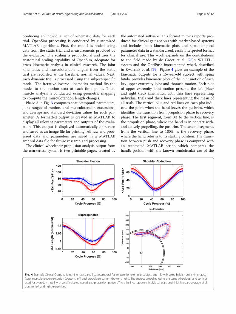
the automated software. This format mimics reports pro-duced for clinical gait analysis with marker-based systemsand includes both kinematic plots and spatiotemporalparameter data in a standardized, easily interpreted formatfor clinical use. This work expands on the contributionsto the field made by de Groot et al. [28]‘s WHEEL-Isystem and the OptiPush instrumented wheel, describedin Kwarciak et al. [29]. Figure 4 gives an example of thekinematic outputs for a 15-year-old subject with spinabifida, provides kinematic plots of the joint motion of eachkey upper extremity joint and thoracic motion. Each plotof upper extremity joint motion presents the left (blue)and right (red) kinematics, with thin lines representingindividual trials and thick lines representing the mean ofall trials. The vertical blue and red lines on each plot indi-cate the point when the hand leaves the pushrim, whichidentifies the transition from propulsion phase to recoveryphase. The first segment, from 0% to the vertical line, isthe propulsion phase, where the hand is in contact with,and actively propelling, the pushrim. The second segment,from the vertical line to 100%, is the recovery phase,where the hand returns to its starting position. The transi-tion between push and recovery phase is computed withan automated MATLAB script, which compares thehand’s position with the known semicircular arc of the
Fig. 4 Example Clinical Outputs. Joint Kinematics and Spatiotemporal Parameters for exemplar subject, age 15, with spina bifida – Joint kinematics(top), musculotendon excursion (bottom, left) and propulsion pattern (bottom, right). The subject propelled using the same wheelchair and settingsused for everyday mobility, at a self-selected speed and propulsion pattern. The thin lines represent individual trials, and thick lines are average of alltrials for left and right extremities
Rammer et al. Journal of NeuroEngineering and Rehabilitation (2018) 15:96 Page 6 of 12
pushrim at each time point, and is then able to estimatewhich data points have hand contact with the pushrimand which points are recovery phase. In the lower left cor-ner of the first page, values are tabulated for range of mo-tion, peak angular velocity, and peak angular accelerationof key joints. The values are averages across all trials, withleft and right extremities presented separately.To analyze the sensitivity of the model for the wheel-
chair propulsion task, sensitivity analysis was used to re-late shoulder and elbow joint motion to musculotendonexcursions for the muscles which cross the respectivejoints. The purpose of this analysis is to mathematicallydetermine the effect of changing joint angle on musculo-tendon motion. This was performed by perturbing themodel throughout the range of shoulder and elbow mo-bility expected in wheelchair propulsion and recordingthe musculotendon response length response for eachmeasurement. Then, a linear regression model was usedto determine the musculotendon sensitivity to joint mo-tion. This method of using sensitivity analysis to assessthe model performance is adapted from the formulas ofRump et al. [30], and while it has not yet been widelyapplied in the field of biomechanics, it has been a usefulprocedure in several other scientific fields, such as auto-motive injury prediction [31–35].To more specifically address the sensitivity of the model
at key points of interest in manual wheelchair propulsion,the shoulder joint is assessed at the start (hand contact)and end (hand release) of a typical wheelchair propulsioncycle, since these transition points represent the mostsignificant potential for injury, as suggested by Rankin etal. [36], who found peak power output at the transitionpoints. To set up the analysis, the model is fixed to thestart and end points (based on typical values collectedfrom subjects), and the other joints not being perturbedare fixed at those values. Thus, only the joint of interest isbeing perturbed for the sensitivity analysis. Analysis isperformed using a dimensionless sensitivity coefficient.This is computed as:((MTL + 5%)-(MTL -5%))/(Initial MTL)]/[((JA +
5%)-(JA-5%))/(Initial JA)Where MTL =musculotendon length, and JA = joint
angle.Inter-trial measurement repeatability was analyzed
using correlation analysis to provide scatterplots andPearson correlation coefficients describing the repeat-ability of the measurement system, to test the hypothesisthat the system reliably measures parameters betweentrials. Thirty wheelchair propulsion assessments ofpediatric manual wheelchair users were conducted aspart of an institutional review board-approved study.The subjects ranged from 6 to 17 years of age with 6females and 24 males, 12 with spina bifida, 3 withCharcot-Marie-Tooth disorder, and 15 with cerebral
palsy, all of whom use a manual wheelchair as a primarymeans of daily mobility.The assessment included spatiotemporal parameters,
such as push time, cycle time, frequency, contact angle,and proportion of push and recovery, similar to thoseused in Vegter et al. [37]. Additionally, joint range ofmotion and musculotendon excursion were analyzed,which differs from the kinetic analysis of Vegter. Eachassessment was performed twice, and the two assess-ments are statistically compared – each subject’s datafrom the first assessment was compared to the samesubject’s data from the second assessment. The subjectswere instructed to perform the same manual propulsiontask during each assessment. Speed and power outputwere results of the subject’s standard, self-selected pat-tern. A Lilliefors test of normality was performed on thedifferences between the data sets to ensure that the nor-mality assumption of the parametric Pearson correlationanalysis was satisfied.
ResultsApplication and kinematic resultsThe system developed during this project has beenrigorously assessed in both laboratory and real-worldapplications. The cost-effectiveness, at approximately$8000 for the complete system, and simplified assess-ment protocol make the system viable in several keyenvironments. Our pilot experience in field-testingthe system suggested that assessments typically canbe completed in under 15 min and training cliniciansto use the system is feasible and can be accomplishedin 30–45 min.
Sensitivity analysisSensitivity analysis was performed on the musculoskel-etal model to determine the relationship between jointmotion and musculotendon excursion. For shoulderelevation, the anterior and posterior deltoid musculo-tendon complexes (− 0.08%/degree and 0.15%/degree,respectively), along with the coracobrachialis (− 0.09%/degree), have high sensitivity to shoulder elevation androtation when compared to the other musculotendoncomplexes studied, mostly in the range of 0.01–0.03%/degree. Sensitivity analysis describing musculotendonresponse to shoulder elevation shows a clear transitionto a point where the joint is not moving very much, butthe muscles are quickly changing in length. The highlinear velocity of the musculotendon complexesstrongly suggests a high potential for injury during thewheelchair propulsion cycle.Thus, each coefficient presented in Table 2 below is
dimensionless, and the higher the coefficient, the moresensitive the muscle is to joint angle changes within thespecified propulsion area. These coefficients are then
Rammer et al. Journal of NeuroEngineering and Rehabilitation (2018) 15:96 Page 7 of 12

categorized as moderately sensitive (0.40 < s < 0.75) orhighly sensitive (s > 0.75).The results in Table 2 show several key points. Muscu-
lotendon complexes are most sensitive at the beginningof the propulsion cycle (hand contact), with fewer mus-culotendon complexes showing high sensitivity at theend of the propulsion cycle (hand release). Further, sev-eral muscles exhibit significantly higher sensitivity thanothers, including the posterior and lateral deltoid, teresmajor, latissimus dorsi, coracobrachialis, and biceps bra-chii. These results suggest that there is a higher risk ofinjury during initial hand contact over hand release, andthat at the hand contact these key muscles are most sen-sitive to the angular changes, and thus at risk for injury.The longer muscles overall appear to have lower sensi-tivity, and hypersensitivity in the shorter musculoten-dons suggests a higher risk of injury. These results maybe limited due to the use of only data from a representa-tive subject for this model analysis. Future work is sug-gested to confirm these results in a larger population.
Repeatability analysisFor each pediatric manual wheelchair propulsion assess-ment, two separate kinematic trials were recorded foreach subject. Statistical correlation analysis was per-formed to determine inter-trial measurement repeatabil-ity of the system, with the results summarized inTable 3. Figure 5 shows that the Pearson correlation
coefficients for spatiotemporal parameters, joint range ofmotion, and musculotendon excursion were high andcorrelations were significant for all parameters, demon-strating inter-trial measurement repeatability of the sys-tem. An additional finding of note is that the metricswith higher Pearson correlation coefficients are the met-rics with the least standard deviation in the data, andvice versa. This may suggest that within-subject variabil-ity is inversely related to the repeatability of inter-trialmeasurements.
DiscussionThe system developed in this project uses a combinationof consumer-grade hardware and open-source musculo-skeletal modeling software to create a unique, cost-effective,efficient, and appropriate analysis technique for clinicalresearch in manual wheelchair biomechanics. TheMicrosoft Kinect was chosen because of its low cost and
Table 2 Sensitivity of Musculotendon Complexes to Shoulder Motion at Start and End Points of Propulsion
Muscle Shoulder Elevation(Start Point)
Shoulder Elevation(End Point)
Shoulder Rotation(Start Point)
Shoulder Rotation(End Point)
Ant Deltoid 0.435* −0.185 0.243 0.810**
Lat Deltoid −1.409** 0.039 −0.779** −0.175
Post Deltoid −2.038** 0.288 −1.137** −1.268**
Supraspinatus 0.118 −0.041 0.068 0.183
Infraspinatus 0.466* −0.025 0.257 0.112
Subscapularis −0.494* 0.028 −0.273 −0.124
Teres Minor 0.828** 0.038 0.456* −0.169
Teres Major 1.493** 0.191 0.819** −0.840**
Pectoralis Major 0.701* −0.038 0.385 0.169
Latissimus Dorsi 1.369** 0.103 0.751** −0.453*
Coracobrachialis 1.719** −0.155 0.952** 0.683*
Triceps-Long 0.372 0.088 0.203 −0.385
Triceps-Medial −0.287 −0.103 − 0.156 0.454*
Biceps-Long 0.778** 0.029 0.406* −0.131
Biceps-Short 1.874** 0.008 1.010** −0.037
Brachialis 0.318 0.077 0.172 −0.341
Values presented as dimensionless sensitivity coefficients with +/− 5% perturbation at the start and end points of propulsion; Shoulder thoracohumeralangles describe the arm position – consistent with the coordinate system used in the musculoskeletal model. The start point represents initial contact ofthe hand with the pushrim, and end point is the instant when the hand leaves the pushrim* = Sensitive (coefficient magnitudes > 0.40); ** = Highly sensitive (coefficient magnitudes > 0.75)
Table 3 Inter-Trial Measurement RepeatabilityMetric Type Pearson Correlation Coefficient Significance (p)
Spatiotemporal Parameters 0.792 0.001*
Joint Range of Motion 0.853 0.001*
Musculotendon Excursion 0.931 0.001*
Results of correlation analysis. Each individual subject was tested twice underself-selected conditions with no control of speed or power output, andthe two measures for each subject are compared* p-value significant at α = 0.05
Rammer et al. Journal of NeuroEngineering and Rehabilitation (2018) 15:96 Page 8 of 12

ease of use. The OpenSim upper extremity model bringssignificant computational power to the system, and theinterface allowing its use with the Kinect and automatingthe protocol is the key development of this work.Characterization of the system in several settings has dem-onstrated its effectiveness for its intended applications.The system adds value to clinical assessments by extract-ing metrics that other methods, such as standardized out-come tools, cannot.Comparison of the markerless wheelchair propulsion
assessment system against other common outcomemeasurement protocols reveals several key differences.When compared to laboratory marker-based motionanalysis techniques [1] the markerless system requiresless space (due to the stationary wheelchair platform),reduced training requirements, and allows faster assess-ment. However, the marker-based systems have higherprecision, and include kinetic assessment and EMG data.Inertial measurement units [4] and instrumented wheels([11], and [10]) have similar ease of use when comparedto the markerless system, and require less time andtraining to implement than marker-based systems. How-ever, inertial measurement units and instrumentedwheels do not provide complete kinematic outputs, butonly partial or supplemental data. Inertial measurementunits and instrumented wheels are possible future ex-pansion options for the markerless system to permit kin-etics to be included in the model. Standardized outcomemeasures [12] have fewer equipment and technologicalrequirements, but do require trained observation. It isthese evaluations that the markerless system is intendedto supplement, by adding objective, quantitative out-comes, while adding minimal time and expense.Manual wheelchair skills capacity is a key therapeutic
outcome and can affect quality of life [38]. Assessmentsusing standardized outcome tools in a communitysetting have documented improvements in response totherapy [39], and shown that levels of physical activitycan impact the risk of shoulder pain [40]. Additionally,clinical guidelines suggest monitoring propulsionpattern and technique employed, daily activities, andexercise or therapeutic activities, as a means to assess
risk of upper limb pain or injury [41]. This monitoringis important to continuing research in the field, giventhe paucity of research into therapeutic outcomes ofmanual wheelchair users [42]. The system proposedhere has several key benefits and limitations relative toboth standardized outcome tools and complex labora-tory motion capture. Clinically, a physical therapist canuse the system to assess the UE kinematics and propul-sion pattern of wheelchair users as part of routine ther-apy visits, to track progress. The system providesreliable quantitative data to track patient progress,which is rarely available in current physiotherapeuticclinical assessments. Propulsion pattern, for instance,impacts upper extremity muscle power and stress inmanual wheelchair users [43], and is produced by thesystem. The system enhances the capability of thera-pists to obtain quantitative data without requiringoverly complex and detailed analysis, such as laboratorymotion capture [1]. Basic data includes speed, cadence,and propulsion pattern, which are difficult to measureaccurately by video or other techniques. The system iseffective in this role because it requires minimal train-ing – the software is largely automated and therapistscan be trained to use it in a short time.Several limitations of this work exist and should be ad-
dressed. The present system does not include power out-put, which would require kinetic detection hardware,and is a common metric presented in wheelchair bio-mechanics literature [37, 44]. The present system alsodoes not measure wheel speed directly, but an algorithmhas been implemented to estimate ground speed basedon a known wheel diameter and hand motion. Cadenceis measured by the system and the resistance level isadjustable based on wheelchair wheel diameter. The lackof speed and power output data reduces the ability tocontrol testing conditions for these metrics, whichwould be desirable for consistency, such as trackingchanges among preferred cadence, wheelchair specifica-tions, and personal differences over time. The additionof speed and power output would increase usability ofthe system, allowing tracking of changes in preferredcadence, wheelchair styles, and personal performance.
Fig. 5 Inter-Trial Pearson Correlation for Categorical Metrics
Rammer et al. Journal of NeuroEngineering and Rehabilitation (2018) 15:96 Page 9 of 12

Inter-trial repeatability was significant for spatiotem-poral parameters, joint kinematics, and musculotendonexcursions. This suggests that the markerless wheelchairpropulsion kinematic assessment system is a repeatablemeasurement tool for pediatric manual wheelchair users,and detects changes that are greater than the inherentnormal variability in the population. Given inter-trial re-peatability with significant correlation, the system isrecommended for further quantitative assessment use inpediatric manual wheelchair users. However, it shouldbe noted that the markerless technology has limitationsin precision of kinematic detection, and more advancedtechnologies may be required to obtain higher precision.
ConclusionsThere is a significant deficit in current literature onmanual wheelchair propulsion biomechanics andphysiotherapeutic treatment for this population. Thesystem is suggested for immediate implementation innovel research to resolve these key deficiencies incurrent literature, leading to more effective point-of-care clinical outcome assessments for manual wheel-chair users, provided the limitations of markerless tech-nology are taken into account. In the future, home useand telerehabilitation development are suggested aspossible directions for the project.
AcknowledgmentsThe authors acknowledge the support of Bay Cliff Health Camp Children’sTherapy and Wellness Center and the Orthopaedic and Rehabilitation EngineeringCenter (OREC) at Marquette University. The contents of this paper weredeveloped under a grant from the National Institute on Disability, IndependentLiving, and Rehabilitation Research (NIDILRR Grant number 90RE5006-01-00, Formerly H133E100007). NIDILRR is a Center within the Administration forCommunity Living (ACL), Department of Health and Human Services(HHS). The contents of this paper do not necessarily represent the policyof NIDILRR, ACL, HHS, and you should not assume endorsement by theFederal Government.
FundingThe contents of this paper were developed under a grant from the NationalInstitute on Disability, Independent Living, and Rehabilitation Research(NIDILRR Grant number 90RE5006-01-00, Formerly H133E100007). NIDILRRis a Center within the Administration for Community Living (ACL), Departmentof Health and Human Services (HHS). The contents of this paper do notnecessarily represent the policy of NIDILRR, ACL, HHS, and you shouldnot assume endorsement by the Federal Government.
Availability of data and materialsNot applicable. This study focuses on methodological development using apublicly available musculoskeletal model.
Authors’ contributionsJR and SR performed the data collection and analysis. GH, BS, JW, and JKcontributed to the interpretation of results and writing of the manuscript.All authors read and approved the final manuscript.
Ethics approval and consent to participateAll human subjects interaction in this study was approved by the MarquetteUniversity Institutional Review Board.
Consent for publicationNot applicable.
Competing interestsThe authors declare that they have no competing interests.
Publisher’s NoteSpringer Nature remains neutral with regard to jurisdictional claims in publishedmaps and institutional affiliations.
Author details1Orthopaedic and Rehabilitation Engineering Center (OREC), MarquetteUniversity, Olin Engineering Suite 323, Milwaukee, WI 53201-1881, USA.2Department of Biomedical Engineering, Marquette University, OlinEngineering Suite 323, Milwaukee, WI 53201-1881, USA. 3Department ofOrthopaedic Surgery, Medical College of Wisconsin, Milwaukee, WI53201-1881, USA. 4Shriners Hospitals for Children, Chicago, IL, USA.5Midwestern University, Physical Therapy Program, 555 31st St., Alumni Hall340C, Downers Grove, IL 60515, USA. 6University of Wisconsin-Milwaukee,2400 E Hartford Ave, Rm. 983, Milwaukee, WI 53211, USA. 7MarquetteUniversity, Biomedical Engineering, Milwaukee, WI 53201-1881, USA.
Received: 7 March 2018 Accepted: 18 October 2018
References1. Schnorenberg AJ, Slavens BA, Wang M, Vogel LC, Smith PA, Harris GF.
Biomechanical model for evaluation of pediatric upper extremity jointdynamics during wheelchair mobility. J Biomech. 2014;47(1):269. https://doi.org/10.1016/j.jbiomech.2013.11.014.
2. van der Woude LH, Veeger D, Dallmeijer AJ, Janssen TW, Rozendaal LA.Biomechanics and physiology in active manual wheelchair propulsion. MedEng Phys. 2001;23(10):713–33. https://doi.org/10.1016/S1350-4533(01)00083-2.
3. Vegter RJK, Hartog J, de Groot S, Lamoth CJ, Bekker MJ, van der Scheer JanW, Veeger Dirkjan HEJ. Early motor learning changes in upper-limbdynamics and shoulder complex loading during handrim wheelchairpropulsion. J Neuroeng Rehabil. 2015;12. Retrieved from https://doi.org/10.1186/s12984-015-0017-5.
4. Bergamini E, Morelli F, Marchetti F, Vannozzi G, Polidori L, Paradisi F, Delussu,AS. Wheelchair propulsion biomechanics in junior basketball players: a methodfor the evaluation of the efficacy of a specific training program. BioMed Res Int.2015;275965. Retrieved from https://doi.org/10.1155/2015/275965.
5. Leving M, Horemans H, Vegter R, de Groot S, Bussmann J, van derWoude L. Validity of consumer-grade activity monitor to identifymanual wheelchair propulsion in standardized activities of daily living.PLoS One. 2018;13(4):e0194864.
6. van der Slikke R, Berger M, Bregman D, Lagerberg A, Veeger H. Opportunitiesfor measuring wheelchair kinematics in match settings: reliability of a threeinertial sensor configuration. J Biomech. 2015;48:3398–405.
7. van der Slikke R, Berger M, Bregman D, Veeger H. From big data to richdata: the key features of athlete wheelchair mobility performance. JBiomech. 2016;49:3340–6.
8. van der Slikke R, Mason B, Berger M, Goosey-Tolfrey V. Speed profiles inwheelchair court sports: comparison of two methods for measuringwheelchair mobility performance. J Biomech. 2017;65:221–5.
9. van der Slikke R, Bregman D, Berger M, de Witte A, Veeger D. The future ofclassification in wheelchair sports: can data science and technologicaladvancement offer an alternative point of view? Int J Sports PhysiolPerform. 2018;13:742–9.
10. Dellabiancia F, Porcellini G, Merolla G. Instruments and techniques for theanalysis of wheelchair propulsion and upper extremity involvement inpatients with spinal cord injuries: current concept review. MusclesLigaments Tendons J. 2013;3(3):150–6.
11. Conger SA, Scott SN, Fitzhugh EC, Thompson DL, Bassett DR. Validity ofphysical activity monitors for estimating energy expenditure duringwheelchair propulsion. J Phys Act Health. 2015;12(11):1520. Retrieved fromhttps://doi.org/10.1123/jpah.2014-0376.
12. Kenny S, Gowran RJ. Outcome measures for wheelchair and seating provision:a critical appraisal. Br J Occup Ther. 2014;77(2):67–77. https://doi.org/10.4276/030802214X13916969447119.
13. Boninger M, Koontz A, Sisto S, Dyson-Hudson T, Chang M, Price R, CooperR. Pushrim biomechanics and injury prevention in spinal cord injury:recommendations based on CULP-SCI investigations. J Rehabil Res Dev.2005;42(3):9–20.
Rammer et al. Journal of NeuroEngineering and Rehabilitation (2018) 15:96 Page 10 of 12

14. Bonnechère, B., Jansen, B., Salvia, P., Bouzahouene, H., Omelina, L., Moiseev, F., .. . Van Sint Jan, S. (2014). Validity and reliability of the kinect within functionalassessment activities: comparison with standard stereophotogrammetry. GaitPosture, 39(1), 593–598. doi:https://doi.org/10.1016/j.gaitpost.2013.09.018.
15. Dutta T. Evaluation of the Kinect sensor for 3-D kinematic measurement inthe workplace. Appl Ergon. 2012;43:645–9.
16. Galna B, Barry G, Jackson D, Mhiripiri D, Olivier P, Rochester L. Accuracy ofthe microsoft kinect sensor for measuring movement in people withparkinson's disease. Gait Posture. 2014;39(4):1062–8. https://doi.org/10.1016/j.gaitpost.2014.01.008.
17. Otte K, Kayser B, Mansow-Model S, Verrel J, Paul F, Brandt AU, Schmitz-Hübsch T. Accuracy and reliability of the kinect version 2 for clinicalmeasurement of motor function. PLoS One. 2016;11(11):e0166532. https://doi.org/10.1371/journal.pone.0166532.
18. van Diest M, Stegenga J, Wortche HJ, Postema K, Verkerke GJ, Lamoth CJC.Suitability of kinect for measuring whole body movement patterns duringexergaming. J Biomech. 2014;47(12):2925–32. https://doi.org/10.1016/j.jbiomech.2014.07.017.
19. Huber ME, Seitz AL, Leeser M, Sternad D. Validity and reliability of kinectskeleton for measuring shoulder joint angles: a feasibility study. Physiotherapy.2015;101(4):389–93. https://doi.org/10.1016/j.physio.2015.02.002.
20. Lee SH, Yoon C, Chung SG, Kim HC, Kwak Y, Park H, Kim K. Measurement ofshoulder range of motion in patients with adhesive capsulitis using a kinect.PLoS One. 2015;10(6):e0129398. https://doi.org/10.1371/journal.pone.0129398.
21. Mobini A, Behzadipour S, Saadat M. Test-retest reliability of kinect'smeasurements for the evaluation of upper body recovery of stroke patients.Biomed Eng Online. 2015;14:75 Retrieved from https://doi.org/10.1186/s12938-015-0070-0.
22. Rammer J, Krzak J, Riedel S, Harris G. Evaluation of upper extremity movementcharacteristics during standardized pediatric functional assessment with aKinect®-based Markerless motion analysis system. IEEE Eng Med Biol Conf.2014:2525–8. https://doi.org/10.1109/EMBC.2014.6944136.
23. Delp SL, Anderson FC, Arnold AS, Loan P, Habib A, John CT, Thelen DG.OpenSim: open-source software to create and analyze dynamic simulationsof movement. IEEE Trans Biomed Eng. 2007;54(11):1940–50. https://doi.org/10.1109/TBME.2007.901024.
24. Holzbaur K, Murray W, Delp S. A model of the upper extremity for simulatingmusculoskeletal surgery and analyzing neuromuscular control. Ann BiomedEng. 2005;33(6):829–40. https://doi.org/10.1007/s10439-005-3320-7.
25. Saul K, Hu X, Goehler C, Vidt M, Daly M, Velisar A, Murray W. Benchmarkingof dynamic simulation predictions in two software platforms using anupper limb musculoskeletal model. Comput Methods Biomech BiomedEngin. 2014;18(13):1–14.
26. DiGiovine CP, Cooper RA, Boninger ML. Dynamic calibration of a wheelchairdynamometer. J Rehabil Res Dev. 2001;38(1):41 Retrieved from http://www.ncbi.nlm.nih.gov/pubmed/11322470.
27. Rammer J, Riedel S, Harris G. A portable, low-cost wheelchair ergometerdesign based on a mathematical model of pediatric wheelchair dynamics.In: Proceedings of 2015 Annual Meeting of the Rehabilitation Engineeringand Assistive Technology Society of North America; 2015. p. 4.
28. de Groot S, Vegter R, Vuijik C, van Dijk F, et al. WHEEL-I: development of awheelchair propulsion laboratory for rehabilitation. J Rehabil Med. 2014;46:493–503.
29. Kwarciak A, Turner J, Guo L, Richter W. Comparing handrim biomechanics fortreadmill and overground wheelchair propulsion. Spinal Cord. 2011;49:457–62.
30. Rump S. Rigorous sensitivity analysis for systems of linear and nonlinearequations. Math Comput. 1990;54(190):721–36.
31. Autrique L, Lormel C. Numerical design of experiment for sensitivityanalysis—application to skin burn injury prediction. IEEE Trans Biomed Eng.2008;55(4):1279–90.
32. Fang X, Yang T. Regression methodology for sensitivity analysis of solarheating walls. Appl Therm Eng. 2008;28:2289–94.
33. Small D. Sensitivity analysis for instrumental variables regression withoveridentifying restrictions. J Am Stat Assoc. 2007;102(49):1049–58.
34. Suchao X, Hongqi T. Influencing factors and sensitivity analysis of occupantimpact injury in passenger compartment. Traffic Injury Prevention. 2013;14:816–22.
35. Tondel K, Vik J, Martens J, Indahl U, Smith N, Omholt S. Hierarchicalmultivariate regression-based sensitivity analysis reveals complexparameter interaction patterns in dynamic models. Chemom Intell LabSyst. 2013;120:25–41.
36. Rankin J, Richter W, Neptune R. Individual muscle contributions to pushand recovery subtasks during wheelchair propulsion. J Biomech. 2011;44:1246–52.
37. Vegter R, de Groot S, Lamoth C, Veeger D, van der Woude L. Initial skillacquisition of handrim wheelchair propulsion: a new perspective. IEEE TransNeural Syst Rehabil Eng. 2014;22(1):104–13.
38. Hosseini SM, Oyster ML, Kirby RL, Harrington AL, Boninger ML. Manualwheelchair skills capacity predicts quality of life and community integrationin persons with spinal cord injury. Arch Phys Med Rehabil. 2012;93(12):2237.https://doi.org/10.1016/j.apmr.2012.05.021.
39. Best K, Kirby R, Smith C, MacLeod D. Wheelchair skills training forcommunity-based manual wheelchair users: a randomized controlled trial.Arch Phys Med Rehabil. 2005;86:2316–24.
40. Mulroy SJ, Hatchett P, Eberly VJ, Haubert LL, Conners S, Requejo PS.Shoulder strength and physical activity predictors of shoulder pain inpeople with paraplegia from spinal injury: prospective cohort study. PhysTher. 2015;95(7):1027–38. https://doi.org/10.2522/ptj.20130606.
41. Consortium for Spinal Cord Medicine. Preservation of upper limb functionfollowing spinal cord injury: a clinical practice guideline for health-careprofessionals; 2005. p. 48.
42. Zwinkels, M. G. J., Verschuren, O. W., Janssen, T., Ketelaar, M., Takken, T.,Backx, F. J. G., . . . Volman, M. (2014). Exercise training programs to improvehand rim wheelchair propulsion capacity: a systematic review. Clin Rehabil,28(9), 847–861. doi:https://doi.org/10.1177/0269215514525181.
43. Slowik J, Requejo P, Mulroy S, Neptune R. The influence of wheelchairpropulsion hand pattern on upper extremity muscle power and stress. JBiomech. 2016;49:1554–61.
44. Dallmeijer A, Kilkens O, Post M, de Groot S, Angenot E, van Asbeck F, vander Woude L. Hand-rim wheelchair propulsion capacity during rehabilitationof persons with spinal cord injury. J Rehabil Res Dev. 2005;42(3 Suppl 1):55Retrieved from http://www.ncbi.nlm.nih.gov/pubmed/16195963.
45. Clark RA, Bower KJ, Mentiplay BF, Paterson K, Pua Y. Concurrentvalidity of the microsoft kinect for assessment of spatiotemporalgait variables. J Biomech. 2013a;46(15):2722–5. https://doi.org/10.1016/j.jbiomech.2013.08.011.
46. Clark RA, Pua Y, Fortin K, Ritchie C, Webster KE, Denehy L, Bryant AL. Validityof the microsoft kinect for assessment of postural control. Gait Posture.2012;36(3):372–7. https://doi.org/10.1016/j.gaitpost.2012.03.033.
47. Clark, R. A., Pua, Y., Oliveira, C. C., Bower, K. J., Thilarajah, S., McGaw, R., . . .Mentiplay, B. F. (2015a). Reliability and concurrent validity of the microsoft xboxone kinect for assessment of standing balance and postural control. GaitPosture, 42(2), 210–213. doi:https://doi.org/10.1016/j.gaitpost.2015.03.005.
48. Clark, R. A., Vernon, S., Mentiplay, B. F., Miller, K. J., McGinley, J. L., Pua, Y. H., .. . Bower, K. J. (2015b). Instrumenting gait assessment using the kinect inpeople living with stroke: reliability and association with balance tests. JNeuroeng Rehabil, 12(1), 15. doi:https://doi.org/10.1186/s12984-015-0006-8.
49. Boninger M, Souza A, Cooper R, Fitzgerald S, Koontz A, Fay B. Propulsionpatterns and Pushrim biomechanics in manual wheelchair propulsion. ArchPhys Med Rehabil. 2002;83:718–23.
50. Clark RA, Pua Y, Bryant AL, Hunt MA. Validity of the microsoft kinect forproviding lateral trunk lean feedback during gait retraining. Gait Posture.2013b;38(4):1064–6. https://doi.org/10.1016/j.gaitpost.2013.03.029.
51. de Groot S, Veeger H, Hollander A, van der Woude L. Consequence offeedback-based learning of effective hand rim wheelchair force productionon mechanical efficiency. Clin Biomech. 2002;17:219–26.
52. de Groot S, de Bruin M, Noomen S, van der Woude L. Mechanical efficiencyand propulsion technique after 7 weeks of low-intensity wheelchair training.Clin Biomech. 2008;23:434–41.
53. Finley M, Rodgers M. Prevalence and identification of shoulder pathology inathletic and nonathletic wheelchair users with shoulder pain: a pilot study. JRehabil Res Dev. 2004;38:395–402.
54. Gonzalez-Jorge H, Rodríguez-Gonzálvez P, Martínez-Sánchez J, González-Aguilera D, Arias P, Gesto M, Díaz-Vilariño L. Metrological comparisonbetween kinect I and kinect II sensors. Measurement. 2015;70:21–6. https://doi.org/10.1016/j.measurement.2015.03.042.
55. Guo L, Kwarciak A, Rodriguez R, Sarkar N, Richter W. Validation of abiofeedback system for wheelchair propulsion training. Rehabil Res Pract.2011;2011:590780 7p.
56. Kertis JD, Fritz JM, Long JT, Harris GF. Static and dynamic calibration of aneight-camera optical system for human motion analysis. Crit Rev PhysRehabil Med. 2010;22(1–4):49–60.
Rammer et al. Journal of NeuroEngineering and Rehabilitation (2018) 15:96 Page 11 of 12

57. Knippenberg E, Verbrugghe J, Lamers I, Palmaers S, Timmermans A,Spooren A. Markerless motion capture systems as training device inneurological rehabilitation: a systematic review of their use, application,target population, and efficacy. J NeuroEng Rehabil. 2017;14:61 11p.
58. Morrow M, Van Straaten M, Murthy N, Braman J, Zanella E, Zhao K. Detailedshoulder MRI findings in manual wheelchair users with shoulder pain.Biomed Res Int. 2014;2014:1–7.
59. Rankin J, Kwarciak A, Richter W, Neptune R. The influence of wheelchairpropulsion technique on upper extremity muscle demand: a simulationstudy. Clin Biomech. 2012;27:879–86.
60. Rice I, Gagnon D, Gallagher J, Boninger M. Hand rim wheelchair propulsiontraining using biomechanical real-time visual feedback based on motorlearning theory principles. J Spinal Cord Med. 2010;33:33–42.
61. Rice I, Pohlig R, Gallagher J, Boninger M. Handrim wheelchair propulsiontraining effect on Overground propulsion using biomechanical real-timevisual feedback. Arch Phys Med Rehabil. 2013;94:256–63.
62. Richter W, Kwarciak A, Guo L, Turner J. Effects of single-variablebiofeedback on wheelchair Handrim biomechanics. Arch Phys MedRehabil. 2011;92:572–7.
63. Rodgers M, Keyser R, Gardner E, Russell P, Gorman P. Influence of trunkflexion on biomechanics of wheelchair propulsion. J Rehabil Res Dev. 2000;37(3):283–95.
64. Sabick M, Korajarvi B, An K. A new method to quantify demand on theupper extremity during manual wheelchair propulsion. Arch Phys MedRehabil. 2004;85:1151–9.
65. Veeger H, Rozendaal L, van der Helm F. Load on the shoulder in low-intensity wheelchair propulsion. Clin Biomech. 2002;17:211–8.
66. Wu G, van der Helm F, Veeger H, Makhsous M, Van Roy P, Anglin C, NagelsJ, Karduna A, McQuade K, Wang X, Werner F, Buchholz B. ISBrecommendation on definitions of joint coordinate systems of various jointsfor the reporting of human joint motion—part II: shoulder, elbow, wrist,and hand. J Biomech. 2005;38:981–92.
67. Xu X, McGorry RW. The validity of the first and second generation microsoftkinect™ for identifying joint center locations during static postures. ApplErgon. 2015;49:47–54. https://doi.org/10.1016/j.apergo.2015.01.005.
68. Xu X, McGorry RW, Chou L, Lin J, Chang C. Accuracy of the microsoft kinectfor measuring gait parameters during treadmill walking. Gait Posture. 2015;42(2):145–51. https://doi.org/10.1016/j.gaitpost.2015.05.002.
Rammer et al. Journal of NeuroEngineering and Rehabilitation (2018) 15:96 Page 12 of 12