assessment of coma · • meningoencephalitis. general neurological examination • tone, power,...
TRANSCRIPT

Assessment of Coma
Dr Martin HughesApril 2010

Example 1
• 68 year old woman, found collapsed by son• Was well about 1 hour previously• No medical history available• Asked to take for scan, GCS 3• Airway patent, breathing 35 / minute, 130/
80, 90/min

Example 2
• 58 yo alcoholic• Recovering from pneumococcal pneumonia• Sedation stopped, not waking up

Example 3
• 46 year old man with abdominal pain• Smoker and drinker, FH cerebrovascular
disease• ?gastritis, in medical wards• Sudden LOC (spoken to by nurses 5
minutes earlier)

Example 4
• 48 y.o man• OOH cardiac arrest• Admitted for cooling• Post cooling still GCS 3• Facial twitching ? myoclonus
Plan
• Basic approach to comatose patient• System to allow accurate assessment of
coma and diagnosis of the cause of coma• Allow appropriately directed further
investigation• No CT interpretation• Not about outcome• Not about management of specific diseases
Basic Approach
• Airway with cervical spine: patency and protection. Act if necessary
• Breathing: SpO2, respiratory rate, respiratory examination. Act if necessary
• Circulation: BP, HR, capillary refill. Act if necessary
• Get history• Glucose, ECG, CxR, Bloods, ABGs, tox screen
General examination
• Skin: rash, anaemia, cyanosis, jaundice, spiders, track marks
• Temp: infection, hypothermia• CVS: infection, Addison's, arrhythmia• RS: hypoxia or hypercarbia. Remember
neurogenic pulmonary oedema• Abdomen: ascites, organomegaly• Thyroid

Basic Approach - Trauma
• GCS 15, age < 65, no clinical evidence skull #, no retrograde amnesia, no seizure, no significant mechanism of injury or serious assault
• GCS 13 or 14 but 15 within 2 hours of injury and above
• Otherwise CT

Consciousness
• Wakefulness (arousal, vigilance, alertness)• Awareness (of self or environment)• No awareness without wakefulness• Wakefulness can occur without awareness
(vegetative state)
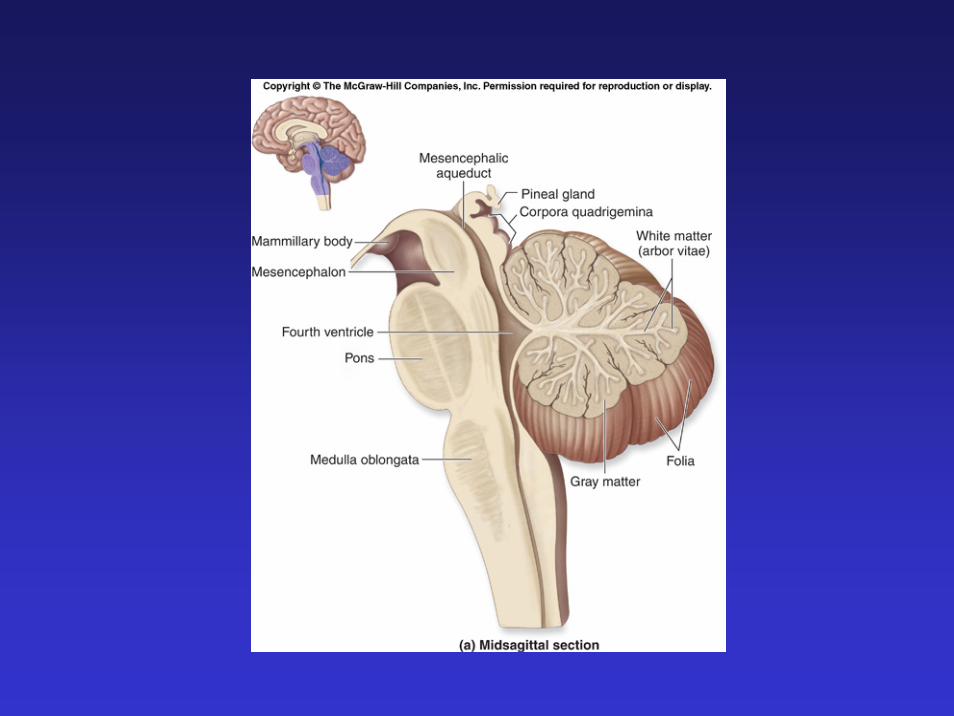
Wakefulness
• Linked to ARAS• In pons and midbrain• Projects to diencephalon (thalamus and
hypothalamus) and cortex
Awareness
• Needs functioning cortex and subcorticalconnections
• Needs wakefulness
What is Coma?
• No awareness or arousal• Lasts > 1h (cf syncope and concussion)• No spontaneous speech or movement, eyes shut• No eye opening to verbal command (E2) • Noxious stimuli: vocalisation limited or absent
(V2)• Noxious stimuli: motor activity
absent/abnormal/reflexive (not purposeful or defensive) (M4)

Coma
• No sleep wake cycles• Transitional• Injury or functional disruption of bilateral
cortical structures or ARAS

How can we classify it?
• Direct brainstem• Indirect brainstem• Generalised neuronal dysfunction

What are the causes?
• Commonly stroke, cranial trauma and drugs• ‘Medical’: CVA 50%, hypoxic / ischaemic 20%,
infective / metabolic encephalopathies the rest• Depends on location: when looked for subclinical
status epilepticus up to 8% in ICU coma• HOW DO WE MAKE THE DISTINCTION,
AND WHY?: direct brainstem, indirect brainstem, generalised neuronal dysfunction

Generalised Neuronal Dysfunction
• Drugs• Anoxia• Epilepsy: may be
subclinical• CO• SIRS• Hypoxia• Hypercapnia• Glucose
• Sodium• Liver• Kidneys• Hypothermia• Hypertension• Hypotension• Hypercalcaemia• Wernicke’s• Thyroid, adrenal, pituitry
Examination


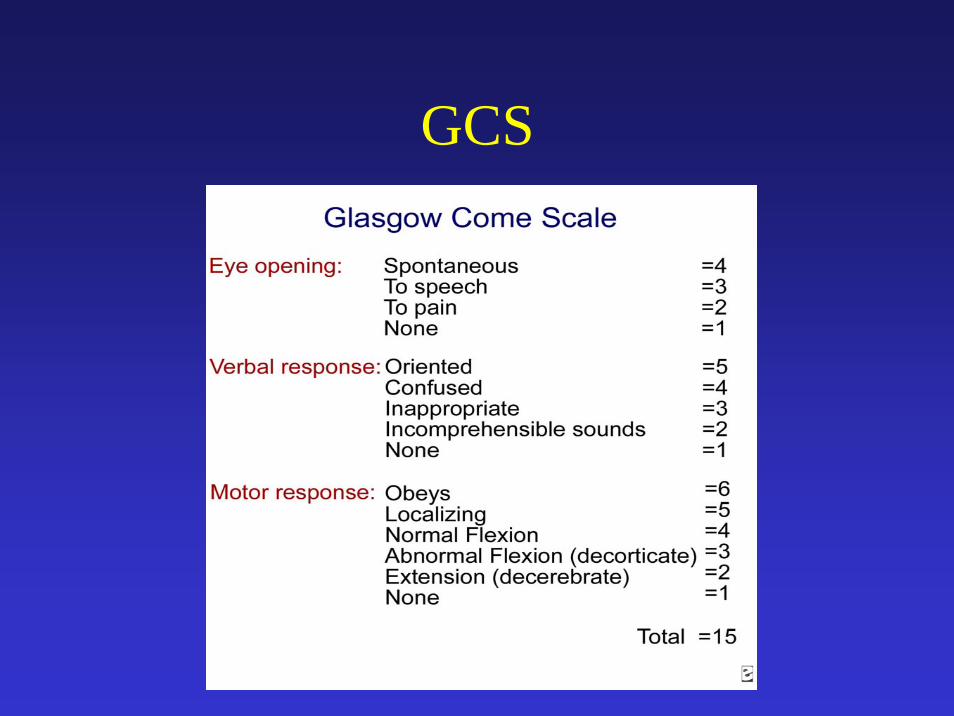
GCS

EYES
• ARAS near centres for pupillary function and eye movements
• Pupillary responses• Oculocephalic reflexes• Corneal reflexes• Eye deviation and movements• Fundus
Pupillary responses
• Preserved in lesions above thalamus and below pons
• If they are normal it is unlikely to be direct or indirect brainstem injury (as the cause of coma)
• Beware drugs (tricyclics, anticholinergics, amphetamines, carbamazepine)

Pupillary responses
• Large fixed bilateral: tectal (dorsal mid brain)
• Mid size fixed bilateral: mid brain• Pin point bilateral: pons• Small reactive: diencephalon• Large fixed unilateral: III nerve
Pupillary responses
• Need bright light and sometimes a magnifying glass
• Generally intact in metabolic/toxic/generalised neuronal dysfunction
• Local injury and drugs can interfere

Oculocephalic reflexes
• Centres for eye movement adjacent to centres for arousal: useful guide to brainstem disease
• ‘Doll’s eye’ reflex• Only in a coma• Remain fixed on the current point of focus
initially• Then follow the head

Oculocephalic reflexes
• If normal pontomedullary junction to level of oculomotor nucleus in the brainstem intact
• Abnormality means brainstem dysfunction, normal means brainstem ok
• Can substitute caloric reflexes (neck disorders, more sensitive. More effort. Identifies INO)

Corneal reflexes
• Intact pons: closure• Intact pons and midbrain: Bell’s
phenomenon
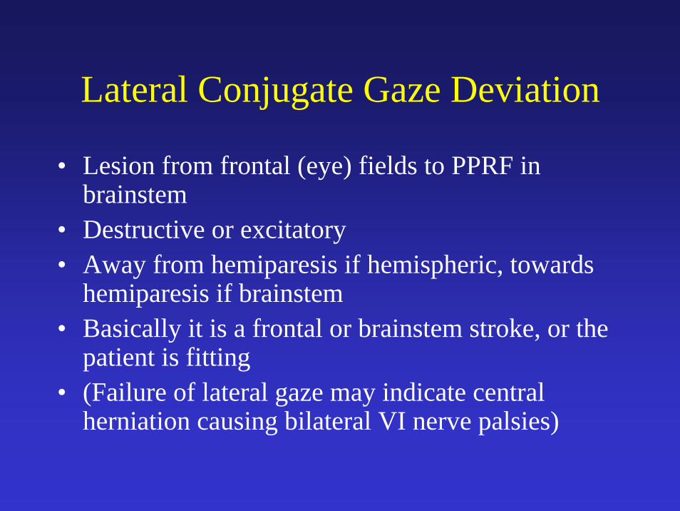
Lateral Conjugate Gaze Deviation
• Lesion from frontal (eye) fields to PPRF in brainstem
• Destructive or excitatory• Away from hemiparesis if hemispheric, towards
hemiparesis if brainstem• Basically it is a frontal or brainstem stroke, or the
patient is fitting• (Failure of lateral gaze may indicate central
herniation causing bilateral VI nerve palsies)
Disconjugate gaze deviation
• III nerve• VI nerve• Brainstem

Downward deviation
• Not especially helpful• Bilateral thalamic and subthalamic (dorsal
mid brain) lesions• E.g. acute obstructive hydrocephalus• Some metabolic encephalopathies, e.g
hepatic encephalopathy• Ocular bobbing is pontine

Upward Deviation
• Not helpful• Sleep• Brainstem• Seizures• Bilateral hemispheric damage

Roving eye movements
• Slow, conjugate, lateral, to and fro• Generalised neuronal dysfunction, usually
DRUGS• (Occasionally bilateral hemispheric)

Ping pong movements
• Bilateral hemispheric
INO
• Caloric stimulation• Near eye moves to cold water, far eye
stationary• Damage to MLF i.e. pons

Fundus

Fundus

Fundus
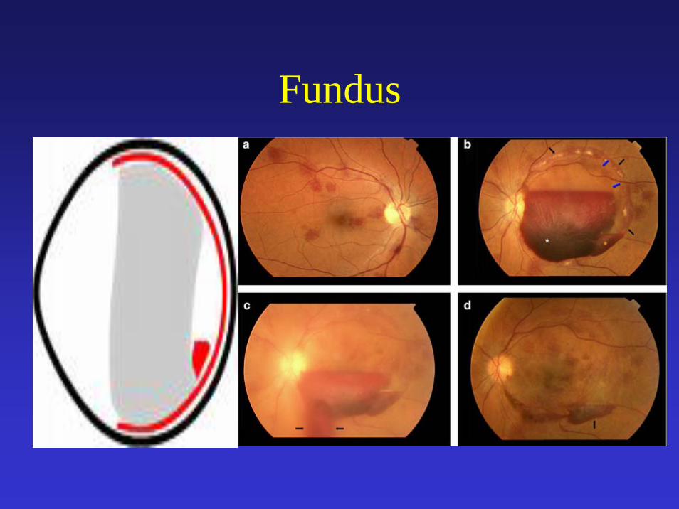
Fundus
Other brainstem signs
• Grimacing• Gag• Breathing
Meningism
• SAH• Meningitis• Meningoencephalitis
General Neurological Examination
• Tone, power, reflexes• Identifies lateralising signs: hemispheric
lesions• In general plantars do not help• Signifies hemispheric lesions but may be
old• Decerebrate, decorticate

Respiratory pattern
• Cheyne –Stokes: bilateral diencephalic or hemispheric injury (LVF, COPD, drugs depressing response to CO2, ventilatory over support)
• Hyperventilation: pons or midbrain injury (ARF, shock, fever, acidosis, psychiatric disease)
• Apneustic breathing: pons• Ataxic: medulla, usually preterminal


In reality you need: good torch, cotton wool or swab, opthalmoscope, tendon hammer
• Pupils, size and reaction • Eye deviation and eye movements• Oculocephalic reflex• Corneal reflex• Cough/gag, grimacing, breathing• Fundi• Meningism• Neurolgy: GCS, tone, power, reflexes• About 4 minutes
Examination
Why examine?
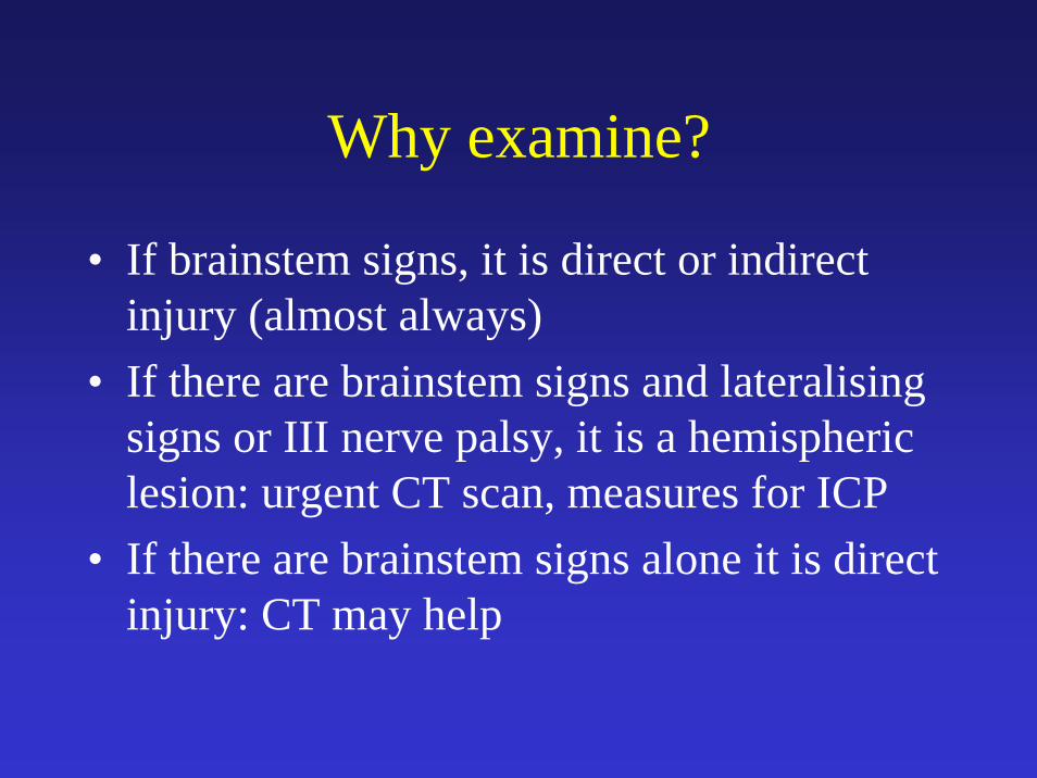
• If brainstem signs, it is direct or indirect injury (almost always)
• If there are brainstem signs and lateralising signs or III nerve palsy, it is a hemispheric lesion: urgent CT scan, measures for ICP
• If there are brainstem signs alone it is direct injury: CT may help
Why examine?
• If there are no brainstem signs it is a generalised cause of coma: look for it, identify it
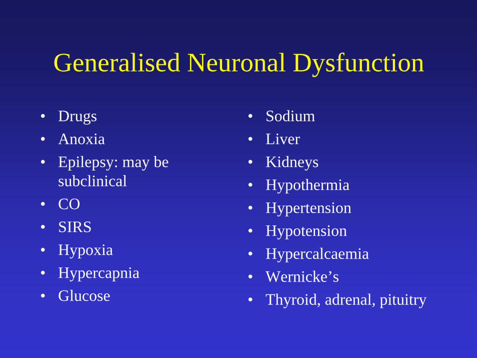
Generalised Neuronal Dysfunction
• Drugs• Anoxia• Epilepsy: may be
subclinical• CO• SIRS• Hypoxia• Hypercapnia• Glucose
• Sodium• Liver• Kidneys• Hypothermia• Hypertension• Hypotension• Hypercalcaemia• Wernicke’s• Thyroid, adrenal, pituitry

Why examine?
• You may identify seizures, including sub clinical status
• You may identify meningism• More commonly you can direct your
investigation appropriately• Even more commonly you can do nothing
except watchful waiting while treating the underlying disease
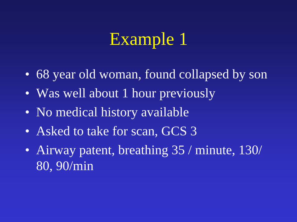
Example 1
• 68 year old woman, found collapsed by son• Was well about 1 hour previously• No medical history available• Asked to take for scan, GCS 3• Airway patent, breathing 35 / minute, 130/
80, 90/min
Example 1
• No peripheral focal neurology identifiable• Pupils pinpoint• No corneal or oculocephalic relexes• DIAGNOSIS

Example 2
• 58 yo alcoholic• Recovering from pneumococcal pneumonia• Sedation stopped, not waking up

Example 3
• 46 year old man with abdominal pain• Smoker and drinker, FH cerebrovascular disease• ?gastritis, in medical wards• Sudden LOC (spoken to by nurses 5 minutes
earlier)• GCS 3, no focal neurology, brainstem signs or
meningism, CVS stable• DIAGNOSIS

Example 4
• 48 y.o man• OOH cardiac arrest• Admitted for cooling• Post cooling still GCS 3• Facial twitching ? myoclonus• Lateral conjugate gaze deviation• DIAGNOSIS
Summary
• Clinical assessment of coma is useful• It can lead to unexpected diagnoses• It leads to the appropriate investigation,
including no investigation• It should be as routine as respiratory
examination in respiratory failure