assessment of marginal leakage around class ii composite ... · pdf fileassessment of marginal...
TRANSCRIPT

PEDIATRIC DENTISTRY/Copyright © 1990 byThe American Academy of Pediatric Dentistry
Volume 12~ Number !
Assessment of marginal leakage around Class IIcomposite restorations in retrieved primary molarsAnna B. Fuks, CD Aubrey Chosack, BDS (Rand), MSD
Eliezer Eidelman, Dr Odont, MSD
Abstract
The aim of the present investigation was to evaluate, bymeans of dye penetration, the microleakage around Class IIcomposite restorations, in retrieved primary molars thatfunctioned in the mouth for at least one year. Theexperimental material consisted of 13 exfoliated primarymolars that had been restored with Herculite ® (KerrCorporation, Romulus, Michigan 48174 USA) at least oneyear previously, utilizing an incremental or a bulk fillingtechnique.
The retrieved teeth were insulated with utility wax andnail polish, immersed in 2% basic fuchsin, embedded inacrylic resin, and ground off to various depths. The marginalleakage was assessed according the degree of dye penetrationat the occlusal and cervical margins.
No difference was observed between the two fillingtechniques. In most teeth, no leakage at the occlusal marginswas observed; minimal leakage, limited to the enamel, wasobserved at the occlusal margins of two teeth, one of eachfilling technique. Severe penetration was evident at thecervical margin of three restorations, two of them filledincrementally and the third using the bulk technique. Mild tomoderate penetration was observed at the cervical margin inthe majority of the other restorations.
It was concluded that an incremental filling techniquecould not eliminate microleakage at the cervical margins ofClass II composite restorations.
Introduction
Several "posterior composites" have becomeavailable in recent years. However, their application forClass II restorations has not been fully accepted. Poorsealing of the margins is a major problem of thesematerials (Browne and Tobias 1986; Moore and Vann1986a; Moore and Vann 1986b) occurring to a greaterextent at the gingival margin (Lui et al. 1987). Gaps mayappear at the tooth-resin interface as a result of
polymerization shrinkage of the setting resin (Jensenand Chan 1985). These gaps are subsequently affectedby other factors such as masticatory forces (Ericksonand Jensen 1986), thermal changes, and water sorption.
Several methods have been suggested to decreasemarginal leakage, such as beveling the cavosurfaceenamel (Moore and Vann 1986a; Moore and Vann1986b), application of dentin adhesive agents, or fillingthe cavity incrementally (Donly and Jensen 1986;Leclaire et al. 1986).
The penetration of a dye, one of the oldest techniquesavailable, has been utilized widely in several in vitrostudies to assess microleakage (Fuks and Shey 1983;Fuks et al. 1984; Fuks et al. 1986; Holan et al. 1986). Othertechniques, such as silver nitrate, air pressure, andradioactive isotopes, also have been employed (Fuksand Shey 1983). Recently, no differences were foundwhen these techniques for measuring marginal leakagewere evaluated (Strange and Hembree 1987).
Thermocycling is performed in in vitro studies in anattempt to simulate the in vivo conditions. However, inaddition to temperature changes, restorative materialsare subjected, among others, to masticatory forces(Erickson and Jensen 1986). In vitro simulation of in vivoconditions is difficult, if not impossible, and theconclusions are questionable.
The aim of the present investigation was to evaluatethe microleakage around Class II compositerestorations, utilizing two filling techniques, inretrieved primary molars.
The microleakage assessment, although done as anin vitro study, had the benefit of the preparation of theteeth for staining in vivo, with natural thermal changesand stresses of normal function in the mouth for at leastone year. This eliminated the need for thermocycling,usually done in in vitro experiments.
24 CL^ss [I COMPOSITES IN PRIMARY TEETH: FUKS, CHOSACK, AND EIDELMAN
Materials and MethodsThe experimental material consisted of 13 exfoliated
primary molars restored with Herculite one yearpreviously.
The retrieved teeth were part of a comprehensivestudy in which 60 primary molars had been restoredwith the mentioned composite resin using either a bulkor an incremental filling technique, and had beenassessed clinically and radiographically after one year.Visual and tactile assessment of 16 proximal surfaces ofthe 13 retrieved teeth (three had MOD fillings) also hadbeen done (Eidelman et al. 1989).
The furcation area of the 13 teeth and any eventualroot remnants were insulated with IRM and triple-coated with a layer of varnish, melted utility wax, and asecond layer of varnish, excluding the restoration andapproximately 0.5-1 mm of the surrounding enamelmargin (Holan et al. 1986). The coated teeth then wereimmersed in a 2% solution of basic fuchsin for 24 hr.After removal from the dye, the coatings were peeled offthe teeth, washed thoroughly in water, and embeddedin acrylic resin. Longitudinal mesiodistal sections wereobtained by grinding off the embedded teeth. Thesections were polished and blindly examined by thesenior author, as described before (Holan et al. 1986). Noreplication was performed, since the reliability of thistechnique was assessed in previous works, and inter-and intraexaminer agreement was found to be morethan 96% (Fisbein et al. 1988; Koenigsberg et al. 1988).Grinding and polishing was repeated to allow four tofive observations of each restoration. The depth of dyepenetration was considered as an indicator of marginalleakage. Six degrees of leakage were distinguished,utilizing a standardized system suggested by Going etal. (1960) and modified by Fuks and Shey (1983).
Degrees of leakage at the occlusal margin and at thecervical margin are represented by Arabic and Romannumerals, respectively.
Degree 0: No penetration of dyeDegree 1 or I: Penetration of dye along the occlusal or
gingival wall limited to the enamelDegree 2 or II: Penetration of dye along the entire
length of the occlusal or gingival wall but notalong the pulpal wall
Degree 3 or III: Penetration of dye along the pulpalwall
Degree 4 or IV: Diffusion of the dye into the dentinunder the pulpal wall
Degree 5 or V: Penetration of dye through the dentininto the pulp chamber.
Every section was rated separately for the occlusaland cervical margins. The highest rating for eachmargin of the four or five observations was taken as thefinal score of the evaluated margin.
ResultsLeakage at the margins was assessed from the degree
of dye penetration in the sections. No difference wasobserved between the two filling techniques, when theocclusal and cervical surfaces of the two groups werecompared. Minimal leakage was observed at theocclusal margin (Degree 1) of two restorations, one fromeach filling technique group. All the other teeth showedno leakage (Table 1). The opposite was observed at thecervical margins.
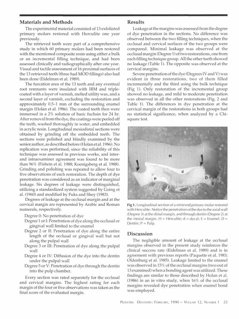
Severe penetration of the dye (Degrees IV and V) wasevident in three restorations, two of them filledincrementally and the third using the bulk technique(Fig 1). Only restoration of the incremental groupshowed no leakage, and mild to moderate penetrationwas observed in all the other restorations (Fig. 2 andTable 1). The differences in dye penetration at thecervical margin of the restorations in both groups hadno statistical significance, when analyzed by a Chi-square test.
Fig1. Longitudinal section of a retrieved primary molar restoredwith Herculite. Noticethe penetration ofthedyeto the axial wall(Degree 3) at the distal margin, and through dentin (Degree 2) atthe mesial margin. H = Herculite; d = dycal; E = Enamel; D =Dentin; P= Pulp.
DiscussionThe negligible amount of leakage at the occlusal
margins observed in the present study reinforces theclinical success rate (Eidelman et al. 1989) and is inagreement with previous reports (Paquette et al. 1983;Oldenburg et al. 1985). Leakage limited to the enamelwas observed in 15% of the occlusal margins (two out of13 examined) when a bonding agent was utilized. Thesefindings are similar to those described by Holan et al.(1986) in an in vitro study, when 16%c of the occlusalmargins revealed dye penetration when enamel bondwas employed.
PEDIATRIC DENTISTRY: FEBRUARY, 1990 ~ VOLUME 12, NUMBER 1 25

TABLE 1: Marginal Leakage at the Occlusal and Cervical Margins
Depth of DyePenetration
01 or I2 or II3 or III4 or IV5 o r V
Total0* + 1"2 + 3 + 4 + 5*"
Number of SurfaceIncremental
Occlusal
71
———-
880
Cervical
1—3211
817
s ExaminedBulk
Occlusal
71
————
880
Cervical
—232—1
826
* No dye penetration;of dve into dentin.
penetration of dye limited to enamel; *** penetration
Fig 2. Longitudinal section of another retrieved primary molarfilled incrementally with Herculite. No penetration of dye isevident at the mesial margin, and minimal leakage (Degree 1)can be observed at the distal margin. H = Herculite; d = dycal;E = Enamel; D = Dentin; P = Pulp.
In a study involving small restorations surroundedby enamel on facial tooth surfaces, smaller microgapswere found af ter incremental f i l l ing than afterapplication of the resin in bulk (Herrin and Berry 1986).An incremental filling technique did not improve thequality of the cervical margin in the present study. Thiscould be due to a combination of factors such as thinenamel, poor adherence of the material at the cervicalmargin, and the difficulty of condensation of thematerial to the gingival wall. In addit ion,polymerization contraction is related directly tomarginal leakage, and increases with the size of therestorations. Shrinkage of small restorations in facialsurfaces could be of lesser significance than in largeClass II restorations.
Donly and Jensen (1986) assessed the stresses createdby three d i f fe ren t f i l l ing techniques duringpolymerization of posterior resin restorations. They
found that a buccolingual filling techniqueresulted in significantly lower strain valuesthan bulk polymerization, but no significantdifferences were observed using agingivoocclusal incremental filling.
In the present study a gingivoocclusalincremental filling technique could noteliminate microleakage at the cervicalmargins of Class II composite restorationsplaced in primary molars. Dye penetrationwas observed in all but one of the cervicalmargins of all the teeth in both groups.
Dr. Fuks is an associate professor; Dr. Chosack is aprofessor; and Dr. Eidelman is a professor and chairman;all are from the Department of Pedodontics, HadassahFaculty of Dental Medicine, Hebrew University,
Jerusalem, Israel. Requests for reprints and all other correspondenceshould be addressed to: Dr. Anna B. Fuks, Department ofPedodontics, Hadassah Faculty of Dental Medicine, HebrewUniversity, Jerusalem, Israel, P.O.B. 12000.
Browne RM, Tobias RS: Microbial leakage and pulpal inflammation.A review. Endod Dent Traumatol 2:177-83,1986.
Donly KJ, Jensen ME: Posterior composite polymerization shrinkagein primary teeth: an in vitro comparison of three techniques.Pediatr Dent 8:209-12, 1986.
Eidelman E, Fuks AB, Chosack A: A clinical, radiographic and SEMevaluation of Class II composite restorations in primary teeth.Operative Dent 14:58-63, 1989.
Erickson J, Jensen ME: Effect of pressure cycling on microleakage ofcomposite restoration margins. J Dent Res 65:Abstract 895,1986.
Fisbein B, Holan G, Grajower J, Fuks AB: The effect of VLCScotchbond" and an incremental filling technique on leakagearound Class II composite restorations. ASDC J Dent Child 55:29-33,1988.
Fuks AB, Shey Z: In vitro assessment of marginal leakage of combinedamalgam-sealant restorations on occlusal surfaces of permanentposterior teeth. ASDC J Dent Child 50:425-29, 1983.
Fuks AB, Grajower R, Shapira J: In vitro assessment of marginalleakage of sealants placed in permanent molars with differentetching times. ASDC J Dent Child 51:425-27, 1984.
Fuks AB, Grajower R, Eidelman E: Assessment of marginal leakage ofClass II combined amalgam-sealant restorations in primarymolars. ASDC J Dent Child 53:343-45, 1986.
Going RE, Massler M, Dute HL: Marginal penetration of dentalrestorations by different radioactive isotopes. J. Dent Res 39:273-84,1960.
Herrin HK, Berry EA: Variables affecting the microgap of the enamel-composite interface. J Dent Res 65:Abstract 777, 1986.
Holan G, Fuks AB, Grajower R, Chosack A: In vitro assessment of theeffect of Scotchbond" on the marginal leakage of Class IIcomposite restorations in primary molars. ASDC J Dent Child53:18892, 1986.
Jensen ME, Chan DCN: Polymerization shrinkage and microleakage,in posterior composite resin dental restorative materials,Vannherle and Smith, DC editors, Minnesota Mining Mfg. Co.,1985. St. Paul, MN, pp 243-62.
Koenigsberg S, Fuks A, Grajower R: The effect of three fillingtechniques on marginal leakage around Class II compositerestorations in vitro. Quintessence Int 20:117-121,1988.
LeclaireCC, Blank LW, Hargrave LW, Pellen GB: A 2-stagecompositeresin fi l l technique and microleakage below the CEJ. J Dent Res65:Abstract 799,1986.
26 CLASS II COMPOSITES IN PRIMARY TEETH: FUKS, CHOSACK, AND EIDELMAN

LuiJL, Shigeyuki M, Setcos JC, Lutz F, Schwartz ML, Phillips RW:Margin quality and microleakage of Class 1| composite resinrestorations, J Am Dent Assoc 114:49-54, 1987.
Moore DH, Vann WF: A method to study marginal leakage ofposterior composite restorations. J Dent Res 65:Abstract 778,1986a.
Moore DH, Vann WF: Effect of the cavosurface bevel on posteriorcomposite marginal leakage. J Dent Res 65:Abstract 898, 1986b.
Oldenburg TR, Vann WF, Dilley DC: Composite restorations forprimary molars: two-year results. Pediatr Dent 7:96-103, 1985.
Paquette DE, Vann WF, Oldenburg TR, Leinfelder KF: Modifiedcavity preparations for composite resins in primary molars.Pediatr Dent 5:246-51, 1983.
Strange DC, Hembree JH: Comparison of three techniques formeasuring marginal leakage of restorative materials. J Dent Res66:Abstract 1494, 1987.
What if OSHA pays a visit?
OSHA has authority over two areas in the dental office: hazard communication (right-to-know laws) and infection control.
The hazard communication rule sets standards designed to ensure that products containinghazardous chemicals are handled properly by employees. The rules requires chemical manufac-turers and importers to convey hazard information to employers through labels on containersand material safety data sheets. All employers must pass this information on to their employees.
In the area of infection control, OSHA is utilizing its general authority to regulate workplacesafety by imposing basic infection control requirements on dental workers, as well as on otherhealthcare workers.
In order to enforce its workplace regulations, OSHA is authorized by statute to conductworkplace inspections. Because of the large number of workplaces, inspections of dental officeswill involve almost exclusively those offices in which an employee complains to OSHA.
If an OSHA inspector makes an unannounced visit to a dental office to make an inspection,the dentist has the right to demand that the inspector obtain a warrant prior to entry. Theadvantages of requesting this warrant are that the warrant should define the scope of the inspec-tion; and the time it takes the inspector to get a warrant may allow the dentist more time toprepare for the inspector’s arrival. The disadvantage is that the inspector may have, or mayacquire, a broadly worded warrant allowing widespread inspection.
OSHA determines penalties based on the seriousness of the violation, the employers’ goodfaith in attempting to comply with the regulations, size of the business and history of previousviolations. A citation informs the dentist employer and employees of the alleged violations,proposed penalties, and how and when to correct the problems. The citation, which may includea fine, is mailed to the employer from the OSHA area office. An OSHA booklet that explains theemployers’ rights and responsibilities following an inspection should be included with thecitation.
The dentist employer has 15 days from the time any citation and proposed penalty arereceived to contest either or both. Any notice of citation must be posted in a prominent locationin the workplace for employees to see, or must be given personally to each employee, regardlessof whether the employer intends to contest the matter.
OSHA poster 2203, "Job Safety & Health Protection," is available from any of OSHA’s tenregional offices. If you’re an employer of 11 or more employees (total of all employees in alloffices), you should also obtain a copy of OSHA form 200, an accident.
PEDIATRIC DENTISTRY: FEBRUARY, 1990 -- VOLUME 12, NUMBER 1 27