assessment of mitral left heart · withpredominant stenosis andsomewithpredominant regurgitation....
TRANSCRIPT

ASSESSMENT OF MITRAL VALVE DISEASE BYLEFT HEART CATHETERIZATION
BY
ERNEST W. HANCOCK*From the Cardiac Department, Guy's Hospital, and the Thorndike Memorial Laboratory and the Second and FourthMedical Services (Harvard), Boston City Hospital, and the Department of Medicine, Harvard Medical School, Boston,
MassachusettsReceived October 24, 1958
Many studies of the pressures in the left atrium and the left ventricle of patients with mitralvalve disease have been published. Initially obtained by direct puncture of the cardiac chambersat operation, such recordings have since been made possible by the development of techniques of leftatrial puncture as a diagnostic procedure before operation. Direct determinations of the left atrialpressure, the left ventricular diastolic pressure, the diastolic pressure gradient across the mitral valve,and the duration of the diastolic filling period render unnecessary several assumptions that mustbe made when mitral disease is assessed by right heart catheterization. When mitral stenosis isassociated with aortic valve disease, left ventricular failure, or significant mitral regurgitation, thelimitations of right heart catheterization may be great, and precise information may be obtainableonly by left heart catheterization.
Assessment of the relative importance of mitral stenosis and regurgitation when present in com-bination has remained a difficult problem. Various opinions of the diagnostic value particularlyof the left atrial pulse form (Gunning and Linden, 1958; Marshall et al., 1957; and Morrow et al.,1957) and of dye dilution curves (Korner and Shillingford, 1955; Shillingford, 1958; and Woodwardet al., 1957) have been expressed. The present report summarizes experience with left heartcatheterization in 75 patients with mitral valve disease, with particular reference to the assessmentof combined stenosis and regurgitation.
METHODSLeft atrial puncture was carried out by the right posterior transthoracic route (Biork et al., 1953) in the
prone position. A thin-walled cannula of 1-5 mm. outer diameter, containing a short-bevelled needle, wasused. The left ventricle was catheterized with a polyethylene catheter of 090 mm. internal diameter.Simultaneous left atrial and left ventricular pressures were recorded in many cases by means of a secondleft atrial needle. Pressures were recorded with strain gage (Statham P23D) or capacitance electromano-meters (Southern Insts. Ltd.) on a direct writing oscillograph (Sanborn) or a mirror galvanometer photo-graphic recorder (New Electronic Products Ltd.). Pressures were referred to a zero level at mid-thorax.
The mitral diastolic pressure gradient, defined as the mean difference between left atrial and left ventricularpressure during the diastolic filling period, was measured on superimposed pressure tracings, either as directlyrecorded (Fig. 1-2) or as redrawn. A significant mitral diastolic pressure gradient has been defined as agradient equivalent to at least 2 mm. Hg throughout the diastolic filling period. If left atrial and left ventri-cular pressures became equalized during diastole, there has been considered to be no significant gradient,although such patients sometimes showed a small gradient for the initial 004-008 sec. of diastole (Fig. 8A).
Mitral valve area was calculated from a modification of the formula of Gorlin and Gorlin (1951).Diastolic filling period was measured directly on superimposed left atrial and left ventricular pressuretracings, rather than on the brachial arterial tracing, and the directly measured mitral diastolic pressure* Research Fellow of the American Heart Association, and Research Fellow, Cardiac Department, Guy's Hospital.
389
on 21 March 2019 by guest. P
rotected by copyright.http://heart.bm
j.com/
Br H
eart J: first published as 10.1136/hrt.21.3.389 on 1 July 1959. Dow
nloaded from
ERNEST HANCOCK
4 1 Second Po40-
20 -
LEFTATRIUM
LEFTVENTRICLE
ECG
0
,t PHONO
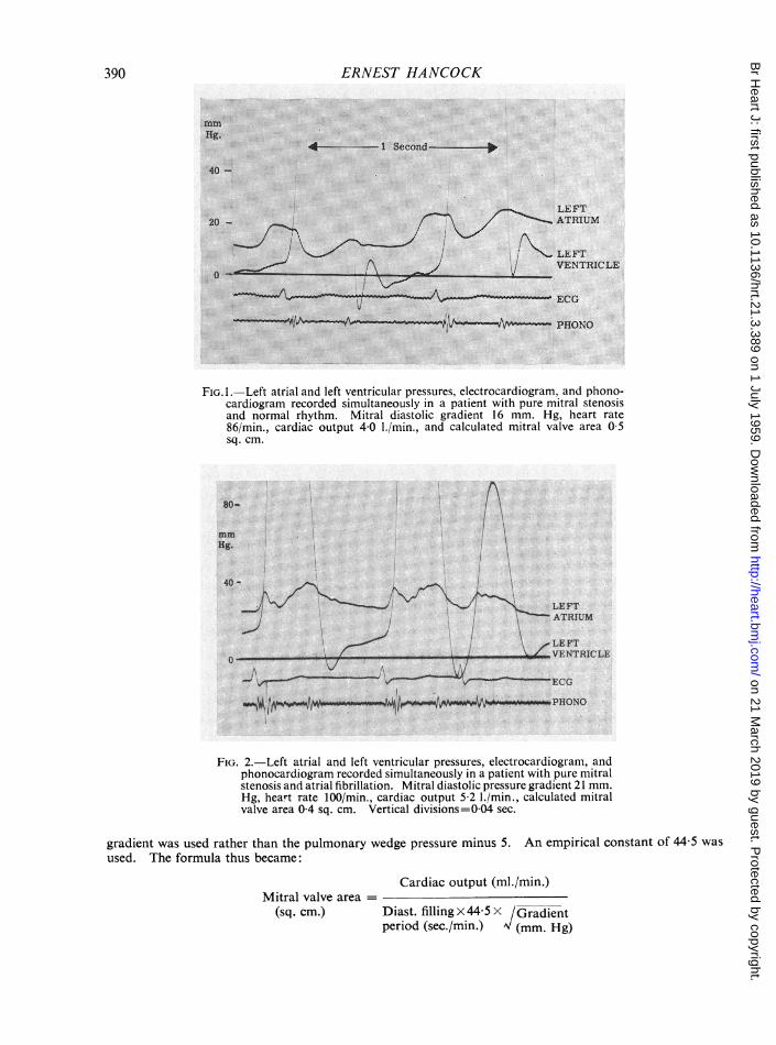
FiG.1.-Left atrial and left ventricular pressures, electrocardiogram, and phono-cardiogram recorded simultaneously in a patient with pure mitral stenosisand normal rhythm. Mitral diastolic gradient 16 mm. Hg, heart rate86/min., cardiac output 40 1./min., and calculated mitral valve area 0 5sq. cm.
80-
mm
Hg.
40-
_LE FT\ ATRIUM
zLEFTVENTRICLE
E CG
FIG. 2.-Left atrial and left ventricular pressures, electrocardiogram, andphonocardiogram recorded simultaneously in a patient with pure mitralstenosis and atrial fibrillation. Mitral diastolic pressure gradient 21 mm.Hg, heart rate 100/min., cardiac output 52 1./min., calculated mitralvalve area 0-4 sq. cm. Vertical divisions=0 04 sec.
gradient was used rather than the pulmonary wedge pressure minus 5. An empirical constant of 44 5 was
used. The formula thus became:
Cardiac output (mi./min.)Mitral valve area =
(sq. cm.) Diast. filling x 44-5 X /Gradientperiod (sec./min.) (mm. Hg)
390
mm
on 21 March 2019 by guest. P
rotected by copyright.http://heart.bm
j.com/
Br H
eart J: first published as 10.1136/hrt.21.3.389 on 1 July 1959. Dow
nloaded from

LEFT HEART CATHETERIZATION
Mitral valve area at operation was determined by measurement of drawings made by the surgeon. Ingeneral, valves 1 cm. in length and admitting only a finger tip had areas of 1 0 sq. cm. or less. Valves 1-2 cm.in length, admitting a finger to the first joint with difficulty, were usually 1 O-1 5 sq. cm., and valves admittinga finger freely were greater than 1 5 sq. cm. The regurgitant jet at operation was judged 1+ if only a finelocalized jet, 2+ if a considerable stream but primarily felt only at the orifice, 3+ if a more powerful streampalpable widely in the atrium, and 4+ if very gross. The last category was reserved for the two patientsoperated on for pure mitral regurgitation.
Cardiac output was determined in most cases by the dye dilution technique. Multiple samples were takenfrom the brachial artery at intervals of two seconds following injection of Evans Blue dye into the leftatrium.
Technique and Complications. After initial trials of right and left lateral positions, the prone position hasbeen used exclusively. This is well tolerated even by patients who are severely orthopnceic when supine.Barbiturate, meperidine, or morphine premedication is used in addition to local anesthesia as deep as thepericardium. The appropriate intercostal space, usually the ninth, is located fluoroscopically as a finalpreliminary, and the needle is passed to the left atrium as described by Biork. A characteristic pulsatilesensation is always felt when the needle comes against the atrium, and no other guidance is needed. In-advertent puncture of the right atrium requires reinsertion of the needle in a more medial direction. Theleft atrium has been successfully punctured in every case.
The left ventricle has been successfully catheterized in all but three, with no failures in the last 60 con-secutive cases. This is facilitated by direction of the needle caudally as well as medially. Experience hasshown that 15-20 minutes of manipulation with constant oscilloscopic observation of the pressure wave formresults in successful catheterization of the left ventricle in almost every case.
There have been no deaths attributable to the procedure, and no instance of cardiac tamponade. Slightbleeding from the left atrium occurs, as sero-sanguinous pericardial fluid is found at subsequent operationin the great majority. Injury to the right lung, usually by the preliminary local anrsthetic needle, hasresulted in pneumothorax in eight and hemoptysis in eight cases: the pneumothorax would usually goundetected were not a chest X-ray taken as a routine immediately after the procedure. Neither of thesecomplications has ever been severe enough to require treatment. There has been only one instance of eachin the last 35 cases.
Mild pleuritic pain in the right anterior chest for 12-24 hours is a usual side effect. Severe pain is veryuncommon. Clinical evidence of pericarditis has not been seen, although in three patients in whom opera-tion was delayed for 6-12 weeks after the procedure, a vascular adhesive pericarditis of apparently recentorigin was found.
Arterial embolism has never occurred during the procedure, but has occurred in two cases, 3 and 5 dayslater. Both patients had established atrial fibrillation, one with multiple emboli previously. The embolus,to the superior mesenteric artery, was fatal in one: the other had a successful embolectomy from the aorticbifurcation, but died suddenly of unknown cause two weeks later. At autopsy, both had mural thrombusin the left atrial appendage, but none at the site of puncture, which was scarcely visible.
Atrial fibrillation began during the procedure in one of 20 patients with normal rhythm, and began thefollowing day in two others. All three had a simultaneous right heart catheterization. Ventricular fibrilla-tion or arrest has not occured.
CLASSIFICATION OF PATIENTS
The patients have been divided into three groups, according to the nature of the mitral valvelesion: (1) pure mitral stenosis, 35 cases, (2) combined mitral stenosis and regurgitation, 24 cases,and (3) pure mitral regurgitation, 14 cases. Patients with a significant gradient across the valveand no regurgitation clinically or at operation were included in group 1. Patients with a significantgradient across the valve and also regurgitation were included in group 2, which thus included somewith predominant stenosis and some with predominant regurgitation. Patients have been includedin group 3 if mitral regurgitation was diagnosed clinically, and no significant pressure gradientfound across the valve. Two patients with clinical signs of pure mitral stenosis but no significantpressure gradient are considered separately.
The presence of a significant diastolic pressure gradient across the mitral valve, as previouslydefined, appears to be diagnostic of at least a certain degree of anatomical mitral stenosis. It
391
on 21 March 2019 by guest. P
rotected by copyright.http://heart.bm
j.com/
Br H
eart J: first published as 10.1136/hrt.21.3.389 on 1 July 1959. Dow
nloaded from

ERNEST HANCOCK
probably implies a valve area of 3 sq. cm. or less. Three of the patients found to have no gradienthave later come to autopsy, and each has had a valve area of at least 3 5 sq. cm., the orifice admittingthree fingers (Fig. 3). Thus no evidence has been found that mitral regurgitation is associatedwith a significant diastolic gradient unless there is also some degree of stenosis.
FIG. 3. The mitral valve of a patient with pure mitral regurgitation. There wasno diastolic pressure gradient across the valve. At operation, for mitralregurgitation, a gross (4+) regurgitant jet was present.
ASSESSMENT OF THE DEGREE OF STENOSISThe calculated valve area in pure mitral stenosis correlated closely with that observed at operation
(Fig. 4) and there were no discrepancies of clinical importance. The gradient across the valve wasnot nearly so well correlated with the degree of stenosis, and the left atrial pressure even less well.Thus, the desirability of simultaneous determination of cardiac output with the pressure measure-ments, to allow calculation of the mitral valve area, is reaffirmed. The necessity of flow measure-ment is particularly great when the gradient is small, as this may be due to mild stenosis with anormal or high flow, or to severe stenosis with a low flow.
In combined stenosis and regurgitation, assessment of the degree of stenosis is less certainbecause of the unknown factor of regurgitant flow. As this cannot be measured, the mitral valvearea has been calculated simply from the cardiac output, as in pure stenosis. In the majority ofpatients with combined stenosis and regurgitation in whom there is anatomical confirmation, thecalculated valve area proved to be a reasonably accurate prediction (Table I). This suggests thatregurgitant flow is only a small fraction of the cardiac output in these patients. Indeed, the degreeof stenosis in this group was in the same range as in those with pure stenosis, even though clinicalsigns of regurgitation were often very prominent, and the regurgitant jet at operation 2-3 +.Three patients, however, proved to have valve areas more than twice the predicted size (Fig. 4).They must have had predominant regurgitation, and a much greater backflow than the other 11,so that use of the systemic cardiac output alone in calculating valve area underestimated the actualforward flow across the mitral valve and hence underestimated the valve size.
Thus, in patients with the clinical signs of mitral stenosis and regurgitation, investigated withpresent techniques of left heart catheterization, a division may first be made into (1) those with nopressure gradient across the valve, therefore having mitral regurgitation without significant stenosis,
392
on 21 March 2019 by guest. P
rotected by copyright.http://heart.bm
j.com/
Br H
eart J: first published as 10.1136/hrt.21.3.389 on 1 July 1959. Dow
nloaded from

LEFT HEART CATHETERIZATION
L'n
U
LUJ
-i
.oO
1.o
2.0 1.0
MITRAL VALVE AREA, AT OPERATION (SQ. CM.)Z.o
FIG. 4.-Calculated mitral valve area, on the ordinate in sq. cm., and mitral valve area observed at operation,on the abscissa in sq. cm. Pure mitral stenosis, solid circles, on the left, and combined mitral stenosis andregurgitation, open circles, on the right. Mitral valve area calculated from the systemic cardiac outputonly, without regard for regurgitant flow.
TABLE ICOMPARISON OF CATHETERIZATION AND OPERATION OR AUTOPSY FINDINGS IN PATIENTS WITH COMBINED MITRAL
STENOSIS AND REGURGITATION
Mitral valve area,sq. cm. Regurg. Downslope of
Patient jet at dye dilutionoperation curve. (sec.)
Calc. Obs.
V.B. 09 09 + + 9-2H.H. 1 5 1-3 + + 6-2E.P. 1 2 0 9 + + 12-2M.R. 0-7 0-6 + + 18-2M.W. 1-2 10 (PM) 101L.M. 0-6 0-8 + + 18 4E.H. 0-8 0 7 + + 16 2A.S. 0 3 0 5 (PM) 28-8A.N. 10 10 ++ 7-8M.B. 12 10 + 13 6E.C. 14 1-6 +++ 19 5A.M. 1 2 2 5 + + + 13-1J.S. 0-6 1-7 ++ + 41-6I.J. 0 8 2-0 + + + 51 8
and (2) those with a significant gradient, who have a combination of stenosis and regurgitation.In the latter group, calculation of the mitral valve area on the basis of the systemic cardiac outputwill yield in most cases a reasonably accurate prediction of the degree of stenosis, but in a few cases,the regurgitant flow will be so large that the valve size will be significantly underestimated. In anycase, the calculated valve area may be considered a minimal valve size, potentially an underestimateaccording to the degree of regurgitation judged to be present. For example, if the calculated valvearea is 0-4 sq. cm., it is evident that stenosis is severe, as the valve size would still be less than
0~~~~~~~~~~
'00. 0~~~~~~~~06\* *\ 0
* \ a
* "\ 0\* ". "0
0',
01
* "S-MMS " MS-MR
I ,
393
on 21 March 2019 by guest. P
rotected by copyright.http://heart.bm
j.com/
Br H
eart J: first published as 10.1136/hrt.21.3.389 on 1 July 1959. Dow
nloaded from

394 ERNEST HANCOCK
1 sq. cm. if regurgitant flow exceeded the forward output. On the other hand, if the calculatedvalve area is 2-5 sq. cm., it is obvious that stenosis is not severe whatever the regurgitant flow.
In spite of the limitation imposed by inability to measure regurgitant flow, left heart catheteriza-tion has been of much value in assessing patients with combined stenosis and regurgitation. Of33 patients with this clinical problem in the present group, nine proved to have no gradient acrossthe valve, 10 had calculated valve areas in excess of 1'5 sq. cm. and have not been operated on, and14 have been operated upon for mitral stenosis on the basis of a substantial pressure gradient and acalculated valve area less than 1-5 sq. cm. The findings in the patients confirmed at operation areshown in Table I. Only three showed a disappointingly slight degree of stenosis at operation, andeven in these valvotomy was considered worthwhile by the surgeon.
0
c3 0
E 0
0~~~~~~~~~~
R o o~~~~~~~~a 0 ~~~~~~~00'.3 * *~~~ ~0 0U ~~~~~~~~~~0
0 0
10 00 0
.0 ~~0
E* MS j MS-MR
1.0 2.0 1.0 2.0MITRAL VALVE AREA, AT OPERATION (SQ. CM.)
FIG. 5.-Mean mitral diastolic pressure gradient in mm. Hg on the ordinate; mitral valvearea observed at operation in sq. cm. on the abscissa. Pure mitral stenosis, solid circles,on the left; combined stenosis and regurgitation, open circles, on the right.
The concept of predominant stenosis or predominant regurgitation is difficult to apply to somepatients who seem to have a very significant degree of both. A patient with a mitral valve area of2-5 sq. cm. has, at the most, moderate stenosis. Yet, regurgitation may so increase the flow acrossthe valve as to require a pressure gradient of 15-20 mm. Hg. The resulting elevation of left atrialpressure, and the necessary further rise with effort, tachycardia, etc. is a hemodynamic fault identicalwith that of tight mitral stenosis. While surgical relief of the stenosis alone may be beneficial, sucha valve is generally much less favourable than one with tight stenosis. While the decision to operatefor mitral stenosis involves many considerations, the presence of anatomically severe stenosis isobviously one of the most important. It is not enough merely to demonstrate a high left atrialpressure or a substantial gradient across the valve. A calculation of the valve area and someestimate of the degree of regurgitation are required in order to make the best possible predictionof the actual valve size.
LEFT ATRIAL PULSE FORMMany ways of analyzing the left atrial pulse form have been proposed to distinguish predominant
stenosis from predominant regurgitation. Most of these are based on the fact that in mitralregurgitation the systolic v wave is higher, the systolic x descent less prominent, the early diastolicy descent more rapid, and the diastolic and mean pressure lower than in mitral stenosis (Fig. 6).
on 21 March 2019 by guest. P
rotected by copyright.http://heart.bm
j.com/
Br H
eart J: first published as 10.1136/hrt.21.3.389 on 1 July 1959. Dow
nloaded from

LEFT HEART CATHETERIZATION
PURE STENOSIS STENOSIS PLUSREGURGITATION PURE REGURGITATU01q
40 4 4TjtVV7T:..
A A
deind or an tw grup fro one anthe. Ingnea, threare. dsicveiffencsin th
left atrial pulse forms of patients with no significant gradient across the valve as opposed to thosewith a gradient. The diastolic portion of the tracing is the most useful. Of those patients withatrial fibrillation, the left atrial pressure showed a continuous fall throughout diastole in 33 of 38patients with a significant gradient across the valve, whereas in 15 of 16 patients without a gradient,left atrial pressure remained the same or rose during diastole. It is this phenomenon that in adamped and delayed form is reflected in the Ry/V ratio in the pulmonary wedge tracing (Owen andWood, 1953), and is a useful guide to the presence or absence of mitral obstruction, i.e. a gradientacross the valve. It does not, however, necessarily distinguish predominant stenosis from pre-dominant regurgitation.
The systolic x descent was present in the majority of patients in all groups. As the deflectionwas less than 3 mm. Hg in most of those with pure mitral stenosis, it was difficult to recognizeimpairment of the x descent as a feature of regurgitation. A large systolic wave with no discerniblex descent was a sign of regurgitation, however, occurring in 5 of 11 patients with pure regurgitation,2 of 20 with combined stenosis and regurgitation, and none of 19 with pure stenosis, consideringonly those with atrial fibrillation. Only one of these 7 patients had predominant stenosis.
. l 1 . ,+vvti
4~~ ~ ~~~~i4
FIG. 7.-Leftatical adt lef vnr ina3 tient wit c mifitbriaIstenosi andyretogrgonp Miotra diastol.ps gradient '1t m. Hg,.caiac outputn44 1 the
cetaralcpulated mitral valv parieat0w6tsqcm. Miniitraltgainarshvalvearestoeaio7s.c.reguritan jto3+os
withagraien.< The diasto....lipoto of th trcn is the !mo:.s~t.usef-u.l. Of ths paiet with
paiet with' a significant gradien acos th vave whra in of 16 paietwIthout aEgrdint--t:letatil:^{tprssrereaie the sam or rosedurin-diasol. I^^..t is this phnmeo tha in adape and delaedXfor is relete in th Ry V rai in th pulmonary-weg train -(OIwe and-Woo, -l953adia usful gud to th prsec or abec f mira obtucin i.e. a grdin
ac-*:-rosthe+valve. I^'t-doe jn--t', however '', necesaril ditigus prdmnn stnoi--from pr-ido Inan 1ttreugta}tio. i. IX.The sys-toli xtdesce-k'n''vt--twas wp tresent'in--the majorit of ptientIsin al grus As th deflec~ tio
was}lesta 3 m.Hgin mot of thos with pur mItrl stenosi, it was difcl to....................:.recogn. ;4;!1izimpairment of -thex esen as'-.a............... .fe-^turo reugtation A-----lag sysoli wav wit no discrnible
xIGdesen Left atrialgnd leftregurgituatin howeessres occurrdeicnseuivinafpatients withpureinegurgitratin2o20wtcobndstenosisand regurgitation,Mitra diasoli ofressthurestenonsismmH,cardacoupud44e./ingonlycthosedwitatrial firlveation- s. Only oneralfvalveeareaatioeratsohad predominanseugtentojei3s.
395
on 21 March 2019 by guest. P
rotected by copyright.http://heart.bm
j.com/
Br H
eart J: first published as 10.1136/hrt.21.3.389 on 1 July 1959. Dow
nloaded from

396 ERNEST HANCOCK
Of the three patients who were found at operation to have severe regurgitation with onlymoderate stenosis, two showed evidence of regurgitation in the left atrial pulse form, i.e. a largepositive systolic wave with no x descent in one (Fig. 7), and flat pressure during diastole in the other.
ASSESSMENT OF REGURGITATION BY DYE DILUTION CURVESMuch attention has been given to the use of dye dilution curves in the assessment of valvular
regurgitation since the observations of Korner and Shillingford (1955) that valvular regurgitationresults in a lower peak concentration and a more gradual disappearance slope of the dye curvethan would be expected. Attempts to measure regurgitant flow by this method have been unsatis-factory, however. The form of dye dilution curves is affected by many factors, including thecardiac output, velocity of the circulation, site of injection, and the size of various cardiac chambers,particularly the size of the left atrium in the case of mitral regurgitation.
Fig. 8 shows the disappearance slopes of the dye dilution curves of the patients in the presentstudy, expressed as the number of seconds required for a tenfold drop in concentration on thedownslope, when extrapolated as a straight line on semilogarithmic paper. This measurement isthus independent of absolute units of concentration. More complicated indices, involving peakconcentration, appearance time, central volume, cardiac output, etc. have not proved more usefulthan this simple expression of the downslope. With this and other indices expressing similaraspects of the shape of dye dilution curves there is considerable overlap among patients with and
30sk
Zo 0
FIG. 8.-Frequency distribution of the downslope of thedyedilution curve, expressed as the number ofsecondsrequired for a tenfold fall in concentration. Puremitral stenosis, left; combined stenosis and regur-gitation, centre; pure mitral regurgitation, right.
0
0
0
C.0
* 30
0* 0
0
r .* r
on 21 March 2019 by guest. P
rotected by copyright.http://heart.bm
j.com/
Br H
eart J: first published as 10.1136/hrt.21.3.389 on 1 July 1959. Dow
nloaded from

LEFT HEART CATHETERIZATION
without regurgitation. The shape of the curve may be valuable evidence in assessing the degreeof regurgitation, but is not necessarily reliable in an individual case.
The downslope of the dye dilution curve is systematically steeper in patients with pure mitralstenosis than in those with mitral regurgitation. There is, however, no systematic difference betweenpatients with pure regurgitation and those with combined stenosis and regurgitation, although themajority of the latter have predominant stenosis. In other words, in a patient with mitral regurgita-tion, the contour of the dye curve has little or no correlation with the presence or absence ofsignificant mitral stenosis in that patient. The data in Fig. 8 might suggest that mitral regurgitantflow is as large on the whole when there is associated stenosis of marked degree as it is in pure
mitral regurgitation. On the other hand, it may be that the shape of the dye dilution curve is reflect-ing primarily the degree of left atrial enlargement, which tends to be greater in mitral regurgitation,with or without stenosis, than in pure stenosis.
SPECIAL FEATURES OF PURE MITRAL REGURGITATION
Left heart catheterization has made it possible to define the clinical entity of pure mitral regurgita-tion on a more objective basis than was previously possible-namely, the entity of mitral regurgita-tion with no significant diastolic pressure gradient across the valve. Excluding those with aorticvalve disease and those with regurgitation that was surgically induced, there were 11 suchpatients. Most were advanced cases with much cardiac enlargement, and chronic or recurrentcongestive heart failure. Eight had atrial fibrillation. The characteristic physical signs includeda normal or soft first heart sound at the apex, a loud apical holosystolic murmur, and an apicaldiastolic rumbling murmur, soft and diminuendo but often of considerable length. An openingsnap was present in only one, and that possibly tricuspid rather than mitral in origin, as a smallpressure gradient was demonstrated across the tricuspid valve. Electrocardiograms usually showedlow voltage, an intermediate or horizontal QRS axis, and some evidence of both right and leftventricular hypertrophy in the pracordial leads. Right ventricular hypertrophy was doubtlessdue to the pulmonary hypertension which was significant in six of the seven in whom right heartcatheterization was carried out. The pulmonary vascular resistance ranged up to 8-5 units. Thesystemic cardiac index ranged from 1 6 to 4-3l./min./m2. The three patients with normal rhythmall had P mitrale, but did not have giant a waves in the left atrial pulse form and did not havecrescendo presystolic murmurs.
These patients were, of course, selected for study because it was suspected that importantmitral stenosis might be present. Patients with more classical signs of pure mitral regurgitation,e.g. a loud systolic murmur only with a third heart sound and left ventricular enlargement, havenot been studied.
MITRAL STENOSIS OF NO PHYSIOLOGICAL SIGNIFICANCE
As in aortic stenosis, the clinical signs of mitral stenosis may be present when the lesion is so
mild as to be undetectable by direct physiological assessment. Two patients have been seen withclinical signs of pure mitral stenosis, in whom no significant mitral diastolic pressure gradientcould be demonstrated. Clinical features in the two patients were similar. Both were women
50 years of age, who had atrial fibrillation, and who complained in a neurotic manner of palpitationand to a lesser extent of dyspncea and fatigue. Both had a faint but prolonged mitral diastolicrumbling murmur, and one had a loud opening snap of the mitral valve (Fig. 9). Both had normalheart size, but slight left atrial enlargement. The cardiograms were normal except for atrialfibrillation. An obstructive lesion having been ruled out, it was concluded that there was onlymitral valvulitis of little or no functional significance, and that symptoms were due to atrial fibrilla-tion and psychoneurosis, and possibly myocardial insufficiency. The left ventricular end-diastolicpressure was normal in one (5 mm. Hg) and slightly raised in the other (13 mm. Hg).2D
397
on 21 March 2019 by guest. P
rotected by copyright.http://heart.bm
j.com/
Br H
eart J: first published as 10.1136/hrt.21.3.389 on 1 July 1959. Dow
nloaded from

3V8ERNEST HANCOCK
tX~~~~~~~~~~~~~~~~~~~4.
Sc 0
20-
ECG
4 .--- l Second -
ECG
*~~~~~~~~~~~~~~~c1 S.
3RD. ICS
APEX W \ U
FIG. 9.-(A) Consecutive left atrial and left ventricular pressures in a patientwith clinical signs of mitral stenosis but no significant pressure gradientacross the valve. (B) Phonocardiogram of the same patient, showing theopening snap of the mitral valve.
THE MURMUR OF MITRAL STENOSIS
It has already been mentioned that a long rumbling mitral diastolic murmur may be present inthe absence of a significant pressure gradient across the valve, with or without associated regurgita-tion. There is only a rough correlation between the loudness of the rumble and the size of thegradient. Five patients have been seen with no audible diastolic murmur in spite of a large gradient.In three of these, mitral stenosis was an incidental discovery during left heart catheterization forthe investigation of aortic stenosis. Two of those with no murmur had atrial fibrillation, pulmonaryhypertension, and congestive heart failure, circumstances in which disappearance of the murmurof mitral stenosis is well-recognized. In two, with normal rhythm and associated aortic stenosis,short presystolic murmurs were perhaps obscured by the loud first sound and aortic systolic murmurfollowing. The fifth had pure mitral stenosis, in normal rhythm without heart failure, but withpulmonary hypertension.
The long recognized influence of cardiac rhythm on the murmur of mitral stenosis is well docu-mented by pressure records. In normal rhythm, with a crescendo presystolic murmur, atrialcontraction produces a giant a wave in the left atrium, and the gradient is abruptly increased in theimmediate presystolic period (Fig. 1). In atrial fibrillation, with a long diminuendo diastolic rumble,the gradient is always largest at the beginning of diastole, and diminishes steadily to its smallestvalue in end-diastole (Fig. 2). It has been difficult to correlate the length of murmurs as heard orrecorded phonocardiographically with the presence or absence of a gradient persisting throughoutdiastole. In several cases the murmur has seemed clearly to end before the first sound, yet a largegradient was present throughout diastole. Likewise, in patients with normal rhythm and showingonly a short presystolic murmur, the gradient is not merely presystolic, but is substantial throughoutthe diastolic filling period.
308
on 21 March 2019 by guest. P
rotected by copyright.http://heart.bm
j.com/
Br H
eart J: first published as 10.1136/hrt.21.3.389 on 1 July 1959. Dow
nloaded from

LEFT HEART CATHETERIZATION 399
MYOCARDIAL FAILURE IN MITRAL VALVE DISEASEThe importance of myocardial function as opposed to the mechanical obstructive factor in mitral
valve disease has been controversial for a long time. Cardiologists generally agree that myocardialfailure is an important factor in many cases, but specific proof of this is difficult to obtain. It washoped that measurement of the left ventricular diastolic filling pressure by left heart catheterizationmight enable differentiation of patients suffering primarily from mechanically obstructive mitralstenosis from those suffering primarily from rheumatic heart disease with myocardial failure, inwhich case the left ventricular diastolic filling pressure might be raised. Studies of patients withaortic valve disease have often shown a gross rise of filling pressure in the left ventricle, and thepulmonary wedge pressure may be grossly raised in patients with heart failure due to hypertension,coronary artery disease, etc. In the present study, however, no patient with mitral stenosis has hadan end-diastolic pressure in the left ventricle higher than 14 mm. Hg, except one who also had severeaortic stenosis (Fig. 10). Only one patient with pure mitral regurgitation without significant aorticvalve disease had much rise of left ventricular filling pressure. The differences between the threegroups do suggest that, on the whole, mitral regurgitation is associated with higher filling pressuresin the left ventricle, and hence with left ventricular failure. However, the rise of left atrial pressure
15 [-
1.0 [-
0
FIG. 10.-Frequency distribution of the left ventricular end-diastolic pressure, in mm. Hg referred to mid-thoraciclevel. Pure mitral stenosis, left; combined stenosis andregurgitation, centre; pure mitral regurgitation, right.Solid circles represent patients without aortic valvedisease. Open circles represent patients with significantaortic stenosis as well as mitral valve disease.
0
o 0
o
*0
@0
0@
0*.-0*-*
o@ 0
* '-* *0
* *-o--- 0000 0
@90000 @ 0
* 00
@0000 .0
s H
on 21 March 2019 by guest. P
rotected by copyright.http://heart.bm
j.com/
Br H
eart J: first published as 10.1136/hrt.21.3.389 on 1 July 1959. Dow
nloaded from

ERNEST HANCOCK
in mitral regurgitation is generally not due to this, but rather (1) to the diastolic pressure gradientacross the valve resulting from the associated stenosis, or (2) to the v wave in systole, which rangesup to 65 mm. Hg in height.
Cardiac failure was always treated first, and left heart catheterization only undertaken when thesigns and symptoms of congestion were controlled as well as possible. Very few patients werestudied while in a manifest state of congestive heart failure, and this fact may have been the causeof failure to demonstrate rises of left ventricular diastolic pressure. It appears, nevertheless, thatleft ventricular myocardial failure in patients with mechanically significant mitral stenosis is un-common.
SUMMARY AND CONCLUSIONSLeft heart catheterization by the Biork technique has been carried out in 75 patients with mitral
valve disease. There have been no deaths or instances of cardiac tamponade due to the procedure.Hxemoptysis and pneumothorax occurred in 12 per cent, but less frequently with experience.Delayed arterial embolism occurred twice. The left atrium was successfully punctured in every case,and the left ventricle successfully catheterized in 96 per cent.
In pure mitral stenosis the severity of the lesion may be accurately assessed if the pressure gradientacross the valve and the cardiac output are determined together. Clinical signs of mitral stenosismay be present in the absence of a significant pressure gradient. Five patients had severe mitralstenosis in the absence of a diastolic murmur.
In pure mitral regurgitation there is no significant pressure gradient across the valve in diastole.These patients cannot always be distinguished from those with predominant mitral stenosis by theclinical signs. Diastolic murmurs and pulmonary hypertension are commonly present.
In combinations of mitral stenosis and regurgitation, large diastolic pressure gradients arecommonly present, even when regurgitation is the dominant lesion. Exact assessment of the valvesize in this group is limited by inability to measure regurgitant flow. Estimation of a minimalvalve area is of considerable value, and further information is gained from the left atrial pulse formand from dye dilution curves.
Left heart catheterization is a practical and useful diagnostic procedure in patients with mitralvalve disease in whom clinical assessment is difficult.
Surgical observations were made by Dr. D. E. Harken, Dr. G. W. B. Starkey, Dr. J. S. Strieder, Dr. I. Madoff,Mr. R. H. S. Brain, Mr. D. N. Ross, and Sir Russell Brock.
Dr. W. H. Abelmann, Dr. W. M. Madison, Jr., Dr. M. H. Proctor, and Dr. A. M. Johnson collaborated in thesestudies.
The guidance of Dr. Laurence B. Ellis and Dr. Charles Baker throughout this work is gratefully acknowledged.
REFERENCESBiork, V. O., Malmstrom, G., and Uggula, L. G. (1953). Ann. Surg., 138, 718.Gorlin, R., and Gorlin, S. G. (1951). Amer. Heart J., 41, 1.Gunning, A. J., and Linden, R. J. (1958). Circulation, 17, 354.Korner, P. I., and Shillingford, J. P. (1955). Clin. Sci., 14, 553.Marshall, H., Connally, D. C., and Wood, E. H. (1957). Circulation, 16, 913.Morrow, A. G., Braunwald, E., Haller, J. A., and Sharp, E. H. (1957). Circulation, 16, 399.Owen, S. G., and Wood, P. (1955). Brit. Heart J., 17, 41.Shillingford, J. P. (1958). Brit. Heart J., 20, 229.Woodward, E., Burchell, H. B., and Wood, E. H. (1957). Proc. Staff Meetings Mayo Clinic, 32, 518.
400
on 21 March 2019 by guest. P
rotected by copyright.http://heart.bm
j.com/
Br H
eart J: first published as 10.1136/hrt.21.3.389 on 1 July 1959. Dow
nloaded from