assessment report for scintimun - european …€¦ · · 2013-07-212.2 quality aspects ... •...
TRANSCRIPT
European Medicines Agency Evaluation of Medicines for Human Use
7 Westferry Circus, Canary Wharf, London, E14 4HB, UK Tel. (44-20) 74 18 84 00 Fax (44-20) 74 18 85 45
E-mail: [email protected] http://www.emea.europa.eu © European Medicines Agency, 2009. Reproduction is authorised provided the source is acknowledged.
Doc. Ref.: EMEA/743339/2009
ASSESSMENT REPORT
FOR
Scintimun
International Nonproprietary Name: besilesomab
Procedure No. EMEA/H/C/001045
Assessment Report as adopted by the CHMP with
all information of a commercially confidential nature deleted.

TABLE OF CONTENTS
1. BACKGROUND INFORMATION ON THE PROCEDURE........................................... 3 1.1 Submission of the dossier ........................................................................................................ 3 1.2 Steps taken for the assessment of the product.......................................................................... 3
2 SCIENTIFIC DISCUSSION................................................................................................. 4 2.1 Introduction.............................................................................................................................. 4 2.2 Quality aspects ......................................................................................................................... 5 2.3 Non-clinical aspects ................................................................................................................. 9 2.4 Clinical aspects ...................................................................................................................... 15 2.5 Pharmacovigilance................................................................................................................. 42 2.6 Overall conclusions, risk/benefit assessment and recommendation ...................................... 45
Page 2 of 48

1. BACKGROUND INFORMATION ON THE PROCEDURE 1.1 Submission of the dossier The applicant CIS bio international submitted on 02 July 2008 an application for Marketing Authorisation to the European Medicines Agency (EMEA) for Scintimun, through the centralised procedure falling within the Article 3(1) and point 1 of Annex of Regulation (EC) No 726/2004. The legal basis for this application refers to: Article 8.3 of Directive 2001/83/EC, as amended - complete and independent application. The application submitted is a complete dossier composed of administrative information, complete quality data, non-clinical and clinical data based on applicants’ own tests and studies and/or bibliographic literature substituting/supporting certain tests or studies. The applicant applied for the following indication: • Scintigraphic imaging for the detection or investigation of sites of inflammation and /or infection
in the following cases: − detection of chronic peripheral osteomyelitis − investigation of infected diabetic foot − detection of infected joint prosthesis − investigation of endocarditis when other imaging modalities are unconclusive − investigation of fever of unknown origin when other imaging modalities are unconclusive
• Scintigraphic imaging of bone marrow involvement (detection and extent of carcinoma metastasis,
as cold spots). Licensing status: Scintimun has been given a Marketing Authorisation in Czech Republic, Hungary and Sweden on 24 November 1993, 22 July 1993 and 22 April 1994, respectively. The Rapporteur and Co-Rapporteur appointed by the CHMP were: Rapporteur: Ian Hudson Co-Rapporteur: Concepción Prieto Yerro 1.2 Steps taken for the assessment of the product • The application was received by the EMEA on 02 July 2008. • The procedure started on 23 July 2008. • The Rapporteur's first Assessment Report was circulated to all CHMP members on 9 October
2008. The Co-Rapporteur's first Assessment Report was circulated to all CHMP members on 17 October 2008.
• During the meeting on 17-20 November 2008, the CHMP agreed on the consolidated List of Questions to be sent to the applicant. The final consolidated List of Questions was sent to the applicant on 20 November 2008.
• The applicant submitted the responses to the CHMP consolidated List of Questions on 23 April 2009.
• The Rapporteurs circulated the Joint Assessment Report on the applicant’s responses to the List of Questions to all CHMP members on 8 June 2009.
• During the CHMP meeting on 22-25 June 2009, the CHMP agreed on a list of outstanding issues to be addressed in writing and in an oral explanation by the applicant.
• Written explanations were provided by the Applicant on 24 August 2009. • Experts of the Scientific Advisory Group on Diagnostics (SAG-D) were convened on 7
September 2009 to address questions raised by the CHMP. • During the CHMP meeting on 21-24 September 2009, outstanding issues were addressed by the
applicant in an oral explanation.
Page 3 of 48

• During the meeting on 19-22 October 2009, the CHMP, in the light of the overall data submitted and the scientific discussion within the Committee, issued a positive opinion for granting a Marketing Authorisation for Scintimun on 22 October 2009. The applicant provided the letter of undertaking on the follow-up measures to be fulfilled post-authorisation on 16 October 2009.
2 SCIENTIFIC DISCUSSION
2.1 Introduction Inflammatory diseases are associated with non-specific and specific inflammatory markers. In clinical practice, confirmation of diagnosis of infections relies on a combination of clinical features, microbiological tests and imaging techniques. In case of suspected infectious and non-infectious inflammatory lesions, timely identification and localisation of pathology might be of critical importance in the appropriate management of patients. CT, MRI and ultrasound scans are usually relied upon for standard imaging tests but have their limitations as they provide morphological and anatomical information but do not inform the treating clinician on the nature of pathology. In addition, knowledge on potential localisation of the pathology is required in order to effectively use these techniques. Therefore, there is a need for a method to identify clustering of neutrophils to the site of infection or inflammation where the nature of the pathology requires further identification or where there are no apparent clinical features to suggest the location of the disease. The gold standard of labelling of neutrophils has been ex-vivo labelling using indium (111In) oxinate. This technique has some drawbacks as it carries various risks: misadministration to the wrong patient, contamination of the blood by environmental substances/organisms, and contamination of workers during radiolabelling. There are also other radiopharmaceuticals which have been used to detect infection/inflammation such as gallium (67Ga) citrate, technetium (99mTc) human immunoglobulin, technetium (99mTc) exametazime labelled leucocytes, or fludeoxyglucose (18F). Scintimun (technetium (99mTc) besilesomab) has the theoretical advantage of labelling neutrophils specifically in vivo. A similar product, an anti-NCA90 antibody (sulesomab; LeukoScan) is currently approved in Europe for “determining the location and extent of infection/inflammation in patients with suspected osteomyelitis, including patients with diabetic foot ulcers”. Besilesomab is a mouse monoclonal antibody of IgG1 kappa isotype that binds to antigenic structures shared by a surface glycoprotein NCA-95 (non specific cross-reacting antigen 95) of granulocytes and the tumour marker, carcinoembryonic antigen (CEA). In infection and inflammation, it is postulated that the accumulation of radiolabelled antibody bound to neutrophils provides ‘hot spots’ upon scintigraphic imaging. This medicinal product is for diagnostic use only. This medicinal product is for use in designated nuclear medicine facilities only, and should only be handled by authorised personnel. The indications initially proposed by the applicant were the following:
• Scintigraphic imaging for the detection or investigation of sites of inflammation and /or infection in the following cases: − detection of chronic peripheral osteomyelitis − investigation of infected diabetic foot − detection of infected joint prosthesis − investigation of endocarditis when other imaging modalities are unconclusive − investigation of fever of unknown origin when other imaging modalities are unconclusive
• Scintigraphic imaging of bone marrow involvement (detection and extent of carcinoma metastasis,
as cold spots).
Page 4 of 48

The current recommended proposed indication is the following: • This medicinal product is for diagnostic use only. Scintigraphic imaging, in conjunction with other
appropriate imaging modalities, for determining the location of inflammation/infection in peripheral bone in adults with suspected osteomyelitis. Scintimun should not be used for the diagnosis of diabetic foot infection.
The recommended activity of technetium (99mTc) besilesomab should be between 400 MBq and 800 MBq. This corresponds to the administration of 0.25 to 1 mg of besilesomab. For repeated use, see SPC section 4.4. 2.2 Quality aspects Introduction The drug substance besilesomab is a murine monoclonal antibody that specifically binds to NCA-95 (non specific cross-reacting antigen 95), an epitope expressed at the cell membrane of granulocytes and granulocyte precursors. Besilesomab is produced by conventional hybridoma technology using a murine myeloma cell line X63Ag8.653 and spleen cells from Balb/c mice previously immunised with CEA antigen (from human liver metastasis). A two-tiered cell banking system of Master Cell bank (MCB) and Working Cell Bank (WCB) is used for the manufacture of the drug substance. Fermentation and purification consist of a 13-step process. The fermentation process is operated by continuous perfusion to obtain a cell-free culture harvest which is then purified by a series of chromatography steps, virus inactivation and filtration steps and ultrafiltration/diafiltration steps. Manufacture of the drug product includes partial reduction of besilesomab to allow covalent binding of the radiolabel, filtration steps, formulation steps, freeze-drying of the formulated drug substance and filling into vial. The solvent to be used with the formulated drug substance is prepared and filled in a separate vial. Scintimun is presented as a kit for radiopharmaceutical preparation and is administered by intravenous injection following reconstitution with a saline solution of technetium 99m (99mTc). Drug Substance Nomenclature INN Name: besilesomab Compendial Name: not applicable Chemical name: monoclonal antibody BW 250/183 Laboratory Code Name: MAb BW 250/183 CAS Registry Number: 537694-98-7 Other Names: not applicable Description of the drug substance Besilesomab is a murine IgG1 kappa monoclonal antibody of 150 kDa. It binds to NCA-95 (non specific cross-reacting antigen 95), an epitope expressed at the cell membrane of granulocytes and granulocyte precursors. Besilesomab is glycosylated. Manufacture Cell banking, fermentation and harvesting, purification, Quality Control testing and batch release of besilesomab drug substance take place at GLYCOTOPE Biotechnology GmbH, Czerny-Ring 22; D-
Page 5 of 48

69115, Heidelberg, Germany. This site is EU-GMP compliant and a valid manufacturing authorisation was provided. Development genetics Besilesomab is produced by conventional hybridoma technology. Immune spleen cells and X63Ag 8.653 myeloma cells were mixed and the mixture was incubated in a medium containing foetal bovine serum. Following pre-selection of hybridoma, cloning and subcloning steps were performed to select one high-producing clone (MAb BW 250/183). Cell bank system A two-tiered cell banking system of Master Cell Bank (MCB) and Working Cell Bank (WCB) was developed and maintained in accordance to cGMP and ICH guidelines. Seed cells corresponding to the high-producing clone were adapted to growth in a medium containing foetal bovine serum, leading to the establishment of the original MCB and WCB. Procedures followed for the preparation of MCB and WCB were appropriately described. An extensive range of tests was performed for their characterisation, in accordance with ICH guidelines, including identity, viability, stability, presence of adventitious agents. Fermentation process Pre-culture is initiated from a single WCB vial and consists of the expansion in T-flasks followed by cultivation in spinner bottles. Cells are then transferred into a production bioreactor containing a chemically-defined medium. The fermentation is operated by continuous perfusion. The cell culture harvest is collected from the bioreactor and cells and particles are removed by micro-filtration. Purification process The downstream process encompasses chromatography steps, virus treatment steps and ultrafiltration/diafiltration steps. The final formulation of the purified besilesomab bulk is a phosphate buffered saline solution containing sorbitol as a stabilising agent. The bulk solution is stored frozen at less than -20 °C. Manufacturing process development and process validation A comprehensive process manufacturing history was provided, showing the different changes introduced, and the corresponding batches involved, as well as the use of these batches. Comparability studies were conducted to support the introduction of the manufacturing changes. The commercial manufacturing process was validated in order to demonstrate that the process consistently maintains process parameters within specified ranges and meets pre-established acceptance criteria for performance indicators. Process validation studies included the cell culture process, the purification process, resin re-use studies for chromatography steps, cleaning procedures. The history of the product which has been manufactured commercially since 1993 was relevant to the overall validation of the manufacturing process. Characterisation A) Elucidation of structure and other characteristics: A series of tests was performed to confirm the primary structure, molecular weight and higher order structure of the antibody. The carbohydrate structure was adequately characterised. Characterisation of biological activity of besilesomab was adequate. B) Impurities: Potential process-related impurities including cell substrate derived impurities (host cell proteins, host cell DNA), media components and downstream-derived impurities are adequately addressed and proposed specifications justified. Product-related substances correspond to aggregates and fragments.
Page 6 of 48
Specifications The drug substance release specifications, including tests for identity, impurities, potency, quantity and general attributes, are acceptable and well justified. Stability The design of the stability program, including the testing intervals and temperature storage conditions, are in accordance with current guidelines. The tests chosen are a subset of tests from the release specifications selected for stability-indicating properties. The stability data provided were within the specifications and support the proposed shelf life for the drug substance. Drug Product Pharmaceutical Development Scintimun is presented as a kit for radiopharmaceutical preparation intended for intravenous injection following reconstitution with a saline solution of technetium-99m (99mTc), which corresponds to the radiolabelling of besilesomab. Scintimun is intended to be administered once and one kit can be used for several patients. This kit is composed of one or two sets of two multidose vials: - Vial “Scintimun” (10 ml, colourless, type I glass vial, closed with chlorobutyl rubber stopper and aluminium crimped capsule (green)) containing besilesomab drug product: lyophilised powder in which the drug substance (1 mg/vial) is formulated with sodium dihydrogen phosphate and disodium hydrogen phosphate (buffer) and sorbitol (stabiliser). - Vial “Solvent for Scintimun” (6 ml, colourless, type I glass vial, closed with chlorobutyl rubber stopper and aluminium crimped capsule (yellow)) corresponding to a lyophilised powder containing propane tetraphosphonic acid (PTP) (tranchelator agent), tin (II) chloride (or stannous chloride) (reducing agent), sodium hydroxide and hydrochloric acid (pH adjustment). Stannous chloride is commonly used with technetium (99mTc) radiopharmaceuticals for the reduction of pertechnetate (99mTcO4-) into technetium (99mTc). Stannous chloride is known to interact with the disulphide bridges of an antibody; therefore, in order to prevent this, stannous chloride is chelated to 1.1.3.3 –propane tetraphosphonic acid, tetrasodium salt (PTP) and conditioned in a separate vial. PTP is the only non compendial excipient used in Scintimun. Its synthesis, characterisation, analysis and specifications were well documented; data provided to support the use of PTP were considered acceptable. The formulation of Scintimun has not changed since the first clinical studies performed by Behring. Adventitious Agents The only raw material of animal or human origin used for the manufacture of Scintimun is foetal calf serum (FCS) for the cryopreservation of the cell banks manufactured in 1998-2000. Ph. Eur. TSE certificates of suitability for FCS were provided. Safety concerning TSE is considered sufficiently assured. Control of potential contamination by other non-viral adventitious agents (mycoplasma, bioburden, endotoxins) was considered adequate. Extensive screening for viruses was performed. Raw material controls minimise the risk of viral contamination, and clearance studies provide assurance that any potential virus will be removed or inactivated by the manufacturing process. Types A and C endogenous retroviruses particles were detected in the MCB. However, no ERV particles were detected in bulk fermentation harvests from 5 validation batches with a threshold of detection of 1.4 x 10⁵ VLP/ml. Negative results on the XC plaque assay and the S+L- Focus assay (on
Page 7 of 48

the MCB) demonstrate that they are not replication competent retroviruses. Small scale validation of the capacity of the process to remove or inactivate these particles has been carried out using four model viruses. Validation studies demonstrated acceptable levels of virus reduction by the manufacturing process. Considering that the validation is most likely significantly underestimating the potential to inactivate these particles, and they have been shown to be replication incompetent, then the detection of these particles in the cell bank is not considered to result in a safety concern. Manufacture of the product The manufacture and quality control of the drug product (vial “Scintimun” and vial “Solvent for Scintimun”) are performed at a manufacturing site compliant with EU-GMP. CIS bio international, BP 32, 91192 Gif sur Yvette cedex, France, is responsible for EU batch release of the drug product. The manufacture of vial “Scintimun” comprises the following steps: The frozen formulated bulk drug substance is thawed, pooled, mixed, filtered and concentrated and purified. The antibody solution is then treated by a reducing agent whose excess is removed by ultrafiltration. This solution is then filtered and sorbitol is added. Following adjustment of besilesomab concentration, the antibody solution is sterile filtered and aseptically filled into vials. The solution is then freeze-dried. The flip-off caps are crimped onto the stoppered vials which are then labelled. The manufacture of vial “Solvent for Scintimun” starts with the preparation of an oxygen-free PTP solution, addition of a stannous chloride dihydrate solution, followed by pH and volume adjustment steps. The formulated solution is then sterile filtered and filled into vials. The solution is then freeze-dried. The flip-off caps are crimped onto the stoppered vials which are then labelled. The proposed critical (in-process control) and non-critical (monitoring) parameters sufficiently control the manufacturing process. In particular, reduction of the antibody as well as integrity and immunoreactivity of drug product and radioactivity of radiolabelled product in use are adequately controlled. Process validation provided a documented, thorough understanding of the ability of the manufacturing process to consistently and reliably meet predetermined product specifications and quality attributes. Adequate data were provided to support the changes to the drug product manufacturing process implemented during development. Product specifications The specifications for Scintimun include control for each component of the drug product, i.e. vial “Scintimun” and vial “Solvent for Scintimun”, and control of the reconstituted and radiolabelled product. Adequate tests are in place and the proposed specification limits are acceptable. Stability of the product Real-time and accelerated stability studies were initiated in accordance with ICH guidelines and per protocol to monitor the time-temperature stability of cGMP lots of drug product. On the basis of the data provided, the approvable shelf life for the drug product is two years at 2-8°C and 3 hours not above 25°C after radiolabelling. Discussion on chemical, pharmaceutical and biological aspects The generation of the original cell line, from the expression construct to the selection process was well described. Cell banks have been established and adequately characterised. The drug substance manufacturing process is well defined and adequately controlled, with appropriate in-process controls and acceptance criteria in place. The manufacturing process has been adequately
Page 8 of 48

validated using relevant parameters and good consistency has been shown. Reprocessing for the drug substance manufacturing process was considered acceptable provided that only one round of re-processing per batch is permitted and that in the event of a failure in sterility, the route cause is investigated prior to batch release. Comparability studies to support the introduction of several changes in the manufacturing process of the drug substance in 1999 and 2007 were considered adequate and the results satisfactory. The new pivotal Phase III study, conducted to address the major clinical objections raised with the initial marketing authorisation application, used “1999 material” and not “2007 product”. Although no specific extra validation was performed due to these changes (except virus removal validation), batch release results remain within specification and after considering the nature of the process changes, this was deemed sufficient to demonstrate comparability of commercial product to that used in the new clinical study. The stability data on drug substance from the 1999 and 2007 processes indicate that the proposed shelf life for the drug substance is acceptable. In relation to the drug product, the minor changes to the manufacturing process since the initial Phase III study are described and well supported by data. Analytical methods are described and acceptable. For this imaging reagent, an assay measuring percentage of immunoreactivity is described. This determines the proportion of besilesomab which binds to “antigen containing material” (ACM) derived from CaCo2 human colon tumour cells displaying the NCA95 and CEA epitopes, under defined conditions. Although the assay is acceptably defined and validated, the applicant was asked to justify the use of human tumour cells in a binding assay where the indication is binding to human granulocytes. This major objection was adequately addressed by demonstrating that the antibody crossreacts to ACM as well as granulocytes. The applicant adequately justified why the ACM antigen was chosen instead of human granulocytes, because with the technology available at the time of development, it would not have been possible to validate the granulocyte-detecting assay. The drug product specifications (vial “Scintimun” and vial “Solvent for Scintimun”) are acceptable. The binding of the antibody at the drug product level is not monitored or specified. Although the percentage of radioimmunoactivity is specified, this monitors the binding of radioactivity and does not directly measure antibody binding. This format was justified since it overcomes a disadvantage of directly detecting immunoreactivity of radiolabelled antibody by ELISA. This disadvantage (of using an ELISA as an immunoreactivity assay for radiolabelled product) is that only a very small proportion of the monoclonal antibody is actually radiolabelled. Most remains unlabelled. Therefore, if the act of radiolabelling the monoclonal antibody was to damage it and reduce/eliminate its immunoreactivity, this fact would be hidden because the cold antibody, which is present in a great excess, would continue to be as immunoreactive and the detection method cannot distinguish between labelled and unlabelled antibody. The proposed shelf life of 2 years for the drug product before reconstitution and 3 hours after reconstitution/radiolabelling is considered acceptable. Viral safety and safety concerning other adventitious agents including TSE are sufficiently assured. 2.3 Non-clinical aspects Introduction The product is a kit for radiopharmaceutical preparation, consisting of two vials, each containing a freeze-dried powder. The first vial contains 1 mg of the active substance, anti-granulocyte monoclonal antibody (besilesomab, BW250/183) and excipients sodium phosphate and sorbitol. The second vial contains 1,1,3,3-propane tetraphosphonic acid, tetrasodium salt, dehydrate (PTP), tin (II) chloride dehydrate, and sodium hydroxide and hydrochloric acid for pH adjustment.
Page 9 of 48

The antibody is a mouse monoclonal anti-granulocyte IgG1 antibody directed against an epitope of the membrane protein NCA 95 (non-specific cross-reacting antigen of 95kDa) on human granulocytes and has been chemically modified to allow effective labelling with the radionuclide technetium-99m. In the second vial, the chelator PTP couples the reduced technetium-99m-pertechnate onto the antibody. The non-clinical safety studies were conducted using the kits made up with decayed technetium. They were conducted in mice, rats and the cynomolgus monkey. The majority of the studies were conducted in the early 1990’s. Antibodies from different manufacturers were used for the Ames tests and a local tolerance study in rabbits. However, chemical and biological equivalence studies reported in the quality and clinical dossiers stated that the products (antibody and PTP) are chemically equivalent to that manufactured by previous manufacturers. The applicant has not requested any formal scientific advice from the EMEA. The non-clinical safety studies were conducted in compliance with GLP and in accordance with the guidance that at the time when the studies were undertaken. A GLP inspection was not considered necessary. Pharmacology The monoclonal antibody is a mouse IgG1 antibody with a molecular weight about 150 kDa. There are two identical kappa (κ)- light polypeptide chains (about 25 kDa each) and two identical γ1-heavy polypeptide chains (about 50 kDa each) linked together by disulfide bonds. There is limited information on the specificity of the antibody for granulocytes. The information is almost 20 years old and relates to an antibody manufactured by a different method from that intended for marketing. There is no further updated information on the specificity of the antibody. • Primary pharmacodynamics In a study investigating the specificity of binding to cryo-preserved human tissues using an indirect alkaline phosphatase anti-alkaline phosphatase (APAAP) technique, the antibody from hybridoma supernatants showed staining to cytoplasmic, membranous and interstitial areas of primary colon carcinoma tissue, to single granulocytic cells in normal human liver and lung and to a large proportion of granulocytic cells in normal human bone marrow but not to blood vessels or connective tissue. Additional tissues investigated that showed binding of the antibody to granulocytic cells were breast, kidney, parotid gland, pituitary, lymph nodes and spleen, as well as colonic, pancreatic, some lung and some breast carcinomas. A negative control using supernatants derived from a non-secretor myeloma cell line showed no such binding in the same tissues. The purified antibody and the prepared kit bound similarly to the granulocytes in normal bone marrow, lung, liver, spleen and colorectal carcinomas. The prepared kit produced some additional staining in some connective tissue fibres in normal lung, some muscle fibres in normal colon and in liver parenchymal cells. Binding of the antibody to blood cells from cynomolgus monkey was also investigated. The antibody bound strongly to granulocytes from peripheral blood from cynomolgus monkeys and from humans. • Secondary pharmacodynamics No secondary pharmacodynamic studies have been submitted. • Safety pharmacology programme Study No. 158-35 investigated safety pharmacology of the product (prepared kit with decayed Tc-99m generator) in anaesthetised dogs. Ascending intravenous doses of 0.5, 1.0 and 5.0mg/animal were administered at 5 minute intervals. There were no effects on cardiovascular or respiratory systems (including arterial pressure, heart rate, respiratory rate, respiratory volume, cardiac output and peripheral resistance), nor on haematology, coagulation or clinical chemistry parameters • Pharmacodynamic drug interactions No studies have been submitted.
Page 10 of 48

Pharmacokinetics Two studies were conducted. One was a combined dosimetry and pharmacokinetic study in rats. The second was a pharmacokinetic study in cynomolgus monkeys, the latter using the kit prepared with decayed technetium. In the rat pharmacokinetic and dosimetry study, radioactivity was measured in a scintillation counter or an ionisation chamber, depending on the level of activity. An ELISA was used for measurement of besilesomab in the monkey pharmacokinetic study. Absorption Pharmacokinetics were evaluated in cynomolgus monkeys in Study No. 158-07 following intravenous administration of the kit product with decayed technetium, at a dose of 0.5mg antibody/kg. Serum levels were highest at the first time point at 10 minutes, and were 14 and 15 µg/ml in the two animals, respectively. The distribution occurred within 20 minutes and the terminal half-life of the antibody was 26 and 36 hours in the two animals, respectively (Table 5). Table 5 Pharmacokinetics of technetium (99mTc) besilesomab in monkeys Study ID
Species N Dose (mg/kg)
Route Terminal t1/2 (h)*
Vd (ml)*
Cl (ml/min)*
AUC* (µg.h/ml)
158-07 Cynomolgus monkey 2 0.5 IV 25.5
35.6 162 156
0.098 0.068
355 412
* - only two animals used; values for both are provided In a rat pharmacokinetic study, the technetium (99mTc) besilesomab kit was prepared, and radioactivity in blood and tissues as well as that excreted in urine and faeces was measured after intravenous administration of 2.5µg of antibody with an activity of 4 to 5 MBq. Time points were 1, 7, 17, 30 and 48 hours after administration. At 1 hour, the highest levels of radioactivity were evident in the blood, followed by the liver, compact bone, kidney and small intestine. Elimination from most organs appeared to be biphasic with the half-lives of the initial and terminal phases being 2-7 hours and less than 100 hours, respectively. The plasma half-life was 30-35 hours, which was comparable with the value obtained for monkeys. In the kidney (both sexes) and the testes, radioactivity appeared to peak at 7 hours post dose. It remained at similar levels thereafter in the kidney but declined in the testes. This also occurred to a lesser extent in pancreas and thymus of male rats. Radioactivity levels also increased in skeletal muscle for both sexes (7 hours in males and 30 hours in females) and in the uterus and intestine (17 hours after the dose). Radioactivity was not detected in mammary gland. Distribution Distribution in normal animals (dosimetry) was investigated in rats. Most of the organs showed biphasic elimination with an initial half-life of 2-4h, followed by a slower phase with a half-life of 5-6h, the latter mainly determined by the half-life of the labelling nuclide Tc-99m. Blood and plasma values recorded were with a half life of 3-4 hours and 30 – 65 hours respectively. During the first 7 hours after administration accumulation of radioactivity is observed in kidneys and testes and with less magnitude in pancreas and thymus. An increase during the first seven hours was observed in skeletal muscle in male rats and thirty hours in female rats. In the uterus and intestine the maximal accumulation was 17 hours, and no detection was observed in the mammary gland. A GLP compliant pharmacokinetic study (study 158-07) was carried out in cynomolgus monkeys. The test compound was administered IV at 0.5 mg/kg. Only two animals we used during the studies (2 males). Blood samples where taken at baseline 10, 20, 30 min, 1, 3, 9, 15, 24. 48 hours, and examined IgG levels by ELISA. Distribution was achieved in 20 min followed by a monoexponential decrease with a terminal t1/2 of 26-36 hours. The distribution volume (162 and 156 ml) was below plasma volume. AUC (0-48 h) measured was 355 and 412 µg/ml x h respectively.
Page 11 of 48
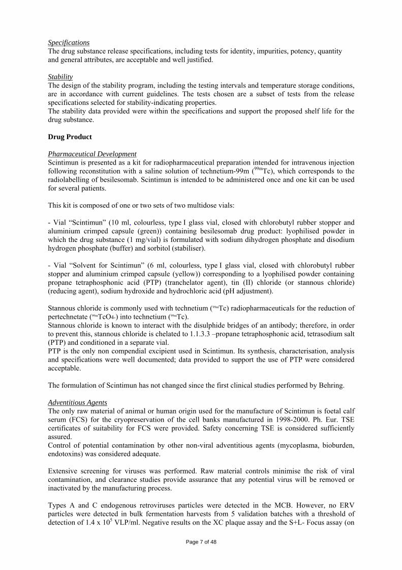
Metabolism The breakdown of antibodies has been reported in the literature. The murine antibody is expected to be broken down into individual amino acids similarly to other immunoglobulins. Information was provided on the effect of different radionuclides on the catabolism of antibodies, using a murine antibody labelled with iodine (131I), indium (111In), technetium (99mTc) or selenium (75Se), the latter being incorporated as a metabolic marker and behaving as an unlabelled MAb. The native and radiolabelled murine antibodies are initially taken up into the liver to a similar extent, but the retention of radioactivity in the liver is dependent upon the label used, with 75Se, 111In and 99mTc (with a high sulphydryl content) being retained for 5 days and 99mTc (with a low number of sulphydryl groups) and 131I showing decreasing within 2 days. Excretion The elimination of radioactivity from the body was measured in the rat pharmacokinetic study. Over 30 hours, 31-34% of the radioactivity was excreted in the urine and 7-13% in the faeces. Faecal elimination was noted mainly from the 17h time point. Pharmacokinetic drug interactions No studies have been submitted. Toxicology • Single-dose toxicity Acute toxicity studies were conducted in mice and rats. Results are shown in Table 6 Table 6 Acute toxicity in mice and rats Study ID Species/
Sex/Number/ Group
Dose/Route Approx. lethal dose / observed max non-lethal dose
Major findings
158-11 Mouse 5/sex/group
0, 0.5, 1, 5 mg/kgIV
Max non-lethal dose > 5mg/kg None
158-12 Rat 5/sex/group
0, 0.5, 1, 5 mg/kgIV
Max non-lethal dose > 5mg/kg None
• Repeat-dose toxicity (with toxicokinetics) Repeated-dose studies were conducted in rats and cynomolgus monkeys. The results are summarised in Table 7 Table 7 Repeat-dose toxicity in the rat and cynomolgus monkey Study ID Species/Sex/
Number/Group Dose/Route Duration NOEL/ NOAEL
(mg/kg/day) Major findings
158-42 Rat 5/sex/group
0, 0.5, 1, 2 mg/kg/day IV
30 days
Dose-dependent round cell infiltration in the liver
6555 TSP Cynomolgus monkey 3/sex/group
0.1, 0.5, 1.0 mg/kg/day IV
30 days NOEL 0.5mg/kg/day
Increased spleen weight in males at 1mg/kg/day; accumulation of acidophilic substance in Kupffer cells in liver of one male at 1mg/kg/day
Page 12 of 48

In the rat, there was a dose-dependent round cell infiltration in the portal area of the liver seen in 6/10, 9/10, 9/10 and 10/10 rats in the control, low-, intermediate- and high-dose groups, respectively. There was an increase in incidence and severity of the infiltration. In the monkey, treatment-related findings were confined to the high-dose males (increased spleen weight with no histopathological correlates). In 1/3 animals, accumulation of an acidophilic substance in Kupffer cells in the portal area of the liver was observed, with some instances of single cell necrosis and focal vasculitis in the liver. After 14 days, most monkeys had developed antibodies against the murine antibodies. However, antibodies were also detected in some control animals at week 4. Both repeat-dose studies included ophthalmoscopic examination and ECG measurements. The results showed no effect on ECG or ophthalmoscopy in either the rat or the monkey. • Genotoxicity A number of genotoxicity studies have been conducted. Technetium (99mTc) besilesomab does not show genotoxic potential. • Carcinogenicity No carcinogenicity studies were submitted. • Reproduction Toxicity There were no studies submitted on reproduction toxicity, fertility and early embryonic development, embryo-fetal development, pre-natal and post-natal development nor in juvenile animals. . • Toxicokinetic data No toxicokinetic studies were submitted. • Local tolerance A report of a local tolerance study (Study 158-24) in rabbits using the intravenous (i.v.), intra-arterial (i.a.) and paravenous (p.v.) routes was submitted. The study was conducted using kit produced by the original manufacturers of antibody and PTP. Animals received 1mg/3ml intravenous (i.v.) and intra-arterial (i.a.) or 0.033mg/0.1ml paravenous (p.v.) into the ear, with saline injected into the contra-lateral ear as control. There was slight perivascular reddening within the first 2 days of the study following i.a. and p.v. administration. Similar reactions were seen with the saline control. The i.v. administration did not produce any irritation. There were very slight to slight haemorrhages in single animals in the i.v., i.a. and p.v. groups, with similar findings at the control sites. A further series of studies investigating local tolerance were conducted in rabbits using the kit containing the antibody produced by the drug substance manufacturer and PTP from the previous manufacturer and prepared with decayed technetium and saline as a control in the contra-lateral side. Following injection of 0.5ml into the marginal ear vein, technetium (99mTc) besilesomab had no local effects besides reddening around the injection site in one animal, which was reversible in 8 days (study No. TSXT20000284). Injection of 0.5ml of technetium (99mTc) besilesomab into the central artery of rabbit ear was well tolerated and there were no histopathological findings (study No. TSXT20000285). Injection of 1.0ml of technetium (99mTc) besilesomab paravenously to the vena saphena lateralis of the right hind leg of rabbits showed reversible local irritation in 2/4 rabbits (study No. TSXT20000287). Examination on day 7 revealed hardening of subcutaneous tissue in one rabbit and mild inflammatory cell infiltration in paravenous subcutaneous tissue and adjacent muscle in 4/4 rabbits. Injection of 1.0ml of technetium (99mTc) besilesomab intramuscularly into the sacrospinal muscle in rabbits showed no clinical or histopathological signs of local irritation following intramuscular (i.m.) administration (study No. TSXT20000286). • Other toxicity studies The applicant conducted a number of studies with various materials used in the manufacturing process or as excipients in the product (Triton X-100, PTP and Protein A). Each of the studies was stated to be
Page 13 of 48

GLP-compliant. Triton X-100 was lethal at a high dose (125 mg/kg in mice; 20 mg/kg in anaesthetised cats) but well tolerated at lower doses (2.5 mg/kg in mice and 1 mg/kg in cats). At high doses of besilesomab in human blood in vitro (180 and 360 µg/ml blood), there was decreased platelet aggregation. Concentrations of 18 µg/ml and higher caused haemolysis, increased the proportion of segmented neutrophils and decreased lymphocytes. PTP was not found to be mutagenic in a reverse mutation test at any concentration tested (10 to 2500 µg/plate), either in the presence or absence of metabolic activation, nor clastogenic in a micronucleus test in mice at an intraperitoneal dose of 30mg/kg. For protein A, mice were given a single intravenous dose at 10, 100 or 500 µg/kg, with saline as control. Clinical signs (aggravated respiration, shaggy coats and partially closed eyes) were seen in the high dose group for 2 hours after administration. There were no other clinical signs, body weight changes or gross pathological findings in any group. The NOEL was therefore 100µg/kg. • Antigenicity There were no studies submitted on antigenicity. In the repeat-dose study in the monkey, antibodies against technetium (99mTc) besilesomab were detected. • Immunotoxicity There were no studies submitted on immunotoxicity. Ecotoxicity/environmental risk assessment The environmental risk assessment is based on the relevant components of technetium (99mTc) besilesomab, namely PTP, Tc and the radioactivity associated with Tc-(99m). The antibody itself is expected to be broken down by normal physiological processes and does not present a risk to the environment. The values for Predicted Environment Concentration (PEC) for PTP and Tc are 0.15 x 10⁻³ ng/l and 0.0021 x 10⁻³ ng/l respectively. The initial PEC for radioactivity from the Tc-(99m) was calculated to be 0.0027µCi/l, taking no account of the short half-life of this isotope. Discussion on the non-clinical aspects In vitro binding of technetium (99mTc) besilesomab was demonstrated in granulocytes in a number of normal human tissues as well as to some human carcinomas. The antibody also bound to granulocytes in peripheral blood from cynomolgus monkeys and from humans, which validated the use of the cynomolgus monkey as a species for toxicity studies. Binding of the antibody was shown for hybridoma supernatants as well as for the purified antibody and the technetium (99mTc) besilesomab kit product. Non-clinical studies were conducted in rats, dogs and cynomolgus monkeys. The rat species does not have the appropriate epitope for binding and was used to measure distribution in normal animals (dosimetry). The radioactivity associated with technetium (99mTc) besilesomab was being measured, either in the body fluids or distributed to the various tissues, as free technetium (99mTc) besilesomab as there was no binding of the antibody to any epitope. Pharmacokinetics were evaluated in monkeys. The terminal half-life of the antibody was 26 and 36 hours (only 2 animals used). The plasma half-life was 30-35 hours, which was comparable in the rat and monkey. This suggests a similar terminal half-life in man and animals (23h, 26-36h and 30-35h in man, monkey and rat, respectively). However urinary excretion appears to be lower in man (13-15% over 24hours) compared with rats (30% in 30h). Section 5.2 (Pharmacokinetic properties) of the SPC contains human data. Safety pharmacology was investigated in anaesthetised dogs, with no effect on cardiovascular or respiratory systems, haematology, coagulation or clinical chemistry parameters. There were no changes noted in ECG in rats or monkeys during the repeated-dose toxicity studies. The absence of a study to investigate effects on the CNS is justified as antibodies, high molecular weight compounds, do not cross the blood brain barrier. Repeated dose studies were conducted in rats and cynomolgus monkeys. These were 30-day studies and this duration is more than adequate to support the single diagnostic administration of besilesomab to humans. Besilesomab was generally well tolerated, although there were findings in the liver in both species. Toxicokinetic analysis was not conducted in either of the repeated-dose studies. The studies were conducted in the early 1990’s, before the introduction of guidance on the collection of
Page 14 of 48

toxicokinetic data. The absence of such data is not considered to be a concern for this product, which is administered intravenously as a single dose. A NOAEL was not established in rats, but in monkeys the safety findings were restricted to the high dose. Therefore, the NOAEL in the monkey was 0.5 mg/kg/day. Toxicokinetic analysis was not undertaken and safety margins cannot be calculated on the basis of systemic exposure. However the maximum administered dose in man is 1mg (or 0.016 mg/kg for a 60 kg person), which is 30 times lower than the NOAEL (0.5 mg/kg/day) in the monkey and represents a 30-fold safety margin in terms of applied dose (10 in terms of body surface area). Based on the genotoxicity studies submitted besilesomab does not have genotoxic potential. The lack of carcinogenicity studies is justified for this type of product (a diagnostic containing an antibody for a single use). There was no evidence of toxicity to the reproductive organs in the repeat-dose toxicity studies and the absence of reproductive and developmental toxicity studies has been adequately justified. Technetium (99mTc) besilesomab is a radioactive diagnostic agent and should not be given to pregnant women. The label (technetium-99m) has a half-life of about 6 hours and most of the radioactivity will have decayed within 24 hours of the dose. The SPC reflects the absence of reproductive toxicity data and contraindicates the product in pregnancy. A series of local tolerance studies in rabbits employing single i.v., i.a., p.v. or i.m. administrations of technetium (99mTc) besilesomab (with decayed technetium) demonstrated that the product is generally well tolerated. The finding of anti-mouse antibodies in the monkey study was not unexpected and human anti-mouse antibodies (HAMA) have been found in man which might affect the diagnostic ability of technetium (99mTc) besilesomab if HAMAs are present in patients. Repeated administration might not be effective in the presence of HAMA. Antibodies to the murine antibody were not assayed in the rat study, but were present in the monkeys in the repeated-dose study. Based on the values for Predicted Environment Concentration (PEC) for PTP and Tc, the use of the product is not considered to pose a risk to the environment. In summary, many of the non-clinical studies were performed approximately 15 years ago and assessment of the MAA raised a number of questions. All questions raised were resolved during the assessment and there are no remaining issues in the non-clinical section. 2.4 Clinical aspects Introduction Pharmacotherapeutic group: Diagnostic Radiopharmaceuticals ATC code: V09HA03 A previous application was submitted to the EMEA in 2005, but it was withdrawn before the end of the assessment. The clinical data submitted include the documentation of clinical studies performed by Behring-Werke AG more than 10 years ago with besilesomab manufactured according to the old manufacturing process, data of 3 recent studies performed by CIS bio international using besilesomab manufactured according to the new manufacturing process and published literature to demonstrate evidence of diagnostic performance. Studies 306340 and AG-NP were carried out to assess the comparability of the products derived from old process (used in the old studies) and new process (commercial product). A new pivotal phase III study, which according to the applicant follows the “Points to consider on the evaluation of diagnostic agents” (CPMP/EWP/1119/98), is the only new clinical documentation submitted with this second application. No scientific advice was requested from the CHMP. This study was performed with the commercial product. The applicant’s initial proposed indications were the following:
Page 15 of 48

• Scintigraphic imaging for the detection or investigation of sites of inflammation and /or infection in the following cases: − detection of chronic peripheral osteomyelitis − investigation of infected diabetic foot − detection of infected joint prosthesis − investigation of endocarditis when other imaging modalities are unconclusive − investigation of fever of unknown origin when other imaging modalities are unconclusive
• Scintigraphic imaging of bone marrow involvement (detection and extent of carcinoma metastasis, as cold spots).
The current proposed indication is the following: This medicinal product is for diagnostic use only. Scintigraphic imaging, in conjunction with other appropriate imaging modalities, for determining the location of inflammation/infection in peripheral bone in adults with suspected osteomyelitis. Scintimun should not be used for the diagnosis of diabetic foot infection. The complete list of the clinical studies is presented in chronological order in Table 8. Studies AG-NP, 306340 and AG-PH3 were performed with the commercial product. Table 8 Tabulated summary of all clinical studies
Study Study dates Phase No. of
patients Design
BI 71.015 05/87 – 09/90 pilot 379 open-label, non-randomized, multi-centre clinical study, diagnostic efficacy and safety
7MN-301SZ-A 12/91 – 12/93 phase III 690 open-label, non-randomized, multi-centre study in patients with inflammatory diseases, diagnostic efficacy and safety
7D-101SZ-A 04/92 – 05/93 phase I 24
single injection of either 0.25 or 1.0 mg of SCINTIMUN, parallel group design, 2 centres, pharmacokinetic parameters, safety, and diagnostic efficacy
7MN-302SZ-A 05/92 – 06/93 phase III 4
repeated administration of SCINTIMUN, 5 centres, open-label, 4 patients evaluable, pharmacokinetic parameters, safety, and diagnostic efficacy
7D-201 SZ-A 07/92 – 12/93 phase II 49
open-label, non-randomized, multi-centre study in patients with suspected inflammatory processes, diagnostic efficacy and safety
AG-NP 07/00 – 12/00 phase IV 20
randomised, open-label, comparative study (old versus new manufacturing process), patients with clinical findings suggesting infected ulcer and/or osteomyelitis of the foot (diabetic foot)
306340 05/02 – 06/02 phase I 19 comparison between old and new manufacturing process, randomized, open-label, comparative study
AG-PH3 06/06 – 01/08 phase III 120 cross-over, randomized, multicentre study in patients with osteomyelitis (SCINTIMUN versus 99mTc-WBCs)
GCP The clinical trials were performed in accordance with GCP as claimed by the applicant. There was no major concern with regard to GCP compliance of the new pivotal trial. Pharmacokinetics Pharmacokinetic (PK) parameters were obtained in 3 studies involving a total of 52 patients (Table 9). Two studies (7D-101SZ-A and 7MN-302SZ-A) were performed with 99mTc-MAb BW 250/183 (technetium (99mTc) besilesomab) manufactured according to the “old process” and study (Study 306340) was carried out upon regulatory request to compare both manufacturing processes with regard to bio-distribution and kinetics. Pharmacokinetic measurements were obtained in patients with suspected inflammatory processes.
Page 16 of 48

Table 9 Tabulated summary of pharmacokinetic studies
Study (study period)
Phase No. of patients
7D-101SZ-A (4/92 – 5/93)
Phase I 24 Single injection of either 0.25 or 1.0 mg of 99mTc MAb BW 250/183, parallel group design, 2 centres
7MN-302SZ-A (5/92 - 6/93)
phase III 9
(4 evaluable)
Repeated administration of 99mTc-MAb BW 250/183, 5 centres, open-label.
306340 (5/02 – 6/02)
Phase I 19 Comparison between old and new manufacturing process, randomized, open-label, comparative study, 2 centres
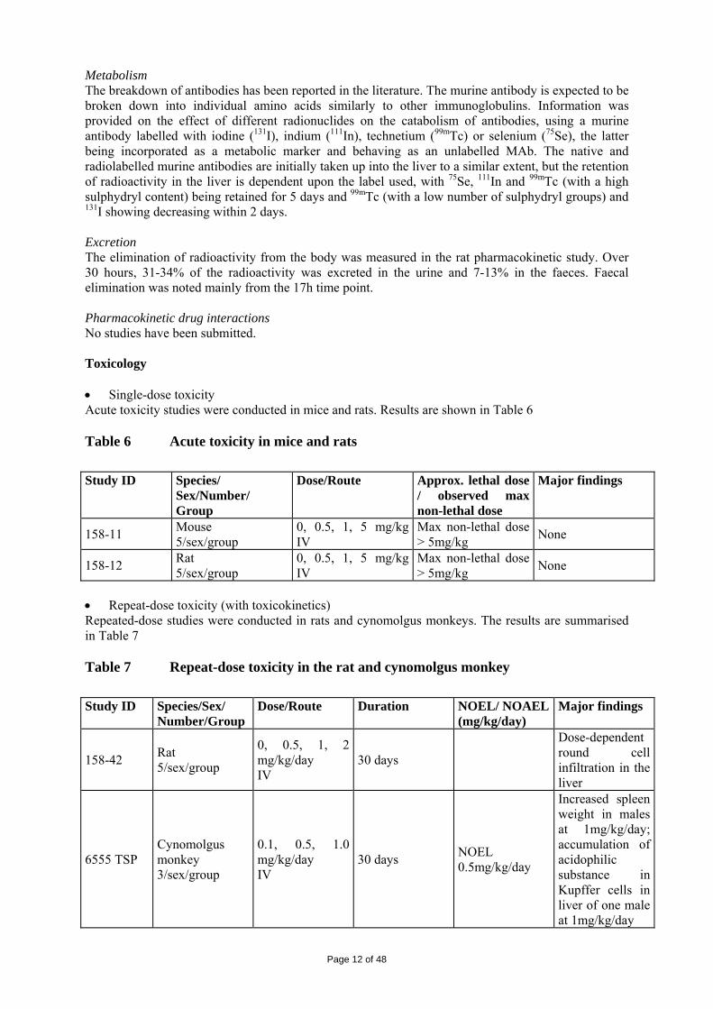
Radioactivity levels were measured with a gamma counter in plasma and whole blood to demonstrate the binding kinetics of the antibody to cells. The additional quantitative assay of antibody concentration was meant to show the pharmacokinetics of intact labelled antibody. The analytical method was based on a commercial kit (Enzygnost® mIgG micro), now discontinued, which detected mouse IgGs. PK data were analysed according to a two compartment model and in a non-compartmental model for study 306340. The radiation exposure was calculated as per the MIRD concept. Numbers in the original study (7D-101SZ-A) as well as the comparability study (Study 306340) were chosen arbitrarily as variability of results were unknown to estimate the number of patients that would be required to fulfil statistical requirements. • Absorption There were no absorption study reports submitted. The bioavailability is 100% as the product is administered intravenously. • Distribution The data were obtained mainly in Study 7D-101 SZ-A, an open-label, non-randomized, parallel group design phase I study, conducted in two study centres in 24 patients with suspected inflammatory processes. Half of the patients received a single intravenous injection of either 0.25 mg or 1 mg of 99mTc-MAb BW 250/183. The mean administered 99mTc radioactivity was 492 MBq (range: 448 to 560 MBq) in the 0.25 mg group and 623 MBq (range: 449 to 1,000 MBq) in the 1 mg group. Primary study objectives were pharmacokinetics and radiation exposure of the patients. Blood samples and urine samples for measurement of 99mTc radioactivity and the concentration of the antibody were taken over 24 hours after injection. Further samples for determination of antibody concentration were taken up to 30 days after injection. The concentration-time curves obtained in these measurements showed two phases: an early α phase (0-2 h) and a late β phase (5-24 h). The results are presented in Table 10.
Page 17 of 48
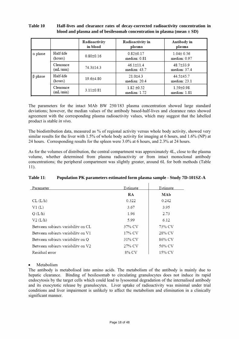
Table 10 Half-lives and clearance rates of decay-corrected radioactivity concentration in blood and plasma and of besilesomab concentration in plasma (mean ± SD)
The parameters for the intact MAb BW 250/183 plasma concentration showed large standard deviations; however, the median values of the antibody based-half-lives and clearance rates showed agreement with the corresponding plasma radioactivity values, which may suggest that the labelled product is stable in vivo. The biodistribution data, measured as % of regional activity versus whole body activity, showed very similar results for the liver with 1.5% of whole body activity for imaging at 6 hours, and 1.6% (NP) at 24 hours. Corresponding results for the spleen were 3.0% at 6 hours, and 2.3% at 24 hours. As for the volumes of distribution, the central compartment was approximately 4L, close to the plasma volume, whether determined from plasma radioactivity or from intact monoclonal antibody concentrations; the peripheral compartment was slightly greater, around 6L for both methods (Table 11). Table 11: Population PK parameters estimated form plasma sample - Study 7D-101SZ-A
• Metabolism The antibody is metabolised into amino acids. The metabolism of the antibody is mainly due to hepatic clearance. Binding of besilesomab to circulating granulocytes does not induce its rapid endocytosis by the target cells which could lead to lysosomal degradation of the internalised antibody and its exocytotic release by granulocytes. Liver uptake of radioactivity was minimal under trial conditions and liver impairment is unlikely to affect the metabolism and elimination in a clinically significant manner.
Page 18 of 48

• Elimination Technetium (99mTc) decays with a half-life of 6 hours. The total blood radioactivity is the result of the contribution of radioactive intact labelled antibody and other radioactive species like antibody fragments, smaller metabolites and free pertechnetate (99mTc). These radioactive species display large differences in how they are cleared from blood. Whereas the radioactivity associated with intact antibody will stay in the vascular compartment for a long time, radioactive fragments, small radiometabolites and free pertechnetate (99mTc) will clear rapidly from blood and will accumulate in the kidneys and further in urine. In all studies, about 14 % of the injected radioactivity was recovered in urine, which was only collected for 24 hours after administration. This radioactivity can be attributed to the elimination of free technetium (99mTc) and labelled low-molecular-weight antibody fragments and small radiometabolites. No information was provided about the fate of the injected radioactivity after the first 24 hours post-administration of Scintimun. However, the radioactivity absorbed by the kidneys (0.022 mGy/MBq on average) lies in the range of other approved 99mTc-radiopharmaceuticals. Furthermore, due to the short half-life of technetium (99Tc), technical limitations would prevent any meaningful determination after 24 hours. • Bioequivalence Study 306340 was carried out to assess the comparability between the old process (OP) and new process (NP). This was a randomised, open-label, comparative study on kinetics and biodistribution, conducted at two study centres in 19 patients (OP; N=10; NP; N=9) with suspected or documented inflammatory processes. In both groups, the maximum administered antibody dose was 1 mg, labeled with a mean 99mTc radioactivity of 828±106 MBq (OP; range: 670 to 1001 MBq) and of 836±114 MBq (NP; range: 731 to 1034 MBq). Bioequivalence at the 5% significance level was to be established by demonstrating that the 90% confidence intervals (CI) for the ratio of the geometric means of Cmax (ratio NP/OP) and the ratio of the geometric means of AUC0-tlast (ratio NP/OP) are included in the interval from 80% to 125%. The ratios NP/OP and their confidence intervals are summarised in Table 12. Taking into account body weight and gender as covariates, the size of the CI’s was reduced and the upper limit of 125% was only marginally exceeded. Table 12 Ratio of geometric means (NP/OP) for Cmax and AUC0 – tlast , and confidence
intervals (CI) – Study 306340
Without body weight and gender as covariates
With body weight and gender as covariates (retrospective analysis)
Ratio of means CI [LL, UL] CI [LL, UL]
Cmax 1.1251 [88.32%, 143.33%] [99.79%, 126.86%]
AUC0 – tlast 1.1010 [88.72%, 136.62%] [95.40%, 127.06%] LL – lower limit of CI, UL – upper limit of CI
• Dose proportionality and time dependencies Tables 13 and 14 give details of relationship of radioactivity to administered dose for early and late phases, with and without correction for decay.
Page 19 of 48

Table 13 99mTc activity in plasma: PK parameters in early (α) phase – Study 7D-101SZ-A
Table 14 Tc-99m activity in plasma: PK parameters in late (β) phase – Study 7D-101 SZ-A
Page 20 of 48

• Special populations There were no specific studies carried out in any special population. • Pharmacokinetic interaction studies There were no studies submitted on PK interaction. Pharmacodynamics Pharmacodynamics (PD) data on technetium (99mTc) besilesomab include 2 studies in patients (Studies 7D-101SZ-A and 7MN-302 SZ-A), 2 published papers and 4 in vitro studies. • Mechanism of action There were no studies submitted on mechanism of action. Although the mechanism of accumulation of besilesomab at the site of infection/inflammation has not been fully elucidated, it is postulated that it is mainly passive (increased vascular permeability) and partly active (migration of human granulocytes carrying besilesomab to the infection/inflammation site) because only 10% to 20% of the injected radiolabelled antibody bind in vivo to human circulating granulocytes. Specific binding of besilesomab to already migrated and activated granulocytes may be the major part of the detection signal. • Primary and Secondary pharmacology In 6 healthy subjects, 60-97% of peripheral blood granulocytes and less than 5% of other peripheral blood cells were stained by the antibody in vitro (study BW 250/183-BS-9). In a most recent study performed with the new product, this percentage was found to be 97.45% and 96.58% of peripheral blood granulocytes, for males and females respectively. Another study showed that besilesomab binds to 99.6% of mature human granulocytes in whole blood samples analysed by flow cytometry but does not bind to early granulocyte precursors in human bone marrow CD34+ cells. No significant binding of besilesomab to other human peripheral blood cells was observed: erythrocytes (0.1% to 1.3%), platelets (0% to 0.9%), lymphocytes (0.2% to 5%) and monocytes (0.4% to 3.5%). Another study confirmed the non cross-reactivity of besilesomab with human platelets (<3%). Tissue specificity of besilesomab was evaluated on a broad panel of human cryopreserved normal as well as cancer tissues derived from various individuals using immunohistochemistry. Besilesomab showed strong binding to colon carcinomas and granulocytic cells in normal tissues (i.e. normal liver, lung and bone marrow). Furthermore, besilesomab binds to pancreatic, some lung and some breast carcinomas but does not bind to blood vessels and connective tissue. Finally, CD66 expression on bone marrow cells of patients with multiple myeloma was shown ex vivo by flow cytometry using besilesomab as anti CD66. Therefore, false positive images are possible in case of CEA expressing tumours or haematological malignancies such as myeloma. Despite its binding capacity to human granulocytes, besilesomab does not significantly influence granulocyte-mediated functions (enzyme release, pinocytosis, chemiluminescence) and does not induce any lytic effects to epitope positive cells via complement-dependent or antibody-dependent cell-mediated cytotoxicity (CDC or ADCC). It does not impair bone marrow cell proliferation. Although no preliminary dose finding study was carried out, two studies have compared different injected amounts of monoclonal antibody (0.25 mg, 0.5 mg and 1 mg) and did not show any difference in terms of efficacy (see Table 15).
Page 21 of 48

Table 15 Studies 7MN-301 SZ-A and 7D-101 SZ-A: sensitivity and specificity for detection of infection / inflammation with different doses of technetium (99mTc) besilesomab
As for the dose of radioactivity, the retrospective analysis of study 7MN-301 SZ-A revealed no statistically significant differences in sensitivity and specificity between the (retrospectively defined) groups receiving different amounts of radioactivity (see Table 16). However, sensitivity was lower in the group of patients receiving less than 400 MBq of technetium (99mTc) besilesomab, whereas the sensitivity and specificity were at comparably high levels in all other activity groups. Table 16 Sensitivity and specificity for detection of infection / inflammation with different
radioactivity – Study 7MN-301 SZ-A:
Based on these studies, the recommended radioactivity is between 400 and 800 MBq. Since the maximum radioactivity that can be added to one vial containing 1 mg of antibody is 1,800 MBq, this corresponds to the administration of 0.25 to 1 mg of besilesomab. Further parameters were examined to analyze diagnostic efficacy more precisely such as the occurrence of unusual accumulations. Results are shown for study 7MN-301 SZ-A in Table 17. Table 17 Unusual accumulations at 5 h and 24 h after injection – Study 7MN-301 SZ-A
Page 22 of 48

Clinical efficacy For the current application, the clinical data submitted consists of clinical studies performed more than 10 years ago with besilesomab manufactured according to the old manufacturing process, data from 3 recent studies using besilesomab manufactured according to the new manufacturing process and data from published literature. The new single phase III study AG-PH3, is considered as the pivotal study for efficacy. This is a comparative study with technetium (99mTc) exametazime labelled leukocytes. • Dose response study(ies) There was no prospective dose-response study carried out. A retrospective analysis of a correlation between radioactivity and diagnostic efficacy (sensitivity and specificity) was submitted (see previous section). Based on these results, the selected radioactivity for the pivotal study AG-PH3 was 800 MBq. In study 7MN-301 SZ-A, images were acquired at 5 h and 24 h post-injection. Although the sensitivity and specificity at each time timepoint and by class of diagnoses did not change significantly, acquiring images at 5 h and 24 h increased the percentage of lesions found. In three percent of patients lesions were found at 5 h post-injection (p.i.) but not found at 24 h. Five percent of patients had no lesion at 5 h p.i.; however, a lesion was detectable at 24 h p.i. Based on these results planar acquisition was done at 4 h and 24 h p.i. in the pivotal study AG-PH3. • Main study(ies) AG-PH3: Phase III, multicentre, randomised, open-label, clinical study on the agreement of Scintimun and labelled 99mTc-WBC in diagnosing infection/inflammation by scintigraphy in peripheral bone and joints with suspected osteomyelitis. METHODS Study Participants Main Inclusion criteria: • Patients aged at least 18, both sexes, suffering from suspected or documented osteomyelitis
(acute, subacute, chronic) in the peripheral skeleton including patients with loosening of joint prosthesis and patients with diabetic foot
• Patients with at least one of the following signs or symptoms was required: localised pain, nonhealing skin ulceration, fever > 37.8°C for at least 3 days, leukocyte count in excess of the upper normal limits, erythrocyte sedimentation rate in excess of the upper normal limits, radiographic findings suggestive of osteomyelitis, or positive blood or wound cultures.
Main exclusion criteria: • Pregnant and breastfeeding women • Patients with a positive HAMA test prior to the first study drug administration • Severe disease or surgery (except for orthopedic reasons) within the last 4 weeks prior to the first
study drug administration • Patients with leukocyte count < 4.109/L (4,000/mm3) • The use of non-steroid anti-inflammatory drugs and corticosteroids within 3 days prior to the first
injection and up to 24 h after the last injection • Receipt of cancer chemotherapy and immunosuppressive drugs or immunomodulators within 4
weeks prior to study entry Treatments The comparator was Ceretec, 99mTc-exametazime (also known as hexamethyl propylene amine oxime (HMPAO)). The white blood cells (WBCs) preparation, labelling and quality control was done according to the SPC of Ceretec or the own validated procedures of the study sites.
Page 23 of 48

Administration of technetium (99mTc) besilesomab was a single intravenous dose of 800 MBq (22 mCi) of technetium (99mTc) besilesomab and one single intravenous dose of 330 MBq (9 mCi) of 99mTc-WBCs in a randomised order. However, the German Radiation Protection Committee asked to reduce the cumulative activities to be administered to German patients in order to not exceed the maximal effective dose of 10 mSv after injection of the two tracers. The study protocol was amended for German patients to reduce the activity of technetium (99mTc) besilesomab from 800 MBq to 650 MBq and the activity of 99mTc-WBCs from 330 MBq to 300 MBq leading to a reduction of the maximal effective dose from 11.7 mSv to 10 mSv. The wash-out period between the two administrations was 2 – 4 days. Drugs inhibiting inflammation which may lead to false negative results were not allowed during the last 3 days before injection and till the acquisition of the last images of the study. Chemotherapy, immunosuppressive drugs and immunomodulators were not allowed within 4 weeks of study entry until one month after the last images acquisition. Objectives The primary objective was to assess the agreement rate of the immunoscintigraphy with technetium (99mTc) besilesomab and scintigraphy with 99mTc-WBCs with regard to the diagnosis of infection/inflammation in patients suspected of osteomyelitis. The agreement rate was based on the evaluations of three blinded and independent readers in a blinded read. The secondary objectives were to assess the image quality and the safety of the two products, in particular, the possible HAMA induction after technetium (99mTc) besilesomab administration. Outcomes/endpoints Primary efficacy variable The primary efficacy variable was the agreement rate as an average across the results of the three blinded readers ("average reader") based on the adjudication of a fourth blinded reader. Secondary variables The secondary variables are:
• Agreement rate per blinded reader based on adjudication of 4th blinded reader • Agreement rate across the three blinded readers and per reader based on the final evaluation of
the clinical investigators at Month 1 taking into account all available findings such as medical history, physical examination, results of biology, biopsy and /or imaging and the clinical follow-up
• Image quality as assessed by the three blinded readers (using a categorical scale 1-4/non evaluable)
• Safety of the two products and, in particular, the possible HAMA factor induction after administration of technetium (99mTc) besilesomab
Sample size This was a non-inferiority trial with an intra-individual comparison design. The following assumptions were set:
• assumed agreement rate = 0.80 • limit of clinical relevance = -0.10 • α = 0.025 • power = 80%
The assumption of an agreement rate of 80% was based on published data with technetium (99mTc) besilesomab (sensitivity between 69 and 100% for the diagnosis of infection). For this study, a sensitivity of 85% was assumed to represent a fair estimate; the same value was assumed for 99mTc-WBCs. With regard to specificity, false positive findings in the reported literature (for infection) probably represent inflammation without infectious agent. In the context of this study, these cases would be regarded as true positive cases. A value of 95% was considered appropriate for specificity of technetium (99mTc) besilesomab and 99mTc-WBCs. Based on the assumptions for sensitivity and
Page 24 of 48

specificity an overall accuracy of 90% was expected. For 2 independent methods with an accuracy of 90% each, the agreement rate was calculated as 82% (0.9 x 0.9 + 0.1 x 0.1). A one-sample χ²-test with 80% would require evaluable 153 patients, if only one observation per patient would be obtained. This had to be corrected for the effect of clustered data (i.e., 1 area of infection/inflammation and 1 not affected area for each patient). The inflation factor (IF) was estimated at 0.70. Therefore, the number of evaluable patients needed was n = 153 * 0.7 = 108. Taking into account a drop-out rate of 10% and a rate of not evaluable images of 10%, a total sample size of 108 plus 20% = 130 patients were to be randomised. Randomisation The order of administration of technetium (99mTc) besilesomab and 99mTc-WBCs was centrally allocated randomly according to a randomisation list. Set of images (both of technetium (99mTc) besilesomab and 99mTc-exametazime WBC) for each patient was read in a randomised order. Blinding (masking) The conduct of the test procedures was not blinded. The images of technetium (99mTc) besilesomab and 99mTc-WBCs were evaluated in a blinded and randomised way by 3 independent nuclear physicians with expertise in scintigraphy of infection/inflammation. The 3 readers were blinded to the patient, to the study site and to the injected radiotracer, and were not provided with any details concerning the patient population (demographic data, age, indication, clinical question, details of the study protocol) in order to reduce possible bias. In order to test intra-reader variability, a randomly selected sample (10%) of completed cases was added to the total number of cases to be read, with a different random code. Statistical methods The analyses were performed in all-subject-treated sample (AST) for safety and in per protocol sample (PP) for efficacy where AST and PP consisted of:
• All-Subjects-Treated (AST): all randomised patients who received at least one of the treatments (technetium (99mTc) besilesomab and/or 99mTc-WBCs). The AST set served as the Full Analysis Set for safety.
• Per-Protocol (PP) set: all patients who received both treatments and performed the required imaging and who did not have major protocol violations affecting the validity of analysis. Major and minor protocol violations were identified at a Blind Review meeting. The PP sample served as the Full Analysis Set for the primary and secondary analysis of agreement rate, and for the analysis of image quality.
For the primary objective, the agreement rate was analysed using a modified adjusted Chi2-test to cover clustered data and multiple measurements per cluster. The primary efficacy variable was calculated as an average across the results of the 3 blinded and independent readers ("average reader") based on the adjudication results of the fourth blinded reader who compared each reader’s evaluation and matched the regions of interest. The primary analysis of the single primary variable was planned as a confirmatory analysis. Therefore no adjustment for multiple testing was necessary. The agreement rate was calculated from the results of a 2x2 cross table and defined as follows:
Number of agreed affected sites + Number of agreed not affected sites Total number of all affected and not affected sites
The agreement rate was supposed to be sufficient if its one-sided 97.5% confidence interval was positioned above 0.70.
Page 25 of 48

RESULTS Participant flow
Recruitment Patients were recruited from September 2006 until October 2007. There were 130 patients randomised and 120 patients evaluable for the primary efficacy variable. Conduct of the study Six protocol amendments to the study protocol were produced during the study period; the main changes were: • Changes in the planned end date of the study due to low recruitment rate • Decrease in the threshold of leukocyte count as exclusion criterion (< 4 instead of < 6.109/l) • Correction of the error in the dose of antibody (already mentioned) • A negative scintigraphy to be analysed as one affected site (and not two for both sides) • Reduction in the dose of radioactivity in Germany (already mentioned) • Finally, an additional analysis of the agreement rates was planned, which took into account all
completely imaged body regions, and not only those with affected sites at least on one side, as assessed by the three blinded readers. A fifth reader was asked to check all body parts that were imaged completely for both sides.
Baseline data There were 79 males and 50 females (1 had no CRF completed); their mean age was 61.3 years. All patients had a previous history of either joint prostheses (66 patients) or chronic osteomyelitis (34 patients), or diabetic foot (27 patients), or atherosclerosis with suspicion of osteomyelitis (2 patients). At study entry, they all presented either clinical symptoms and/or biological signs and/or imaging findings of osteomyelitis. The baseline characteristics are shown in Table 18.
Page 26 of 48

Table 18 Baseline characteristics – Study AG-PH3
Numbers analysed There were 130 patients randomised in the trial. The PP analysis set included in principle 120 patients. However, an additional patient was excluded because his images were only assessed by 2/3 readers and all the agreement rate analyses were conducted on 119 patients. It should be noted that two patients were not injected the tracers in the allocated order due to an error of the investigator; these patients were analysed according to the actual tests performed. For the agreement rate based on the final assessment of the investigator at Month 1, 7 patients from the PP population were excluded from the analysis because the final assessment of the investigator at Month 1 was missing. Outcomes and estimation Primary efficacy variable: Agreement rate based on 4th blinded reader The agreement rate between three blinded and independent readers for technetium (99mTc) besilesomab and 99mTc-WBCs with regards to the diagnosis of infection/inflammation by immunoscintigraphy based on the adjudication of a fourth blinded reader is presented in Table 19. Of the 119 patients, 22 patients were completely negative. Each of these patients counted as one agreed non-affected (or negative) site. In the other 97 patients, at least one affected site was found by any of the three readers with any of the two tracers. The value of the agreement across readers was 0.83 with a lower limit of the 95% confidence interval of 0.80, which is above the lower limit of clinical relevance of 0.70. The agreement rate was also estimated per reader based on the adjudication of the fourth blinded reader. All three readers had an agreement rate between 0.81 and 0.85 and a 95% CI lower bound equal or higher to 0.76. Table 19 Agreement rates across readers and per reader based on evaluation of the 4th
reader - Study AG-PH3
Page 27 of 48

Secondary variables The agreement rate was also calculated by body region based on the evaluation of the 3 blinded independent readers, not taking into account the adjudication of the fourth reader (Table 20). Table 20 Agreement rate by body region per reader for the whole patient – Study AG-PH3
This analysis was also performed per separate body region. The sixteen body regions included the upper limbs: shoulder girdle, arm, elbow, forearm, wrist, carpus, metacarpus, fingers and the lower limbs: hip girdle, thigh, knee, leg, ankle, tarsus, metatarsus, and toes (Table 21). As some patients had lesions in more than one segment, the sum of clusters across the segments is higher than the total number of patients and the number of clusters for the whole body evaluation. The three body regions with the best agreement rate were: elbow, ankle and leg and those with the worst agreement rate were toes, knee, and tarsus. Table 21 Agreement rate across readers per whole patients by body region - Study AG-
PH3
Finally, the agreement rate was calculated by average reader and per reader according to the final assessment of the investigator at Month 1. Of the 112 patients analysed, 73 patients were diagnosed by the investigator as positive (with at least one affected site) and 39 patients as negative (with no affected site). The results are shown in Table 22.
Page 28 of 48

Table 22 Agreement rate based on the final assessment of the investigator at Month 1- Study AG-PH3
The assessment of the image quality showed better results for technetium (99mTc) besilesomab compared to 99mTc-WBCs. The results are shown in Table 23. Table 23 Frequency distribution of image quality - Study AG-PH3
The intra-variability within readers was assessed. The reproducibility for image quality (four-point scale) ranged between 0.18 and 0.38 using Cohen’s kappa. A kappa value of 0.18 represents a slight agreement and a kappa value between 0.21 to 0.40 represents a fair agreement between the results of the initial read and those of the re-read. The kappas were statistically significantly different from zero for 2 of the three readers. Also the reproducibility of the readers to assess diagnosis (affected/not affected) was tested. Cohen’s kappa ranged from 0.57 to 0.71, which means moderate to substantial agreement in assessing the diagnosis. All p values were highly significant. The kappa values of the first 2 readers were higher than the kappa value of the third reader. In addition, kappa values were calculated separately for technetium (99mTc) besilesomab and 99mTc-WBCs for each reader's assessment. The reproducibility of diagnosis did not differ clearly between the two products. Ancillary analyses In addition to the agreement rate, sensitivity and specificity with respect to the final assessment of the investigator at Month 1 as standard of reference (SOR) was calculated for technetium (99mTc) besilesomab and 99mTc-WBCs and results are shown in Table 24.
Page 29 of 48

Table 24 Sensitivity and specificity of technetium (99mTc) besilesomab and 99mTc-WBCs using the final diagnosis of the investigator as standard of reference - Study AG-PH3
Post-hoc analyses A post-hoc analysis based on a surrogate standard of truth, which was an evaluation by an expert panel of all data available from the study CRFs plus all additional raw data available from the investigators was provided (Table 25). This expert panel did not have access to the imaging results with technetium (99mTc) besilesomab and 99mTc WBCs. The expert panel provided a diagnostic as normal/abnormal in 74 patients out of 119 (62%) and inconclusive diagnosis for the 45 remaining patients where no additional data had been received. The overall diagnostic performance of technetium (99mTc) besilesomab in this analysis, sensitivity (76%) and specificity (69%) appeared similar to that of the previous analysis based on the investigator diagnosis at 1 month, 75% and 72%, respectively. Of note, contrary to the expert panel, the investigators had access to the results of the test imaging results. For 99mTc-WBCs, sensitivity was 63% and specificity 76%, which compared to 59% and 79%, respectively, in the previous analysis. Confidence intervals were wide and no analysis by indication (e.g. chronic osteomyelitis, infection of joint prosthesis, diabetic foot) or clinical setting (e.g. acute vs. chronic) was presented. Table 25 Average reader diagnostic performance with truth panel assessment as SOR -
Patient-based analysis (at least one body region affected vs. no body region affected)
Tracer Number of patients
Estimate and 95 % Confidence Intervals
(99mTc)-besilesomab 0.756 [0.647 – 0.866] 99mTc-WBCs 0.626 [0.501 – 0.751]
Sensitivity
Difference
41
0.130 [0.031 – 0.229] (99mTc)-besilesomab 0.687 [0.554 – 0.820] 99mTc-WBCs 0.758 [0.633 – 0.882]
Specificity
Difference
33
-0.071 [-0.184 – 0.042] (99mTc)-besilesomab 0.725 [0.641 – 0.810] 99mTc-WBCs 0.685 [0.595 – 0.774]
Accuracy
Difference
74
0.041 [-0.037 – 0.118] • Analysis performed across trials (pooled analyses and meta-analysis) There was no meta-analysis performed by the applicant as the population included in the clinical trials was not homogeneous and the reference method was different from one study to another. The Applicant has also provided meta-analyses on the diagnostic performance of technetium (99mTc) besilesomab, 99mTc-WBCs, Magnetic Resonance Imaging (MRI), and LeukoScan (sulesomab) in the
Page 30 of 48

indication of suspected osteoarticular infection (osteomyelitis, joint prosthesis infection and diabetic foot). These studies used microbiology/histology confirmation from a biopsy/surgical sample or clinical follow-up for at least 5-6 months as standard of truth. The estimated sensitivity and specificity results are shown in Table 26. However, these meta-analyses suffer from potential major biases and can only be considered supportive. Table 26 Meta-analyses (weighted sensitivity and specificity with 95% CI)
Procedure Weighted sensitivity (%) Weighted specificity (%) (99mTc)-besilesomab 89.5 (83.7, 95.2) 80.8 (74.2, 87.4) LeukoScan 85.2 (79.6, 90.8) 82.4 (75.3, 89.6) 99mTc-WBCs 90.7 (85.9, 95.3) 91.3 (84.8, 97.8) MRI 94.1 (89.5, 98.8) 83.8 (74.9, 92.6)
• Clinical studies in special populations There were no specific clinical studies submitted to evaluate the performance of technetium (99mTc) besilesomab in patients with impaired renal function or impaired hepatic function. During the clinical development only 6 children were recruited in clinical trials (Study BI 71.015). A publication describing a series of 30 children with fever of unknown origin is available. • Supportive study(ies) Several clinical studies were submitted as part of the initial application: Study BI 71015, Study 7D-201SZ-A and Study 7MN-301SZ-A. They were all conducted with the product manufactured according to the old process and enrolled patients in a multiplicity of indications, most of which were withdrawn by the applicant during this procedure. These studies applied the standards of more than 10 years ago and were considered by the CHMP as “seriously methodologically flawed, rendering the results to be of poor quality” (Withdrawal Public Assessment Report, 28 June 2006). Therefore, they were not considered supportive of the diagnostic performance of technetium (99mTc) besilesomab in the current application. Literature support for efficacy The clinical expert report also includes data on efficacy from literature on osteomyelitis1,2,3,4,5,6,7,8,9,10,11,12,13 diabetic foot14, inflammatory bowel disease15,16,17,18, fever of unknown origin19,20,21, endocarditis22,23 and bone marrow metastasis24,25,26,27,28. • Discussion on clinical efficacy Pharmacokinetics The analytical methods, the statistical aspects and the number of patients assessed for the PK measurements appear acceptable. Since all the subjects in the PK studies were patients suffering from an inflammatory process, it is reasonable to consider that the results would be representative of a population with the target indication. The parameters for technetium (99mTc) besilesomab plasma concentration showed large standard deviations and this wide inter-individual variability in results was discussed. The parallel arm design for PK measurements was acceptable in order to avoid repeated administration of technetium (99mTc) besilesomab in the same individual, increasing the risk of HAMA formation. This design implies that there may be a degree of inter-individual variance in addition to intra-individual variance, reflecting the broad standard deviations. There were no notable differences between the 2 doses of besilesomab (0.25 mg and 1.0 mg) in terms of half-life, AUC, volume of distribution and clearance. In the population studied, about 15% of the radioactivity was excreted in the urine in 24 hours. It is considered that in patients with impaired renal function, this is not likely to pose a clinical problem. In addition, liver impairment is unlikely to affect the metabolism and elimination in a clinically significant manner. The issues are adequately covered in the SPC in section 4.2
Page 31 of 48

Efficacy The pivotal study, AG-PH3, was a comparative study with leukocytes labelled with technetium (99mTc) exametazime. The selected comparator was considered appropriate and reflects current medical practice. Recruited patients in the pivotal study presented with peripheral osteomyelitis but very heterogeneous in nature. A major deficiency identified in the pivotal trial AG-PH3 was the lack of an adequate standard of truth or surrogate, as recommended in the guideline CPMP/EWP/1119/98. Therefore, diagnostic parameters like sensitivity and specificity could not be calculated and only agreement rate could be estimated. Considering that technetium (99mTc) besilesomab has characteristics very similar to existing agents, it is acceptable to consider the agreement rate as a suitable alternative endpoint if thoroughly justified. The CHMP noted the justification for not having bone biopsies as standard of truth as this may not have been feasible. Using the pre-specified non-inferiority margin defined in the trial protocol, the trial met its primary objective where all the lower bounds of the 95% CIs for the various agreement rates, whether average or per reader, were above the prespecified 70% lower limit. However, this lower limit was defined based on assumptions regarding patient recruitment and overall feasibility of the trial rather than clinical relevance. The overall results were better when a 4th reader matched the affected regions identified by the readers (primary analysis) than when the analysis was performed on the readings without this adjudication. It is considered that the analysis without adjudication is the most relevant although it produced slightly lower agreement rates. The supplementary analysis based on all completely imaged body regions only added negative readings (for all readers) to the previous analysis which increased all agreement rates. Regarding the agreement rates by body region, the best agreement rates were considered those based on the smallest number of clusters (leg, ankle, elbow) whereas the worst agreement rate was exhibited for the knee, the largest cluster. The applicant provided further discussion on the discordant findings, which did not follow the guidance document on radiopharmaceuticals (CPMP/EWP/1119/98) but was eventually considered acceptable although it was not possible to draw any unbiased conclusion with respect to the comparator. As a secondary endpoint, the final diagnosis of the investigator was used as standard of reference to establish the agreement rate and diagnostic performance of technetium (99mTc) exametazime and technetium (99mTc) besilesomab. It is of note that the clinical and imaging data available for the investigator were not standardised in all patients and not independent of the evaluated imaging procedures. The lack of standard of truth in the AG-PH3 trial was addressed in a post-hoc analysis based on a surrogate, which was an evaluation by an expert panel of all data available from the study CRFs plus all additional data available from the investigators. This expert panel did not have access to the imaging results with technetium (99mTc) besilesomab and 99mTc WBCs. This approach is based on the CHMP guideline: “… a surrogate standard of truth, such as an appropriate combination of tests, clinical data, repeat testing and clinical follow-up may be used to provide a good approximation of the disease state.” This approach has obvious limitations since the surrogate was not defined prospectively and additional data were not available for all patients. In spite of these intrinsic weaknesses, the CHMP noted that the various analyses (pivotal trial and meta-analyses) provided a consistent pattern of results for technetium (99mTc) besilesomab and the comparator 99mTc WBCs which, even if the absolute numbers were slightly different, suggested similar (or slightly higher) sensitivity for technetium (99mTc) besilesomab at the expense of a lower specificity. The CHMP expressed concern over this loss of specificity as in the context of its potential clinical use in osteomyelitis, specificity is an important endpoint. Therefore, the issue was whether this loss of specificity outweighed the advantages of the method, i.e. simpler and safer for the patient and health care professional. In addition, there were no adequate data showing the impact of the specificity of technetium (99mTc) besilesomab on diagnostic thinking and patient management. The CHMP acknowledged that a lower specificity could be acceptable if technetium (99mTc) besilesomab would be used not as a sole technique but in conjunction with other imaging modalities to determine the appropriate diagnosis. To resolve the issue, the applicant committed to perform a randomised parallel-group trial in the approved indication, the primary objective of which would be to evaluate the impact of technetium (99mTc)-besilesomab on diagnostic thinking and patient management in clinical practice and in comparison to an acceptable comparator.
Page 32 of 48

The literature meta-analysis submitted was only considered supportive given its potential major biases. The evidence submitted for the indication of scintigraphic imaging of bone marrow involvement for detection and extent of carcinoma metastasis was considered insufficient by the CHMP and therefore, the indication was withdrawn. In addition, the evidence submitted for the indication of scintigraphic imaging of diabetic foot infection was considered insufficient by the CHMP and therefore, Scintimun should not be used for the diagnosis of diabetic foot infection (see SPC section 4.1). No prospective dose-response studies have been performed for the requested indications of technetium (99mTc) besilesomab. However, the amount of antibody administered is driven by the radioactivity which is necessary to achieve the expected diagnosis. As a result the amount of antibody administered in study AG-PH3 ranged from 0.27 to 0.96 mg (0.60 mg in average), which was considered acceptable. The proposed optimal timing for imaging acquisition, 3-6 h and 24 h, was considered acceptable. There were no data in the paediatric population apart from literature which is not relevant to the indication of osteomyelitis. Thus, technetium (99mTc) besilesomab is not recommended for use in children below 18 years of age due to insufficient data on safety and efficacy (see SPC section 4.2, 5.1 and 5.2). There were no subanalyses performed in the inflammatory situations (eg. acute versus chronic inflammation, multifocal osteomyelitis, etc…) likely to affect diagnostic performance of technetium (99mTc) besilesomab. The CHMP, after evaluating the responses to the D120 list of questions, sought the advice of the Scientific Advisory Group on Diagnostics (SAG-D) to further examine and discuss the data submitted by the applicant and to consider the evidence presented on the diagnostic performance and impact on diagnostic thinking and therapeutic decision of technetium (99mTc) besilesomab. Following the CHMP request, a SAG meeting was convened on the 7th September 2009 to provide advice on the list of questions adopted at the June 2009 CHMP meeting. Based on the totality of the data presented by the applicant in the MAA, the SAG-D agreed that the diagnostic performance of technetium (99mTc) besilesomab could be considered as established for the following indication “scintigraphic imaging for determining the location of inflammation/infection in peripheral bone in patients with suspected osteomyelitis”. The SAG-D suggested deleting the term “joints” from the indication as it is considered that the pathophysiology of an inflammation reaction is different in the presence of an exogenous implant such as a joint prosthesis. There were slightly divergent positions between SAG-D members regarding whether the product could be used in acute or chronic osteomyelitis. The majority of the SAG-D considered that technetium (99mTc) besilesomab could be used in both suspected chronic as well as acute osteomyelitis while one member disagreed and commented that the clinical utility of technetium (99mTc) besilesomab in acute osteomyelitis was not fully justified in the data presented. The SAG-D were unanimous in their agreement that technetium (99mTc) besilesomab should not be used in diabetic foot infection as the product will not distinguish inflammation, in particular Charcot arthropathy, infection in the soft tissues and infection in the bone, also in relation with the limited resolution of scintigraphy vs. MRI or PET scan. This distinction has important consequences on the management of the foot in diabetic patients. In addition to the answers to the CHMP questions, the SAG-D commented that the trial performed by the applicant, AG-PH3, was lacking in robustness and did not fulfil the requirements according to the guideline “Points to consider on the evaluation of diagnostic agents” (CPMP/EWP/1119/98). The follow-up period of patients, which is considered standard practice in such trials, was not part of the trial protocol and was performed post-hoc at the request of the CHMP. However, it was stated that additional data would not have affected the outcome of the results from the expert panel. The SAG-D supported the recommendation of an additional post-marketing confirmatory study to evaluate the
Page 33 of 48

clinical relevance of technetium (99mTc) besilesomab in relation to the standard diagnostic work-up of the patient and the impact on diagnostic thinking and patient management. Finally, the SAG-D recognised that technetium (99mTc) besilesomab is a product which could be of a certain importance given the advantages already expressed in the assessment reports, i.e. that in vivo labelling of granulocytes reduces the risk of misadministration to patients of exogenous in vitro labelled leucocytes and reduces the level of exposure of hospital technical personnel to radioactivity and to exogenous blood. This could be especially relevant in hospitals across Europe with small nuclear medicine centres for which there is no proper facility for in vitro radioactive labelling of leucocytes. For all patients, the radiation exposure must be justifiable by the expected diagnostic achieved with the lowest possible radiation dose. For the elderly, no dose adjustment is required. Formal studies have not been performed in patients with renal or hepatic impairment. However, due to the nature of the molecule and the short half-life of technetium (99mTc), dose adjustment was considered not necessary in such patients. This medicinal product should be reconstituted and radiolabelled before administration to the patient with sodium technetium (99mTc) pertechnetate to obtain a clear and colourless technetium (99mTc) besilesomab solution for injection. The radiolabelled solution should be administered intravenously as a single dose only. This medicinal product should be administered exclusively by authorised personnel (see “General warnings” in the SPC section 4.4). For instructions for preparation of the radiopharmaceutical, see SPC section 12. For disposal and other handling, see SPC section 6.6. For patient preparation, see SPC section 4.4. Images acquisition should start 3 to 6 hours after administration. An additional acquisition 24 hours after initial injection is recommended. Acquisition can be performed using planar imaging. Scintimun should be given to sufficiently hydrated patients. In order to obtain images of best quality and to reduce the radiation exposure of the bladder, patients should be encouraged to drink sufficient amounts and to empty their bladder prior to and after the scintigraphic examination (see SPC section 4.4). An interval of at least 2 days must be observed between any previous scintigraphy with other technetium (99mTc)-labelled agents and administration of Scintimun. There are currently no criteria to distinguish infection and inflammation by means of Scintimun imaging. Scintimun images should be interpreted in the context of other appropriate anatomical and/or functional imaging examinations (see SPC section 4.4). Only limited data is available about binding of technetium (99mTc) besilesomab to CarcinoEmbryonic Antigen (CEA) expressing tumours in vivo. In vitro, besilesomab cross-reacts with CEA. False positive findings in patients with CEA expressing tumours cannot be excluded. False results may be obtained in patients with diseases involving neutrophil defects and in patients with haematological malignancies including myeloma. Clinical safety Safety data for technetium (99mTc) besilesomab include adverse events, follow-up of vital signs, laboratory parameters, HAMA from clinical trials and post marketing experience. In addition, safety parameters include dosimetry.
Page 34 of 48
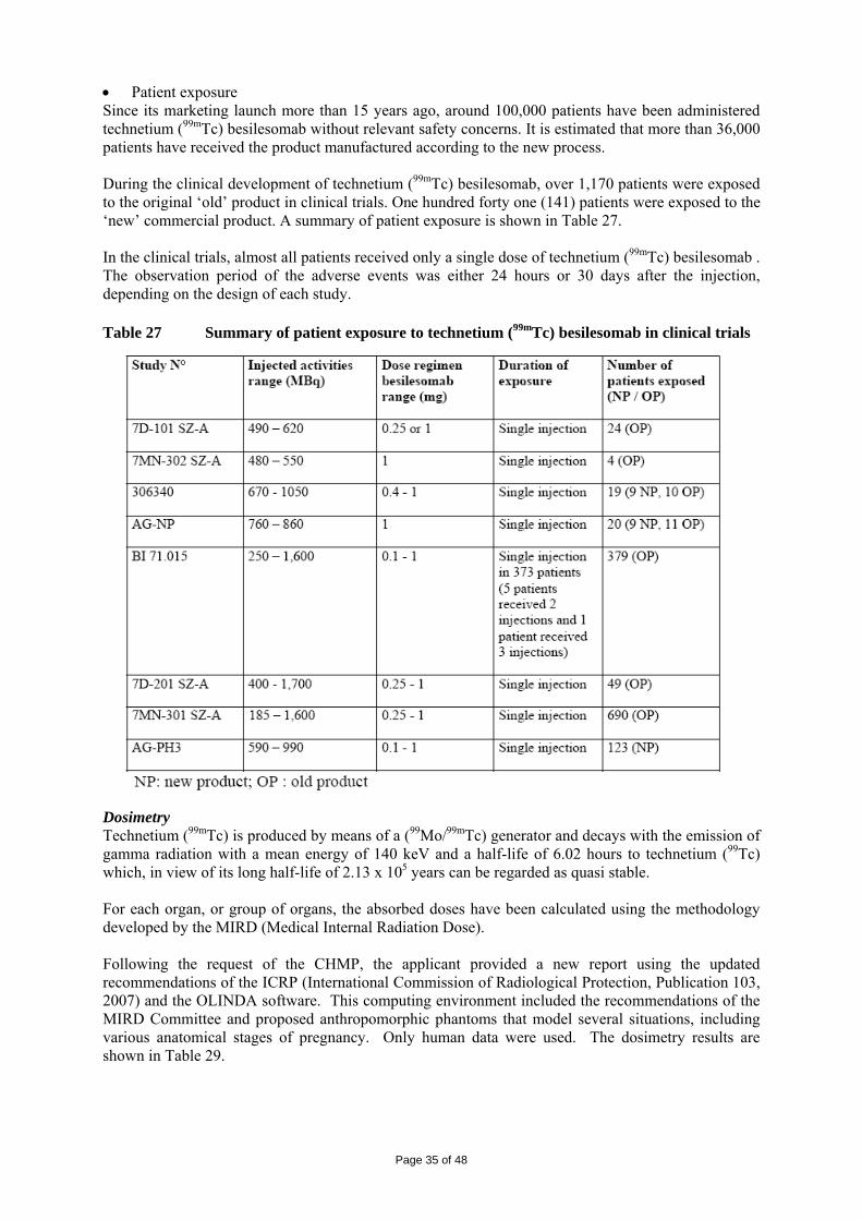
• Patient exposure Since its marketing launch more than 15 years ago, around 100,000 patients have been administered technetium (99mTc) besilesomab without relevant safety concerns. It is estimated that more than 36,000 patients have received the product manufactured according to the new process. During the clinical development of technetium (99mTc) besilesomab, over 1,170 patients were exposed to the original ‘old’ product in clinical trials. One hundred forty one (141) patients were exposed to the ‘new’ commercial product. A summary of patient exposure is shown in Table 27. In the clinical trials, almost all patients received only a single dose of technetium (99mTc) besilesomab . The observation period of the adverse events was either 24 hours or 30 days after the injection, depending on the design of each study. Table 27 Summary of patient exposure to technetium (99mTc) besilesomab in clinical trials
Dosimetry Technetium (99mTc) is produced by means of a (99Mo/99mTc) generator and decays with the emission of gamma radiation with a mean energy of 140 keV and a half-life of 6.02 hours to technetium (99Tc) which, in view of its long half-life of 2.13 x 10⁵ years can be regarded as quasi stable. For each organ, or group of organs, the absorbed doses have been calculated using the methodology developed by the MIRD (Medical Internal Radiation Dose). Following the request of the CHMP, the applicant provided a new report using the updated recommendations of the ICRP (International Commission of Radiological Protection, Publication 103, 2007) and the OLINDA software. This computing environment included the recommendations of the MIRD Committee and proposed anthropomorphic phantoms that model several situations, including various anatomical stages of pregnancy. Only human data were used. The dosimetry results are shown in Table 29.
Page 35 of 48

Table 28 Values of the absorbed doses calculated for the individual male and female of reference
Organ mSv/MBq
Reference male Reference female Brain 2.36 E-03 3.12 E-03 Heart 4.95 E-03 5.97 E-03 Colon 4.50 E-03 5.76 E-03 Stomach 4.45 E-03 5.35 E-03 Liver 1.00 E-02 1.26 E-02 Small Intestine 4.80 E-03 5.75 E-03 Bone marrow (red) 2.42 E-02 2.29 E-02 Muscles 3.17 E-03 3.91 E-03 Ovaries 5.94 E-03 Pancreas 6.90 E-03 8.26 E-03 Skin 1.78 E-03 2.16 E-03 Lungs 1.25 E-02 1.60 E-02 Spleen 2.71 E-02 3.24 E-02 Kidney 2.10 E-02 2.34 E-02 Breast 3.01 E-03 Adrenal 7.59 E-03 9.37 E-03 Testis 1.82 E-03 Thymus 3.51 E-03 4.23 E-03 Thyroid 2.79 E-03 3.21 E-03 Bone 1.77 E-02 2.27 E-02 Uterus 5.01 E-03 Gallbladder 5.91 E-03 6.81 E-03 Bladder 3.05 E-03 3.80 E-03 Whole body 4.45 E-03 5.52 E-03 Effective Dose 8.63 E-03mSv / MBq
The effective dose as defined in ICRP 103 was estimated at 8.63x10⁻³ mSv per MBq of technetium (99mTc) besilesomab administered. In an adult, this examination would cause low to moderate exposure of a few mSv (6.9 mSv for 800 MBq administered). This is in the range of other diagnostic nuclear medicine techniques, e.g. LeukoScan (6-9 mSv), but more than Ceretec (~3 mSv). This exposure is not considered to constitute a safety concern except in pregnancy, which is a contraindication. For all patients, the radiation exposure must be justifiable by the expected diagnostic achieved with the lowest possible radiation dose. Exposure to ionising radiation is linked with cancer induction and a potential for development of hereditary defects. For diagnostic nuclear medicine investigations the frequency of these adverse reactions is not known. • Adverse events Overall, few adverse events were reported in the clinical trials. They are summarised in Table 29. The most frequent adverse events (AEs) were related to the underlying disease, e.g. fever (18 cases), purulent discharge, arthritis, osteomyelitis or worsening of osteomyelitis, erysipela with dermatitis, surgical operations in the clinical studies for suspected infection of peripheral bones or joints. The most other frequent AEs were cardiovascular events: myocardial infarction (3), cardiovascular failure (2), circulatory collapse (1), thrombophlebitis (1), pulmonary embolism (1), arterial occlusive disease (4). Other significant AEs were GI haemorrhages (3) and angioedema (1). The events of fever, hypertension, and abnormal laboratory tests were retrospectively identified by the applicant based on vital signs and lab test results.
Page 36 of 48
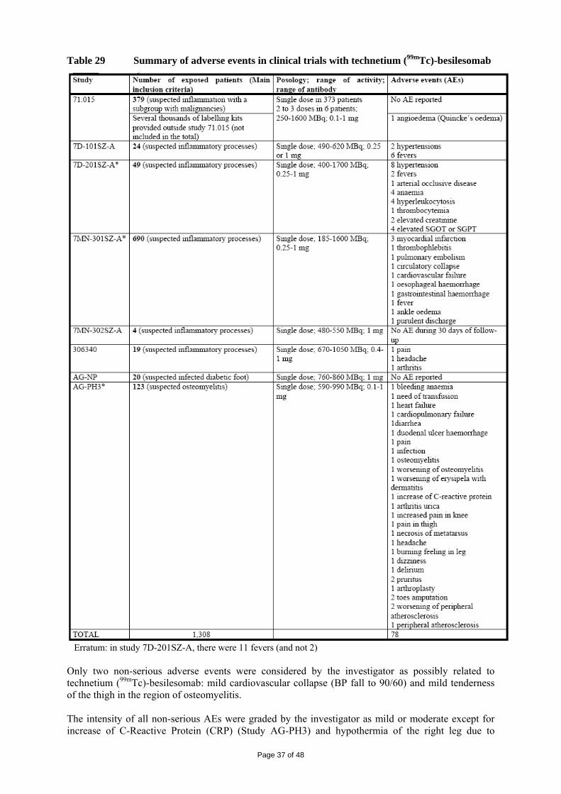
Table 29 Summary of adverse events in clinical trials with technetium (99mTc)-besilesomab
Erratum: in study 7D-201SZ-A, there were 11 fevers (and not 2) Only two non-serious adverse events were considered by the investigator as possibly related to technetium (99mTc)-besilesomab: mild cardiovascular collapse (BP fall to 90/60) and mild tenderness of the thigh in the region of osteomyelitis. The intensity of all non-serious AEs were graded by the investigator as mild or moderate except for increase of C-Reactive Protein (CRP) (Study AG-PH3) and hypothermia of the right leg due to
Page 37 of 48

occlusive arterial disease (Study 7D-201SZ-A) which were considered mild to severe. Both these adverse reactions were considered unrelated to the study drug. A non interventional safety survey was conducted between April 2007 and July 2009 (SG-NISS) and its final results submitted during the procedure. A prospective cohort of 1112 subjects suspected of inflammation/infection were administered technetium (99mTc) besilesomab in the Czech Republic, Hungary, and Austria while 966 subjects received any another radiopharmaceutical used for the same indication (mostly 111In-WBCs, 67Ga, diphosphonates, 99mTc- WBCs) in France and Austria. Of the 966 patients included in the control arm, 119 patients (12.3 %) reported at least one adverse event with a total of 171 adverse events; 61 were considered related to the injected radiopharmaceutical and affected 49 patients. The events most frequently reported were feeling hot, nausea, headache, and paresthesia. Of the 1112 patients that received technetium (99mTc) besilesomab, 31 patients (2.8%) reported at least one adverse event with a total 61 adverse events; 12 were considered related to technetium (99mTc) besilesomab and affected 6 patients, who all recovered:
• anaphylactic reaction with nausea, vomiting, eyelid oedema, hypotension, and circulatory collapse (1)
• anaphylactoid reaction with nausea, vertigo, discomfort (1) • periorbital oedema (1) • urticaria (1) • arthralgia (1) • myalgia (1)
These data are consistent with the clinical data otherwise collected and have been used, with the data from the pivotal clinical trial AG-PH3, as the basis of section 4.8 of the SPC. • Deaths Overall, there were 9 deaths reported but none of the deaths were attributed to technetium (99mTc) besilesomab administration. The causes were cancer (3), myocardial infarction (3), cardiovascular failure (2) and peritonitis with gastrointestinal haemorrhage (1). • Laboratory findings Laboratory tests were performed in some of the trials including study AG-PH3. Immunogenicity In the early clinical studies (71.015, 7D101 SZ-A, 7D-201 SZ-A, 7MN-301 SZ-A, 7MN-302 SZ-A) determination of HAMA was performed using the “Enzygnost HAMA micro” enzyme assay supplied by Behringwerke, Marburg, Germany but were not always performed systematically. The overall percentage of positive HAMA samples was less than 5%. In the recent clinical studies (AG-NP, 306340, AG-PH3) determination of HAMA was performed using the “HAMA-ELISA medac” test supplied by Medac, Wedel, Germany which detects IgG antibodies. HAMA concentrations higher than 40 μg/L were reported as positive. The overall percentage of positive HAMA samples in these studies was 13%. In the new phase III study (AG-PH3), blood samples were collected at screening and twice after treatment (1 and 3 months). Only patients with negative HAMA test at screening were included in the study. Of the 116 patients who had at least one HAMA assessment after administration of technetium (99mTc) besilesomab, 16 patients (14%) showed positive HAMA levels. Seropositivity was observed at 1 month in 13 patients and at 3 months in 3 patients. Of the 13 patients who were positive at 1 month, 3 had no assessment at 3 months, 6 became negative, and 4 remained positive with increasing values in 2 patients and decreasing values in the other 2 patients.
Page 38 of 48

• Safety in special populations Children A retrospective analysis of a series of 30 children with fever of unknown origin is available as a publication22. Dosage was performed according to the recommendations of the Paediatric Task Group of the European Association of Nuclear Medicine; the children received between 100 and 150 μg of the antibody labelled with 185-259 MBq of technetium (99mTc) besilesomab. The publication does not mention any safety concern. Women of childbearing potential When it is necessary to administer radioactive medicinal products to women of childbearing potential, information should always be sought about pregnancy. Any woman who has missed a period should be assumed to be pregnant until proven otherwise. Where uncertainty exists, it is important that radiation exposure should be the minimum consistent with achieving the desired clinical information. Alternative techniques which do not involve ionising radiation should be considered. Pregnancy Scintimun is contraindicated in pregnancy (see section 4.3). Available data is insufficient to address the effects of the product during pregnancy. No reproductive studies have been carried out in animals. Radionuclide procedures carried out on pregnant women also involve radiation doses to the fetus. The dose delivered to the fetus varies with the stage of pregnancy and is reported in the following table:
Stage of pregnancy Equivalent dose in mSv/MBq Three months 0.00379 Six months 0.00335 Nine months 0.00300
Breastfeeding It is not known if the product is excreted in human milk. A risk to a breast-fed child cannot be excluded. Before administering a radioactive medicinal product to a mother who is breast-feeding, consideration should be given as to whether the investigation could reasonably be delayed until the mother has ceased breast-feeding and as to whether the most appropriate choice of radiopharmaceutical has been made, bearing in mind the secretion of activity in breast milk. If the administration is considered necessary, breast-feeding should be interrupted for three days and the expressed milk discarded. These three days correspond to 10 half-lives of technetium (99mTc) (60 hours). At that time the remaining activity represents about 1/1000 of the initial activity in the body. A close contact with the child should also be avoided during the first 12 hours after the injection. • Safety related to drug-drug interactions and other interactions Active substances which inhibit inflammation or affect the haematopoietic system (such as antibiotics and corticosteroids) may lead to false negative results. Such substances should therefore not be administered together with, or a short time before the injection of Scintimun. • Post marketing experience Over the period 1996-2002, 21,023 kits of technetium (99mTc) besilesomab (old product) were sold worldwide, which is assumed to correspond approximately to 63,000 patients exposed to technetium (99mTc) besilesomab. Three spontaneous ADRs were reported: one anaphylactoid reaction (sneezing, facial swelling and breathing difficulties), one death with limited information (pulmonary oedema and acute respiratory insufficiency during an hospitalisation for necrosis of the toe) and one cardiac arrest with respiratory insufficiency 10 minutes after an injection of technetium (99mTc) besilesomab. Over the period 2002-2008, it is estimated that 36,610 patients were administered technetium (99mTc) besilesomab (new product). During this period, three SAEs were reported in three patients: a stroke
Page 39 of 48

one day post-injection of technetium (99mTc) besilesomab injection and two deaths from sepsis, all attributed to the underlying disease. One non-serious cutaneous reaction was also reported. • Discussion on clinical safety There is sufficient exposure of technetium (99mTc) besilesomab to single administration of the old and new product in clinical trials and supplemented with substantial post-marketing data/non interventional survey to assess the safety profile of technetium (99mTc)-besilesomab. In general, technetium (99mTc) besilesomab appears to be well tolerated with few reported AEs. The most serious and fatal events reported were related to the underlying diseases, post-operative complications and/or cardiovascular history of the patients, with no indication of a causal relationship to the radiotracer. There were some cases of hypersensitivity reactions that have been reported both in clinical trials and in post-marketing experience although allergic reactions appear to be rare. However, acute blood pressure declines occurring shortly after the injection of technetium (99mTc) besilesomab were observed in several patients, including three in the AG-PH3 trial; whether mediated by hypersensitivity mechanisms or independently of them, the causal role of technetium (99mTc) besilesomab in this phenomenon could not be ruled out. As shown in the recent clinical trials, there is a significant risk of development of HAMAs to besilesomab; the higher rate of development of HAMAs in the new studies compared with the old studies is likely due to higher sensitivity of the quantitative “HAMA-ELISA medac” test used in the new studies. However, this rate is consistent with literature data after a single injection of murine antibody. In line with the clinical trials, a screening for HAMA should be performed before any administration of besilesomab and a positive test is regarded as a contraindication (see SPC section 4.3). Likewise, repeated administration is not recommended as immunogenicity is an issue, which has been reflected in the SPC section 4.4. The safety issue associated with hypersensitivity reactions, acute hypotension and immunogenicity are adequately addressed in the RMP and in the Direct Healthcare Professional Communication (DHPC) letter. The laboratory parameters (haematology and coagulation) do not appear to be affected by the administration of technetium (99mTc) besilesomab since no clinically relevant findings were observed. The information on the safety profile in children is limited, thus no conclusion can be drawn from the information. The use of technetium (99mTc) besilesomab in children is not recommended and this is included in the SPC section 4.2. Taking into account the data of equivalent dose to the fetus at different stages of pregnancy, the exposure would be higher than the limit of 1 mSv if the recommended posology of technetium (99mTc) besilesomab for a pregnant woman is higher than 264 to 334 MBq. Therefore, the use in pregnancy is contraindicated (see SPC section 4.6) Technetium (99mTc) besilesomab is contraindicated in patients who have hypersensitivity to the active substance, to other murine antibodies or to any of the excipients, have a positive screening test for human anti-mouse antibody (HAMA) and in pregnancy (see SPC 4.3 and section 4.6). References 1 Boubaker A, Bischof-Delaloye A, Blanc CH, Dutoit M, Leyvraz PF, Delaloye B. Immunoscintigraphy with antigranulocyte monoclonal antibodies for the diagnosis of septic loosening of hip prostheses. Eur J Nucl Med 1995; 22 (2): 139-147 2 Gallowitsch HJ, Heinisch M, Mikosch P, Kresnik E, Kumnig G, Gomez I, Lind P; Tc-99m ciprofloxacin in clinically selected patients suspected for peripherial osteomyelitis, spondylodiscitis and fever of unknown origin: preliminary results. Nuklearmedizin 2002; 41:30-6. 3 Gratz S, Braun HG, Behr TM, Meller J, Herrmann A, Conrad M, Rathmann D, Bertagnoli R, Willert HG, Becker W. Photopenia in Chronic Vertebral Osteomyelitis with Technetium-99m-Antigranulocyte Antibody (BW 250/183). J Nucl Med 1997; 38 (2): 211-6
Page 40 of 48

4 Guhlmann A, Brecht-Krauss D, Suger G, Glatting G, Kotzerke J, Kinzl L, Reske SN. Fluorine-18-FDG PET and Technetium-99m Antigranulocyte Antibody Scintigraphy in Chronic Osteomyelitis. J Nucl Med 1998; 39 (12): 2145-52 5 Horger M, Eschmann SM, Pfannenberg C, Storek D, Dammann F, Vonthein R, Claussen CD, Bares R. The value of SPET/CT in chronic osteomyelitis. Eur. J. Nucl. Med. Mol. Imaging 2003, 30:1665-73. 6 Hotze AL, Briele B, Overbeck B, Kropp J, Gruenwald F, Mekkawy MA, von Smekal A, et al. Technetium-99m-Labeled Anti-Granulocyte Antibodies in Suspected Bone Infections. J Nucl Med 1992; 33: 526-531 7 Kaim A, Ledermann HP, Bongartz G, Messmer P, Müller-Brand J, Steinbrich W. Chronic post-traumatic osteomyelitis of the lower extremity: comparison of magnetic resonance imaging and combined bone scintigraphy/immunoscintigraphy with radiolabelled monoclonal antigranulocyte antibodies. Skeletal Radiol. 2000; 29:378-386. 8 Klett R, Kordelle J, Stahl U, Khalisi A, Puille M, Steiner D, Bauer R. Immunoscintigraphy of septic loosening of knee endoprosthesis : a retrospective evaluation of the antigranulocyte antibody BW250/183. Eur J Nucl Med Mol Imaging 2003; 30:1463-66. 9 Kordelle J, Klett R, Stahl U, Hossain H, schleicher I, Haas H. Infektdiagnostik nach Knie-TEP-Implantation. Z. Orthop. 2004:142:337-343. 10 Kroiss A, Böck F, Perneczky G, Auinger C, Weidlich G, Kleinpeter G, Brenner H. [Immunoscintigraphy for detection of inflammatory processes in diseases of bones and joints]. Immunszintigraphie zur Aufdeckung von Entzündungsherden bei Knochen- und Gelenkserkrankungen. Wiener Klin Wochenschr 1990; 102: 713-717 11 Peltier P, Potel G, Lovat E, Baron D, Chatal JF. Detection of lung and bone infection with anti-granulocyte monoclonal antibody BW 250/183 radiolabelled with 99Tcm. Nucl Med Comm 1993; 14: 766-774 12 Reuland P, Winker KH, Heuchert T, Ruck P, Müller-Schauenburg W, Weller S, Feine U. Detection of Infection in Postoperative Orthopedic Patients with Technetium-99m-Labeled Monoclonal Antibodies against Granulocytes. J Nucl Med 1991; 32: 2209-2214 13 Sciuk J, Brandau W, Vollet B, Stücker R, Erlemann R, Bartenstein P, Peters PE, Schober O. Technetium-99m polyclonal human immunoglobulin and technetium-99m monoclonal antibodies for imaging chronic osteomyelitis. Eur J Nucl Med 1991;18:401-407 14 Dominguez-Gadea L, Martin-Curto LM, de la Calle H, Crespo A. Diabetic foot infections: scintigraphic evaluation with 99Tcm-labelled anti-granulocyte antibodies. Nucl Med Comm 1993; 14: 212-218 15 Almers S, Granerus G, Franzén L, Ström M. Technetium-99m scintigraphy: more accurate assessment of ulcerative colitis with exametazime-labelled leucocytes than with antigranulocyte antibodies. Eur J Nucl Med 1996; 23 (3): 247-55 16 Papós M, Nagy F, Nárai G, Rajtár M, Szántai G, Láng J, Csernay L. Anti-granulocyte Immunoscintigraphy and [99mTc]Hexamethylpropyleneamine-Oxime-Labeled Leukocyte Scintigraphy in Inflammatory Bowel Disease. Dig Dis Sci 1996; 41 (2): 412-20 17 Segarra I, Roca M, Baliellas C, Vilar L, Ricart Y, Mora J, Puchal R, Martin-Comin J. Granulocyte-specific monoclonal antibody technetium-99m-BW 250/183 and indium-111 oxine-labelled leucocyte scintigraphy in inflammatory bowel disease. Eur J Nucl Med 1991; 18: 715-719 18 Tarján Z, Tóth G, Györke T, Mester A, Karlinger K, Makó EK. Ultrasound in Crohn's disease of the small bowel. Eur J Radiol 2000; 35 (3): 176-182 19 Becker W, Dölkemeyer U, Gramatzki M, Schneider MU, Scheele J, Wolf F. Use of immunoscintigraphy in the diagnosis of fever of unknown origin. Eur J Nucl Med 1993; 20 (11): 1078-1083 20 Meller J, Ivancevic V, Conrad M, Gratz S, Munz DL, Becker W. Clinical Value of Immunoscintigraphy in Patients with Fever of Unknown Origin. J Nucl Med 1998; 39 (7): 1248-53
Page 41 of 48

21 Gratz S, Behr TM, Herrmann A, Meller J, Conrad M, Zappel H, Becker W. Immunoscintigraphy (BW 250/183) in neonates and infants with fever of unknown origin. Nucl Med Commun 1998; 19 (11): 1037-45 22 Morguet AJ, Munz DL, Ivančević V, Werner GS, Kreuzer H. [Clinical value of radioimaging using the murine monoclonal antigranulocyte antibody BW 250/183 in the diagnosis of prosthesic-valve endocarditis]. Klinische Bedeutung der Szintigraphie mit dem murinen monoklonalen Antigranulozyten-Antikörper BW 250/183 für die Diagnose der Prothesenendokarditis. Dtsch med Wschr 1995; 120 (24): 861-6 23 Morguet AJ, Munz DL, Ivančević V, Werner GS, Sandrock D, Bökemeier M, Kreuzer H. Immunoscintigraphy Using Technetium-99m-Labeled Anti-NCA-95 Antigranulocyte Antibodies as an Adjunct to Echocardiography in Subacute Infective Endocarditis. J Am Coll Cardiol 1994; 23 (5): 1171-1178 24 Duncker CM, Carrió I, Berná L, Estorch M, Alonso C, Ojeda B, Blanco R, Germá JR, Ortega V. Radioimmune Imaging of Bone Marrow in Patients with Suspected Bone Metastases from Primary Breast Cancer. J Nucl Med 1990; 31: 1450-1455 (Comment in: J Nucl Med 1991; 32: 549-51) 25 Prior JO, Barghouth G, Delaloye JF, Leyvraz S, Bischof Delaloye A. The value of bone marrow scintigraphy using 99mTc monoclonal antigranulocyte antibodies in complement to bone scintigraphy in detecting bone metastases from primary breast cancer. Nucl Med Comm 2003; 24:29-36 26 Reske SN, Glöckner WM, Schwarz A, Karstens JH, Steinstrasser A, Ammon J, Büll U. Radioimmunoimaging for diagnosis of bone marrow involvement in breast cancer and malignant lymphoma. Lancet 1989; Feb 11: 299-301 27 Reske SN, Karstens JH, Henrich MM, Sohn M, Glöckner W, Jakse G, Büll U. [Detection of skeletal involvement in malignant disease by immunoscintigraphy of bone marrow]. Nachweis des Skelettbefalls maligner Erkrankungen durch Immunszintigraphie des Knochenmarks. Nuklearmedizin 1993; 32: 111-119 28 Sohn SK, Ahn BC, Lee SW, Kim DH, Chun KA, Kim JG, Park SH, et al. Bone marrow immunoscintigraphy using technetium-99m anti-granulocyte antibody in multiple myeloma. Eur J Nucl Med 2002; 29:591-596 2.5 Pharmacovigilance Detailed description of the Pharmacovigilance system The CHMP considered that the Pharmacovigilance system as described by the applicant fulfils the legislative requirements. Risk Management Plan The MAA submitted a risk management plan, which included a risk minimisation plan. Table 30 Summary of the risk management plan Safety concern: Important potential risks
Proposed pharmacovigilance activities (routine and additional)
Proposed risk minimisation activities (routine and additional)
Possible consequences of HAMA positivity
1.Routine Pharmacovigilance 2.Dear Doctor Letter
Dear Doctor Letter to be sent to all nuclear medicine specialists including a warning of the potential risks of human anti-mouse antibody (HAMA) generation Contraindication in Section 4.3 of the SPC: Contraindications: Positive screening test for human anti-mouse antibody (HAMA)
Page 42 of 48

Warnings in Section 4.4 of the SPC : Administration of murine monoclonal antibodies can lead to the development of Human Anti-Mouse Antibodies (HAMA). Patients who are HAMA positive may have a greater risk for hypersensitivity reactions. Inquiry on possible previous exposure to murine monoclonal antibodies and a HAMA test should be made prior to administration of Scintimun; a positive response would contraindicate the administration of Scintimun (see section 4.3). “Scintimun should only be used once in a patient’s lifetime”. Labelled in section 4.8 of the SPC
Radiation exposure during pregnancy
Routine Pharmacovigilance Contraindication in section 4.3 of the SPC of use during pregnancy Warning in section 4.6 of the SPC that “When it is necessary to administer radioactive medicinal products to women of childbearing potential, information should always be sought about pregnancy. Any woman who has missed a period should be assumed to be pregnant until proven otherwise. Where uncertainty exists, it is important that radiation exposure should be the minimum consistent with achieving the desired clinical information. Alternative techniques which do not involve ionising radiation should be considered”. “Scintimun is contraindicated in pregnancy. Available data are insufficient to address the effects of the product during pregnancy. No reproductive studies have been carried out in animals. Radionuclide procedures carried out on pregnant women also involve radiation doses to the foetus.”
Carcinogenicity / hereditary defects
Routine Pharmacovigilance Warning in section 4.4 of the SPC that “For all patients, the radiation exposure must be justifiable by the expected diagnostic achieved with the lowest possible radiation dose.” Information in section 4.8 of the SPC: “For all patients, the radiation exposure must be justifiable by the expected diagnostic achieved with the lowest possible radiation dose.” Exposure to ionising radiation is linked with cancer
Page 43 of 48

induction and a potential for development of hereditary defects. For diagnostic nuclear medicine investigations the frequency of these adverse reactions is not known. After administration of the maximum recommended activity of technetium (99mTc) besilesomab of 800 MBq, the effective dose is about 6.9 mSv.
Acute hypotension 1.Routine Pharmacovigilance 2.Reporting to EMEA of cases (regardless of seriousness) within 15 days
Dear Doctor Letter to be sent to all nuclear medicine specialists including a warning of the risks of acute hypotension Warning in section 4.4 of the SPC that Appropriate cardiopulmonary resuscitation facilities and trained personnel should be available for immediate use in the event of an adverse reaction. Labelled in section 4.8 of the SPC
Missing lesions
1.Routine Pharmacovigilance 2. Reporting to EMEA of cases (regardless of seriousness) within 15 days
Warning in section 4.4 of the SPC regarding the interpretation of Scintium imaging There are currently no criteria to distinguish infection and inflammation by means of Scintimun imaging. Scintimun images should be interpreted in the context of other appropriate anatomical and/or functional imaging examinations. Only limited data is available about binding of technetium (99mTc) besilesomab to CarcinoEmbryonic Antigen (CEA) expressing tumours in vivo. In vitro, besilesomab cross-reacts with CEA. False positive findings in patients with CEA expressing tumours cannot be excluded. False results may be obtained in patients with diseases involving neutrophil defects and to patients with haematological malignancies including myeloma.
Administration errors due to mixing up the vials
1.Routine Pharmacovigilance 2. Reporting to EMEA of cases (regardless of seriousness) within 15 days
Information in section 4.2 of the SPC The following sentence will be printed on the carton box label: “Reconstitute Scintimun with its solvent first and then radiolabel with a sodium pertechnetate (99mTc) solution. Read the leaflet for further information before use.”
Repeat use Routine Pharmacovigilance Warning in section 4.4 - Special warnings and precaution of use – Re-administration: “Data on repeated dosing of Scintimun are very limited. Scintimun should only
Page 44 of 48
be used once in a patient’s lifetime”. Safety and efficacy of Scintimun in patients under the age of 18
Routine Pharmacovigilance Information in Section 4.2 - Posology and method of administration that: Scintimun is not recommended for use in children below age 18 due to insufficient data on safety and efficacy
Use in patients with liver or renal impairment
Routine Pharmacovigilance
Information in Section 4.2 - Posology and method of administration): “Formal studies have not been performed in patients with renal or liver impairment. However, due to the nature of the molecule and the short half-life of technetium (99mTc) besilesomab, dosage adjustment is not necessary in such patients”.
The CHMP, having considered the data submitted in the MA application is of the opinion that the risk minimisation activities described in the above table are necessary for the safe and effective use of the medicinal product. 2.6 Overall conclusions, risk/benefit assessment and recommendation Quality In general, the different aspects of the chemical, pharmaceutical and biological documentation comply with existing guidelines. The fermentation and purification of the drug substance are adequately described, controlled and validated. The drug substance is well characterised with regard to its physicochemical and biological characteristics, using state-of the-art methods, and appropriate specifications are set. The manufacturing process of the drug product has been satisfactorily described and validated. The quality of the drug product is controlled by adequate test methods and specifications. Viral safety and the safety concerning other adventitious agents including TSE have been sufficiently assured. Except for a number of points, which will be addressed as part of post-approval follow-up measures, the overall Quality of Scintimun is considered acceptable. Non-clinical pharmacology and toxicology Whole blood concentration-time radioactivity curves show a two-phase course, which can be subdivided into an early phase (0-2 h) and a late phase (5-24 h). After correcting for the decay of radionuclide, the calculated half-life of the early phase is 0.5 h whereas the late phase shows a half-life of elimination of 16 h. Six hours after injection, about 1.5% of the whole body radioactivity is found in the liver whereas about 3.0% is found in the spleen. Twenty-four hours after injection, the percentages of radioactivity are 1.6% in the liver and 2.3% in the spleen. Measurement of radioactivity levels in urine shows that up to 14% of the administered activity is excreted via the bladder during the 24 h post-injection. The low renal clearance of activity (0.2 l/h for a glomerular filtration rate around 7 l/h) indicates that the kidney is not the major route of besilesomab elimination. Non pathological unusual accumulations may be observed in the spleen (up to 6% of the patients), in the bowel (up to 4% of the patients), in the liver and bone marrow (up to 3% of the patients), and in the thyroid and kidneys (up to 2% of patients). Preclinical toxicity and safety studies were performed using commercial kits reconstituted with decayed technetium and thus the effect of radiation has not been assessed.
Page 45 of 48

Preclinical data obtained with the non-radioactive compound reveal no special hazard for humans based on conventional studies of safety pharmacology, single-dose and repeated-dose toxicity, although antimurine antibodies were found in all dose groups (including controls) in a repeated-dose study in monkeys. Genotoxicity studies conducted to test for potentially genotoxic impurities were also negative. No animal studies regarding carcinogenic potential and toxicity to reproduction were performed. Efficacy In a randomised cross-over trial comparing blinded reading of Scintimun and 99mTc-White Blood Cells (WBCs) images in 119 patients with suspected osteomyelitis, the agreement rate between the two methods was 83% (lower 95% confidence interval limit: 80%). A major deficiency identified in the pivotal trial AG-PH3 was the lack of an adequate standard of truth or surrogate. Therefore, diagnostic parameters like sensitivity and specificity could not be calculated and only agreement rate could be estimated. The lack of standard of truth in the AG-PH3 trial was addressed in a post-hoc analysis based on a surrogate, which was an evaluation by an expert panel of all data available from the study CRFs plus all additional data available from the investigators. The overall diagnostic performance of technetium (99mTc) besilesomab in this analysis, sensitivity (76%) and specificity (69%) appeared similar to that of the previous analysis based on the investigator diagnosis at 1 month, 75% and 72%, respectively. These results and those from the literature, suggest that the sensitivity of technetium (99mTc) besilesomab is equivalent to that of 99mTc WBCs but that specificity is likely lower. The CHMP acknowledged that a lower specificity could be acceptable if technetium (99mTc) besilesomab would be used in conjunction with other imaging modalities to determine the appropriate diagnosis. However, there are insufficient data on the use of Scintimun for the diagnosis of diabetic foot infection. There are no data in the paediatric population for the indication of osteomyelitis. Thus, technetium (99mTc) besilesomab is not recommended for use in children below 18 years of age due to insufficient data on safety and efficacy (see SPC section 4.2, 5.1 and 5.2). There are insufficient data on the use of Scintimun for the diagnosis of diabetic foot infection. Safety From the evidence from clinical trials and post-marketing surveillance available Scintimun was considered to exhibit a good safety profile. Radiopharmaceuticals should be received, used and administered only by authorised persons in designated settings. Their receipt, storage, use, transfer and disposal are subject to the regulations and the appropriate authorisations of the competent authorities. Scintimun contains sorbitol therefore patients with rare hereditary problems of fructose intolerance should not be administered this product. This medicinal product contains less than 1 mmol sodium (23 mg) per dose administered, i.e. is essentially ‘sodium- free’. Immunogenicity Administration of murine monoclonal antibodies can lead to the development of Human Anti-Mouse Antibodies (HAMA). Patients who are HAMA positive may have a greater risk for hypersensitivity reactions. Inquiry on possible previous exposure to murine monoclonal antibodies and a HAMA test should be made prior to administration of Scintimun; a positive response would contraindicate the administration of Scintimun (see section 4.3). Repeated use Data on repeated dosing of Scintimun are very limited. Scintimun should only be used once in a patient’s lifetime.
Page 46 of 48

Hypersensitivity reactions Anaphylactic or anaphylactoid reactions may occur after administration of the medicinal product. Appropriate cardiopulmonary resuscitation facilities and trained personnel should be available for immediate use in the event of an adverse reaction. Since allergic reactions to the murine protein cannot be excluded, cardiovascular treatment, corticosteroids, and antihistamines must be available during administration of the product. Risks related to radioactivity The administration of radiopharmaceuticals creates risks for other persons from external radiation or contamination from spills of urine, vomiting, expectoration or other biological fluids. Radiation protection precautions in accordance with national regulations must be taken. Any unused product or waste material should be disposed of in accordance with local requirements. No case of overdose has been reported. If an overdose of technetium (99mTc) besilesomab has been administered, the radiation dose delivered to the patient must be reduced by increasing as much as possible the elimination of the radionuclide by forced diuresis and frequent micturitions, and by the use of laxatives to promote faecal excretion. No studies on the effects on the ability to drive and use machines have been performed. At the recommended activity for diagnostic procedures, Scintimun is not likely to have any influence on the ability to drive and use machines. From the safety database all the adverse reactions reported in clinical trials and post-marketing have been included in the Summary of Product Characteristics. Having considered the safety concerns in the risk management plan, the CHMP considered that the proposed activities described in section 2.5 adequately addressed these. • User consultation The CHMP considered the user testing conducted on the package leaflet and its results acceptable in spite of the difficulty of the questionnaire and the methodology employed. The layout of the PL is acceptable. Risk-benefit assessment There is a definite need for a specific imaging technique of neutrophil clustering that is safe and has robust technical performance, diagnostic performance and a defined impact of diagnostic thinking in the case of suspected osteomyelitis. The CHMP noted the benefits of technetium (99mTc) besilesomab administration for in vivo labelling of granulocytes in terms of the good quality of images (technical performance) achieved with technetium (99mTc) besilesomab and the acceptable safety profile of technetium (99mTc) besilesomab. The comparative data against 99mTc-WBCs in the pivotal study AG-PH3 have been generated in the diagnosis of peripheral osteomyelitis (including diabetic foot and joint prosthesis) and had an agreement rate of 80%. Sensitivity and specificity was analysed in a post-hoc analysis and was found within the acceptable margins and limits of other scintigraphic imaging techniques. However, a number of methodological issues were unresolved following the scientific assessment. The applicant, at an oral explanation, presented data addressing remaining issues concerning the lack of internal validity of the pivotal trial AG-PH3, the potential bias and heterogeneity from the literature meta-analysis review, the diagnostic performance in relation to the standard of truth and the impact of diagnostic performance and clinical relevance in relation to the indication. The applicant also addressed the discrepancies in diagnosis between technetium (99mTc) besilesomab and 99mTc-WBCs in a post-hoc analysis. Given the data submitted, the justifications presented by the applicant, the discussion following the oral explanation, the expert views presented by the SAG-D, the CHMP concluded that a consistent diagnostic efficacy has been established for technetium (99mTc) besilesomab, in particular, based on
Page 47 of 48

Page 48 of 48
adequate agreement rate and similar sensitivity for technetium (99mTc) besilesomab compared to 99mTc WBCs, albeit at the expense of a lower specificity. This in vivo technique may be of benefit in clinical practice when used in conjunction with the available diagnostic tools, particularly in situations where in vivo diagnostic methods are preferred compared to ex vivo methods. The CHMP noted that, although technetium (99mTc) besilesomab would require higher radioactivity than 99mTc-WBCs, there is no safety concern with a maximum exposure of 6.9 mSv as this is within the range of other diagnostic nuclear medicine techniques, except in pregnancy, which is a contraindication. Technetium (99mTc) besilesomab was well tolerated. The safety profile of technetium (99mTc) besilesomab has been adequately characterised based on the safety data presented from the clinical trial and the extensive the post-marketing experience. Thus, the CHMP was of the opinion that the benefits of technetium (99mTc) besilesomab outweight the risks in the restricted indication: “Scintigraphic imaging, in conjunction with other appropriate imaging modalities, for determining the location of inflammation/infection in peripheral bone in adults with suspected osteomyelitis.”. Administration of a murine antibody increases the risk of developing HAMAs. Therefore, due to its immunogenicity, technetium (99mTc) besilesomab should be restricted to single use. The precise extent of the impact of technetium (99mTc) besilesomab on diagnostic thinking and patient management in clinical practice cannot be estimated based on the data submitted. The applicant has committed to perform a randomised parallel-group trial in the approved indication to formally address this issue. The CHMP was of the opinion that the evidence in diabetic foot infection was not established from both the pivotal trial and the literature meta-analysis. Furthermore, since MRI has become the imaging tool of choice in this indication, diabetic foot infection should be excluded from the indication for technetium (99mTc) besilesomab. Therefore, the restricted indication for technetium (99mTc) besilesomab includes the wording “Scintimun should not be used for the diagnosis of diabetic foot infection”. The Marketing Authorisation Holder shall ensure that, at launch, all nuclear medicine specialists who are expected to prescribe/use Scintimun are provided with a Direct Healthcare Professional Communication (DHPC; Dear Doctor letter). A risk management plan was submitted. The CHMP, having considered the data submitted, was of the opinion that:
routine pharmacovigilance was adequate to monitor the safety of the product; Additional risk minimisation activities were required: see as detailed in section 2.5. Recommendation Based on the CHMP review of data on quality, safety and efficacy, the CHMP considered by consensus that the risk-benefit balance of Scintimun in the indication: “This medicinal product is for diagnostic use only. Scintigraphic imaging, in conjunction with other appropriate imaging modalities, for determining the location of inflammation/infection in peripheral bone in adults with suspected osteomyelitis. Scintimun should not be used for the diagnosis of diabetic foot infection” was favourable and therefore recommended the granting of the marketing authorisation.