atherectomy and ivus -...
TRANSCRIPT

Atherectomy and IVUS

IVUS Image of Healthy Vessel
600-0100.167/LD
Media
Intima Adventitia
Results are not predictive of future outcomesImage Courtesy of Philips Volcano

Material between the green line (intima) and red line (adventitia) is fibrotic plaque
IVUS Image of a Diseased Vessel
Results are not predictive of future outcomesImage Courtesy of Philips Volcano
600-0100.167/LD

Corresponding IVUS image demonstrates lumen gain at the expense of adventitial injury.
IVUS Post Directional Atherectomy
Results are not predictive of future outcomesIVUS Image Courtesy of Dr. Dippel
600-0100.167/LD
This specific case shows a sub-optimal outcome. However, presented case result may not be indicative of all directional atherectomy case results.

Corresponding IVUS image demonstrates lumen gain at the expense of adventitial injury.
IVUS Post Directional Atherectomy
Results are not predictive of future outcomes.IVUS images obtain from actual cases with consent from the clinician. Data on file at Philips Volcano.
Adventitial Injury
600-0100.167/LD
This specific case shows a sub-optimal outcome. However, presented case result may not be indicative of all directional atherectomy case results.

IVUS Post Directional Atherectomy
Corresponding IVUS image demonstrates lumen gain at the expense of adventitial injury.
Results are not predictive of future outcomes.IVUS Image Courtesy of Dr. Prakash Krishnan
600-0100.167/LD
This specific case shows a sub-optimal outcome. However, presented case result may not be indicative of all directional atherectomy case results.

IVUS Post Directional AtherectomyCorresponding IVUS image demonstrates lumen gain at the expense of adventitial injury.
Adventitial Injury
Results are not predictive of future outcomes.IVUS images obtain from actual cases with consent from the clinician. Data on file at Philips Volcano.
600-0100.167/LD
This specific case shows a sub-optimal outcome. However, presented case result may not be indicative of all directional atherectomy case results.

Corresponding IVUS image demonstrates lumen gain at the expense of adventitial injury.
IVUS Post Orbital Atherectomy
Adventitial Injury
Results are not predictive of future outcomes. IVUS Image Courtesy of Dr. Staniloae
600-0100.167/LDThis specific case shows a sub-optimal outcome. However, there any many instances of orbital atherectomy where
there was a successful result.

Corresponding IVUS image demonstrates lumen gain at the expense of adventitial injury.
IVUS Post Orbital Atherectomy
Results are not predictive of future outcomes.IVUS images obtain from actual cases with consent from the clinician. Data on file at Philips Volcano.
Adventitial Cut
600-0100.167/LD
This specific case shows a sub-optimal outcome. However, presented case result may not be indicative of all orbital atherectomy case results.

Corresponding IVUS image demonstrates lumen gain at the expense of creating a dissection.
Dissection with Orbital Atherectomy
Results are not predictive of future outcomes.IVUS Image Courtesy of Dr. Staniloae
Dissection
600-0100.167/LD
This specific case shows a sub-optimal outcome. However, presented case result may not be indicative of all orbital atherectomy case results.

Corresponding IVUS image demonstrates lumen gain at the expense of creating a dissection.
Lumen Gain at Expense of a Dissection
Results are not predictive of future outcomes.IVUS images obtain from actual cases with consent from the clinician. Data on file at Philips Volcano.
Dissection
600-0100.167/LD
This specific case shows a sub-optimal outcome. However, presented case result may not be indicative of all orbital atherectomy case results.
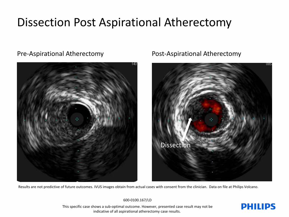
Dissection Post Aspirational Atherectomy
Results are not predictive of future outcomes. IVUS images obtain from actual cases with consent from the clinician. Data on file at Philips Volcano.
Pre-Aspirational Atherectomy Post-Aspirational Atherectomy
Dissection
600-0100.167/LD
This specific case shows a sub-optimal outcome. However, presented case result may not be indicative of all aspirational atherectomy case results.

IVUS performed on the left SFA showed localized dissection in the proximal and mid segments of the vessel.
IVUS demonstrated a significant dissection throughout the length of the right SFA.
Dissection Post Directional AtherectomyIVUS with ChromaFlo was used to assess the result post-atherectomy and balloon dilatation.
Results are not predictive of future outcomes.IVUS images provided by Dr. Dippel
Left SFA Post-Atherectomy
Dissection
Right SFA Post-Atherectomy
Dissection
600-0100.167/LD
This specific case shows a sub-optimal outcome. However, presented case result may not be indicative of all directional atherectomy case results.
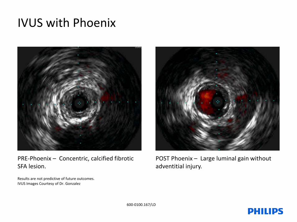
PRE-Phoenix – Concentric, calcified fibrotic SFA lesion.
POST Phoenix – Large luminal gain without adventitial injury.
IVUS with Phoenix
Results are not predictive of future outcomes.IVUS Images Courtesy of Dr. Gonzalez
600-0100.167/LD

PRE-Phoenix – Concentric, non-calcified fibrotic SFA lesion.
POST Phoenix – Large luminal gain without adventitial injury.
IVUS with Phoenix
Results are not predictive of future outcomesIVUS Images Courtesy of Dr. Davis
600-0100.167/LD

PRE-Phoenix POST Phoenix 2.4 - Large luminal gain without adventitial injury.
IVUS with Phoenix
Results are not predictive of future outcomes.IVUS Images Courtesy of Dr. Bennett
600-0100.167/LD

Phoenix is Your Atherectomy Solution
Vessel Injury• Front cutter clears tissue in a way that may help reduce potential trauma to the vessel
Distal Embolization• Continuous capture and passive clearance of debulked material into the catheter resulted in a 1% rate of
symptomatic distal emboli1 in the EASE trial
Ease of Use• Single insertion - no need to remove and clean out debulked material• Battery powered handle operated - no capital equipment or additional procedural accessories required
Deliverability• Low profile, front cutting design allows for direct lesion access without having to first pass a nosecone• OTW design aids in trackability and pushability of catheter
Versatility• Treat a range of tissue types from soft plaque to calcified arteries• Treat most peripheral vasculature with only 3 catheter diameters2
1. Endovascular Atherectomy Safety and Effectiveness Study (EASE), ClinicalTrials.gov Identifier NCT01541774 (accessed 23Oct2015). Results presented at the Vascular Interventional Advances (VIVA) Conference in October of 2013 (Las Vegas, NV) by Stephen Williams, MD 2. Phoenix Atherectomy device is indicated for vessels 2.5mm in diameter and above
600-0100.167/LD

IVUS Can Assist You
Image courtesy of Philips Volcano
AssessCompleteness of Treatment
Is the stent fully apposed?
Did I cover the area of interest?
Did I achieve the necessary luminal gain?
Did I cut into the adventitia?
600-0100.167/LD

IVUS Can Assist You
Images Courtesy of Philips Volcano
Pre-Treatment Strategy
What is the size of the vessel to be treated?
What type of plaque morphology does the patient have?
Where does the lesion start and end?
600-0100.167/LD

600-0100.167/LD