atlas antibodies in breast cancer … 5 (20) monoclonal antibody development atlas antibodies also...
TRANSCRIPT
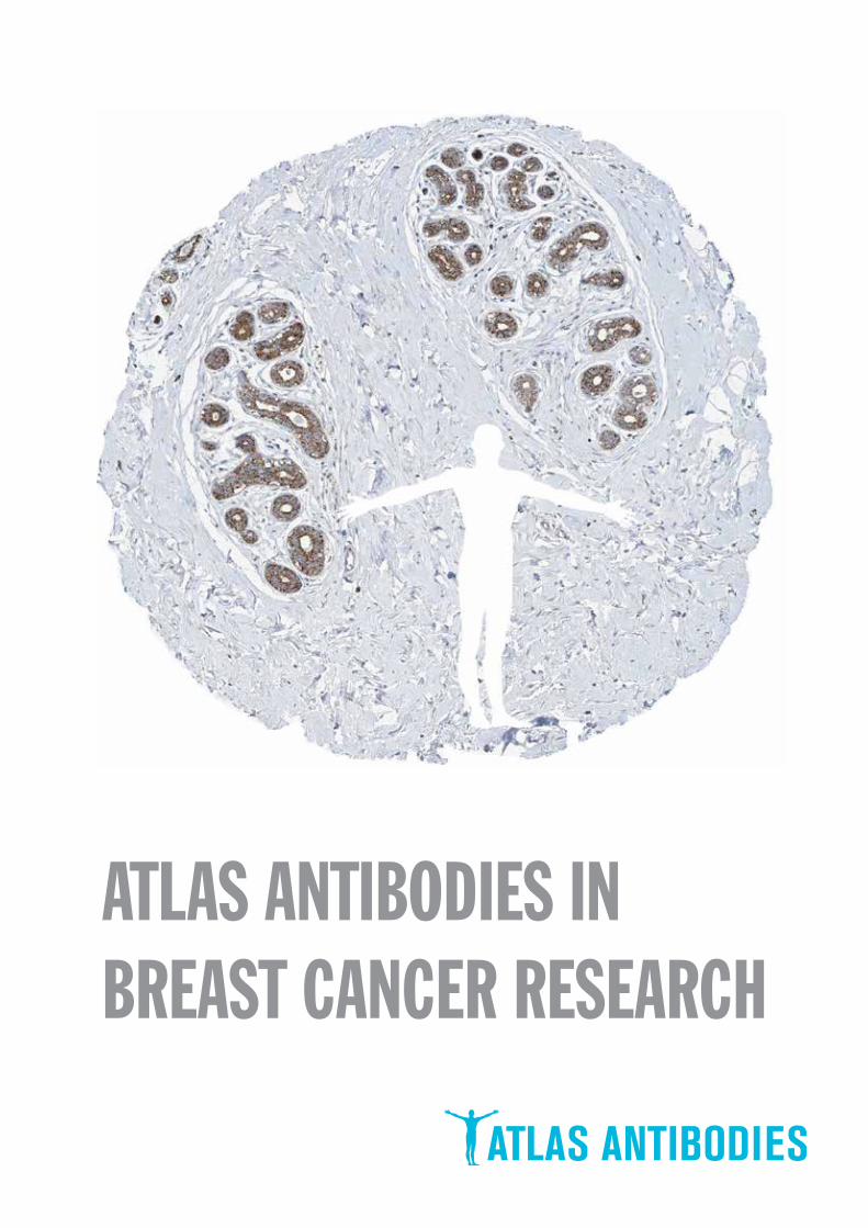
ATLAS ANTIBODIES INBREAST CANCER RESEARCH
Antibodies against MammaPrint and other gene expression test proteins (10-12)
Clinical markers (6)
Antibodies identified in the Human Protein Atlas (13-14)
The Human Protein Atlas, Triple A Polyclonals and monoclonal antibody development (4-5)
Antibodies used in breast cancer research (7-9)
Monoclonal antibody development program (19)
Finding cancer biomarkers, as exemplified by RBM3, granulin and anillin (15-18)
TABLE OF CONTENTS
Contact (20)
Page 2 (20)
Page 3 (20)
proteinatlas.org
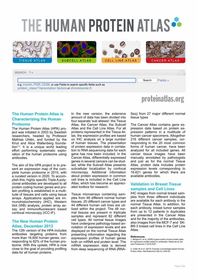
The Human Protein Atlas is Characterizing the Human ProteomeThe Human Protein Atlas (HPA) pro-ject was initiated in 2003 by Swedish researchers, headed by Professor Mathias Uhlén, and funded by the Knut and Alice Wallenberg founda-tion1,2. It is a unique world leading effort performing systematic explo-ration of the human proteome using antibodies.
The aim of the HPA project is to pre-sent an expression map of the com-plete human proteome in 2015, with a curated version in 2020. To accom-plish this, highly specific Triple A poly-clonal antibodies are developed to all protein coding human genes and pro-tein profiling is established in a multi-tude of tissues and cells using tissue arrays. Applications applied are im-munohistochemistry (IHC), Western blot (WB) analysis, protein array as-say and immunofluorescent based confocal microscopy (ICC-IF).
The New Human Protein Atlas, December 2013The 12th version of the HPA includes antibodies targeting proteins from more than 16,600 human genes, cor-responding to 82% of the human pro-teome. With this update, HPA is now close to the goal of providing profiling data for all human proteins.
In the new version, the extensive amount of data has been divided into four separate 'sub atlases': the Tissue Atlas, the Cancer Atlas, the Subcell Atlas and the Cell Line Atlas. For all proteins represented in the Tissue At-las, the expression profiles are based on IHC analysis on a large number of human tissues. The presentation of protein expression data in correla-tion to RNA sequencing data for each gene has now been included. In the Cancer Atlas, differentially expressed genes in several cancers can be stud-ied, while the Subcell Atlas presents subcellular localization by confocal microscopy. Additional information about protein expression in common cell lines is included in the Cell Line Atlas, which has become an appreci-ated toolbox for research.
Tissue microarrays containing sam-ples from 48 different normal human tissues, 20 different cancer types and 44 different human cell lines are uti-lized within the project. The 48 nor-mal tissues are present in triplicate samples and represent 82 different cell types. All normal tissue images have undergone pathology-based an-notation of expression levels and are displayed on the normal Tissue Atlas presenting information regarding the expression profiles of human genes both on mRNA and protein level. The mRNA expression data is derived from deep sequencing of RNA (RNA-
Seq) from 27 major different normal tissue types
The Cancer Atlas contains gene ex-pression data based on protein ex-pression patterns in a multitude of human cancer specimens. Altogether 216 different cancer samples, cor-responding to the 20 most common forms of human cancer, have been analyzed for all included genes. All cancer tissue images have been manually annotated by pathologists and just as for the normal Tissue Atlas, protein data includes protein expression levels corresponding to 16.621 genes for which there are available antibodies.
Validation in Breast Tissue samples and Cell LinesIHC images from normal breast sam-ples from three different individuals are available for each antibody in the normal Tissue Atlas. In addition, for each antibody, breast tumor samples from up to 12 patients in duplicates are presented in the Cancer Atlas and for the majority of the antibodies, also images from the MCF-7 and SK-BR-3 breast cell lines in the Cell Line Atlas.
Page 4 (20)
References:1. Berglund L et al. (2008) A gene-centric human protein atlas for expression profiles based on antibodies. Molecular & Cel-lular Proteomics 7:2019-2027.
2. Uhlén M et al. (2010) Towards a knowledge-based Human Protein Atlas. Nat Biotechnol 28(12):1248-50.
Page 5 (20)
Monoclonal Antibody DevelopmentAtlas Antibodies also provide a se-lected number of mouse monoclonal antibodies. The monoclonal catalog is regularly expanding with new prod-ucts every year.
Unique Features Special care is taken in offering clones recognizing only unique non-overlapping epitopes and/or isotypes. Using the same stringent PrEST production process and characteri-zation procedure as for the Triple A Polyclonals, the monoclonal antibod-ies offer outstanding performance in approved applications, together with defined specificity, secured continu-ity and stable supply. In general they also permit high working dilutions and contribute to more standardized as-say procedures. Clone SelectionFunctional characterization is per-formed on a large number of ELISA positive cell supernatants to select the optimal clones for each applica-
tion prior to subcloning and expan-sion of selected Hybridomas.
Epitope MappingClones are epitope-mapped using synthetic overlapping peptides in a bead-based array format for selec-tion of clones with non-overlapping epitopes only.
IsotypingAll monoclonal antibodies are iso-typed to allow for multiplexing using isotype-specific secondary antibod-ies.
Hybridoma Cell CultivationAtlas Antibodies use in-vitro methods for the production scale-up phase thus replacing the use of mice for pro-duction of ascites fluid.
Antibody CharacterizationThe characterization of Atlas Antibod-ies Monoclonal Antibodies starts with
an extensive literature search to se-lect the most relevant and clinically significant tissues to use for IHC char-acterization. Often there are more than one tissue type displayed in the IHC application data for each anti-body. In addition to positive stained tissue, a negative control tissue stain-ing is also displayed and if relevant, clinical cancer tissue staining.
The Western blot (WB) characteri-zation includes results from endog-enous human cell or tissue protein lysates or optionally recombinant full-length human protein lysates.
Each monoclonal antibody is thus supplied with the most relevant char-acterization data for its specific target.
The product numbers of all Triple A Polyclonals start with "HPA" and of monoclonal antibodies with "AMAb".
Triple A Polyclonals - the Building Blocks of HPAThe uniqueness and low cross reac-tivity of Triple A Polyclonals to other proteins are due to a thorough selec-tion of antigen regions, affinity puri-fication on the recombinant antigen, validation using several methods and a stringent approval process.
DevelopmentThe Triple A Polyclonals are de-veloped against recombinant hu-man Protein Epitope Signature Tags (PrESTs) of approximately 50 to 150 amino acids. These protein fragments are designed, using a proprietary soft-ware, to contain unique epitopes pre-sent in the native protein suitable for triggering the generation of antibod-
ies of high specificity. This is achieved by a complete human genome scan-ning to ensure that PrESTs with the lowest homology to other human pro-teins are used as antigens.
ApprovalThe approval of the Triple A Poly-clonals relies on a combined valida-tion of the experimental results us-ing IHC, WB or ICC-IF, from RNA sequencing and from information obtained via bioinformatics predic-tion methods and literature. Since the literature is often inconclusive, an important objective of the HPA project has been to generate paired antibod-ies with non-overlapping epitopes to-wards the same protein target, allow-ing the results and validation of one
antibody to be used to validate the other one.
Triple A Polyclonal catalogToday, there are more than 17,000 Triple A Polyclonals and 2,000 new antibodies are added each year.
The antibodies developed and characterized within the Human Protein Atlas project are made available to the scientific com-munity by Atlas Antibodies un-der the brand name Triple A Polyclonals.
Triple A Polyclonals
Page 6 (20)
Target protein Product Name Product Number ValidatedApplications
Estrogen receptor Anti-ESR1 HPA000449 IHC,WB
Estrogen receptor Anti-ESR1 HPA000450 IHC,WB
Progesteron receptor Anti-PGR1 HPA004751 IHC,ICC-IF
Progesteron receptor Anti-PGR HPA008428 IHC
Progesteron receptor Anti-PGR HPA017176 IHC
HER2/ERBB2 Anti-ERBB2 HPA001383 IHC,WB
HER2/ERBB2 Anti-HER2 AMAb90627 IHC,WB
Ki67/MKI67 Anti-MKI672 HPA000451 IHC,ICC-IF
Ki67/MKI67 Anti-MKI673 HPA001164 IHC,ICC-IF
Ki67/MKI67 Anti-MKI67 AMAb90870 IHC
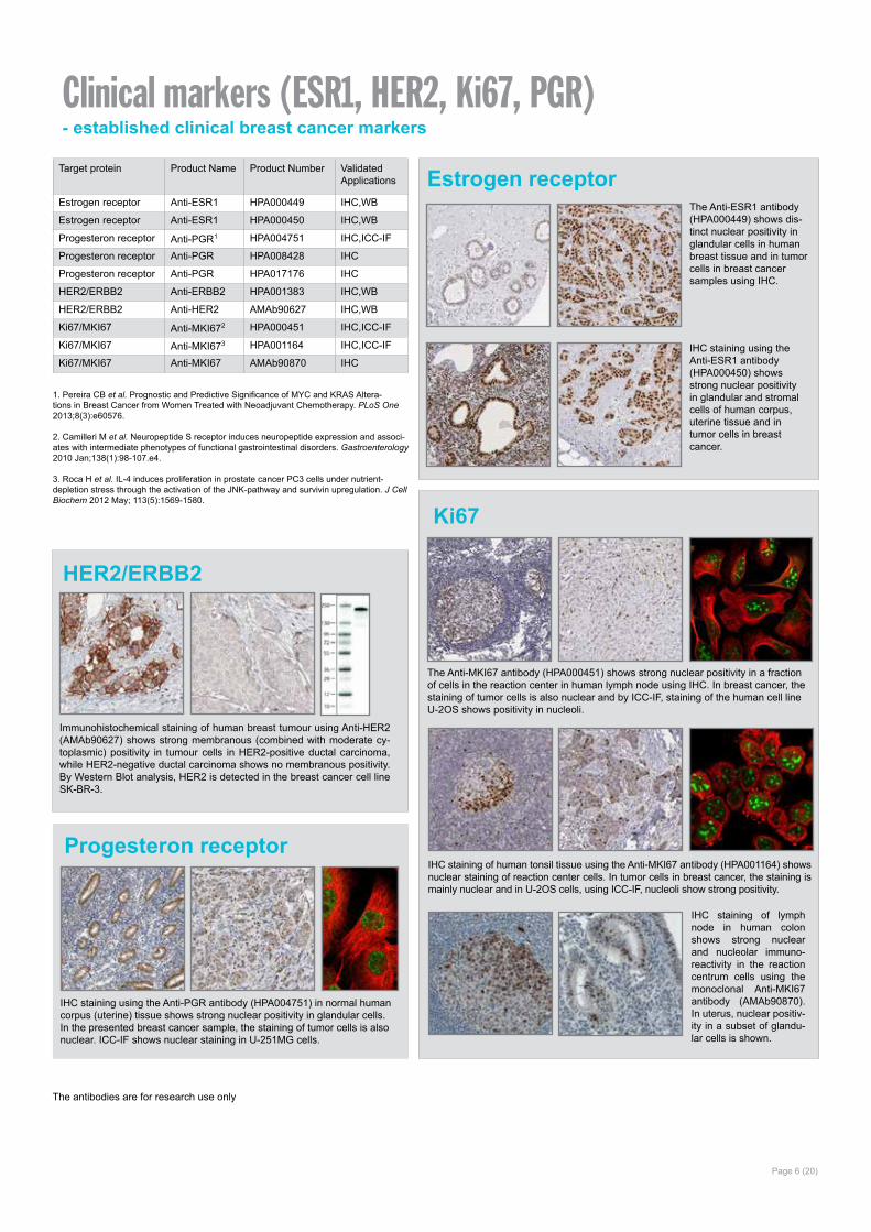
Clinical markers (ESR1, HER2, Ki67, PGR)- established clinical breast cancer markers
Ki67
IHC staining of human tonsil tissue using the Anti-MKI67 antibody (HPA001164) shows nuclear staining of reaction center cells. In tumor cells in breast cancer, the staining is mainly nuclear and in U-2OS cells, using ICC-IF, nucleoli show strong positivity.
Progesteron receptor
The antibodies are for research use only
HER2/ERBB2
Immunohistochemical staining of human breast tumour using Anti-HER2 (AMAb90627) shows strong membranous (combined with moderate cy-toplasmic) positivity in tumour cells in HER2-positive ductal carcinoma, while HER2-negative ductal carcinoma shows no membranous positivity. By Western Blot analysis, HER2 is detected in the breast cancer cell line SK-BR-3.
1. Pereira CB et al. Prognostic and Predictive Significance of MYC and KRAS Altera-tions in Breast Cancer from Women Treated with Neoadjuvant Chemotherapy. PLoS One 2013;8(3):e60576.
2. Camilleri M et al. Neuropeptide S receptor induces neuropeptide expression and associ-ates with intermediate phenotypes of functional gastrointestinal disorders. Gastroenterology 2010 Jan;138(1):98-107.e4.
3. Roca H et al. IL-4 induces proliferation in prostate cancer PC3 cells under nutrient-depletion stress through the activation of the JNK-pathway and survivin upregulation. J Cell Biochem 2012 May; 113(5):1569-1580.
Estrogen receptorThe Anti-ESR1 antibody (HPA000449) shows dis-tinct nuclear positivity in glandular cells in human breast tissue and in tumor cells in breast cancer samples using IHC.
IHC staining using the Anti-ESR1 antibody (HPA000450) shows strong nuclear positivity in glandular and stromal cells of human corpus, uterine tissue and in tumor cells in breast cancer.
IHC staining using the Anti-PGR antibody (HPA004751) in normal human corpus (uterine) tissue shows strong nuclear positivity in glandular cells. In the presented breast cancer sample, the staining of tumor cells is also nuclear. ICC-IF shows nuclear staining in U-251MG cells.
The Anti-MKI67 antibody (HPA000451) shows strong nuclear positivity in a fraction of cells in the reaction center in human lymph node using IHC. In breast cancer, the staining of tumor cells is also nuclear and by ICC-IF, staining of the human cell line U-2OS shows positivity in nucleoli.
IHC staining of lymph node in human colon shows strong nuclear and nucleolar immuno-reactivity in the reaction centrum cells using the monoclonal Anti-MKI67 antibody (AMAb90870). In uterus, nuclear positiv-ity in a subset of glandu-lar cells is shown.
Page 7 (20)
Target Protein Product Name Product Number ValidatedApplications
53BP1 Anti-TP53BP1 HPA008788 IHC,ICC-IF
53BP1 Anti-TP53BP1 HPA022133 IHC,WB*,ICC-IFACAT1 Anti-ACAT11,2 HPA004428 IHC,WB*,ICC-IF
ACAT1 Anti-ACAT12-4 HPA007569 IHC,WB,ICC-IF
AGR2 Anti-AGR25 HPA007912 IHC,WB
AIB1/NCOA3 Anti-NCOA3 HPA024210 IHC,ICC-IFAnillin/ANLN Anti-ANLN AMAb90660 IHC,WBAnillin/ANLN Anti-ANLN AMAb90662 IHC,WBAnillin/ANLN Anti-ANLN6 HPA005680 IHC,WB,ICC-IF
ARG1 Anti-ARG17 HPA024006 IHC,WB
ASAH1 Anti-ASAH18,9 HPA005468 IHC,WB
ATR Anti-ATR HPA028264 IHCBAAT1/BRAT1 Anti-BRAT1 HPA029455 IHCBACH1 Anti-BACH110 HPA003175 IHC,WB,ICC-IF
BAP1 Anti-BAP1 HPA028814 IHC,WB,ICC-IFBARD1 Anti-BARD1 HPA044864 IHC,ICC-IFBeta-Catenin Anti-CTNNB1 HPA029159 IHC,WB*,ICC-IFBeta-Catenin Anti-CTNNB1 HPA029160 IHC, IFBIRC3/API2 Anti-BIRC311 HPA002317 IHC,WB,ICC-IF
BIT1/ PTRH2 Anti-PTRH212,13 HPA012897 IHC,WB,ICC-IF
Blooms Syndrome Prot Anti-BLM HPA005689 IHC,ICC-IFBmi1 Anti-BMI1 HPA030472 IHC,WB*,ICC-IFBRCA1 Anti-BRCA1 HPA034966 IHCBRCA2 Anti-BRCA2 HPA026815 IHC,ICC-IFBRIP1/FANCJ Anti-BRIP1 HPA005474 IHC,WB,ICC-IFC11orf51/ANAPC15 Anti-C11orf51 HPA036596 IHC,WB,ICC-IFCAR/NR1I3 Anti-NR1I3 HPA051365 IHC,ICC-IFCASP8 Anti-CASP8 HPA001302 IHC,WB,ICC-IFCASP8 Anti-CASP8 HPA005688 IHC,WB,ICC-IFCAXII/CA12 Anti-CA1214-17 HPA008773 IHC,WB
Antibodies used in Breast Cancer Research
1. Sanchez-Alvarez R et al. Ethanol exposure induces the cancer-associated fibroblast phenotype and lethal tumor metabolism: Implications for breast cancer prevention. Cell Cycle 2013 Jan 15; 12(2):289-301.
2. Martinez-Outschoorn UE et al. Ketone bodies and two-compartment tumor metabolism: Stromal ketone production fuels mitochondrial biogenesis in epithelial cancer cells. Cell Cycle 2012 Nov 1; 11(21):3956-3963.
3. Martinez-Outschoorn UE et al. Ketone body utilization drives tumor growth and metastasis. Cell Cycle 2012 Nov 1;11(21):3964-71.
4. Chang HT et al. Ketolytic and glycolytic enzymatic expression profiles in malignant glio-mas: implication for ketogenic diet therapy. Nutr Metab (Lond) 1047. Epub 2013/07/05.
5. Hrstka R et al. AGR2 Predicts Tamoxifen Resistance in Postmenopausal Breast Cancer Patients. Dis Markers 2013; 35(4):207-212. Epub 2013/09/03.
6. O´Leary PC et al. Systematic antibody generation and validation via tissue microarray techno-logy leading to identification of a novel protein prognostic panel in breast cancer. BMC Cancer. 2013 Apr 2;13:175.
7. de Boniface J et al. Expression patterns of the immunomodulatory enzyme arginase 1 in blood, lymph nodes and tumor tissue of early-stage breast cancer patients. Oncoimmunology 2012 Nov 1; 1(8):1305-1312.
8. Lucki NC, Sewer MB. Genistein Stimulates MCF-7 Breast Cancer Cell Growth by Inducing Acid Ceramidase (ASAH1) Gene Expression. J Biol Chem 2011 Jun 3; 286(22):19399-19409. Epub 2011 Apr 14.
9. Lucki NC et al. Acid Ceramidase (ASAH1) Represses Steroidogenic Factor 1-Dependent Gene Transcription in H295R Human Adrenocortical Cells by Binding to the Receptor. Mol Cell Biol 2012 Nov; 32(21):4419-4431.
10. Liang Y et al. Transcriptional Network Analysis Identifies BACH1 as a Master Regulator of Breast Cancer Bone Metastasis. J Biol Chem 2012 Sep 28;287(40):33533-44.
11. Almubarak H et al. Zoledronic acid directly suppresses cell proliferation and induces apop-tosis in highly tumorigenic prostate and breast cancers. J Carcinog 2011 Jan 15;10:2.
12. Brunquell C et al. TLE1 is an anoikis regulator and is downregulated by Bit1 in breast cancer cells. Mol Cancer Res 2012 Nov; 10(11):1482-1495. Epub 2012/09/04.
13. Karmali PP et al. Metastasis of tumor cells is enhanced by downregulation of bit1. PLoS One 2011;6(8):e23840.
14. Vermeulen JF et al. Immunophenotyping invasive breast cancer: paving the road for molecular imaging. BMC Cancer 12240. Epub 2012 Jun 13.
15. Davidson B et al. Gene expression signatures differentiate ovarian/peritoneal serous carcinoma from breast carcinoma in effusions. J Cell Mol Med 2011 Mar;15(3):535-44.
16. Vermeulen et al. Differential expression of growth factor receptors and membrane-bound tumor markers for imaging in male and female breast cancer. PLoS One 2013;8(1):e53353.
17. Tafreshi NK et al. Noninvasive detection of breast cancer lymph node metastasis us-ing carbonic anhydrases IX and XII targeted imaging probes. Clin Cancer Res 2012 Jan 1;18(1):207-19.
* WB both in human and rodent samples
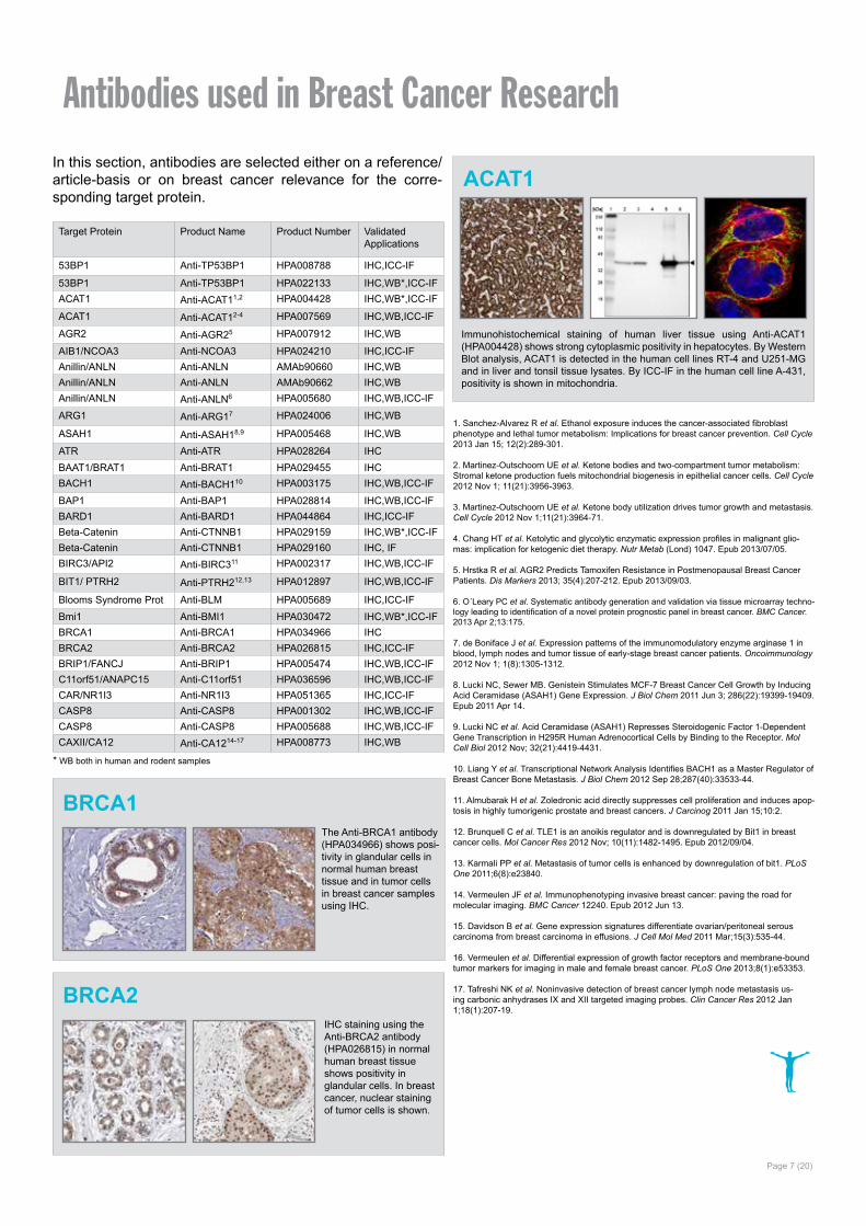
BRCA2
ACAT1
Immunohistochemical staining of human liver tissue using Anti-ACAT1 (HPA004428) shows strong cytoplasmic positivity in hepatocytes. By Western Blot analysis, ACAT1 is detected in the human cell lines RT-4 and U251-MG and in liver and tonsil tissue lysates. By ICC-IF in the human cell line A-431, positivity is shown in mitochondria.
BRCA1The Anti-BRCA1 antibody (HPA034966) shows posi-tivity in glandular cells in normal human breast tissue and in tumor cells in breast cancer samples using IHC.
IHC staining using the Anti-BRCA2 antibody (HPA026815) in normal human breast tissue shows positivity in glandular cells. In breast cancer, nuclear staining of tumor cells is shown.
In this section, antibodies are selected either on a reference/article-basis or on breast cancer relevance for the corre-sponding target protein.
Target Protein Product Name Product Number ValidatedApplications
CD44 Anti-CD4418-22 HPA005785 IHC,WB,ICC-IF
CD82 Anti-CD82 HPA028900 IHC,WB
CDH1 Anti-CDH1 HPA004812 IHC,ICC-IF
CEA/CEACAM5 Anti-CEACAM5 HPA019758 IHC,WB
CHEK2 Anti-CHEK2 HPA001878 IHC,WB
CKB Anti-CKB HPA001254 IHC
CRABP2 Anti-CRABP2 HPA004135 IHC,WB,ICC-IF
CTNND1 Anti-CTNND1 HPA015955 IHC,WB*,ICC-IF
CX32/GJB1 Anti-GJB123 HPA010663 IHC,WB
Cyclin E1 Anti-CCNE1 HPA018169 IHC,WB,ICC-IF
cyklin A2 Anti-CCNA2 HPA020626 IHC,WB
Cytokeratin 14/CK14 Anti-KRT14 HPA023040 IHC,WB*,ICC-IF
Cytokeratin 17/CK17 Anti-KRT1724 HPA000452 IHC
Cytokeratin 17/CK17 Anti-KRT17 HPA000453 IHC,WB,ICC-IF
DACH2 Anti-DACH225 HPA000258 IHC,ICC-IF
DBC1/KIAA1967 Anti-KIAA1967 HPA019907 IHC,WB*,ICC-IFDBC1/KIAA1967 Anti-KIAA1967 HPA019943 IHC,ICC-IFDCAF7 Anti-DCAF726 HPA022962 IHC, WB
Decorin/DCN Anti-DCN27,28 HPA003315 IHC, WB
DIRAS3 Anti-DIRAS3 HPA028483 IHC,WBDIRAS3 Anti-DIRAS3 HPA028557 IHC,WBDIRAS3 Anti-DIRAS3 HPA029384 IHCEGFR Anti-EGFR AMAb90816 IHC,WBEGFR Anti-EGFR AMAb90819 WBEGFR Anti-EGFR29 HPA001200 IHC
EGFR Anti-EGFR30 HPA018530 IHC,WB,ICC-IF
Endoplasmin/ HSP90B1 Anti-HSP90B127,31 HPA003901 IHC,WB,ICC-IF
ERLIN2 Anti-ERLIN232,33 HPA002025 IHC,WB*,ICC-IF
ERFF/C1orf64 Anti-C1orf6434 HPA026676 IHC,WB,ICC-IF
FAAH Anti-FAAH35 HPA007425 IHC,ICC-IF
FGFR2 Anti-FGRF2 HPA035305 IHC,WB,ICC-IFGATA3 Anti-GATA3 HPA029730 IHC,ICC-IFGATA3 Anti-GATA3 HPA029731 IHC, WBGCDFP/PIP Anti-PIP HPA009177 IHC,WBGEF-H1 Anti-ARHGEF236,37 HPA017046 IHC,WB
GGH Anti-GGH35 HPA025226 IHC,WBGranulin Anti-GRN38 HPA008763 IHC,ICC-IFGranulin Anti-GRN38 HPA028747 IHC,ICC-IF
HIF-1 alpha/HIF1A Anti-HIF1A39-42 HPA001275 IHC,ICC-IF
HJURP Anti-HJURP43-45 HPA008436 IHC,WB,ICC-IF
HMGCL Anti-HMGCL2 HPA004727 IHC,WB
HMGCR Anti-HMGCR46 HPA008338 IHCHSD17B14 Anti-HSD17B14 HPA021467 IHC,WB,ICC-IF
18. Vazquez-Martin A et al. Metformin regulates breast cancer stem cell ontogeny by transcriptional regulation of the epithelial-mesenchymal transition (EMT) status. Cell Cycle 2010 Sep 15;9(18):3807-14.
19. Baccelli I et al. Identification of a population of blood circulating tumor cells from breast cancer patients that initiates metastasis in a xenograft assay. Nat Biotechnol 2013 Apr 21;
20. Petit V et al. Optimization of tumor xenograft dissociation for the profiling of cell surface markers and nutrient transporters. Lab Invest 2013 May;93(5):611-21.
21. Twarock S et al. Synthesis of hyaluronan in oesophageal cancer cells is uncoupled from the prostaglandin-cAMP pathway. Br J Pharmacol 2009 May;157(2):234-43.
22. Asplund A et al. Expression profiling of microdissected cell populations selected from basal cells in normal epidermis and basal cell carcinoma. Br J Dermatol 2008 Mar;158(3):527-38.
23. Teleki I et al. The potential prognostic value of connexin 26 and 46 expression in neoadjuvant-treated breast cancer. BMC Cancer 1350. Epub 2013/02/02.
24. Kiflemariam S et al. Scalable in situ hybridization on tissue arrays for validation of novel cancer and tissue-specific biomarkers. PLoS One 2012;7(3):e32927.
25. Nodin B et al. Discovery of dachshund 2 protein as a novel biomarker of poor prognosis in epithelial ovarian cancer. J Ovarian Res 2012 Jan 27;5(1):6.
26. Sircoulomb F et al. ZNF703 gene amplification at 8p12 specifies luminal B breast cancer. EMBO Mol Med 2011 Mar; 3(3):153-166. Epub 2011 Feb 15.
27. Cawthorn TR et al.Proteomic Analyses Reveal High Expression of Decorin and Endo-plasmin (HSP90B1) Are Associated with Breast Cancer Metastasis and Decreased Survival. PLoS One 2012;7(2):e30992.
28. Henke A et al. Stromal Expression of Decorin, Semaphorin6D, SPARC, Sprouty1 and Tsukushi in Developing Prostate and Decreased Levels of Decorin in Prostate Cancer. LoS One 7(8):e42516. Epub 2012 Aug 3.
29. Hudson EP et al. Multiplex epitope mapping using bacterial surface display reveals both linear and conformational epitopes. Sci Rep 2012;2:706.
30. Arabi A et al. Proteomic screen reveals Fbw7 as a modulator of the NF-κB pathway. Nat Commun 2012;3:976.
31. Ito A et al. Novel application for pseudopodia proteomics using excimer laser ablation and two-dimensional difference gel electrophoresis. Lab Invest 2012 Sep;92(9):1374-85.
32. Holland DG et al. ZNF703 is a common Luminal B breast cancer oncogene that dif-ferentially regulates luminal and basal progenitors in human mammary epithelium. EMBO Mol Med 2011 Mar;3(3):167-80.
33. Mulder J et al. Tissue profiling of the mammalian central nervous system using human antibody-based proteomics. Mol Cell Proteomics 2009 Jul;8(7):1612-22.
34. Su D et al. Role of ERRF, a Novel ER-Related Nuclear Factor, in the Growth Control of ER-Positive Human Breast Cancer Cells. Am J Pathol 2012 Mar; 180(3):1189-1201.
35. Shubbar E et al. High levels of γ-glutamyl hydrolase (GGH) are associated with poor prognosis and unfavorable clinical outcomes in invasive breast cancer. BMC Cancer 2013 Feb 1;13:47.
36. Liao YC et al. Overexpressed hPTTG1 promotes breast cancer cell invasion and metas-tasis by regulating GEF-H1/RhoA signalling. Oncogene 2012 Jun 21;31(25):3086-97
37. Cheng IK et al. GEF-H1 over-expression in hepatocellular carcinoma promotes cell motility via activation of RhoA signalling. Pathol 2012 Jul 30;
38. Elkabets M et al. Human tumors instigate granulin-expressing hematopoietic cells that promote malignancy by activating stromal fibroblasts in mice. J Clin Invest 2011 Feb 1;121(2):784-99.
39. Zibert JR et al. Halting angiogenesis by non-viral somatic gene therapy alleviates pso-riasis and murine psoriasiform skin lesions. J Clin Invest 2011 Jan 4;121(1):410-21.
40. Smyth LG et al. Carbonic anhydrase IX expression in prostate cancer. Prostate Cancer and Prostatic Diseases 2009 Dec;13(2):178-181.
41. Paatero I et al. Interaction with ErbB4 promotes hypoxia-inducible factor-1α signaling. J Biol Chem 2012 Mar 23;287(13):9659-71.
42. Zbytek B et al. Putative role of HIF transcriptional activity in melanocytes and melanoma biology. Dermatoendocrinol 2013 Apr 1; 5(2):239-251. Epub 2013/04/01.
43. Hu Z et al. The expression level of HJURP has an independent prognostic impact and predicts the sensitivity to radiotherapy in breast cancer. Breast Cancer Res 2010;12(2):R18
44. Shuaib M et al. HJURP binds CENP-A via a highly conserved N-terminal domain and mediates its deposition at centromeres. Proc Natl Acad Sci U S A 2010 Jan 26;107(4):1349-54
45. de Tayrac M et al. Prognostic Significance of EDN/RB, HJURP, p60/CAF-1 and PDLI4, Four New Markers in High-Grade Gliomas. PLoS One 2013 Sep 11;8(9):e73332.
46. Bjarnadottir O et al. Targeting HMG-CoA reductase with statins in a window-of-opportu-nity breast cancer trial. Breast Cancer Res Treat 2013 Apr;138(2):499-508.
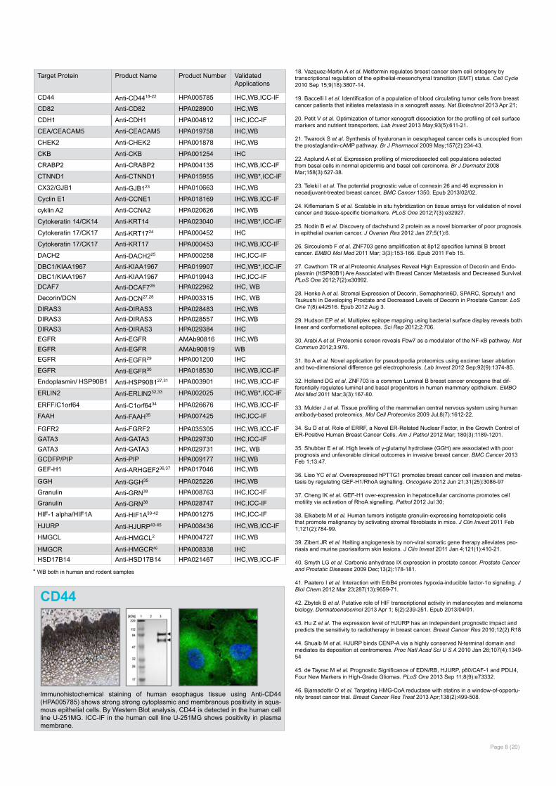
CD44
Immunohistochemical staining of human esophagus tissue using Anti-CD44 (HPA005785) shows strong strong cytoplasmic and membranous positivity in squa-mous epithelial cells. By Western Blot analysis, CD44 is detected in the human cell line U-251MG. ICC-IF in the human cell line U-251MG shows positivity in plasma membrane.
* WB both in human and rodent samples
Page 8 (20)
Target Protein Product Name Product Number ValidatedApplications
KLK3/PSA Anti-KLK347,48 HPA000764 IHC
LSP1 Anti-LSP1 HPA019693 IHC,WB,ICC-IFMMP2 Anti-MMP2 HPA001939 IHCMUC1/CA15-3 Anti-MUC1 HPA004179 IHC,WBMUC1/CA15-3 Anti-MUC1 HPA007235 IHCMUC1/CA15-3 Anti-MUC1 HPA008855 IHC,ICC-IFNBN Anti-NBN HPA001429 IHC,WB,ICC-IFNRP1 Anti-NRP1 HPA030278 IHC, WBOncostatin M Anti-OSM49 HPA029814 IHC,WBp63/TP63 Anti-TP63 HPA006288 IHC,ICC-IFp63/TP63 Anti-TP63 HPA007010 IHC,ICC-IFPHGDH Anti-PHGDH50-52 HPA021241 IHC,WB*,ICC-IFPGD Anti-PGD HPA031314 IHC,WB*PKC alpha/PKCA Anti-PKCA HPA006563 IHC,WB*,ICC-IFPKC alpha/PKCA Anti-PKCA HPA006564 IHC,WB*,ICC-IFPLAT Anti-PLAT HPA003412 IHC,WB,ICC-IFPOLRMT Anti-POLRMT53 HPA006366 IHC
PSPH Anti-PSPH50 HPA020376 IHC,WB
PTMA Anti-PTMA HPA047183 IHC,ICC-IFPTTG1 Anti-PTTG1 HPA008890 IHC,ICC-IFRAP80/UIMC1 Anti-UIMC1 HPA037503 IHC,WBRAP80/UIMC1 Anti-UIMC1 HPA037504 IHC,WB,ICC-IFREST Anti-REST54,55 HPA006079 IHC,ICC-IF
RBM3 Anti-RBM356,57 HPA003624 IHC,WB*,ICC-IF
RBM3 Anti-RBM358-65 AMAb90655 IHC,WB
RRBP1 Anti-RRBP166 HPA009026 IHC,ICC-IF
rs4973768/SLC4A7 Anti-SLC4A7 HPA035857 IHCSIX1 Anti-SIX167-74 HPA001893 IHC,WB,ICC-IF
SNCG Anti-SNCG HPA014404 IHC,WBSTK11 Anti-STK11 HPA017254 IHC,WB,ICC-IFSURVIvin/BIRC5 Anti-BIRC5 HPA002830 IHC,WB,ICC-IFT-STAR/KHDRBS3 Anti-KHDRBS375,76 HPA000500 IHC
Tenascin C/TNC Anti-TNC77-79 HPA004823 IHC,WB
TFF1 Anti-TFF180-82 HPA003425 IHC,WB
THBD Anti-THBD HPA002982 IHCTHEM2/ACOT13 Anti-ACOT13 HPA019881 IHC,WB*,ICC-IFTOP2A Anti-TOP2A HPA006458 IHC,WB,ICC-IFTOP2A Anti-TOP2A HPA026773 IHC,ICC-IFUGT8 Anti-UGT883 HPA014405 IHC,ICC-IF
ULBP1 Anti-ULBP184 HPA007547 IHC
ZNF703 Anti-ZNF70332 HPA023930 IHC,ICC-IF
47. Jaraj SJ et al. GAD1 is a biomarker for benign and malignant prostatic tissue. Scand J Urol Nephrol 2011 Feb;45(1):39-45.
48. Liu H et al. Single-cell clones of liver cancer stem cells have the potential of differentiating into different types of tumor cells. Cell Death Dis 2013 Oct; 4(10):e857-. Epub 2013/10/17.
49. Guo L et al. Stat3-coordinated Lin-28-let-7-HMGA2 and miR-200-ZEB1 circuits initiate and maintain oncostatin M-driven epithelial-mesenchymal transition. Oncogene 2013 Nov 7;32(45):5272-82.
50. Possemato R et al. Functional genomics reveal that the serine synthesis pathway is essen-tial in breast cancer. Nature 2011 Aug 18;476(7360):346-50.
51. Maddocks OD et al. Serine starvation induces stress and p53-dependent metabolic remodel-ling in cancer cells. Nature 2013 Jan 24;493(7433):542-6.
52. Nilsson LM et al. Mouse genetics suggests cell-context dependency for Myc-regulated metabolic enzymes during tumorigenesis. PLoS Genet 2012;8(3):e1002573.
53. Salem AF et al. Mitochondrial biogenesis in epithelial cancer cells promotes breast cancer tumor growth and confers autophagy resistance. Cell Cycle 2012 Nov 15; 11(22):4174-4180.
54. Wagoner MP et al. The transcription factor REST is lost in aggressive breast cancer. PLoS Genet 2010 Jun 10;6(6):e1000979.
55. Prada I et al. REST/NRSF governs the expression of dense-core vesicle gliosecretion in astrocytes. J Cell Biol 2011 May 2;193(3):537-49.
56. Jögi A et al. Nuclear expression of the RNA-binding protein RBM3 is associated with an improved clinical outcome in breast cancer. Mod Pathol 2009 Dec;22(12):1564-74.
57. Hjelm B et al. High nuclear RBM3 expression is associated with an improved prognosis in colorectal cancer. Proteomics Clin Appl 2011 Dec;5(11-12):624-35
58. Ehlén A et al. Expression of the RNA-binding protein RBM3 is associated with a favourable prognosis and cisplatin sensitivity in epithelial ovarian cancer. J Transl Med 2010 Aug 20;8:78
59. Jonsson L et al. High RBM3 expression in prostate cancer independently predicts a re-duced risk of biochemical recurrence and disease progression. Diagn Pathol 2011 Sep 28;6:9
60. Nodin B et al. High MCM3 expression is an independent biomarker of poor prognosis and correlates with reduced RBM3 expression in a prospective cohort of malignant melanoma. Diagn Pathol 782. Epub 2012 Jul 17.
61. Jonsson L et al. Low RBM3 protein expression correlates with tumour progression and poor prognosis in malignant melanoma: an analysis of 215 cases from the Malmö Diet and Cancer Study. J Transl Med 2011 Jul 21;9:114.
62. Ehlén Å et al. RBM3-regulated genes promote DNA integrity and affect clinical outcome in epithelial ovarian cancer. Transl Oncol 2011 Aug;4(4):212-21.
63. Hjelm B et al. High nuclear RBM3 expression is associated with an improved prognosis in colorectal cancer. Proteomics Clin Appl 2011 Dec;5(11-12):624-35
64. Boman K et al. Decreased expression of RNA-binding motif protein 3 correlates with tu-mour progression and poor prognosis in urothelial bladder cancer. BMC Urol 2013 Apr 8;13:17.
65. Nodin B et al. High MCM3 expression is an independent biomarker of poor prognosis and correlates with reduced RBM3 expression in a prospective cohort of malignant melanoma.Diagn Pathol 2012 Jul 17;7:82.
66. Telikicherla D et al. Overexpression of ribosome binding protein 1 (RRBP1) in breast can-cer. Clin Proteomics 9(1):7. Epub 2012 Jun 18.
67. Iwanaga R et al. Expression of Six1 in luminal breast cancers predicts poor prognosis and promotes increases in tumor initiating cells by activation of extracellular signal-regulated kinase and transforming growth factor-beta signaling pathways. Breast Cancer Res 2012 Jul 5;14(4):R100.
68. Smith AL et al. The miR-106b-25 cluster targets Smad7, activates TGF-β signaling, and induces EMT and tumor initiating cell characteristics downstream of Six1 in human breast cancer. Oncogene 2012 Jan 30;
69. Wan F et al. Gene expression changes during HPV-mediated carcinogenesis: a comparison between an in vitro cell model and cervical cancer. Int J Cancer 2008 Jul 1;123(1):32-40
70. McCoy EL et al. Six1 expands the mouse mammary epithelial stem/progenitor cell pool and induces mammary tumors that undergo epithelial-mesenchymal transition. J Clin Invest 2009 Sep;119(9):2663-77.
71. Micalizzi DS et al. The Six1 homeoprotein induces human mammary carcinoma cells to un-dergo epithelial-mesenchymal transition and metastasis in mice through increasing TGF-beta signaling. J Clin Invest 2009 Sep;119(9):2678-90.
72. Farabaugh et al. Eya2 is required to mediate the pro-metastatic functions of Six1 via the in-duction of TGF-β signaling, epithelial-mesenchymal transition, and cancer stem cell properties.Oncogene 2012 Feb 2;31(5):552-62.
73. Ono H et al. SIX1 promotes epithelial-mesenchymal transition in colorectal cancer through ZEB1 activation. Oncogene 2012 Nov 22;31(47):4923-34.
74. Le Grand F et al. Six1 regulates stem cell repair potential and self-renewal during skeletal muscle regeneration. J Cell Biol 2012 Sep 3; 198(5):815-832.
75. Sernbo S et al. Nuclear T-STAR Protein Expression Correlates with HER2 Status, Hormone Receptor Negativity and Prolonged Recurrence Free Survival in Primary Breast Cancer and Decreased Cancer Cell Growth In Vitro. PLoS One 8(7):e70596. Epub 2013/07/29.
76. Ek S et al. From gene expression analysis to tissue microarrays: a rational approach to identify therapeutic and diagnostic targets in lymphoid malignancies. Mol Cell Proteomics 2006 Jun;5(6):1072-81.
77. Schenke-Layland K et al. Cardiomyopathy is associated with structural remodelling of heart valve extracellular matrix. Eur Heart J 2009 Sep;30(18):2254-65.
78. Ghosh Z et al. Dissecting the Oncogenic Potential of Human Embryonic and Induced Pluri-potent Stem Cell Derivatives. Cancer Res 2011 Jul 15; 71(14):5030-5039. Epub 2011 Jun 6.
79. Edlund K et al. CD99 is a novel prognostic stromal marker in non-small cell lung cancer.Int J Cancer 2012 Nov 15;131(10):2264-73.
80. Pontén F et al. The Human Protein Atlas--a tool for pathology. J Pathol 2008 Dec;216(4):387-93.
81. Wu CC et al. Candidate serological biomarkers for cancer identified from the secretomes of 23 cancer cell lines and the human protein atlas. Mol Cell Proteomics 2010 Jun;9(6):1100-17.
82. Davidson B et al. Gene expression signatures differentiate ovarian/peritoneal serous carci-noma from breast carcinoma in effusions. J Cell Mol Med 2011 Mar;15(3):535-44.
83. Dziegiel P et al. Ceramide galactosyltransferase (UGT8) is a molecular marker of breast cancer malignancy and lung metastases. Br J Cancer 2010 Aug 10;103(4):524-31.
84. de Kruijf EM et al. NKG2D ligand tumor expression and association with clinical outcome in early breast cancer patients: an observational study. BMC Cancer 1224. Epub 2012 Jan 18.
* WB both in human and rodent samples
Page 9 (20)
Target Protein Product Name Product Number Validated Applications
AURKA/STK15 Anti-AURKA HPA002636 IHC,WB
AZGP1 Anti-AZGP1 HPA012582 IHC,WB
BAG1 Anti-BAG1 HPA018121 IHC,ICC-IF
BIRC5/Survivin Anti-BIRC5 HPA002830 IHC,WB,ICC-IF
CD68/Macrosialin Anti-CD68 HPA048982 IHC
CDCA7 Anti-CDCA71,2 HPA005565 IHC,WB,ICC-IF
CMC2/C16orf61 Anti-CMC2 HPA006871 IHC
DHCR7 Anti-DHCR7 HPA044280 IHC,WB
DHX58/LGP2 Anti-DHX58 HPA018670 IHC,WB,ICC-IF
DHX58/LGP2 Anti-DHX58 HPA019570 IHC
DIAPH3 Anti-DIAPH3 HPA032152 IHC,WB*
DTL Anti-DTL3 HPA028016 IHC,WB,ICC-IF
ECI2/PECI Anti-ECI2 HPA022130 IHC,WB,ICC-IF
EGLN1/PHD2 Anti-EGLN14 HPA022129 IHC,ICC-IF
ESM1 Anti-ESM1 HPA036660 IHC,WB
Estrogen receptor Anti-ESR1 HPA000449 IHC,WB
Estrogen receptor Anti-ESR1 HPA000450 IHC,WB
Exostosin-1 Anti-EXT1 HPA044394 IHC,WB
FGF18 Anti-FGF18 HPA018795 IHC,WB,ICC-IF
GMPS Anti-GMPS HPA050682 IHC
GNAZ Anti-GNAZ HPA003011 IHC,WB
GPR126/VIGR Anti-GPR126 HPA017346 IHC
GPR180 Anti-GPR180 HPA047250 IHC,ICC-IF
GSTM3 Anti-GSTM3 HPA035190 IHC,WB
GSTM5/GSTM1 Anti-GSTM5 HPA048652 IHC,WB
HER2/ERBB2 Anti-ERBB2 HPA001383 IHC,WB
HER2/ERBB2 Anti-HER2 AMAb90627 IHC,WB
HRASLS Anti-HRASLS HPA051179 IHC
IL6ST/GP130 Anti-IL6ST5 HPA010558 IHC
JHDM1D/KDM7A Anti-JHDM1D HPA012114 IHC,ICC-IF
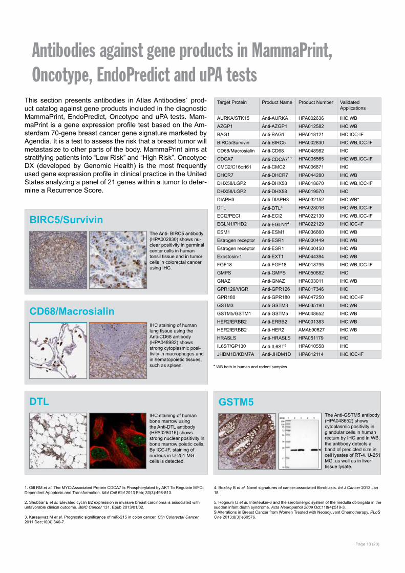
Antibodies against gene products in MammaPrint, Oncotype, EndoPredict and uPA tests
This section presents antibodies in Atlas Antibodies´ prod-uct catalog against gene products included in the diagnostic MammaPrint, EndoPredict, Oncotype and uPA tests. Mam-maPrint is a gene expression profile test based on the Am-sterdam 70-gene breast cancer gene signature marketed by Agendia. It is a test to assess the risk that a breast tumor will metastasize to other parts of the body. MammaPrint aims at stratifying patients into “Low Risk” and “High Risk”. Oncotype DX (developed by Genomic Health) is the most frequently used gene expression profile in clinical practice in the United States analyzing a panel of 21 genes within a tumor to deter-mine a Recurrence Score.
* WB both in human and rodent samples
The Anti- BIRC5 antibody (HPA002830) shows nu-clear positivity in germinal center cells in human tonsil tissue and in tumor cells in colorectal cancer using IHC.
BIRC5/Survivin
IHC staining of human lung tissue using the Anti-CD68 antibody (HPA048982) shows strong cytoplasmic posi-tivity in macrophages and in hematopoietic tissues, such as spleen.
CD68/Macrosialin
IHC staining of human bone marrow using the Anti-DTL antibody (HPA028016) shows strong nuclear positivity in bone marrow poietic cells.By ICC-IF, staining of nucleus in U-251 MG cells is detected.
DTLThe Anti-GSTM5 antibody (HPA048652) shows cytoplasmic positivity in glandular cells in human rectum by IHC and in WB, the antibody detects a band of predicted size in cell lysates of RT-4, U-251 MG, as well as in liver tissue lysate.
GSTM5
1. Gill RM et al. The MYC-Associated Protein CDCA7 Is Phosphorylated by AKT To Regulate MYC-Dependent Apoptosis and Transformation. Mol Cell Biol 2013 Feb; 33(3):498-513.
2. Shubbar E et al. Elevated cyclin B2 expression in invasive breast carcinoma is associated with unfavorable clinical outcome. BMC Cancer 131. Epub 2013/01/02.
3. Karaayvaz M et al. Prognostic significance of miR-215 in colon cancer. Clin Colorectal Cancer 2011 Dec;10(4):340-7.
4. Bozóky B et al. Novel signatures of cancer-associated fibroblasts. Int J Cancer 2013 Jan 15.
5. Rognum IJ et al. Interleukin-6 and the serotonergic system of the medulla oblongata in the sudden infant death syndrome. Acta Neuropathol 2009 Oct;118(4):519-3.S Alterations in Breast Cancer from Women Treated with Neoadjuvant Chemotherapy. PLoS One 2013;8(3):e60576.
Page 10 (20)
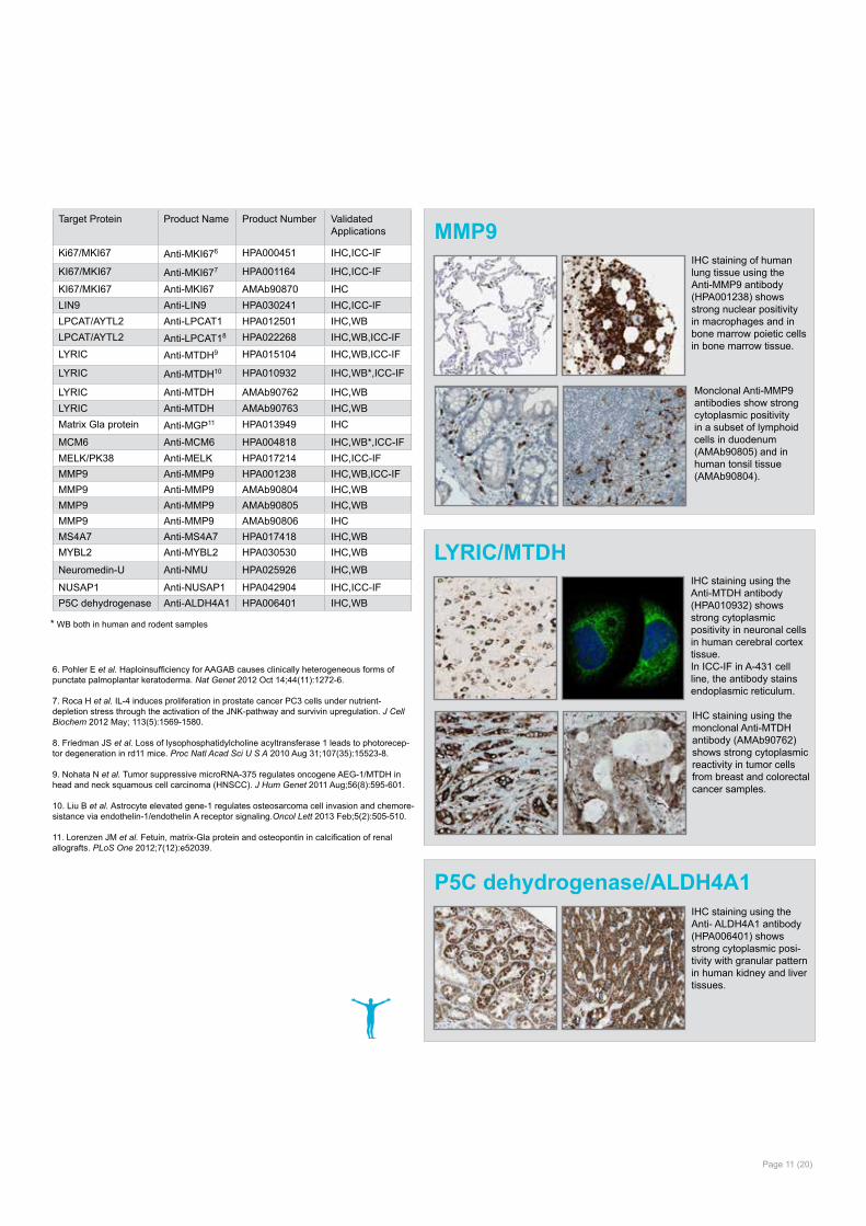
IHC staining using the Anti- ALDH4A1 antibody (HPA006401) shows strong cytoplasmic posi-tivity with granular pattern in human kidney and liver tissues.
P5C dehydrogenase/ALDH4A1
IHC staining using the Anti-MTDH antibody (HPA010932) shows strong cytoplasmic positivity in neuronal cells in human cerebral cortex tissue. In ICC-IF in A-431 cell line, the antibody stains endoplasmic reticulum.
LYRIC/MTDH
IHC staining using the monclonal Anti-MTDH antibody (AMAb90762) shows strong cytoplasmic reactivity in tumor cells from breast and colorectal cancer samples.
IHC staining of human lung tissue using the Anti-MMP9 antibody (HPA001238) shows strong nuclear positivity in macrophages and in bone marrow poietic cells in bone marrow tissue.
MMP9Target Protein Product Name Product Number Validated
Applications
Ki67/MKI67 Anti-MKI676 HPA000451 IHC,ICC-IF
KI67/MKI67 Anti-MKI677 HPA001164 IHC,ICC-IF
KI67/MKI67 Anti-MKI67 AMAb90870 IHCLIN9 Anti-LIN9 HPA030241 IHC,ICC-IFLPCAT/AYTL2 Anti-LPCAT1 HPA012501 IHC,WBLPCAT/AYTL2 Anti-LPCAT18 HPA022268 IHC,WB,ICC-IFLYRIC Anti-MTDH9 HPA015104 IHC,WB,ICC-IF
LYRIC Anti-MTDH10 HPA010932 IHC,WB*,ICC-IF
LYRIC Anti-MTDH AMAb90762 IHC,WBLYRIC Anti-MTDH AMAb90763 IHC,WBMatrix Gla protein Anti-MGP11 HPA013949 IHC
MCM6 Anti-MCM6 HPA004818 IHC,WB*,ICC-IFMELK/PK38 Anti-MELK HPA017214 IHC,ICC-IFMMP9 Anti-MMP9 HPA001238 IHC,WB,ICC-IFMMP9 Anti-MMP9 AMAb90804 IHC,WBMMP9 Anti-MMP9 AMAb90805 IHC,WBMMP9 Anti-MMP9 AMAb90806 IHCMS4A7 Anti-MS4A7 HPA017418 IHC,WBMYBL2 Anti-MYBL2 HPA030530 IHC,WB
Neuromedin-U Anti-NMU HPA025926 IHC,WB
NUSAP1 Anti-NUSAP1 HPA042904 IHC,ICC-IFP5C dehydrogenase Anti-ALDH4A1 HPA006401 IHC,WB
6. Pohler E et al. Haploinsufficiency for AAGAB causes clinically heterogeneous forms of punctate palmoplantar keratoderma. Nat Genet 2012 Oct 14;44(11):1272-6.
7. Roca H et al. IL-4 induces proliferation in prostate cancer PC3 cells under nutrient-depletion stress through the activation of the JNK-pathway and survivin upregulation. J Cell Biochem 2012 May; 113(5):1569-1580.
8. Friedman JS et al. Loss of lysophosphatidylcholine acyltransferase 1 leads to photorecep-tor degeneration in rd11 mice. Proc Natl Acad Sci U S A 2010 Aug 31;107(35):15523-8.
9. Nohata N et al. Tumor suppressive microRNA-375 regulates oncogene AEG-1/MTDH in head and neck squamous cell carcinoma (HNSCC). J Hum Genet 2011 Aug;56(8):595-601.
10. Liu B et al. Astrocyte elevated gene-1 regulates osteosarcoma cell invasion and chemore-sistance via endothelin-1/endothelin A receptor signaling.Oncol Lett 2013 Feb;5(2):505-510.
11. Lorenzen JM et al. Fetuin, matrix-Gla protein and osteopontin in calcification of renal allografts. PLoS One 2012;7(12):e52039.
* WB both in human and rodent samples
Page 11 (20)
Monclonal Anti-MMP9 antibodies show strong cytoplasmic positivity in a subset of lymphoid cells in duodenum (AMAb90805) and in human tonsil tissue (AMAb90804).
Page 12 (20)
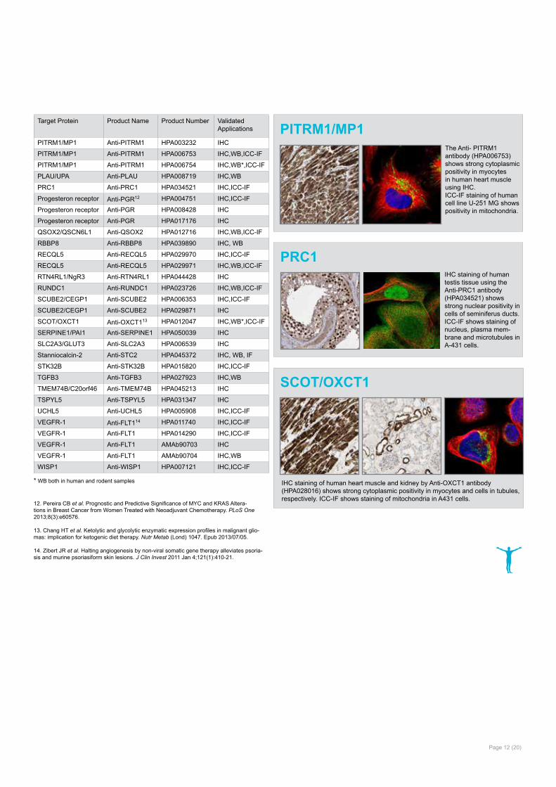
IHC staining of human testis tissue using the Anti-PRC1 antibody (HPA034521) shows strong nuclear positivity in cells of seminiferus ducts.ICC-IF shows staining of nucleus, plasma mem-brane and microtubules in A-431 cells.
PRC1
12. Pereira CB et al. Prognostic and Predictive Significance of MYC and KRAS Altera-tions in Breast Cancer from Women Treated with Neoadjuvant Chemotherapy. PLoS One 2013;8(3):e60576.
13. Chang HT et al. Ketolytic and glycolytic enzymatic expression profiles in malignant glio-mas: implication for ketogenic diet therapy. Nutr Metab (Lond) 1047. Epub 2013/07/05.
14. Zibert JR et al. Halting angiogenesis by non-viral somatic gene therapy alleviates psoria-sis and murine psoriasiform skin lesions. J Clin Invest 2011 Jan 4;121(1):410-21.
Target Protein Product Name Product Number Validated Applications
PITRM1/MP1 Anti-PITRM1 HPA003232 IHC
PITRM1/MP1 Anti-PITRM1 HPA006753 IHC,WB,ICC-IF
PITRM1/MP1 Anti-PITRM1 HPA006754 IHC,WB*,ICC-IF
PLAU/UPA Anti-PLAU HPA008719 IHC,WB
PRC1 Anti-PRC1 HPA034521 IHC,ICC-IF
Progesteron receptor Anti-PGR12 HPA004751 IHC,ICC-IF
Progesteron receptor Anti-PGR HPA008428 IHC
Progesteron receptor Anti-PGR HPA017176 IHC
QSOX2/QSCN6L1 Anti-QSOX2 HPA012716 IHC,WB,ICC-IF
RBBP8 Anti-RBBP8 HPA039890 IHC, WB
RECQL5 Anti-RECQL5 HPA029970 IHC,ICC-IF
RECQL5 Anti-RECQL5 HPA029971 IHC,WB,ICC-IF
RTN4RL1/NgR3 Anti-RTN4RL1 HPA044428 IHC
RUNDC1 Anti-RUNDC1 HPA023726 IHC,WB,ICC-IF
SCUBE2/CEGP1 Anti-SCUBE2 HPA006353 IHC,ICC-IF
SCUBE2/CEGP1 Anti-SCUBE2 HPA029871 IHC
SCOT/OXCT1 Anti-OXCT113 HPA012047 IHC,WB*,ICC-IF
SERPINE1/PAI1 Anti-SERPINE1 HPA050039 IHC
SLC2A3/GLUT3 Anti-SLC2A3 HPA006539 IHC
Stanniocalcin-2 Anti-STC2 HPA045372 IHC, WB, IF
STK32B Anti-STK32B HPA015820 IHC,ICC-IF
TGFB3 Anti-TGFB3 HPA027923 IHC,WB
TMEM74B/C20orf46 Anti-TMEM74B HPA045213 IHC
TSPYL5 Anti-TSPYL5 HPA031347 IHC
UCHL5 Anti-UCHL5 HPA005908 IHC,ICC-IF
VEGFR-1 Anti-FLT114 HPA011740 IHC,ICC-IF
VEGFR-1 Anti-FLT1 HPA014290 IHC,ICC-IF
VEGFR-1 Anti-FLT1 AMAb90703 IHC
VEGFR-1 Anti-FLT1 AMAb90704 IHC,WB
WISP1 Anti-WISP1 HPA007121 IHC,ICC-IF
The Anti- PITRM1 antibody (HPA006753) shows strong cytoplasmic positivity in myocytes in human heart muscle using IHC.ICC-IF staining of human cell line U-251 MG shows positivity in mitochondria.
PITRM1/MP1
IHC staining of human heart muscle and kidney by Anti-OXCT1 antibody (HPA028016) shows strong cytoplasmic positivity in myocytes and cells in tubules, respectively. ICC-IF shows staining of mitochondria in A431 cells.
SCOT/OXCT1
* WB both in human and rodent samples
Page 13 (20)
Product Name Product Number Validated Applications
Anti-AAMDC HPA037918 IHC,WB
Anti-AAMDC HPA037919 IHC
Anti-ABCG4 HPA040312 IHC,ICC-IF
Anti-AC114947.1 HPA007695 IHC,WB,ICC-IF
Anti-AC145676.2 HPA023993 IHC,WB
Anti-ACSF2 HPA024693 IHC,WB
Anti-ADAMTS13 HPA042014 IHC,WB
Anti-AGR3 HPA053942 IHC
Anti-AIF1L HPA020522 IHC,WB
Anti-AJUBA HPA006171 IHC, WB
Anti-ALDH1A3 HPA046271 IHC,WB
Anti-ANKRD46 HPA013758 IHC,WB
Anti-ASB6 HPA004341 IHC,WB,ICC-IF
Anti-ATF6 HPA005935 IHC
Anti-ATP6V1B2 HPA008147 IHC,WB*,ICC-IF
Anti-AVPR2 HPA046678 IHC
Anti-BCL9 HPA020274 IHC
Anti-BTG4 HPA038478 IHC
Anti-C10orf116 HPA026810 IHC,WB
Anti-C10orf54 HPA007968 IHC,WB,ICC-IF
Anti-C12orf76 HPA039713 IHC,WB
Anti-C17orf85 HPA008959 IHC
Anti-C1ORF195 HPA045811 IHC,ICC-IF
Anti-C2orf68 HPA051143 IHC
Anti-C5orf25 HPA037889 IHC,WB
Anti-CAPN8 HPA021480 IHC,WB
Anti-CCDC144NL HPA023457 IHC,WB
Anti-CCDC170 HPA027185 IHC,WB
Anti-CCDC170 HPA027121 IHC,WB
Anti-CDK6 HPA002637 IHC,WB*,ICC-IF
Anti-CLDN3 HPA014361 IHC
Anti-CPNE2 HPA041132 IHC,WBAnti-CRABP2 HPA004135 IHC,WB,ICC-IFAnti-CTNND2 HPA015077 IHCAnti-CXorf67 HPA006128 IHC,WBAnti-CYP4X1 HPA017661 IHCAnti-DACH1 HPA012672 IHC,ICC-IFAnti-DBF4 HPA051589 IHCAnti-DCHS1 HPA050246 IHCAnti-DCLK1 HPA015655 IHCAnti-DECR2 HPA047631 IHCAnti-DOM3Z HPA046708 IHCAnti-DUSP26 HPA018221 IHC,WBAnti-ECD HPA006465 IHC,WB,ICC-IFAnti-EFHD1 HPA049331 IHC,WB,ICC-IFAnti-EPHA6 HPA007397 IHCAnti-FAM101B HPA030879 IHC Anti-FAM189A1 HPA009410 IHCAnti-FKBP7 HPA008707 IHC,WB*,ICC-IFAnti-FMN2 HPA004937 IHCAnti-G6PC HPA052324 IHCAnti-GABRD HPA044371 IHC,WBAnti-GAK HPA027463 IHC,ICC-IF
Antibodies identified in the Human Protein Atlas- showing differential IHC staining patterns in breast cancer samples
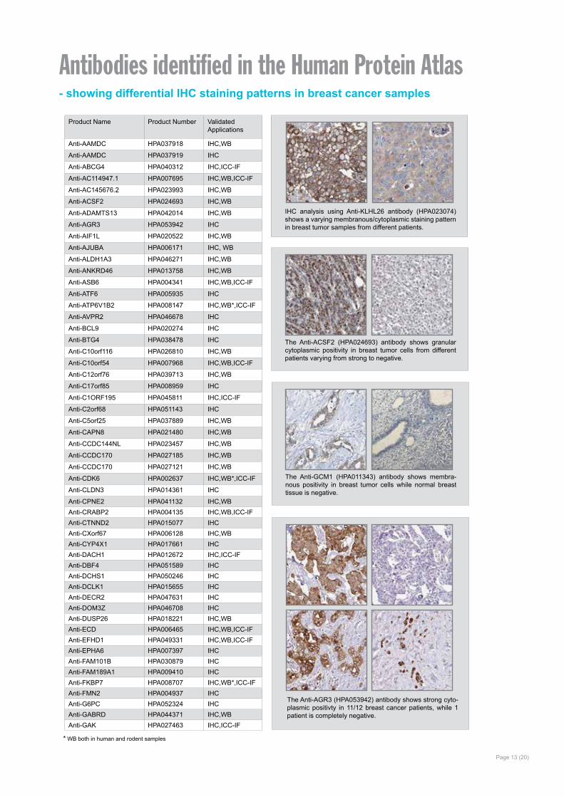
IHC analysis using Anti-KLHL26 antibody (HPA023074) shows a varying membranous/cytoplasmic staining pattern in breast tumor samples from different patients.
The Anti-ACSF2 (HPA024693) antibody shows granular cytoplasmic positivity in breast tumor cells from different patients varying from strong to negative.
The Anti-AGR3 (HPA053942) antibody shows strong cyto-plasmic positivty in 11/12 breast cancer patients, while 1 patient is completely negative.
The Anti-GCM1 (HPA011343) antibody shows membra-nous positivity in breast tumor cells while normal breast tissue is negative.
* WB both in human and rodent samples
Page 14 (20)
* WB both in human and rodent samples
Product Name Product Number Validated Applications
Anti-GCM1 HPA011343 IHC
Anti-GLB1L3 HPA039916 IHC
Anti-GLDC HPA002318 IHC,WB*
Anti-GLYATL1 HPA039501 IHC,WB
Anti-GTF3A HPA007990 IHC,ICC-IF
Anti-HIPK2 HPA007611 IHC,ICC-IF
Anti-HMGCS1 HPA036913 IHC,WB,ICC-IF
Anti-HMGCS2 HPA027423 IHC,WB
Anti-HMGCS2 HPA027442 IHC,WB
Anti-IFITM3 HPA004337 IHC,WB
Anti-IRX2 HPA054669 IHC,WB
Anti-ISYNA1 HPA007931 IHC
Anti-ISYNA1 HPA008232 IHC,WB
Anti-ITGA3 HPA008572 IHC
Anti-ITGBL1 HPA005676 IHC,WB
Anti-ITIH6 HPA000506 IHC
Anti-KLHL2 HPA051637 IHC
Anti-KLHL26 HPA023074 IHC,WB
Anti-KRT31 HPA049550 IHC
Anti-KRT32 HPA040330 IHC
Anti-KRTAP9-3 HPA042482 IHC
Anti-LASP11 HPA012072 IHC,WB*,ICC-IF
Anti-LGR6 HPA008556 IHC
Anti-LRRIQ4 HPA036706 IHC
Anti-MAGEB1 HPA002820 IHC
Anti-MANSC4 HPA039454 IHC,WB
Anti-MROH2B HPA059457 IHC
Anti-MRS2 HPA017642 IHC,WBAnti-MSTO1 HPA005914 IHCAnti-MTMR2 HPA049831 IHCAnti-MYBBP1A HPA005466 IHC,WB,ICC-IFAnti-NAPEPLD HPA024338 IHC,WB,ICC-IFAnti-NASP HPA028136 IHC,WB,ICC-IFAnti-NFIA HPA006111 IHC,WB*,ICC-IFAnti-NKAIN1 HPA006873 IHC
Anti-NPSR12 HPA007489 IHC,ICC-IF
Anti-OR2Z1 HPA048760 IHCAnti-OR9K2 HPA015808 IHCAnti-OTOP2 HPA024524 IHCAnti-PDE4C HPA048975 IHC,WBAnti-PEG10 HPA051038 IHC,ICC-IFAnti-PHLDA2 HPA003994 IHCAnti-PHLPP1 HPA020200 IHCAnti-PHTF2 HPA012312 IHCAnti-PKN3 HPA045390 IHCAnti-PNMA5 HPA044690 IHC,ICC-IFAnti-PPP1R35 HPA051607 IHCAnti-PPR11 HPA023923 IHC,WB,ICC-IFAnti-PVALB HPA048536 IHC
Anti-RAB313 HPA019717 IHC,WB*
Anti-RAC3 HPA047820 IHC,WBAnti-RAD18 HPA008752 IHC,WBAnti-REEP1 HPA058061 IHC
Product Name Product Number Validated Applications
Anti-RIOK2 HPA005681 IHC,ICC-IF
Anti-RNF152 HPA015733 IHC,WB
Anti-RPS13 HPA005985 IHC
Anti-S100A1 HPA006462 IHC,WB,ICC-IF
Anti-S100A13 HPA019592 IHC,WB*
Anti-S100A14 HPA027613 IHC,ICC-IF
Anti-S100A7 HPA006997 IHC,ICC-IF
Anti-SGK196 HPA013321 IHC,WB,ICC-IF
Anti-SH3BGRL HPA051248 IHC,WB
Anti-SHROOM1 HPA037690 IHC
Anti-SLC16A7 HPA005911 IHC,WB
Anti-SLC39A6 HPA042377 IHC,WB
Anti-SPAG1 HPA023748 IHC,ICC-IF
Anti-SQLE HPA018038 IHC,WB
Anti-SRPRB HPA011173 IHC,WB
Anti-SSSCA1 HPA039789 IHC,WB*,ICC-IF
Anti-STAG3 HPA049106 IHC,WB,ICC-IF
Anti-STARD6 HPA042583 IHC,IF
Anti-STX74 HPA001467 IHC,WB*,ICC-IF
Anti-TACC3 HPA005781 IHC,WB
Anti-TAPBP HPA007066 IHC
Anti-TBC1D9 HPA000262 IHC,ICC-IF
Anti-TCTE3 HPA046156 IHCAnti-TGFBI HPA017019 IHCAnti-TMEM110-MUSTN1 HPA051855 IHCAnti-TMEM222 HPA016579 IHC,ICC-IFAnti-TMEM47 HPA046658 IHCAnti-TMEM68 HPA018216 IHC,ICC-IFAnti-TPX2 HPA005487 IHC,WB,ICC-IFAnti-TTLL12 HPA003054 IHC,WBAnti-UBE20 HPA023605 IHC,WB*Anti-WFDC2 HPA042302 IHCAnti-WNT3A HPA050514 IHCAnti-ZBTB7B HPA006811 IHC,WB*,ICC-IFAnti-ZKSCAN3 HPA009637 IHC,ICC-IFAnti-ZNF131 HPA007023 IHCAnti-ZNF627 HPA049770 IHC,WBAnti-ZNF662 HPA039116 IHC,WB
1. Ngan E et al. A complex containing LPP and α-Actinin mediates TGF β-induced migration and invasion of ErbB2-expressing breast cancer cells. J Cell Sci 2013 May 1; 126(0 9):1981-1991. Epub 2013/02/27.
2. Camilleri M et al. Neuropeptide S receptor induces neuropeptide expression and associates with intermediate phenotypes of functional gastrointestinal disorders. Gastroenterology 2010 Jan;138(1):98-107.e4.
3. Bozóky B et al. Novel signatures of cancer-associated fibroblasts. Int J Cancer 2013 Jan 15.
4. Strömberg S et al. Selective expression of Syntaxin-7 protein in benign melano-cytes and malignant melanoma. J Proteome Res 2009 Apr;8(4):1639-46.
RBM3 The RNA-binding motif protein 3 (RBM3) is an RNA- and DNA-binding protein, whose function has not been fully elucidated. It has been shown that the protein is expressed as an early event in mild hypothermia, and also in other conditions relating to cellular stress, such as glucose de-privation and hypoxia1. During stress, RBM3 is thought to protect the cells by aiding in maintenance of protein synthesis needed for survival1. Re-cently, it has also been shown that RBM3 attenuates stem cell-like pro-perties in prostate cancer cells2.
RBM3 was identified via the Human Protein Atlas (HPA) as a potential oncology biomarker through the dif-ferential expression pattern present in several cancers investigated as part of the HPA project (proteinatlas.org)3,4.
The IHC analysis using the Anti-RBM3 antibody HPA003624 showed a weak expression pattern in normal breast tissue, but a stratified pattern in breast cancer tissue (Figure 1). Researchers further investigated the expression in larger breast cancer cohorts and the expression of RBM3 was shown to be associated with a prolonged survival5.
Page 15 (20)
Finding Cancer BiomarkersBreast CancerBreast cancer is the second most common cancer and by far the most frequent cancer among women. The incidence of breast cancer is increas-ing steadily, but without a correspond-ing increase in mortality. If detected at an early stage, the prognosis is rela-tively good for a patient living in a de-veloped country, with a general five-year survival rate of approximately 85%.
Breast Cancer and Treatment Cancer, though often denoted as a singular disease, is truly a multitude of diseases. This understanding has evolved over the years, but many pa-tients are not receiving optimal treat-ment for their disease. For cancer pa-tients to receive a more individualized treatment, there is still a need for new and better ways to stratify patients. The classical prognostic factors such as stage and grade of the tumor are insufficient for a correct estimation of patient prognosis. Additional informa-tion from cancer biomarkers promise to substantially improve this estima-tion, ultimately leading to a more in-dividualized treatment, thus avoiding both under- and over treatment of patients.
The primary curative treatment for breast cancer patients is surgery, often in combination with adjuvant therapy. However, adjuvant therapy is associated with substantial costs and sometimes severe side effects, and physicians have identified reduction of overtreatment as the major clinical need in breast cancer treatment to-day. Thus, the stratification of patients into different prognostic categories is of great importance as it may aid phy-sicians in selecting the most appropri-ate treatment for a given patient.
The majority of breast cancers are hormone receptor responsive, i.e., express the estrogen receptor (ER) and/or the progesteron receptor (PR). Patients with tumors express-ing these receptors may receive ad-juvant endocrine treatment, such as tamoxifen.
Breast cancers may also express the HER2 protein (human epider-mal growth factor receptor 2), and patients with tumors expressing this protein may receive adjuvant therapy with trastuzumab.
Adjuvant treatment may also consist of chemotherapy or radiation therapy.
Cancer tissue - strong
Normal tissue - weak
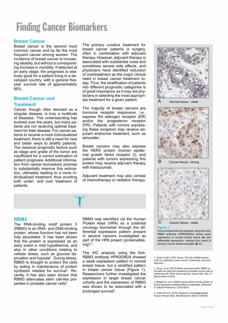
Cancer tissue - weakFigure 1Immunohistochemical analysis using the Anti-RBM3 antibody (HPA003624) shows weak expression in normal breast tissue (A) and differential expression, varying from weak to strong in tumor breast samples (B, C).
A
B
C
1. Ehlén Å (2011) PhD Thesis: The role of RNA-binding motif 3 in epithelial ovarian cancer: A biomarker discovery approach.
2. Zeng Y et al. (2013) Stress response protein RBM3 at-tenuates the stem-like properties of prostate cancer cells by interfering with CD44 variant splicing. Cancer Res. May 10. [Epub ahead of print]
3. Berglund L et al. (2008) A gene-centric human protein at-las for expression profiles based on antibodies. Molecular & Cellular Proteomics 7:2019-2027.
4. Uhlén M et al. (2010) Towards a knowledge-based Human Protein Atlas. Nat Biotechnol 28(12):1248-50.
RBM3 as a prognostic bio-marker in breast cancerAfter identification of RBM3 as a po-tential prognostic biomarker, research-ers further investigated the RBM3 pro-tein expression in larger breast cancer cohorts5. In a cohort of 500 premeno-pausal women with stage II invasive breast cancer, RBM3 expression was found to be associated with small, low-grade, estrogen receptor (ER)-positive tumors. When analyzing the subset of ER-positive patients, RBM3 was an in-dependent predictor of recurrence free survival (RFS). As shown in Figure 2, patients with tumors expressing high levels of the RBM3 protein have an im-proved survival compared to patients with tumors expressing low levels of RBM3.
RBM3 protein expression has further been analyzed in many different pa-tient cohorts from various forms of cancer. Levels of RBM3 expression was found to have a significant con-nection to patient survival in breast5, colon6, ovarian7,8, testicular, urothelial9, and prostate10 cancer as well as in ma-lignant melanoma11.
In conclusion, RBM3 is a marker of good prognosis in breast cancer as well as in several other cancers.
Page 16 (20)
Years from diagnosis
20151050
Recu
rren
ce F
ree
Surv
ival
1.0
0.8
0.6
0.4
0.2
0.0 P=0.06
RBM3 high
RBM3 low
Figure 2Kaplan-Meier (survival) analysis of recurrence free survival (RFS) according to RBM3 expression for ER-positive breast cancer patients. Patients were split into two groups based on high and low RBM3 expression.
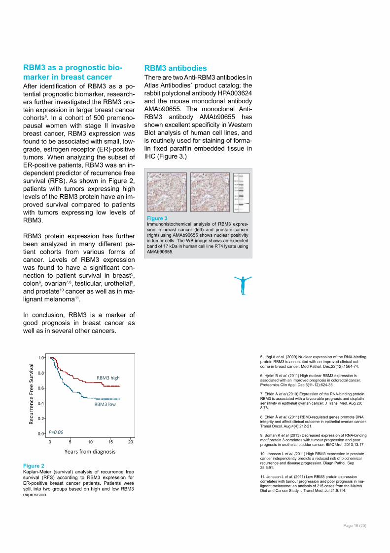
RBM3 antibodiesThere are two Anti-RBM3 antibodies in Atlas Antibodies´ product catalog; the rabbit polyclonal antibody HPA003624 and the mouse monoclonal antibody AMAb90655. The monoclonal Anti-RBM3 antibody AMAb90655 has shown excellent specificity in Western Blot analysis of human cell lines, and is routinely used for staining of forma-lin fixed paraffin embedded tissue in IHC (Figure 3.)
Figure 3Immunohistochemical analysis of RBM3 expres-sion in breast cancer (left) and prostate cancer (right) using AMAb90655 shows nuclear positivity in tumor cells. The WB image shows an expected band of 17 kDa in human cell line RT4 lysate using AMAb90655.
5. Jögi A et al. (2009) Nuclear expression of the RNA-binding protein RBM3 is associated with an improved clinical out-come in breast cancer. Mod Pathol. Dec;22(12):1564-74.
6. Hjelm B et al. (2011) High nuclear RBM3 expression is associated with an improved prognosis in colorectal cancer. Proteomics Clin Appl. Dec;5(11-12):624-35
7. Ehlén Å et al (2010) Expression of the RNA-binding protein RBM3 is associated with a favourable prognosis and cisplatin sensitivity in epithelial ovarian cancer. J Transl Med. Aug 20; 8:78.
8. Ehlén Å et al. (2011) RBM3-regulated genes promote DNA integrity and affect clinical outcome in epithelial ovarian cancer. Transl Oncol. Aug;4(4):212-21.
9. Boman K et al (2013) Decreased expression of RNA-binding motif protein 3 correlates with tumour progression and poor prognosis in urothelial bladder cancer. BMC Urol. 2013;13:17
10. Jonsson L et al. (2011) High RBM3 expression in prostate cancer independently predicts a reduced risk of biochemical recurrence and disease progression. Diagn Pathol. Sep 28;6:91.
11. Jonsson L et al. (2011) Low RBM3 protein expression correlates with tumour progression and poor prognosis in ma-lignant melanoma: an analysis of 215 cases from the Malmö Diet and Cancer Study. J Transl Med. Jul 21;9:114.
GranulinGranulins are a family of secret-ed, glycosylated peptides that are cleaved from a single precursor pro-tein. Cleavage of the signal peptide produces mature granulin which can be further cleaved into a variety of ac-tive peptides. These cleavage prod-ucts are named granulin A, granulin B, granulin C, etc. Both the peptides and intact granulin protein regulate cell growth. Different members of the granulin protein family may act as inhibitors, stimulators, or have dual actions on cell growth. Granulin fam-ily members are important in normal development, wound healing, and tumorigenesis [provided by RefSeq, Jul 2008].
In a paper by Elkabets et al, the role of GRN expression in responding tu-mor instigation was investigated by studying recrution of GRN-expressing bone marrow cells into responding tumors in mice1. Certain tumors can
foster the growth of other tumors or metastatic cells located at distant an-atomical sites, which is referred to as tumor instigation. In this study, rigor-ously growing human breast carcino-ma cells were implanted in mice and it was shown that these cells stimulated the outgrowth of otherwise poorly tu-morigenic, indolent transformed cells. GRN was identified as the most up-regulated gene in the instigating bone marrow cells. The GRN expressing cells induced resident fibroblasts to express genes that promoted malig-nant tumor progression. It was spec-ulated whether anticancer therapies might involve targeting GRN, or the activated GRN expressing cells, and thereby disrupting these cell lines of communication that promote cancer progression.
By using the Anti-GRN antibody HPA028747 in the analysis of tumor tissues from a cohort of breast can-cer patients, high GRN expression
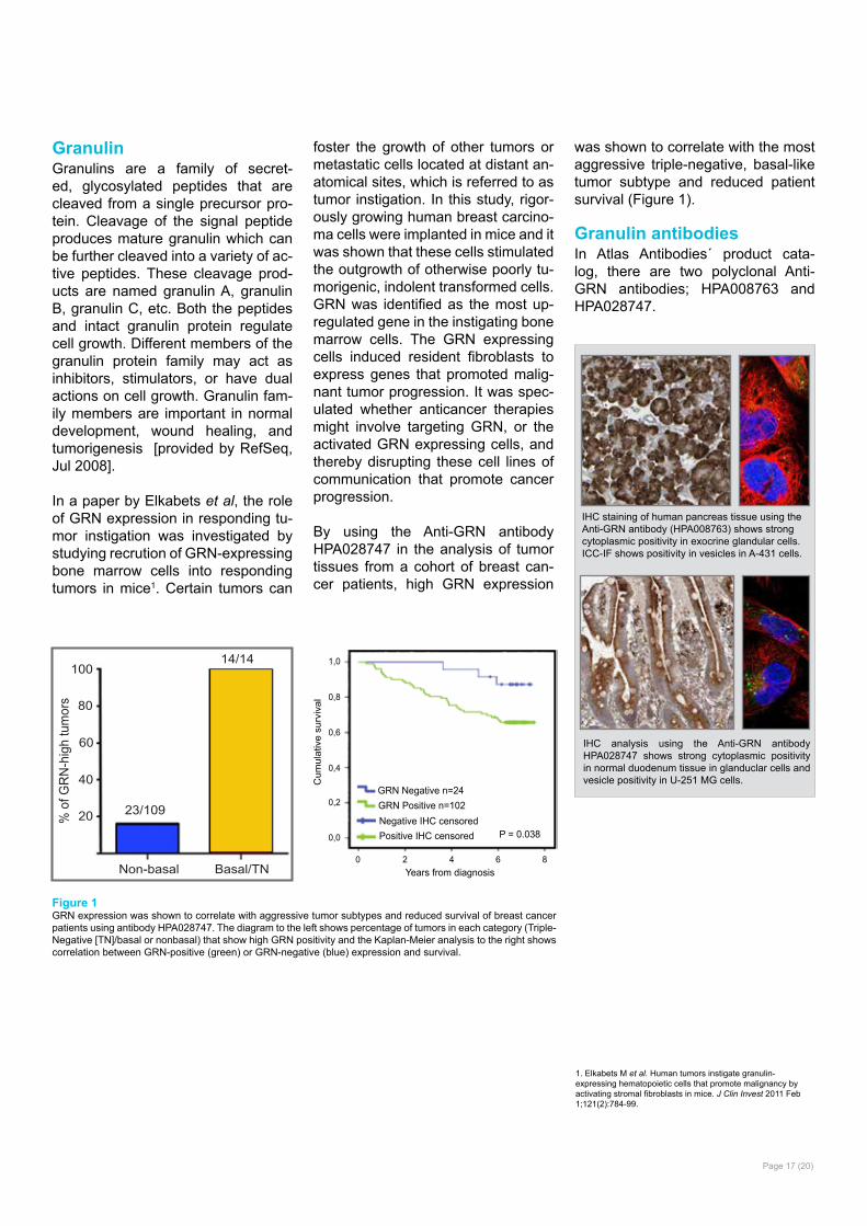
was shown to correlate with the most aggressive triple-negative, basal-like tumor subtype and reduced patient survival (Figure 1).
Granulin antibodiesIn Atlas Antibodies´ product cata-log, there are two polyclonal Anti-GRN antibodies; HPA008763 and HPA028747.
Page 17 (20)
1,0
0,8
0,6
Cum
ulat
ive
surv
ival
0,4
0,2
0,0
0 42 8Years from diagnosis
6
GRN Negative n=24GRN Positive n=102
P = 0.038Negative IHC censoredPositive IHC censored
100
80
60
40
20
Non-basal Basal/TN
% o
f GR
N-h
igh
tum
ors
23/109
14/14
Figure 1GRN expression was shown to correlate with aggressive tumor subtypes and reduced survival of breast cancer patients using antibody HPA028747. The diagram to the left shows percentage of tumors in each category (Triple-Negative [TN]/basal or nonbasal) that show high GRN positivity and the Kaplan-Meier analysis to the right shows correlation between GRN-positive (green) or GRN-negative (blue) expression and survival.
IHC staining of human pancreas tissue using the Anti-GRN antibody (HPA008763) shows strong cytoplasmic positivity in exocrine glandular cells.ICC-IF shows positivity in vesicles in A-431 cells.
IHC analysis using the Anti-GRN antibody HPA028747 shows strong cytoplasmic positivity in normal duodenum tissue in glanduclar cells and vesicle positivity in U-251 MG cells.
1. Elkabets M et al. Human tumors instigate granulin-expressing hematopoietic cells that promote malignancy by activating stromal fibroblasts in mice. J Clin Invest 2011 Feb 1;121(2):784-99.

Page 18 (20)
Anillin Anillin is an actin binding protein that is a subunit of microfilaments, one of the cytoskeleton components. Anil-lin is expressed in most cells and is involved in basic cell functions, e.g. motility, division and signaling. Stud-ies of anillin expression have shown that it is overexpressed in several hu-man tumors.
Anillin as a treatment predictive prognostic biomarker in breast cancerAnillin expression was analyzed in a patient cohort consisting of 467 sam-ples from patients diagnosed with breast cancer, using the Anti-ANLN antibody HPA005680. Patients with tumors expressing high levels of an-
illin had a reduced recurrence free survival (RFS) compared to patients with tumors expressing low levels of anillin (Figure 1A). The same associ-ation between anillin expression and reduced survival could be seen when analyzing breast cancer specific sur-vival (BCSS, Figure 1B). In a study by O´Leary et al, the prognostic impact of anillin was confirmed by Cox regres-sion analysis. High anillin expression was associated with reduced BCSS and RFS in univariate- as well as in multivariate analysis, adjusted for tu-mor size and grade, age at diagnosis, nodal-, ER-, PR-, HER2-, and Ki67 status.
In conclusion, anillin is a marker for poor prognosis in breast cancer.
Anillin antibodiesThere are three Anti-ANLN antibod-ies in Atlas Antibodies product cata-log; the mouse monoclonal antibod-ies AMAb90660 and AMAb90662 and the rabbit polyclonal HPA005680.
Figure 1Kaplan-Meier (survival) analysis of recurrence free- (A) and breast cancer specific survival (B) according to aniliin expression for breast cancer patients. Patients were split into two groups based on high and low anillin expression.
The Anti-ANLN antibody (HPA005680) shows strong nuclear positivity in cells in seminiferous ducts in human testis by IHC. In ICC-IF, nuclei (but not nucleoli) of A-431 cells stain positively and in WB, the antibody detects a band of pre-dicted size in cell lysates of RT-4 and U-251 MG.
Anti-ANLN antibody AMAb90660 shows strong nuclear immunoreactivity in a subset of tumour cells in lung adenocarcinoma and a band of pre-dicted size in human cell line U-251 MG.
AMAb90662 Anti-ANLN antibody shows strong nuclear immunoreactivity in a subset of tumor cells in colorectal cancer and a band of pre-dicted size in human U-251 MG cells.
1. O´Leary PC et al. Systematic antibody generation and valida-tion via tissue microarray technology leading to identification of a novel protein prognostic panel in breast cancer. BMC Cancer. 2013 Apr 2;13:175.
A B
Page 19 (20)
Monoclonal Antibody Development ProgramResearch remains at the heart of At-las Antibodies. We welcome custom-ers to contact us for possible collabo-rations on both existing and future product offerings. One of our collabo-ration programs aims at developing monoclonal antibodies in collabora-tion with our customers.
Atlas Antibodies encourage you to participate in our Monoclonal Anti-body Development Program for hu-man targets. If you are looking for monoclonal antibodies currently not available in our catalog, and if you are interested in developing the antibody together with us, please send in your request to us.
Upon agreement to proceed with a collaboration, Atlas Antibodies will develop and produce the monoclonal
antibody using our standardized pro-cedures. Within this procedure we al-ways epitope map all our clones and this will give you the possible option to obtain multiple clones with unique binding specificity. The selection of the optimal clones will be done in col-laboration with you. Antibodies can either be sent to you for additional characterization in your laboratory or Atlas Antibodies can make the char-acterization at our facilities with our expert input and/or material. If the project results in a commercialized product it will be added to Atlas An-tibodies Monoclonal product portfolio and available to you for a special dis-count price. All our final products will be stained and annotated by the Hu-man Protein Atlas (HPA) project and these results will be available on the HPA web portal.
Benefits of the program Atlas Antibodies take the full develop-ment cost while you get a discounted antibody with proven functionality in your experimental set-up.
For more information and/or requests for participating in the program, you are welcome to contact us at [email protected].
We are looking forward to hearing from you.
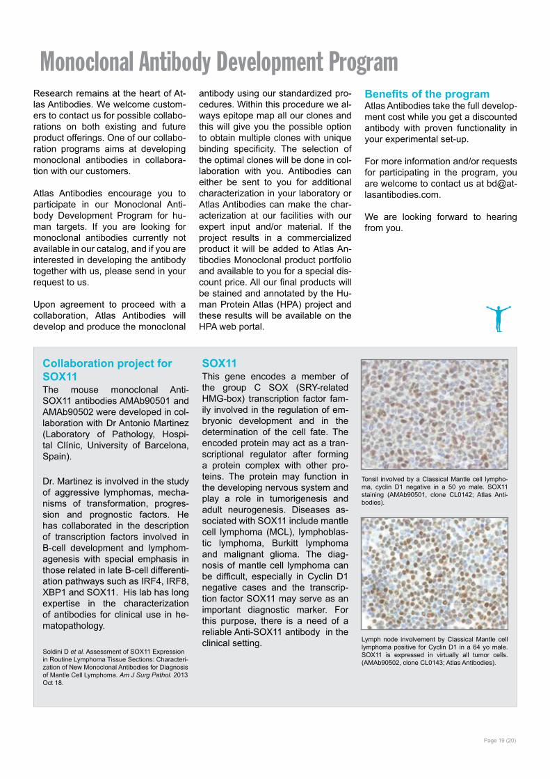
Collaboration project for SOX11The mouse monoclonal Anti-SOX11 antibodies AMAb90501 and AMAb90502 were developed in col-laboration with Dr Antonio Martinez (Laboratory of Pathology, Hospi-tal Clínic, University of Barcelona, Spain).
Dr. Martinez is involved in the study of aggressive lymphomas, mecha-nisms of transformation, progres-sion and prognostic factors. He has collaborated in the description of transcription factors involved in B-cell development and lymphom-agenesis with special emphasis in those related in late B-cell differenti-ation pathways such as IRF4, IRF8, XBP1 and SOX11. His lab has long expertise in the characterization of antibodies for clinical use in he-matopathology.
SOX11This gene encodes a member of the group C SOX (SRY-related HMG-box) transcription factor fam-ily involved in the regulation of em-bryonic development and in the determination of the cell fate. The encoded protein may act as a tran-scriptional regulator after forming a protein complex with other pro-teins. The protein may function in the developing nervous system and play a role in tumorigenesis and adult neurogenesis. Diseases as-sociated with SOX11 include mantle cell lymphoma (MCL), lymphoblas-tic lymphoma, Burkitt lymphoma and malignant glioma. The diag-nosis of mantle cell lymphoma can be difficult, especially in Cyclin D1 negative cases and the transcrip-tion factor SOX11 may serve as an important diagnostic marker. For this purpose, there is a need of a reliable Anti-SOX11 antibody in the clinical setting.
Tonsil involved by a Classical Mantle cell lympho-ma, cyclin D1 negative in a 50 yo male. SOX11 staining (AMAb90501, clone CL0142; Atlas Anti-bodies).
Lymph node involvement by Classical Mantle cell lymphoma positive for Cyclin D1 in a 64 yo male. SOX11 is expressed in virtually all tumor cells. (AMAb90502, clone CL0143; Atlas Antibodies).
Soldini D et al. Assessment of SOX11 Expression in Routine Lymphoma Tissue Sections: Characteri-zation of New Monoclonal Antibodies for Diagnosis of Mantle Cell Lymphoma. Am J Surg Pathol. 2013 Oct 18.
Atlas Antibodies ABAlbaNova University CenterSE-106 91 Stockholm Sweden
Phone +46(0)8 54 59 58 50Fax +46(0)8 54 59 58 [email protected]
atlasantibodies.comOur website provides you with easy access to all characterization data, and online ordering via our web shop. You can also send your order to [email protected].
Or send an e-mail to [email protected] to discuss any matters regarding use of antibodies.You’ll find we’re Totally Human.
Page 20 (20)
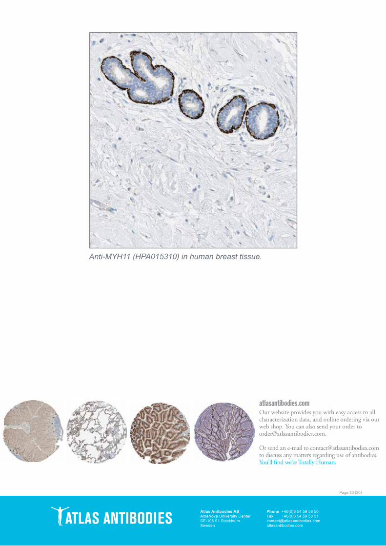
Anti-MYH11 (HPA015310) in human breast tissue.