atlas of osteoarthritis - esceo.org of osteoarthritis15032018... · 8 author biographies ali...
TRANSCRIPT

ATLAS OF OSTEOARTHRITISNigel Arden • Francisco J. Blanco • Olivier Bruyère • Cyrus Cooper • Ali Guermazi • Daichi Hayashi
David Hunter • M. Kassim Javaid • Francois Rannou • Jean-Yves Reginster • Frank W. Roemer
Second edition
This material is copyright of the original publisher Unauthorised copying and distribution is prohibited
Atlas of OsteoarthritisNigel Arden, Francisco J. Blanco, Olivier Bruyère, Cyrus Cooper, Ali Guermazi, Daichi Hayashi, David Hunter, M. Kassim Javaid, Francois Rannou, Jean-Yves Reginster, Frank W. Roemer
This book has been published in partnership with the European Society for Clinical and Economic Aspects
of Osteoporosis, Osteoarthritis and Musculoskeletal Diseases (ESCEO).
ESCEO did not receive any financial support from corporate entities, to support the preparation of this Atlas, with
the exception of an unrestricted educational grant provided by Springer Healthcare.
Second edition
This material is copyright of the original publisher Unauthorised copying and distribution is prohibited

Published by Springer Healthcare Ltd, The Campus, 4 Crinan Street, London, N1 9XW.
www.springerhealthcare.com
© 2018 Springer Healthcare, a part of Springer Nature.
All rights reserved. No part of this publication may be reproduced, stored in a retrieval system or transmitted in any form
or by any means electronic, mechanical, photocopying, recording or otherwise without the prior written permission of
the copyright holder.
British Library Cataloguing-in-Publication Data.
ISBN 978-1-910315-68-2 (Print) 978-1-910315-69-9 (eBook)
A catalogue record for this book is available from the British Library.
Although every effort has been made to ensure that drug doses and other information are presented accurately in this
publication, the ultimate responsibility rests with the prescribing physician. Neither the publisher nor the authors can be
held responsible for errors or for any consequences arising from the use of the information contained herein. Any product
mentioned in this publication should be used in accordance with the prescribing information prepared by the manufacturers.
No claims or endorsements are made for any drug or compound at present under clinical investigation.
This material is copyright of the original publisherUnauthorised copying and distribution is prohibited

Atlas of Osteoarthritis
Nigel ArdenUniversity of Oxford, UKArthritis Research UK Centre for Sport, Exercise and Osteoarthritis
Francisco J. BlancoINIBIC-Instituto de Investigación Biomedica da CoruñaComplejo Hospitalario Universitario A Coruña, Spain
Olivier BruyèreDepartment of Public Health, Epidemiology and Health Economics, University of Liège, Belgium
Cyrus Cooper Medical Research Council Lifecourse Epidemiology UnitUniversity of SouthamptonNuffield Department of Orthopaedics, Rheumatology and Musculoskeletal Sciences, University of Oxford, UK
Ali Guermazi Quantitative Imaging CenterBoston University School of Medicine, USA
Daichi Hayashi Quantitative Imaging Center,Boston University School of Medicine, USA
David Hunter University of SydneyRoyal North Shore HospitalNorth Sydney Orthopaedic and Sports Medicine Centre, Australia
M. Kassim JavaidUniversity of Oxford, UK
Francois RannouDepartment of Rehabilitation, Institute of Rheumatology Cochin Hospital, APHP University, Paris Descartes, France
Jean-Yves ReginsterDepartment of Public Health, Epidemiology and Health Economics, University of Liège, Belgium
Frank W. Roemer Quantitative Imaging Center Boston University School of Medicine, USADepartment of RadiologyUniversity of Erlangen-Nuremberg, Germany
This material is copyright of the original publisherUnauthorised copying and distribution is prohibited

4
Contents
Author biographies 6
1 Introduction: historical and current perspectives on osteoarthritis 11Jean-Yves Reginster
History of osteoarthritis in the literature 11
The changing epidemiology of osteoarthritis 15
References 17
2 Epidemiology of osteoarthritis 18Cyrus Cooper, M. Kassim Javaid and Nigel Arden
Definition of osteoarthritis 18
Classification of osteoarthritis 19
Prevalence and incidence of osteoarthritis 21
Aetiology and risk factors 24
Disease course and determinants of osteoarthritis progression 27
References 30
3 Pathophysiology of osteoarthritis 34Francois Rannou
Anatomy of normal joints 34
Pathophysiology 35
Risk factors for osteoarthritis 44
Molecular mechanisms of osteoarthritis development 46
Osteoarthritis pain 48
References 49
This material is copyright of the original publisherUnauthorised copying and distribution is prohibited
5
Contents
4 Clinical features and diagnosis of osteoarthritis 52Francisco J. Blanco
Clinical criteria for osteoarthritis 52
Symptoms of osteoarthritis 53
Diagnosis of osteoarthritis 55
Staging of osteoarthritis 61
Osteoarthritis in other joints 62
References 63
5 Assessing joint damage in osteoarthritis 66Daichi Hayashi, Frank W. Roemer and Ali Guermazi
Introduction 66
Conventional radiography 67
Magnetic resonance imaging 69
Ultrasound 72
Computed tomography and computed tomography arthrography 73
Nuclear medicine 75
Summary of imaging findings in various osteoarthritis-affected joints 76
Future directions 79
References 79
6 Treatment of osteoarthritis 80David Hunter
Nonpharmacological treatments 80
Pharmacological treatments 85
Surgical treatments 94
References 96
7 SYSADOAs 100Olivier Bruyère, Cyrus Cooper and Jean-Yves Reginster
SYSADOAs in osteoarthritis 100
References 108
This material is copyright of the original publisherUnauthorised copying and distribution is prohibited
6
Author biographies
Nigel Arden, MBBS, FRCP, MSc, MD, , is a Professor in Rheumatic Diseases at
the University of Oxford and Deputy Director: Arthritis Research UK Centre for
Sport, Exercise and Osteoarthritis
Professor Arden trained at St Thomas’s Hospital, London, where he also
completed four years of research into the genetics of osteoporosis. During this
time he gained an MSc in Epidemiology and an MD. In 1998 he spent six months as Visiting Pro-
fessor in Epidemiology at the University of San Francisco. He became a Professor of Rheumatic
Diseases in Southampton in 2008 and at the University of Oxford in 2011.
Professor Arden is based in the Botnar Research Centre, at the University of Oxford with
additional sessions at the MRC Lifecourse Epidemiology Unit at the University of Southampton.
The programme has several major strands: (a) The intrauterine and genetic origins of Osteoarthri-
tis, Osteoporosis and vitamin D metabolism (b) The descriptive Epidemiology of Osteoarthritis
and lower limb Arthroplasty and (c) Clinical trials in the management of common musculoskel-
etal conditions. His research field started in the aetiology of diseases, particularly genetics, but
he has now moved more into the field of treatments and prevention of disease at a population
based level. He has worked with a number of European and International Bodies who produce
guidelines for management, but also looking at implementation policies. He has published over
300 research papers and 5 books.
Francisco J. Blanco, MD, PhD, is Director of Research in the Biomedical Research
Center of A Coruña (INIBIC) and Associate Professor of Medicine at the Universi-
dad de Santiago de Compostela, Galicia, Spain. He was a Research Fellow at the
University of California San Diego, USA. Currently, Dr Blanco works as a rheu-
matologist in clinic at the Hospital Universitario A Coruña. His research group
(GIR) is focused on the cellular and molecular mechanisms of osteoarthritis, and on the search
of biomarkers useful for diagnosis, prognosis and therapeutic response of rheumatic diseases.
He is a member the Proteo-Red (Spanish Network of Proteomics). Dr Blanco is Director of the
Catedra-Bioiberica at A Coruña University. He is editor in chief of the Reumatología Clinica and a
member of the Editorial Board of the Osteoarthritis and Cartilage, Arthritis Research and Therapy,
Open Arthritis Journal and Open Proteomics Journal.
This material is copyright of the original publisherUnauthorised copying and distribution is prohibited

7
Author biographies
Olivier Bruyère, Ph.D., is currently Professor of Clinical Epidemiology in the
Department of Public Health Sciences and of Geriatric Rehabilitation in the
Department of Sport Sciences of the University of Liège in Belgium. He is head
of the Research Unit in Public Health, Epidemiology and Health Economics in
this University. Professor Bruyère is the Chief Executive Officer of the European
Society on Clinical and Economic Aspects of Osteoporosis and Osteoarthritis (ESCEO), President
of the Belgian Ageing Muscle Society (BAMS), General Secretary of the Belgian Bone Club (BBC),
member of the Scientific Advisory Board of the International Osteoporosis Foundation (IOF) as
well as member of the Group for the Respect of Ethics and Excellence in Sciences (GREES). He also
works as expert for the French Agency for Food, Environmental and Occupational Health & Safety
(ANSES). His main fields of interest are prevention, rehabilitation and pharmaco-epidemiology
related to geriatric or rheumatic conditions. Besides being Editor-in-chief of the journal “The
Archives of Public Health”, he is Executive Editor of “Aging Clinical and Experimental Research”,
Associate Editor of “BMC Musculoskeletal Disorders” as well as on the editorial board of various
journals. He is author of more than 250 international scientific publications and book chapters.
Cyrus Cooper OBE, DL, FMedSci, is Professor of Rheumatology and Director of
the MRC Lifecourse Epidemiology Unit; Vice-Dean of the Faculty of Medicine at
the University of Southampton; and Professor of Epidemiology at the Nuffield
Department of Orthopaedics, Rheumatology and Musculoskeletal Sciences,
University of Oxford.
He leads an internationally competitive programme of research into the epidemiology of
musculoskeletal disorders, most notably osteoporosis. His key research contributions have been:
1) discovery of the developmental influences which contribute to the risk of osteoporosis and hip
fracture in late adulthood; 2) demonstration that maternal vitamin D insufficiency is associated
with sub-optimal bone mineral accrual in childhood; 3) characterisation of the definition and
incidence rates of vertebral fractures; 4) leadership of large pragmatic randomised controlled
trials of calcium and vitamin D supplementation in the elderly as immediate preventative strate-
gies against hip fracture.
He is President of the International Osteoporosis Foundation; Chair of the BHF Project
Grants Committee; an emeritus NIHR Senior Investigator; and Associate Editor of Osteoporo-
sis International. He has previously served as Chairman of the Scientific Advisors Committee,
International Osteoporosis Foundation; Chairman, MRC Population Health Sciences Research
Network; Chairman of the National Osteoporosis Society of Great Britain; past-President of the
Bone Research Society of Great Britain; and has worked on numerous Department of Health,
European Community and World Health Organisation committees and working groups. He has
published extensively (over 900 research papers; hi=119) on osteoporosis and rheumatic disor-
ders and pioneered clinical studies on the developmental origins of peak bone mass. In 2015,
he was awarded an OBE for services to medical research.
This material is copyright of the original publisherUnauthorised copying and distribution is prohibited

8
Author biographies
Ali Guermazi, MD, PhD, is a radiologist with expertise in imaging of musculo-
skeletal diseases. Currently, he is Professor of Radiology and Medicine, Vice
Chair of Academic Affairs and Director of the Quantitative Imaging Center at
Boston University School of Medicine. He leads a research group focusing on
the application of magnetic resonance imaging (MRI) to epidemiological studies
and musculoskeletal radiology. He has been involved in developing several original and widely
accepted radiological methods to assess osteoarthritis disease risk and progression. He has also
contributed to a number of large-scale multicentre osteoarthritis trials, such as the Multicentre
Osteoarthritis Study, Health ABC, Framingham Osteoarthritis Study and Osteoarthritis Initiative.
Daichi Hayashi , MBBS, PhD, is a radiologist-in-training and is currently a Research
Assistant Professor of Radiology at Boston University School of Medicine. He
completed his medical degree at King’s College London School of Medicine,
UK, and obtained his doctoral degree from Jikei University School of Medicine,
Tokyo, Japan. He has been involved in musculoskeletal research, focusing on
osteoarthritis and cartilage imaging for several National Institutes of Health (NIH) and pharma-
ceutical sponsored studies. His research interest includes MRI of musculoskeletal diseases, with
a focus on osteoarthritis.
David Hunter, MBBS, PhD, FRACP, is Florance and Cope Chair of Rheumatology,
Professor of Medicine at University of Sydney, Chair of the Institute of Bone and
Joint Research, and Staff Specialist Rheumatologist at Royal North Shore Hospital
and North Sydney Orthopaedic and Sports Medicine Centre. He completed his
medical degree at the University of New South Wales (UNSW), a fellowship in
Rheumatology at the Royal Australian College of Physicians, earned a Masters of Medical Science
(Clinical Epidemiology) from the University of Newcastle, a Masters of Sports Medicine from UNSW
and a PhD from the University of Sydney.
In his current work, Dr Hunter is investigating a number of key elements in osteoarthritis
including the epidemiology of osteoarthritis, genetic epidemiology of osteoarthritis, the role
of biomarkers in understanding osteoarthritis aetiopathogenesis, the application of imaging
to better understand structure and function with application to both epidemiologic research
and clinical trials, the application of novel therapies in disease management and heath service
system delivery of chronic disease management. Dr Hunter has over 400 peer reviewed papers
published in international journals, numerous book chapters, has co-authored a number of books,
including two books on self-management strategies for the lay public.
This material is copyright of the original publisherUnauthorised copying and distribution is prohibited

9
Author biographies
M. Kassim Javaid, MBBS, BMedSci, MRCP, PhD, Senior Research Fellow in Meta-
bolic Bone Disease; Honorary Consultant and Rheumatologist at the University
of Oxford. Dr Javaid completed his medical training at Charing Cross and West-
minster Medical School and specialised in adult rheumatology at the Wessex
Deanery. During that time, he also completed a PhD examining the maternal
determinants of intra-uterine bone growth as part of an Arthritis Research Campaign (ARC)
Clinical Fellowship at the University of Southampton. He was awarded an ARC travelling fellow-
ship and worked with the osteoarthritis group in University of California San Francisco to study
the role of vitamin D and bone in lower limb osteoarthritis.
Dr Javaid further extended his research into the role of vitamin D status in musculoskeletal
disease, improving outcomes after fragility fracture as well as continuing work looking into the
bone phenotypes in osteoarthritis. Balancing clinical and teaching, his direction of research is
evermore linking the basic science with the key clinical issues in osteoarthritis and osteoporosis.
Francois Rannou MD, PhD, is Professor of Medicine at Paris Descartes University
and Cochin Hospital. He is qualified in rehabilitation and rheumatology. He is
the head of the rehabilitation department in the Cochin institute of rheumatol-
ogy, University Paris Descartes. He leads an INSERM team working in the field of
cartilage and intervertebral disc biology. His clinical activity is mainly focussed
on osteoarthritis and low back pain from care to randomised controlled trials.
This material is copyright of the original publisher Unauthorised copying and distribution is prohibited

10
Author biographies
Jean-Yves Reginster, MD, PhD, trained at the University of Liège in Belgium,
and specialised in Physical Medicine and Rehabilitation (Liège), in Public Health
(Nancy, France) and in Epidemiology-Health Policy (Ann Arbor, USA).
As Professor and Chairman, he directs the activities of Public Health, Epidemiol-
ogy and Health Economics of the University of Liège, where he is Honorary Head
at the Center for Investigation in Bone and Articular Cartilage Metabolism. He is also Professor of
Bioethics and Societal Medicine at the same Institution. He is President of the European Society
for Clinical and Economic Aspects of Osteoporosis and Osteoarthritis (ESCEO). He serves at the
IOF (International Osteoporosis Foundation) as a member of the Executive Committee, Board
of Directors and as the President and Chair of the Committee of National Societies, in addition
of serving as a member of the Committee of Scientific Advisors. He is currently serving as the
Director of the WHO Collaborating Center for Public Health Aspects of Musculoskeletal Health
and Aging, University of Liège, Belgium.
He is particularly interested in Metabolic Bone Diseases, in the Epidemiology, Prevention
and Treatment of Postmenopausal Osteoporosis, Osteoarthritis, Frailty and Sarcopenia, in all
aspects of Pharmacoepidemiology, Public Health and Health Economics, Quality of life, and in
the Methodology of Clinical Trials.
Professor Reginster is in the Editorial Board of numerous journals, such as Osteoporosis
International, Bone, Calcified Tissue International. He has written more than 850 scientific articles
and more than 80 books or book chapters.
Frank W. Roemer, MD, is Co-Director of the Quantitative Imaging Center of the
Department of Radiology at Boston University and Section Chief of MRI at the
Department of Radiology at Klinikum Augsburg, a major teaching hospital in
southern Germany. He holds academic appointments as Associate Professor
at Boston University and the University of Erlangen, Germany, and is Associate
Editor of Osteoarthritis Cartilage and BMC Musculoskeletal Disorders.
Dr Roemer is a German board-certified musculoskeletal radiologist with a strong focus on
MRI. His main research interest is imaging of degenerative joint disease, sports imaging and
imaging applications in pre-clinical research.
This material is copyright of the original publisherUnauthorised copying and distribution is prohibited

11
Osteoarthritis is an important issue for both the individual and society [1], and its public health
impact continues to grow due to the ageing population, the rising prevalence of obesity and the lack
of definitive treatments to prevent or halt the progress of the disease [2]. However, osteoarthritis
is difficult to define, and a better understanding of its pathophysiology is required [1,2].
What all forms of osteoarthritis and related disorders have in common is a loss of cartilage
associated with bone features such as osteophytes and subchondral bone sclerosis [3]. However, the
history of osteoarthritis is controversial because of its similarity to conditions such as diffuse idi-
opathic skeletal hyperostosis and ankylosing spondylitis as well as confusion between generalised
osteoarthritis and osteoarthritis secondary to single traumatised joints. The terminology has been
changing as well; over the years, osteoarthritis has been known as osteoarthrosis, degenerative
joint disease, arthrosis deformans and morbus (malum) coxae senilis, among other terms [3].
Despite these difficulties, the occurrence of the disease across history is perhaps one of the
best documented because of the persistence of bones compared with other bodily tissues [3,4].
The earliest examples of osteoarthritis in any animal are preserved in the bones of two dinosaurs
approximately 100 million years old; microscopic examination has revealed increased vascular
spaces and overgrowth of the articular margins [3]. The pathological characteristics of osteoar-
thritis have consequently remained unchanged [3], and it could be argued that the disease is an
immutable part of life [5].
History of osteoarthritis in the literature
From the time of Hippocrates until approximately 250 years ago, all forms of chronic arthritis
were considered to be manifestations of gout (Figure 1.1) [3,6]. The first break with that under-
standing came in 1782, when William Heberden described the nodes that now bear his name,
highlighting that “they have no connexion with gout” [7].
One of the earliest physicians to describe a non-inflammatory erosion of the articular car-
tilage particular to the elderly was Benjamin Brodie in 1829 [8]. A further leap in understanding
came with the description of osteoarthritis of the hip by Robert Smith in 1835 [9]. However,
debate over the nature of the disease continued even after the coining of the term ‘osteoarthritis’
by AE Garrod in 1890 [3].
Chapter 1Introduction: historical and current perspectives on osteoarthritis
Jean-Yves Reginster
This material is copyright of the original publisher Unauthorised copying and distribution is prohibited

12
Atlas of osteoarthritis
The introduction of X-rays at the end of the 19th century further enhanced our understanding
of the disease process [3], while the linking of Heberden noduli with osteoarthritis by Kellgren
and Moore in 1952 allowed the differentiation between generalised osteoarthritis and second-
ary osteoarthritis of a single traumatised joint [10]. The radiographic scoring system developed
by Kellgren and Lawrence later that decade paved the way for them and others to provide
a descriptive epidemiology of the condition [11,12].
Understanding of cartilage in the literature
Crucial to the developing knowledge of the processes of osteoarthritis was an understanding of
the nature and function of articular cartilage. The first recorded description of articular cartilage
Timeline of key events in the history of osteoarthritis
Year
Year
175
1743
1741
1782
1763
1835
1829
1891
1890
1925
1899
First description of cartilage in osteoarthritis given by Joannes Baptista
Morgani in Padua
Benjamin Brodie describes a
non-inflammatory erosion of the articular cartilage particular to the elderly
Aspirin is developed
The first recorded description of articular cartilage and synovial fluid given by Galen
Edward Stone discovers the pain revealing
properties of a dispersion of willow bark
Archibald Edward Garrod coins the
term ‘osteoarthritis’
1543
Event
Event
The first scientific study of articular cartilage
undertaken by William Hunter in London
Description of osteoarthritis of the hip by Robert Smith
Orientation of collagen fibres
and distribution and shape of
chondrocytes in cartilage revealed
Andreas Vesalius expands Galen’s
description of articular cartilage
William Heberden describes the
nodes that now bear his name
First recorded attempt at hip replacement
This material is copyright of the original publisher Unauthorised copying and distribution is prohibited

13
Historical and current perspectives on osteoarthritis
Timeline of key events in the history of osteoarthritis
Demonstration of hyaluronic
acid in cartilage
Proof that cartilage is not nourished
from subchondral vessels
Definitive epidemiology of
osteoarthritis set out by van Saase et al
Heberden noduli linked to osteoarthritis by Kellgren and Moore
Role of hyaluronic acid in proteoglycan structure described
Osteoarthritis proposed as a
disease involving the whole joint
was given by Galen in his treatise from 175 AD titled On the Usefulness of Various Parts of the
Body [13]. Alongside a discussion of synovial fluid, he describes cartilage thus [14]:
“ Cartilages are spread on some parts of them [bones], such as the joints, to make them
smooth, and Nature also uses cartilages occasionally as moderately yielding bodies…
Cartilage serves as a grease for the joints. ” Galen
In the 16th century, Andreas Vesalius substantially added to Galen’s definitions, stating
that cartilage “has no sensation and no marrow”, but his crucial observation was that cartilage
changes with age, such that it hardens and resembles “the fragility and friability of bone” [13].
Figure 1.1 Timeline of key events in the history of osteoarthritis. Data from Dequeker & Luyten [3] and Benedek [6].
1952
1934
1957
1954
1974
1967
1988
1978
2004
1989
First papers detailing total
knee replacement, published by Leslie
Gordon Percival Shiers
Amino acid composition of
collagen discovered
Kellgren and Lawrence describe
a radiographic classification of osteoarthritis
Cyclooxygenase enzyme
first cloned
This material is copyright of the original publisher Unauthorised copying and distribution is prohibited

14
Atlas of osteoarthritis
The first description of cartilage in osteoarthritis was given by Joannes Baptista Morgagni
in Padua in 1741, which was swiftly followed by what is considered to be the first scientific study
of articular cartilage by William Hunter in London in 1743 [13]. Hunter’s description opened up
the debate as to how an apparently nerveless tissue lacking in blood supply could be nourished
and grow. It was only with the development of enzyme chemistry that the pathophysiology of
cartilage deterioration could be properly explored [13].
The first half of the 20th century saw two major discoveries: that cartilage could be divided
into three layers through the orientation of collagen fibres and the distribution and shape of
chondrocytes and that hyaluronic acid was found in cartilage. It is only in the last 30 years that our
sophisticated understanding of collagen could be elucidated, through the use of immunological
and enzyme analyses [13].
Osteoarthritis as a whole-organ disease
Although osteoarthritis has traditionally been primarily characterised by hyaline cartilage loss,
it has more recently been described as a whole organ disease [3], and it has been suggested that
the traditional view of osteoarthritis as a cartilage-only disease is obsolete and should open up
to include the entire joint (Figure 1.2) [15,16]. Paleopathological findings have indicated that
bony involvement in osteoarthritis may involve not only bone sclerosis, but also osteophytes
and enthesophytes, which are ossifications of the insertion sites of ligaments, tendons and joint
Schematic drawing of an osteoarthritic joint
Figure 1.2 Schematic drawing of an osteoarthritic joint. The different tissues involved in clinical and structural changes of the disease are shown on the left. Note that cartilage is the only tissue not innervated. On the right the bidirectional interplay between cartilage, bone and synovial tissue involved in osteoarthritis and the two-way interaction between this interplay and the ligaments and muscles are shown. In the bidirectional interplay, one of the tissues might dominate the disease and as such should be targeted for treatment. Image from Bijlsma et al [15]. © 2011, reproduced with permission from Elsevier.
Weakening and contracture of ligaments and muscles
Inflammation of synovial tissue
Cartilage damage and loss
Outgrowth of bone (osteophytes) and attrition
Changes in subchondral bone (sclerosis and cysts)
Muscle/ligament
Cartilage
Synovial tissue
Bone
Outgrowth of bone
This material is copyright of the original publisher Unauthorised copying and distribution is prohibited

15
Historical and current perspectives on osteoarthritis
capsule to the bone [17,18]. It is therefore likely that common molecular pathways regulate bone
formation in different cellular niches, with osteophytes and enthesophytes potentially triggered
by local joint stresses and abnormal mechanical joint loading [3].
Results from several studies have supported the whole-organ view of osteoarthritis. For
example, synovitis is considered a pivotal factor in the pathogenesis of osteoarthritis, as sug-
gested by the clinical symptoms of inflammation, the presence of histological inflammation in
synovial tissue and early cartilage lesions at the border of the inflamed synovium [16]. There
is also a correlation between degeneration of the anterior cruciate ligament and cartilage,
particularly in the medial compartment of the knee joint [19]. Bone marrow lesions, commonly
resulting from traumatic knee injuries, are significantly associated with pain in people with knee
osteoarthritis [20].
Furthermore, there is growing evidence that subchondral bone plays an important role in
osteoarthritis, with bone remodelling occurring preferentially in the subchondral plate, par-
ticularly in early-stage osteoarthritis [21]. This potentially makes the subchondral plate less
able to absorb and dissipate energy [2]. These changes, alongside increases in bone volume
[21], lead to increases in forces transmitted throughout the joint [2]. The structural progres-
sion of osteoarthritis may also be viewed primarily as an atheromatous vascular disease of
subchondral bone [1].
The changing epidemiology of osteoarthritis
Historical comparisons have indicated that while the prevalence of osteoarthritis has increased
substantially over the last few centuries, the clinical patterns have not. Waldron compared the
prevalence of osteoarthritis in Georgian and early Victorian London with that of today, con-
ducting an analysis of the skeletons of 360 men and 346 women, which were recovered from
a church crypt used for burials between 1729 and 1869 [21]. Osteoarthritis of the large joints
was comparatively uncommon, with osteoarthritis of the hip found in 1.1% of men and 2.9% of
women and osteoarthritis of the knee in 0.8% of men and 5.2% of women [22]. Bilateral knee
osteoarthritis was much more common in women than in men. The right side was affected in five
of nine women and both men with unilateral disease (Figure 1.3, see page 14) [22].
The same author conducted a study of 115 cases and controls, matched for age and sex,
of skeletons with osteoarthritis of the hands that were buried in London in the late 18th and
early 19th centuries. Cases and controls were assessed for the presence of knee osteoarthritis.
The skeletons with osteoarthritis of the hands had an almost sixfold increased likelihood of knee
osteoarthritis versus controls, a significant odds ratio [23]. This pattern confirms the association
observed in contemporary populations [23–25].
Our assumptions about the changing epidemiology of osteoarthritis may also be affected
by discoveries about the pathophysiology of the disease that have led to a potential division of
This material is copyright of the original publisher Unauthorised copying and distribution is prohibited

16
Atlas of osteoarthritis
the disease into distinct phenotypes (Table 1.1) [15]. In addition to improving our understanding
of the disease, classifying the different clinical and structural phenotypes of osteoarthritis
will allow for more direct targeting of treatments, depending on whether the predominate
structural changes are in cartilage, bone, or synovial tissue. Nevertheless, there is currently
no consensus on the subgrouping of osteoarthritis into these phenotypes, and they are not
yet fully characterised [15].
Figure 1.3 Percentage distribution of different sites affected by knee osteoarthritis, by side affected. On the left side, the disease was more or less equally likely to affect only patellofemoral compartment, only the lateral compartment, or both tibiofemoral compartments together. On the right side, the patellofemoral compartment was affected in slightly more than half the cases. The medial tibiofemoral compartment was affected alone in one case only. BTF, Both tibiofemoral compartments; LTF, lateral tibiofemoral compartment; MTF, medial tibiofemoral compartment; PF, patellofemoral compartment. Image from Waldron [22]. © 1991, reproduced with permission from the British Medical Journal Publishing Group.
Table 1.1 Differentiation of clinical osteoarthritis phenotypes. AGE, advanced glycation endproducts; sRAGE, soluble receptor for advanced glycation endproducts. Data from Bijlsma et al [15]. © 2011, reproduced with permission from Elsevier.
Differentiation of clinical osteoarthritis phenotypes
Post-traumatic (acute or repetitive) Metabolic Ageing Genetic Pain
Age Young (<45 years) Middle-aged (45–65 years)
Old (>65 years)
Variable Variable
Main causative feature
Mechanical stress Mechanical stress, adipokines, hyperglycaemia, oestrogen/progesterone imbalance
AGE, chondrocyte senescence
Gene related Inflammation, bony changes, aberrant pain perception
Main site Knee, thumb, ankle, shoulder
Knee, hand, generalised
Hip, knee, hand
Hand, hip, spine
Hip, knee, hand
Intervention Joint protection, joint stabilisation, prevention of falls, surgical interventions
Weight loss, glycaemia control, lipid control, hormone replacement therapy
No specific intervention, sRAGE/AGE breakers
No specific intervention, gene therapy
Pain medication, anti-inflammatory drugs
Percentage distribution of different sites affected by knee osteoarthritis, by side affected
Left
LTF 25.0
PF 31.3
MTF 6.3
BTF 37.5
Right
LTF 28.6
BTF 19.0
PF 52.5
This material is copyright of the original publisher Unauthorised copying and distribution is prohibited

17
Historical and current perspectives on osteoarthritis
References
1 Conaghan PG, Vanharanta H, Dieppe PA. Is progressive osteoarthritis an atheromatous vascular disease? Ann Rheum Dis. 2005;64:1539-1541.
2 Neogi T. Clinical significance of bone changes in osteoarthritis. Ther Adv Musculoskelet Dis. 2012;4:259-267.
3 Dequeker J, Luyten FP. The history of osteoarthritis-osteoarthrosis. Ann Rheum Dis. 2008;67:5-10.4 Bourke J. A review of the paleopathology of the arthritic diseases. In: Brothwell D, Sandison A, eds.
Diseases in Antique. Springfield, IL: Charles Thomas Publishers; 1967:361-370.5 Karsh R, McCarthy J. Archaeology and arthritis. Arch Intern Med. 1960;105:640-644.6 Benedek TG. When did “osteo-arthritis” become osteoarthritis? J Rheumatol. 1999;26:1374-1376.7 Heberden W. Commentaries on the History and Cure of Diseases. London, UK: T. Payne; 1802. 8 Brodie BC. Pathological and Surgical Observations on the Diseases of the Joints. 2nd ed. London, UK:
Longman, Hurst, Rees, Orne and Brown; 1822. 9 Smith R. Malum coxae senilis. Dublin J Med Chem Sci. 1835;6:205.10 Kellgren JH, Moore R. Generalized osteoarthritis and Heberden’s nodes. Br Med J. 1952;1:181-187.11 Kellgren JH, Lawrence JS. Radiological assessment of osteo-arthrosis. Ann Rheum Dis. 1957;16:494-502.12 van Saase JL, van Romunde LK, Cats A, Vandenbroucke JP, Valkenburg HA. Epidemiology of
osteoarthritis: Zoetermeer survey. Comparison of radiological osteoarthritis in a Dutch population with that in 10 other populations. Ann Rheum Dis. 1989;48:271-280.
13 Benedek TG. A history of the understanding of cartilage. Osteoarthr Cartilage. 2006;14:203-209.14 Slack H. Some notes on the composition and metabolism of connective tissue. Am J Med.
1959;26:113-124.15 Bijlsma JWJ, Berenbaum F, Lafeber FPJG. Osteoarthritis: an update with relevance for clinical practice.
Lancet. 2011;377:2115-2126.16 Sellam J, Berenbaum F. The role of synovitis in pathophysiology and clinical symptoms of osteoarthritis.
Nat Rev Rheumatol. 2010;6:625-635.17 Rogers J, Shepstone L, Dieppe P. Is osteoarthritis a systemic disorder of bone? Arthritis Rheum.
2004;50:452-457.18 Rogers J, Shepstone L, Dieppe P. Bone formers: osteophyte and enthesophyte formation are
positively associated. Ann Rheum Dis. 1997;56:85-90.19 Hasegawa A, Otsuki S, Pauli C, et al. Anterior cruciate ligament changes in the human knee joint in
aging and osteoarthritis. Arthritis Rheum. 2012;64:696-704.20 Felson DT, Chaisson CE, Hill CL, et al. The association of bone marrow lesions with pain in knee
osteoarthritis. Ann Intern Med. 2001;134:541-549.21 Burr DB, Gallant MA. Bone remodelling in osteoarthritis. Nat Rev Rheumatol. 2012;8:665-673.22 Waldron HA. Prevalence and distribution of osteoarthritis in a population from Georgian and
early Victorian London. Ann Rheum Dis. 1991;50:301-307.23 Waldron HA. Association between osteoarthritis of the hand and knee in a population of skeletons
from London. Ann Rheum Dis. 1997;56:116-118.24 Cushnaghan J, Dieppe P. Study of 500 patients with limb joint osteoarthritis. I. Analysis by age, sex,
and distribution of symptomatic joint sites. Ann Rheum Dis. 1991;50:8-13.25 Hirsch R, Lethbridge-Cejku M, Scott WW Jr, et al. Association of hand and knee osteoarthritis:
evidence for a polyarticular disease subset. Ann Rheum Dis. 1996;55:25-29.
This material is copyright of the original publisher Unauthorised copying and distribution is prohibited

18
Chapter 2Epidemiology of osteoarthritis
Definition of osteoarthritis
“ A group of overlapping disorders with different aetiologies but similar biologic,
morphologic and clinical outcomes. The disease processes affect articular cartilage,
subchondral bone, synovium, capsule and ligaments. Ultimately, cartilage degenerates
with fibrillation, fissures, ulceration and full thickness loss of joint surface. ” Nigel Arden
This definition is itself developed from one coined by the Diagnostic and Therapeutic Criteria
Committee of the American Rheumatism Association for the development of criteria for clas-
sifying and reporting osteoarthritis in 1986 [1]. It also made the distinction between subclinical,
non-symptomatic defects in articular cartilage, which is poorly innervated, and the clinical
syndrome, which includes pain, that may develop from such defects [1].
“ Knee osteoarthritis is characterised clinically by usage-related pain and/or functional
limitation. It is a common complex joint disorder showing focal cartilage loss, new bone
formation and involvement of all joint tissues. Structural tissue changes are mirrored in
classical radiographic features. ” The European League Against Rheumatism
“ A heterogeneous group of conditions that lead to joint symptoms and signs which are
associated with defective integrity of articular cartilage, in addition to related changes in
the underlying bone at the joint margins. ” American College of Rheumatology
A specific definition of knee osteoarthritis was developed in 2010 for the European League Against
Rheumatism (EULAR) evidence-based recommendations for the diagnosis of knee osteoarthritis
[2]. The EULAR recommendations, which emphasise that knee osteoarthritis may associate with
osteoarthritis at other joints due to shared genetic and constitutional risk symptoms, also high-
light that the definition of knee osteoarthritis may change based on the different levels of care
needed and the clinical requirements [2].
Cyrus Cooper, M. Kassim Javaid and Nigel Arden
This material is copyright of the original publisher Unauthorised copying and distribution is prohibited

19
Epidemiology of osteoarthritis
Classification of osteoarthritis
In 1957, Kellgren and Lawrence developed a classification system that sets out a series of radio-
logical features that are considered evidence of osteoarthritis, and divides the disease into five
grades (Figure 2.1) [3]:
• 0 – None
• 1 – Doubtful
• 2 – Minimal
• 3 – Moderate
• 4 – Severe
Grade 0 indicates a definite absence of osteoarthritis changes on a single anteroposterior X-ray,
while grade 2 represents definite osteoarthritis, albeit of minimal severity [3]. Although the system
is widely used, it has limitations, particularly when assessing individual radiographic features.
Radiographic classification of osteoarthritis
Figure 2.1 Radiographic classification of osteoarthritis. A, Grade 1: doubtful joint space narrowing (JSN) and possible osteophytic lipping. B, Grade 2: definite osteophytes and possible JSN. C, Grade 3: moderate multiple osteophytes, definite JSN, some sclerosis, possible bone end deformity. D, Grade 4: large osteophytes, marked JSN, severe sclerosis definite deformity of bone ends. Image from Kellgren & Lawrence [3]. © 1957, reproduced with permission from BMJ Publishing Group Ltd.
A B
C D
This material is copyright of the original publisher Unauthorised copying and distribution is prohibited

20
Atlas of osteoarthritis
The radiological features of knee osteoarthritis were refined by the Osteoarthritis Research
Society International in 2007 [4], and divided into: the presence of marginal osteophytes in
the medial femoral condyle, medial tibial plateau, lateral femoral condyle and lateral tibial
plateau (Figure 2.2) [5] and joint space narrowing (JSN) of the medial compartment and lateral
compartment. Each of these are graded for degree of change:
• 0 – Normal
• 1 – Mild change
• 2 – Moderate change
• 3 – Severe change
Figure 2.2 Femoral osteophytes. This coronal magnetic resonance image of an osteoarthritis knee is a T1-weighted spin-echo image that shows femoral osteophytes on the medial and lateral aspects of the joint. The bright signal within the osteophytes is produced by marrow fat. Reproduced with permission from Myers [5].
Femoral osteophytes
Recently, a Delphi exercise was undertaken to develop definitions of osteoarthritis on mag-
netic resonance imaging (MRI), which suggested that, while MRI changes of osteoarthritis may
occur in the absence of radiographic findings, MRI changes in isolation and single MRI changes,
are not diagnostic of osteoarthritis [6]. Nevertheless, a definition of tibiofemoral osteoarthritis
on MRI was developed (Figure 2.3, see page 22) [7], which was either the presence of two features
from group A, or one group A feature plus at least two group B features, where:
• Group A, after exclusion of joint trauma within the last 6 months and exclusion of
inflammatory arthritis:
− Definite osteophyte formation
− Full thickness cartilage loss
• Group B:
− Subchondral bone marrow lesion or cyst not associated with meniscal or
ligamentous attachments
− Meniscal subluxation, maceration or degenerative (horizontal) tear
− Partial thickness cartilage loss (where full thickness loss is not present)
− Bone attrition
This material is copyright of the original publisher Unauthorised copying and distribution is prohibited

21
Epidemiology of osteoarthritis
A composite model was created using the above features to assess the ability of MRI to detect
radiographic osteoarthritis compared with Kellgren and Lawrence (KL) grade 2, which yielded
a C statistic of 0.59, which was described by the authors as “disappointing” [6]. Nevertheless,
MRI retains the potential to diagnose osteoarthritis earlier than the current reference standard
of radiography [6].
Prevalence and incidence of osteoarthritis
The prevalence of osteoarthritis has been assessed in a number of studies spanning several
decades. van Saase et al examined the prevalence of mild and severe radiological osteoarthritis
in a single Dutch village, finding that increased radiological osteoarthritis is strongly linked to
age, regardless of whether small or large weight-bearing joints are considered, and holds for
both men and women (Figure 2.4) [8].
The highest prevalence for osteoarthritis is seen in the cervical spine, the lumbar spine and
the distal interphalangeal joints (DIP) [8]. Severe radiological osteoarthritis is uncommon under
age 45 years, and the prevalence does not exceed 20% in the elderly aside from in the cervical
and lumbar spine and DIP and, in women, the joints of the hands and the knees [8]. Significant sex
differences are seen in the knees, in the hips among those aged at least 65 years and in the DIP
of the hands [8]. Comparison with other populations shows that, although there are substantial
differences between populations for individual joints, the slope of the majority of lines is similar
for individual and groups of joints, with no one population having a low or high prevalence of
osteoarthritis for all joints [8].
Figure 2.3 Magnetic resonance imaging of the knee: remodelling and sclerosis. This magnetic resonance image reveals considerable subchondral bone remodelling and sclerosis. Posteriorly, the cartilage of the lateral compartment is thickened with thinning and irregular cartilage in the medial compartment. Reproduced with permission from Altman [7].
Magnetic resonance imaging of the knee: remodelling and sclerosis
This material is copyright of the original publisher Unauthorised copying and distribution is prohibited

22
Atlas of osteoarthritis
The incidence of osteoarthritis increases with age, and women have higher incidences than
men, especially after age 50 (Figure 2.5, see page 24) [9]. The incidence of knee osteoarthritis
is twice that of hand or hip osteoarthritis, and the female:male sex ratio for hand, hip and knee
osteoarthritis is approximately 2:1. The trend of increasing osteoarthritis incidence continues
until age 80 after which there is a levelling off or decline in the rates for all joints, which may be
linked to sedentary activity in older age groups [9].
The lifetime risk of undergoing total hip replacement (THR) or total knee replacement (TKR) is
lower than that of developing symptomatic knee or hip osteoarthritis [10]. The mortality-adjusted
lifetime risk of undergoing THR at age 50 years is estimated, using 2005 data, at 11.6% for women
and 7.1% for men, while the risks of undergoing TKR are 10.8% and 8.1%, respectively [10].
The risk decreases with increasing age for THR and TKR in both men and women, such that, at 80
years of age, the lifetime risk of THR is 3.8% for women and 2.7% for men, while that for TKR is
3.3% and 2.7%, respectively [10].
Figure 2.4 Prevalence of osteoarthritis. A random sample of a Dutch village demonstrated the high prevalence of radiological osteoarthritis, which increases progressively with age. Mild radiological osteoarthritis is more prevalent in women (B) than in men (A), while severe radiological osteoarthritis is substantially more prevalent in women. DIP, distal interphalangeal joints. Data from van Saase et al [8]. © 1989, reproduced with permission from BMJ Publishing Group Ltd.
Prevalence of osteoarthritis
DIP Knee Hip
80
60
40
20
0
20 30 40 50 60 70 80
Age (years)
Prev
alen
ce o
f ost
eoar
thri
tis
(%)
A Men
80
60
40
20
0
20 30 40 50 60 70 80
Age (years)
Prev
alen
ce o
f ost
eoar
thri
tis
(%)
B Women DIP Knee Hip
This material is copyright of the original publisher Unauthorised copying and distribution is prohibited

23
Epidemiology of osteoarthritis
Incidence of osteoarthritis of the hand, hip and knee by age and sex
Figure 2.5 Incidence of osteoarthritis of the hand, hip and knee by age and sex. The data represents incidence in members of the Fallon Community Health Plan, 1991–1992. A, The equivalent figures for men were 5 per 100,000 person-years and 619 per 100,000 person-years. B, Among women, the incidence rates for knee osteoarthritis ranged from 0 per 100,000 person-years among those aged 20–29 years to 1082 per 100,000 person-years for those aged 70–79 years. The overall age- and sex-standardised incidence rate for knee osteoarthritis was 240/100,000 person-years (95% CI 218–262). Adapted from Oliveria et al [9].
Interestingly, the rates of primary TKR have increased substantially over the last two
decades, much more so than for THR (Figure 2.6) [11]. This may reflect the more recent matura-
tion of TKR as an efficacious treatment for osteoarthritis, or be because the number TKRs per-
formed each year is below that which would be appropriate for the burden of osteoarthritis of
the knee [11].
Hand Knee Hip
Inci
denc
e of
ost
eoar
thri
tis
(%)
900
800
700
600
500
400
300
200
100
0
B Women
20 30 40 50 60 70 80
Age (years)
A Men
1200
1100
1000
900
800
700
600
500
400
300
200
100
0
20 30 40 50 60 70 80
Age (years)
Inci
denc
e of
ost
eoar
thri
tis
(%)
This material is copyright of the original publisher Unauthorised copying and distribution is prohibited

24
Atlas of osteoarthritis
Aetiology and risk factors
In order to understand the influence that risks factors for osteoarthritis have on the pathogenesis,
a conceptual framework for the disease has been developed in recent years that consists of the
following tenets (Figure 2.7) [12–18]:
Trends in primary total knee replacement rates
Figure 2.6 Trends in primary total knee replacement rates. During the study period (1991–2006), the estimated age-standardised rates of primary total knee replacement (TKR) increased from 42.5 (95% CI 37.0–48.0) to 138.7 (95% CI 132.3–145.0) in women and from 28.7 (95% CI 23.9–33.6) to 99.4 (95% CI 93.9–104.8) in men. Interestingly, there was a marked plateau in TKR rates from the mid-1990s, followed by a sharp rise from 2000. Data from Culliford et al [11]. © 2012, reproduced with permission from The British Editorial Society of Bone and Joint Surgery.
Female Male
160
140
120
100
80
60
40
20
0
Years
Rat
e (p
er 1
00,0
00 p
erso
n-ye
ars)
19911992
19931994
19951996
19971998
19992000
20012002
20032004
20052006
Figure 2.7 Risk factors for osteoarthritis. Several systemic factors have been identified as risk factors for knee osteoarthritis, which may act by increasing the susceptibility of joints to injury, via direct damage to joint tissues, or by impairing the repair process in damaged joint tissue. Local biomechanical factors are, in contrast, believed primarily to determine the exposure of individual joints to injury and to excess loading that leads to joint degeneration. Adapted from [16–18].
Risk factors for osteoarthritis
Susceptibility to osteoarthritis or to its progression
Systemic factors:1. Age2. Gender3. Ethnic4. Hormonal status5. Genetic factors6. Bone density7. Nutritional factors
(vitamin C and D are protective)8. Inflammation
Local joint factors:1. Previous damage2. Muscle weakness3. Joint deformity/
incongruity4. Ligamentous laxity
Extrinsic factors acting on joints:1. Obesity2. Specific injurious activities:
• Sport and physical activities (excess)
• Occupational factors (eg, farming)
• Cartilage, bone, muscles, ligaments and other joint tissues and structures function as
a biomechanical organ system that maintains proper movement and prevents excessive
joint loading;
• Systemic factors that increase overall susceptibility to joint degeneration, and local
biomechanical factors that impair the optimal functioning of a joint both play an important
role in determining the risk of developing osteoarthritis; and
This material is copyright of the original publisher Unauthorised copying and distribution is prohibited

25
Epidemiology of osteoarthritis
• Systemic factors interact with mechanical factors operating within the local joint
environment to determine which joints develop osteoarthritis and how rapidly the disease
progresses in an affected joint.
It is suggested that several of the pathological features of osteoarthritis, including proliferative
bone changes, may represent attempts to repair the injured joint [19]. For example, osteophytes
may arise from a reactive response of cartilage and bone to abnormal mechanical loading, thus
reducing instability to protect the damaged joint [12]. Systemic and local factors may act in a
joint-specific manner to determine whether such a response is normal or aberrant, and whether
it succeeds or fails in protecting the joint [12]. There are a number of factors associated with
osteoarthritis of the knee, hip and hand.
Age
The age-related increases in osteoarthritis prevalence and incidence are particularly pronounced
in the commonly affected joints, such as the knee, hip and hand. It is thought that the relation-
ship between age and the risk of osteoarthritis is mediated by age-related increases in a range
of systemic and biomechanical risk factors [12].
Sex
Female gender amplifies the age-related increase in osteoarthritis risk in the hands and knees,
as well as osteoarthritis in multiple joints, such that, after 50 years of age, the prevalence and
incidence is significantly greater in women than men [9,20]. While hip osteoarthritis appears to
progress more rapidly in women [21,22], there appears to be no gender impact on knee [23,24],
or hand osteoarthritis progression [12].
Ethnicity
The prevalence of osteoarthritis and patterns of affected joints vary among racial and ethnic
groups [25]. Osteoarthritis is, in general, more prevalent in Europe and the USA than other parts of
the world [26]. Osteoarthritis of the knee is more common in African-American women than white
women [27], but that is not the case for the hip [28]. Osteoarthritis of the hip is more common
in European whites than in Jamaican blacks [29], African blacks [30] or Chinese [31]. The Beijing
Osteoarthritis Study indicated that hip and hand osteoarthritis was less frequent among Chinese
than in whites in the Framingham Study, although the prevalence of radiographic and symptomatic
knee osteoarthritis was significantly higher in Chinese women than in white women [32,33].
Menopause
As the increase in the age-related rise in osteoarthritis occurs following menopause, it would
suggest that sex hormones, particularly oestrogen deficiency, play a role in the systemic pre-
disposition to osteoarthritis [12]. While many studies have looked at the possibility of lowering
osteoarthritis risk through oestrogen use, any associations may be misleading, as oestrogen
use is linked to a healthy lifestyle and osteoporosis, which lowers the risk of osteoarthritis [12].
This material is copyright of the original publisher Unauthorised copying and distribution is prohibited

26
Atlas of osteoarthritis
Genetic factors
Genetic vulnerability appears to account for approximately half the variability of susceptibility
to hand, hip and knee osteoarthritis in women [34–40] and men [38,39]. These studies suggest
that not only are multiple genes likely to be involved in osteoarthritis susceptibility but also
that environmental factors have an important role in progression [12]. The search for candidate
genes has focused on genes encoding type II collagen (the primary collagen in articular cartilage),
structural proteins of the extracellular cartilage matrix, the vitamin D and oestrogen receptor
genes, as well as encoding bone and cartilage growth factors [41].
Obesity
Obesity is one of the most well-established and strongest risk factors for knee osteoarthritis [13], and
precedes the development of knee osteoarthritis by many years [42–44]. In addition, obesity acceler-
ates the progression of knee osteoarthritis [45,46]. The primary mechanism for the impact of obesity
of knee osteoarthritis is likely to be excess weight on overloading of the joints during weight-bearing
activities, leading to breakdown of cartilage and damage to ligaments and other support structures
[12]. Metabolic factors, such as circulating adipocytokines, adiposity-linked glucose and lipid abnor-
malities and chronic inflammation, may also play a role in the pathogenesis of osteoarthritis [12].
Mechanical and occupational factors and trauma
Acute knee injuries, including meniscal and cruciate ligament tears in the knee, fractures and disloca-
tions [12], substantially increase the risk of any subsequent osteoarthritis, as well that of more severe
disease [45]. In addition, the risk of osteoarthritis is increased by weekly participation in sports for a
decade or longer after leaving school [44]. Specifically, repetitive and excessive joint loading due to
specific physical activities increases the risk of developing osteoarthritis in the stressed joints [12].
Congenital and developmental diseases
The risk of developing osteoarthritis is substantially increased as a result of congenital abnormali-
ties that result in abnormal load distributions within the joint [47]. As the mechanical alignment
of the knee, as determined by the hip/knee/ankle angle, is an important determinant of load
distribution of the knee during ambulation [48], varus and valgus malalignment are found with
a high frequency in knees with evidence of osteoarthritis involvement of the medial and lateral
components, respectively [49]. Osteoarthritic knees with varus malalignment have a three- to
fourfold increased risk of further joint space narrowing in the medial compartment, which is
similar to the increased risk of further lateral compartment joint space narrowing in osteoarthritis
knees with valgus malalignment [50]. Discoveries about the pathophysiology of the disease have
led to a potential division of the disease into distinct phenotypes (see Table 1.1) [51]. In addition
to improving our understanding of the disease, classifying the different clinical and structural
phenotypes of osteoarthritis allows for more direct targeting of treatments, depending on where
the predominate structural changes are, eg, cartilage, bone or synovial tissue. However, there is
currently no consensus on the subgrouping of osteoarthritis into these phenotypes [51].
This material is copyright of the original publisher Unauthorised copying and distribution is prohibited

27
Epidemiology of osteoarthritis
Disease course and determinants of osteoarthritis progression
There are a number of biomarkers under investigation for the assessment of osteoarthritis
progression, as the identification of rapid progressors would assist in the development and
targeting of therapies. Imaging technologies such as MRI appear promising in the assessment of
disease progression, and combining biochemical and MRI-based biomarkers may offer effective
diagnostic and prognostic tools for identifying osteoarthritis patients at high risk of progression
(Figure 2.8) [52]. While cartilage roughness is a good diagnostic marker, with an area under the
receiver operating characteristics curve (AUC) of 0.80, and cartilage homogeneity performs well
as a prognostic marker, with an AUC of 0.71, an aggregate marker of cartilage matrix breakdown
and cartilage volume, thickness, area, congruity, roughness and homogeneity performs well both
diagnostically and prognostically, at respective AUCs of 0.84 and 0.77 [52].
Figure 2.9 Clinical and epidemiological studies on the progression of knee osteoarthritis. Circles represent the timings of the visits for the Chingford study. Figure courtesy of Dr K Leyland. Data from [45,46,53–58].
Clinical and epidemiological studies on the progression of knee osteoarthritis
Pain
Structure
Years
5 10 15
Cooper 2000 [45] Spector 1992 [54]
Massardo 1989 [53] Thorstensson 2008 [55]
Hernborg & Nilson 1977 [56]
Schouten 1992 [46]
Other
Lachance 2002 [57] Felson 1995 [58] Chingford study
Osteoarthritis stages, biomarkers and interventions
Figure 2.8 Osteoarthritis stages, biomarkers and interventions. Figure courtesy of Dr C Cooper.
Osteoarthritis progression
Cart
ilag
e qu
anti
ty
Congruity
Homogeneity
Smoothness
Focal thickness
Volume/thickness
Prevention
Cartilage regeneration
Stabilisation
Disability
Pain
This material is copyright of the original publisher Unauthorised copying and distribution is prohibited

28
Atlas of osteoarthritis
There have been a number of studies that have examined the progression of osteoarthri-
tis over follow-up periods of up to 15 years, including the recently published Chingford study
(Figure 2.9) [45,46,53–58].
The evolution of knee osteoarthritis is slow, it typically takes several years and can remain
stable for several years [21]. Radiographic deterioration is seen in a third to two-thirds of osteo-
arthritis patients and radiographic improvement is unusual (Table 2.1) [45,46,53,54,59–65].
Table 2.1 Natural history of knee osteoarthritis. C, Clinical; R, Radiographic. Table adapted with permission from Dennison & Cooper [65]. Data from [45,46,53,54,59–64].
Natural history of knee osteoarthritis
Study N Measure Years Deterioration (%)
Hernborg & Nilson (1977) [56] 94 C
R
15
15
55
56
Danielsson (1970) [59] 106 R 15 33
Massardo (1989) [53] 31 R 8 42
Dougados (1992) [60] 353 C
R
1
1
28
29
Schouten (1992) [46] 142 R 12 34
Spector (1992) [54] 63 R 11 33
Spector (1994) [61] 58 R 2 22
Ledingham (1995) [62] 350 R 2 72
McAlindon (1999) [63] 470 R 4 11
Cooper et al (2000) [45] 354 R 5 22
Felson (2004) [64] 323 R 2.5 28
Odds ratio of incidence and progression of knee osteoarthritis
Figure 2.10 Odds ratio of incidence and progression of knee osteoarthritis. The odds ratio (OR) was calculated over 5 years among patients with Kellgren and Lawrence grade 1+ disease. OR are adjusted for age and sex in all cases. In addition, OR for BMI, knee pain and Heberden’s nodes are mutually adjusted. OR for knee injury and sports participation are adjusted for age, sex, BMI, knee pain and Heberden’s nodes. Obesity was a strong predictor of incidence knee osteoarthritis (P<0.001) and a significant predictor of progression (P<0.05). BMI, Body mass index; CI, confidence interval. *Significant increase in risk. Data from Cooper et al [45].
Incidence Progression100
10
1
0.1BMI
(kg/m2)Knee pain (baseline)
Heberden’s nodes
Previous knee injury
Regular sport
OR
(95%
CI) * *
*
*
*
*
This material is copyright of the original publisher Unauthorised copying and distribution is prohibited

29
Epidemiology of osteoarthritis
While there are several factors significantly associated with the incidence of osteoar-
thritis, only obesity is significantly individually linked to the progression of grade 1+ disease
(Figure 2.10) [45]. In addition, the coexistence of Heberden’s nodes with knee osteoarthritis
increases the risk of knee deterioration by almost sixfold [21].
The Chingford study looked at the progression of individual KL grades over 15 years
Table 2.2 Progression of individual Kellgren and Lawrence grades over 15 years. Data from Leyland et al [66].
Progression of individual Kellgren and Lawrence grades over 15 years
Baseline Kellgren and
Lawrence grade N
Year 15 Kellgren and Lawrence grade
0 1 2 3 4 5
0 905 60.1% (548) 9.9% (90) 15.7% (142) 12.5% (113) 0.1% (1) 1.2% (11)
1 57 19.3% (11) 5.3% (3) 40.4% (23) 29.8% (17) 0.0% (0) 5.3% (3)
2 60 0 (0.0%) 1.7% (1) 50.0% (30) 41.7% (25) 0.0% (0) 6.7% (4)
3 26 0.0% (0) 3.8% (1) 15.4% (4) 65.4% (17) 11.5% (3) 3.8% (1)
(Table 2.2) [66], which revealed that approximately half of knees had a KL grade of 0 throughout,
while two-fifths worsened by at least one grade. Knees with baseline KL grade 1 had a higher
percentage of progression, at almost three-quarters, than knees with any other KL grade at base-
line. Less than 2% of knees were scored as having regressed to a lower KL grade by year 15 [43].
The prevalence of long-term knee pain is dependent on whether there was any pain at
baseline (Figure 2.11) [67]. The presence of knee osteoarthritis increases the risk of persistent
Figure 2.11 Prevalence of self-reported knee pain. Bars show the means with 95% confidence intervals. Individuals without knee pain at baseline (year 3) had an increase in pain prevalence with duration of follow-up, such that, at year 15, the prevalence was 35.2% for those reporting any days of pain. Data from Soni et al [67].
Prevalence of self-reported knee pain
100
90
80
70
60
50
40
30
20
10
0Year 3 Year 5 Year 10 Year 15
Visit
Prev
alen
ce o
f kne
e pa
in (%
)
No pain at year 3 Any pain at year 3
This material is copyright of the original publisher Unauthorised copying and distribution is prohibited
30
Atlas of osteoarthritis
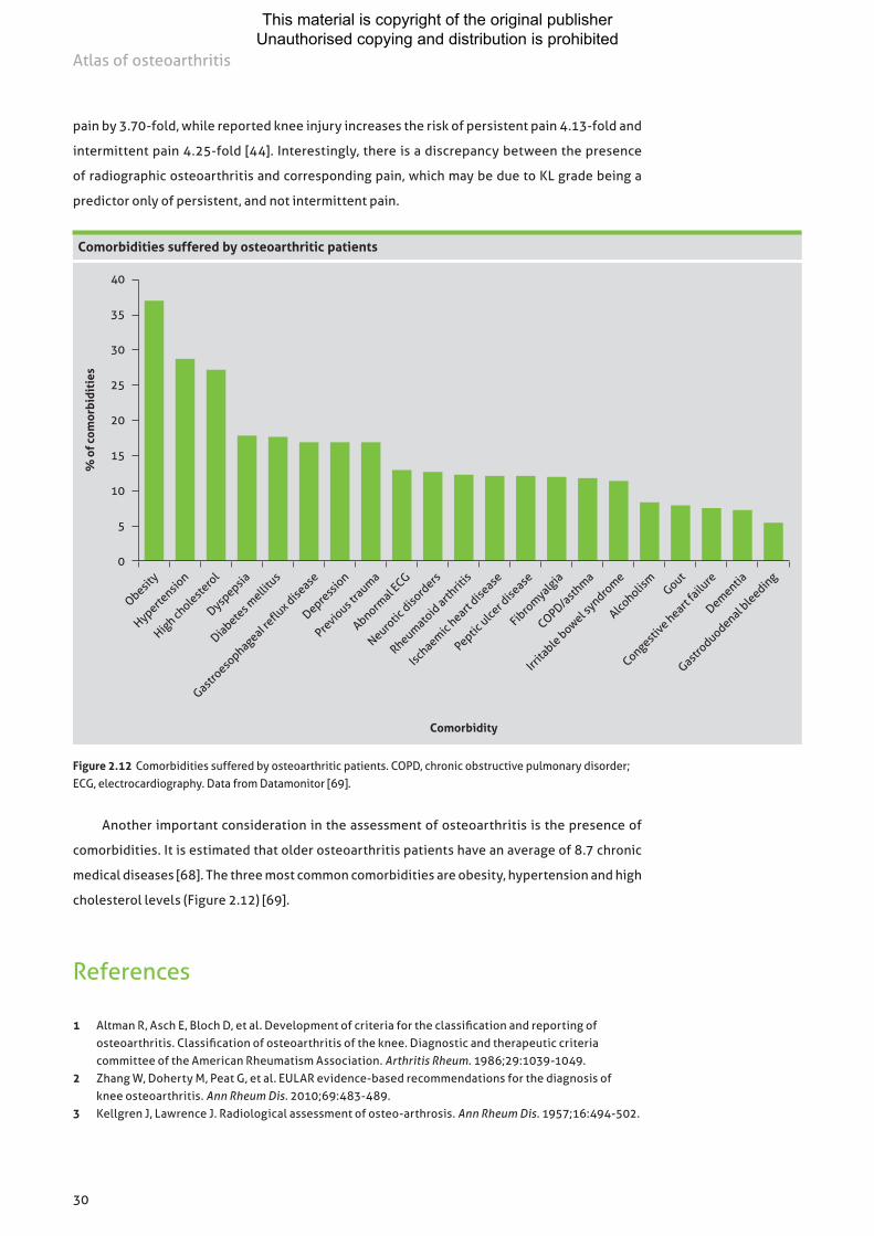
Figure 2.12 Comorbidities suffered by osteoarthritic patients. COPD, chronic obstructive pulmonary disorder; ECG, electrocardiography. Data from Datamonitor [69].
Comorbidities suffered by osteoarthritic patients
40
35
30
25
20
15
10
5
0
Comorbidity
% o
f com
orbi
diti
es
Obesity
Hypertensio
n
High choleste
rol
Dyspepsia
Diabetes m
ellitu
s
Gastroeso
phageaI reflu
x disease
Depress
ion
Previo
us tra
uma
Abnormal E
CG
Neurotic
disord
ers
Rheumatoid
arthrit
is
Ischaemic
heart dise
ase
Peptic ulce
r dise
ase
Fibro
myalgia
COPD/asth
ma
Irrita
ble bowel syndro
me
Alcoholis
mGout
Congestive heart
failu
re
Dementia
Gastroduodenal b
leeding
pain by 3.70-fold, while reported knee injury increases the risk of persistent pain 4.13-fold and
intermittent pain 4.25-fold [44]. Interestingly, there is a discrepancy between the presence
of radiographic osteoarthritis and corresponding pain, which may be due to KL grade being a
predictor only of persistent, and not intermittent pain.
Another important consideration in the assessment of osteoarthritis is the presence of
comorbidities. It is estimated that older osteoarthritis patients have an average of 8.7 chronic
medical diseases [68]. The three most common comorbidities are obesity, hypertension and high
cholesterol levels (Figure 2.12) [69].
References
1 Altman R, Asch E, Bloch D, et al. Development of criteria for the classification and reporting of osteoarthritis. Classification of osteoarthritis of the knee. Diagnostic and therapeutic criteria committee of the American Rheumatism Association. Arthritis Rheum. 1986;29:1039-1049.
2 Zhang W, Doherty M, Peat G, et al. EULAR evidence-based recommendations for the diagnosis of knee osteoarthritis. Ann Rheum Dis. 2010;69:483-489.
3 Kellgren J, Lawrence J. Radiological assessment of osteo-arthrosis. Ann Rheum Dis. 1957;16:494-502.
This material is copyright of the original publisher Unauthorised copying and distribution is prohibited
31
Epidemiology of osteoarthritis
4 Altman R, Gold G. Atlas of individual radiographic features in osteoarthritis, revised. Osteo Cart. 2007;15 Suppl A:A1-56.
5 Myers S. Osteoarthritis and crystal-associated synovitis. In: Hunder G, ed. Atlas of Rheumatology. 4th ed. Philadelphia: Current Medicine Group; 2005:54-64.
6 Hunter D, Arden N, Conaghan P, et al. Definition of osteoarthritis on MRI: results of a Delphi exercise. Osteo Cart. 2011;19:963-969.
7 Altman R. Osteoarthritis in the elderly population. In: Nakasato Y, Yung R, eds. Geriatric Rheumatology. A Comprehensive Approach. New York: Springer; 2011:187-196.
8 van Saase JL, Van Romunde LK, Cats A, Vandenbroucke J, Valkenburg H. Epidemiology of osteoarthritis: Zoetermeer survey. Comparison of radiological osteoarthritis in a Dutch population with that in 10 other populations. Ann Rheum Dis. 1989;48:271-280.
9 Oliveria S, Felson D, Reed J, Cirillo P, Walker A. Incidence of symptomatic hand, hip, and knee osteoarthritis among patients in a health maintenance organization. Arthritis Rheum. 1995;38:1134-1141.
10 Culliford D, Maskell J, Kiran A, et al. The lifetime risk of total hip and knee arthroplasty: results from the UK General Practice Research Database. Osteo Cart. 2012;20:519-524.
11 Culliford D, Maskell J, Beard D, Murray D, Price A, Arden N. Temporal trends in hip and knee replacement in the United Kingdom: 1991 to 2006. J Bone Joint Surg Br. 2010;92:130-135.
12 Arden N, Nevitt M. Osteoarthritis: Epidemiology. Best Pract Res Clin Rheumatol. 2006;20:3-25.13 Felson D, Lawrence R, Dieppe P, et al. Osteoarthritis: new insights. Part 1: The disease and its risk factors.
Ann Intern Med. 2000;133:635-646.14 Dieppe P. The classification and diagnosis of osteoarthritis. In: Kuettner K, Vm G, eds. Osteoarthritic
Disorders. Rosemont, IL: American Academy of Orthopedic Surgeons; 1995:5-12.15 Sharma H, Hanna A, Titterington L, Stephens R. Effect of MAK-4 and MAK-5 on endothelial cell and
soyabean lipoxygenase-induced LDL oxidation. Adv Exp Med Biol. 1994;366:441-443.16 Garstand S. Osteoarthritis: epidemiology, risk factors and pathophysiology. Am J Phys Med Rehabil.
2006;85: S2-S11.17 Zhang Y, Jordan J. Epidemiology of osteoarthritis. Clin Geriatr Med. 2010;26:355-369.18 Woolf A, Pfleger B. Burden of major musculoskeletal conditions. WHO Bulletin. 2003;81:646-656.19 Dieppe P. Subchondral bone should be the main target for the treatment of pain and disease
progression in osteoarthritis. Osteo Cart. 1999;7:325-326.20 Kellgren J, Moore R. Generalized osteoarthritis and Heberden’s nodes. Br Med J. 1952;1:181-187.21 Dougados M, Gueguen A, Nguyen M, et al. Radiological progression of hip osteoarthritis: definition,
risk factors and correlations with clinical status. Ann Rheum Dis. 1996;55:356-362.22 Loeser R, Shakoor N. Aging or osteoarthritis: which is the problem? Rheum Dis Clin North Am.
2003;29:653-673.23 Felson D, Naimark A, Anderson J, Kazis L, Castelli W, Meenan R. The prevalence of knee osteoarthritis
in the elderly. The Framingham osteoarthritis study. Arthritis Rheum. 1987;30:914-918.24 Ledingham J, Dawson S, Preston B, Milligan G, Doherty M. Radiographic progression of hospital
referred osteoarthritis of the hip. Ann Rheum Dis. 1993;52(4):263-267.25 Zhang Y, Jordan J. Epidemiology of osteoarthritis. Clin Geriatr Med. 2010;26:355-369.26 Woolf A, Pfleger B. Burden of major musculoskeletal conditions. Bull World Health Organ.
2003;81:646-656.27 Anderson J, Felson D. Factors associated with osteoarthritis of the knee in the first national Health
and Nutrition Examination Survey (HANES I). Evidence for an association with overweight, race, and physical demands of work. Am J Epidemiol. 1988;128:179-189.
28 Tepper S, Hochberg M. Factors associated with hip osteoarthritis: data from the first National Health and Nutrition Examination Survey (NHANES-I). Am J Epidemiol. 1993;137:1081-1088.
29 Bremner J, Lawrence J, Miall W. Degenerative joint disease in a Jamaican rural population. Ann Rheum Dis. 1968;27:326-332.
30 Solomon L, Beighton P, Lawrence J. Rheumatic disorders in the South African negro. Part II. Osteo-arthrosis. S Afr Med J. 1975;49:1737-1740.
31 Hoaglund F, Yau A, Wong W. Osteoarthritis of the hip and other joints in southern Chinese in Hong Kong. J Bone Joint Surg Am. 1973;55:545-557.
32 Nevitt M, Xu L, Zhang Y, et al. Very low prevalence of hip osteoarthritis among Chinese elderly in Beijing, China, compared with whites in the United States: the Beijing osteoarthritis study. Arthritis Rheum. 2002;46:1773-1779.
This material is copyright of the original publisher Unauthorised copying and distribution is prohibited
32
Atlas of osteoarthritis
33 Zhang Y, Xu L, Nevitt M, et al. Lower prevalence of hand osteoarthritis among Chinese subjects in Beijing compared with white subjects in the United States: the Beijing osteoarthritis study. Arthritis Rheum. 2003;48:1034-1040.
34 Macgregor A, Antoniades L, Matson M, Andrew T, Spector T. The genetic contribution to radiographic hip osteoarthritis in women: results of a classic twin study. Arthritis Rheum. 2000;43:2410-2416.
35 Spector T, Cicuttini F, Baker J, Loughlin J, Hart D. Genetic influences on osteoarthritis in women: a twin study. BMJ. 1996;312:940-943.
36 Kaprio J, Kujala U, Peltonen L, Koskenvuo M. Genetic liability to osteoarthritis may be greater in women than men. BMJ. 1996;313:232.
37 Felson D, Couropmitree N, Chaisson C, et al. Evidence for a Mendelian gene in a segregation analysis of generalized radiographic osteoarthritis: the Framingham study. Arthritis Rheum. 1998;41:1064-1071.
38 Lanyon P, Muir K, Doherty S, Doherty M. Assessment of a genetic contribution to osteoarthritis of the hip: sibling study. BMJ. 2000;321:1179-1183.
39 Ingvarsson T, Stefansson S, Hallgrimsdottir I, et al. The inheritance of hip osteoarthritis in Iceland. Arthritis Rheum. 2000;43:2785-2792.
40 Jonsson H, Manolescu I, Stefansson S, et al. The inheritance of hand osteoarthritis in Iceland. Arthritis Rheum. 2003;48:391-395.
41 Loughlin J. Genetic epidemiology of primary osteoarthritis. Curr Opin Rheumatol. 2001;13:111-116.42 Felson D, Zhang Y, Hannan M, et al. Risk factors for incident radiographic knee osteoarthritis in
the elderly: the Framingham study. Arthritis Rheum. 1997;40:728-733.43 Spector T, Hart D, Doyle D. Incidence and progression of osteoarthritis in women with unilateral knee
disease in the general population: the effect of obesity. Ann Rheum Dis. 1994;53:565-568.44 Gelber A, Hochberg M, Mead L, Wang N, Wigley F, Klag M. Body mass index in young men and the risk
of subsequent knee and hip osteoarthritis. Am J Med. 1999;107:542-548.45 Cooper C, Snow S, McAlindon T, et al. Risk factors for the incidence and progression of radiographic
knee osteoarthritis. Arthritis Rheum. 2000;43:995-1000.46 Schouten J, Van Den Ouweland FA, Valkenburg H. A 12 year follow up study in the general population
on prognostic factors of cartilage loss in osteoarthritis of the knee. Ann Rheum Dis. 1992;51:932-937.47 Harris W. Etiology of osteoarthritis of the hip. Clin Orthop Relat Res. 1986;213:20-33.48 Andriacchi T. Dynamics of knee malalignment. Orthop Clin North Am. 1994;25:395-403.49 Felson D, Nevitt M, Zhang Y, et al. High prevalence of lateral knee osteoarthritis in Beijing Chinese
compared with Framingham caucasian subjects. Arthritis Rheum. 2002;46:1217-1222.50 Sharma L, Song J, Felson D, Cahue S, Shamiyeh E, Dunlop D. The role of knee alignment in disease
progression and functional decline in knee osteoarthritis. JAMA. 2001;286:188-195.51 Bijlsma JWJ, Berenbaum F, Lafeber FPJG. Osteoarthritis: an update with relevance for clinical
practice. Lancet. 2011;377:2115-2126.52 Dam E, Loog M, Christiansen C, et al. Identification of progressors in osteoarthritis by combining
biochemical and MRI-based markers. Arthritis Res Ther. 2009;11:R115.53 Massardo L, Watt I, Cushnaghan J, Dieppe P. Osteoarthritis of the knee joint: an eight year prospective
study. Ann Rheum Dis. 1989;48:893-897.54 Spector TD, Dacre JE, Harris PA, Huskisson EC. Radiological progression of osteoarthritis: an 11 year
follow up study of the knee. Ann Rheum Dis. 1992;51:1107-1110.55 Thorstensson CA, Andersson ML, Jönsson H, Saxne T, Petersson IF. Natural course of knee osteoarthritis
in middle-aged subjects with knee pain: 12-year follow-up using clinical and radiographic criteria. Ann Rheum Dis. 2009;68:1890-1893.
56 Hernborg JS, Nilsson BE. The natural course of untreated osteoarthritis of the knee. Clin Orthop Relat Res. 1977;123:130-137.
57 Lachance L, Sowers MF, Jamadar D, Hochberg M. The natural history of emergent osteoarthritis of the knee in women. Osteoarthr Cartil. 2002;10:849-854.
58 Felson DT, Zhang Y, Hannan MT, et al. The incidence and natural history of knee osteoarthritis in the elderly. The Framingham Osteoarthritis Study. Arthritis Rheum. 1995;38:1500-1505.
59 Danielsson L, Hernborg J. Morbidity and mortality of osteoarthritis of the knee (gonarthrosis) in Malmö, Sweden. Clin Orthop Relat Res. 1970;69:224-226.
60 Dougados M, Gueguen A, Nguyen M, et al. Longitudinal radiologic evaluation of osteoarthritis of the knee. J Rheumatol. 1992;19:378-384.
This material is copyright of the original publisher Unauthorised copying and distribution is prohibited
33
Epidemiology of osteoarthritis
61 Spector TD, Hochberg MC. Methodological problems in the epidemiological study of osteoarthritis. Ann Rheum Dis. 1994;53:143-146.
62 Ledingham J, Regan M, Jones A, Doherty M. Factors affecting radiographic progression of knee osteoarthritis. Ann Rheum Dis. 1995;54:53-58.
63 McAlindon TE, Wilson PW, Aliabadi P, Weissman B, Felson DT. Level of physical activity and the risk of radiographic and symptomatic knee osteoarthritis in the elderly: the Framingham study. Am J Med. 1999;106:151-157.
64 Felson DT, Neogi T. Osteoarthritis: is it a disease of cartilage or of bone? Arthritis Rheum. 2004;50:341-344.65 Dennison E, Cooper C. The natural history and prognosis of osteoarthritis. In: Brandt K, Doherty M,
Lohmander M, eds. Textbook of Osteoarthritis. 2nd ed. Oxford: Oxford University Press; 2003:227-233.66 Leyland KM, Hart DJ, Javaid MK, et al. The natural history of radiographic knee osteoarthritis:
a fourteen-year population-based cohort study. Arthritis Rheum. 2012;64:2243-2251.67 Soni A, Kiran A, Hart D, et al. Prevalence of reported knee pain over twelve years in a community-based
cohort. Arthritis Rheum. 2012;64:1145-1152.68 Bayliss E, Ellis J, Steiner J. Barriers to self-management and quality-of-life outcomes in seniors with
multimorbidities. Ann Fam Med. 2007;5:395-402.69 Datamonitor. Stakeholder Insight: Osteoarthritis. Drug development lags behind rising osteoarthritis
population. Datamonitor Europe: London, UK; December 2009.
This material is copyright of the original publisher Unauthorised copying and distribution is prohibited

34
Francois Rannou
Anatomy of normal joints
Human movement is made possible by synovial fluid, or freely moving, and cartilaginous, or fixed,
joints [1]. The synovial joint is a functional connective tissue unit that allows two opposed limb
bones to move freely in relation to each other. The bone−cartilage−synovial fluid−cartilage−bone
assembly can be regarded as a continuum, with the load-bearing structures organised differentially
depending on site and function, resulting in a specialised joint structure [1].
There are five basic types of structures in the knee (Figures 3.1 and 3.2, see page 36) [2–5]:
• ligaments, which are passive elastic structures that can be loaded in tension only;
• musculotendinous units, which are active elastic structures that act only under tension;
• cartilage and subchondral bone, which accommodate the compressive loads of the joint;
• menisci, which are crescentic fibrocartilaginous pads that attach to the intercondylar area
and periphery of the tibial plateau; and
• the bursae.
Chapter 3Pathophysiology of osteoarthritis
Anterior and lateral view of the normal knee anatomy
A
FemurPatella
TibiaFibulaPosterior cruciate ligament
Medial collateral ligament
Anterior cruciate ligament
Lateral collateral ligament
Femur
Tibia
B
Pes anserinus
Pes anserine bursa
Prepatellar bursa
Deep infrapatellar bursa
Superficial infrapatellar bursa
Pretibial bursa
Figure 3.1 Anterior and lateral view of the normal knee anatomy. A, image showing the basic structure of the knee. Ligaments can be divided into intra-articular and extracapsular. B, The major bursae around the knee. Adapted with permission from Niitsu [5].
This material is copyright of the original publisher Unauthorised copying and distribution is prohibited

35
Pathophysiology of osteoarthritis
Pathophysiology
Osteoarthritis is considered an organ disease that involves the whole joint structure. A gradual loss
of articular cartilage in synovial joints is combined with subchondral bone sclerosis, osteophytes
at the joint margins and mild, chronic nonspecific synovial inflammation [6,7]. A hypothetical
model of the development of osteoarthritis is shown in Figure 3.3 [6].
Figure 3.2 Plain radiograph of the normal right knee. This radiograph clearly shows the femur, tibia and fibula. The patella can be seen as faint circular outline overlapping the femur, centred at the widest part of the femur. Image from Abdul-Jabar et al [4].
Plain radiograph of the normal right knee
Figure 3.3 Hypothetical model for initiation and perpetuation of osteoarthritis. Accumulation of risk factors on ageing cartilage triggers the initiation of the osteoarthritic process. For didactic reasons, two phases are described, early osteoarthritis and late osteoarthritis, but the passage from one to the other is progressive and generally lasts many years. KS, keratan sulphate. Reproduced with permission from Berenbaum [6].
Hypothetical model for initiation and perpetuation of osteoarthritis
Risk factors• Excessive weight• Injury and occupation• Developmental deformities• Joint laxity
Ageing cartilage• Cartilage fissure• Shorter glycosaminoglycan• Increased KS6 concentration/decreased KS4 concentration• Decrease in chondrocyte number• Accumulation of advanced glycation end products• Decrease in water concentration
Initiation• Deleterious mechanical stresses• Genetic factors• Hormonal factors?
Early osteoarthritis• Increased chondrocyte proliferation• Increased synthesis of matrix by chondrocytes• Alteration in collagen synthesis (decrease in type II/type I collagen ration)• Chondrocyte dedifferentiation• Increased synthesis of proteinases by chondrocytes• Increased synthesis of cytokines by chondrocytes• Subchondral bone demineralisation with microfractures• Inflamed synovial tissue
Late osteoarthritis• Decreased chondrocyte proliferation• Chondrocytes apoptosis• Hypertrophic differentiation
of chondrocytes• Osteophyte formation• Bone sclerosis• Persistence of proteinases and
cytokines synthesis
REVERSIBLE
IRREVERSIBLE
Femur
Patella
Tibia
Fibula
This material is copyright of the original publisher Unauthorised copying and distribution is prohibited
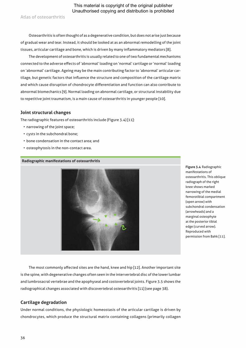
36
Atlas of osteoarthritis
Osteoarthritis is often thought of as a degenerative condition, but does not arise just because
of gradual wear and tear. Instead, it should be looked at as an abnormal remodelling of the joint
tissues, articular cartilage and bone, which is driven by many inflammatory mediators [8].
The development of osteoarthritis is usually related to one of two fundamental mechanisms
connected to the adverse effects of ‘abnormal’ loading on ‘normal’ cartilage or ‘normal’ loading
on ‘abnormal’ cartilage. Ageing may be the main contributing factor to ‘abnormal’ articular car-
tilage, but genetic factors that influence the structure and composition of the cartilage matrix
and which cause disruption of chondrocyte differentiation and function can also contribute to
abnormal biomechanics [9]. Normal loading on abnormal cartilage, or structural instability due
to repetitive joint traumatism, is a main cause of osteoarthritis in younger people [10].
Joint structural changes
The radiographic features of osteoarthritis include (Figure 3.4) [11]:
• narrowing of the joint space;
• cysts in the subchondral bone;
• bone condensation in the contact area; and
• osteophytosis in the non-contact area.
Figure 3.4 Radiographic manifestations of osteoarthritis. This oblique radiograph of the right knee shows marked narrowing of the medial femorotibial compartment (open arrow) with subchondral condensation (arrowheads) and a marginal osteophyte at the posterior tibial edge (curved arrow). Reproduced with permission from Bahk [11].
Radiographic manifestations of osteoarthritis
The most commonly affected sites are the hand, knee and hip [12]. Another important site
is the spine, with degenerative changes often seen in the intervertebral disc of the lower lumbar
and lumbrosacral vertebrae and the apophyseal and costovertebral joints. Figure 3.5 shows the
radiographical changes associated with discovertebral osteoarthritis [11] (see page 38).
Cartilage degradation
Under normal conditions, the physiologic homeostasis of the articular cartilage is driven by
chondrocytes, which produce the structural matrix containing collagens (primarily collagen
This material is copyright of the original publisher Unauthorised copying and distribution is prohibited

37
Pathophysiology of osteoarthritis
type II), and proteoglycans [6,13]. Despite the involvement of multiple joint tissues, osteoarthritis
has long been mainly characterised by a breakdown of the repair process of damaged cartilage
as a result of biochemical and biomechanical changes in the joint [12]. The changes in cartilage
structure as a result of osteoarthritis are shown in Figure 3.6 [13].
In osteoarthritis, the chondrocytes within the joint fail to synthesise a resistant and elastic
matrix and therefore cannot maintain the balance between synthesis and degradation of the
extracellular matrix [6]. Inflammatory mediators such as interleukin (IL)-1 and mechanical
Figure 3.5 Discovertebral osteoarthritis of the spine. Anteroposterior radiograph of the lower lumbar spine shows endplate-based sclerosis in the L4 lower and L5 upper end-plates with narrowing of the disc space between them (arrow). Small claw-like spurs (arrowheads) are seen at the right lateral edges. Reproduced with permission from Bahk [11].
Discovertebral osteoarthritis of the spine
Figure 3.6 Cartilage defects in osteoarthritis. Lapine model of osteoarthritis using safranin O with fast green counterstain. A, Normal: smooth surface, heavy red stain of proteoglycans, no increase or decrease in chondrocytes and one well-defined tidemark. B, Osteoarthritis: disrupted cartilage surface, proliferation of chondrocytes with many pyknotic chondrocytes (indicating cell death), sparse red stain of proteoglycans that is only present around chondrocytes, and duplicated tidemark invaded by blood vessels. Reproduced with permission from Altman [13].
Cartilage defects in osteoarthritis
A Normal
B Osteoarthritis
This material is copyright of the original publisher Unauthorised copying and distribution is prohibited

38
Atlas of osteoarthritis
stress then drive chondrocytes to produce less functional collagen (collagen type I), smaller
and less space-occupying proteoglycans, more degradative enzymes and multiple mediators
of inflammation, including nitric oxide and additional IL-1 [13]. This causes a vicious cycle in
which breakdown exceeds synthesis of the extracellular matrix [12], leading to loss of articular
cartilage (Figure 3.7) [14]. As articular cartilage is aneural, these changes do not result in clinical
signs unless innervated tissues become involved [12].
Figure 3.7 Loss of articular cartilage in osteoarthritis. In this magnetic resonance image of a knee with advanced osteoarthritis, the triangular posterior horn of the medial meniscus is in contact with the cortical margin of the subchondral bone, which appears black. This suggests that little or no articular cartilage remains on the posterior aspect of the femoral condyle. Reproduced with permission from Myers [14].
Loss of articular cartilage in osteoarthritis
Some of the molecular changes seen in cartilage from osteoarthritic joints may be the result
of the ageing process itself. While ageing does cause the wear and tear that precipitates osteoar-
thritis, there are also theories that suggest that there are programmed changes in chondrocytes
that take years to manifest (eg, apoptosis). These changes may leave cartilage more vulnerable
to degeneration even in the absence of undue joint stress [10].
The role of subchondral bone changes in osteoarthritis
The role of subchondral bone is currently believed to be of particular importance in the pathogenesis
of osteoarthritis. Subchondral bone performs shock-absorbing and support duties in normal joints
and supplies nutrients to cartilage [15]. It lies immediately beneath the calcified cartilage and is a
plate of cortical bone that is physiologically and mechanically similar to cortical bone in other skeletal
locations but is not as stiff as diaphyseal cortical bone. Distal to this cortical bone plate is subchondral
cancellous bone that is more porous and metabolically active and has a lower density, volume and
stiffness. The term ‘subchondral bone’ refers to both these cortical and cancellous parts [16].
Both early-stage increased bone remodelling and subchondral bone loss, and late-stage
slow remodelling and subchondral sclerosis (a long-recognised hallmark of osteoarthritis) are
important components of the pathogenetic process that leads to osteoarthritis [12,16]. However,
it remains unclear as to whether changes in the subchondral bone occur before cartilage deg-
radation or result from it. Data from various animal studies demonstrate that microstructural
subchondral bone alterations may occur before, during or after cartilage damage [16].
This material is copyright of the original publisher Unauthorised copying and distribution is prohibited
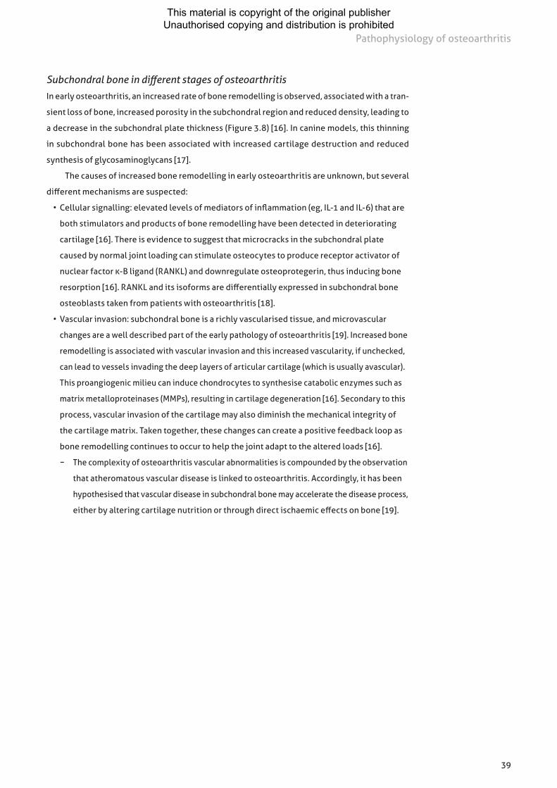
39
Pathophysiology of osteoarthritis
Subchondral bone in different stages of osteoarthritis
In early osteoarthritis, an increased rate of bone remodelling is observed, associated with a tran-
sient loss of bone, increased porosity in the subchondral region and reduced density, leading to
a decrease in the subchondral plate thickness (Figure 3.8) [16]. In canine models, this thinning
in subchondral bone has been associated with increased cartilage destruction and reduced
synthesis of glycosaminoglycans [17].
The causes of increased bone remodelling in early osteoarthritis are unknown, but several
different mechanisms are suspected:
• Cellular signalling: elevated levels of mediators of inflammation (eg, IL-1 and IL-6) that are
both stimulators and products of bone remodelling have been detected in deteriorating
cartilage [16]. There is evidence to suggest that microcracks in the subchondral plate
caused by normal joint loading can stimulate osteocytes to produce receptor activator of
nuclear factor -B ligand (RANKL) and downregulate osteoprotegerin, thus inducing bone
resorption [16]. RANKL and its isoforms are differentially expressed in subchondral bone
osteoblasts taken from patients with osteoarthritis [18].
• Vascular invasion: subchondral bone is a richly vascularised tissue, and microvascular
changes are a well described part of the early pathology of osteoarthritis [19]. Increased bone
remodelling is associated with vascular invasion and this increased vascularity, if unchecked,
can lead to vessels invading the deep layers of articular cartilage (which is usually avascular).
This proangiogenic milieu can induce chondrocytes to synthesise catabolic enzymes such as
matrix metalloproteinases (MMPs), resulting in cartilage degeneration [16]. Secondary to this
process, vascular invasion of the cartilage may also diminish the mechanical integrity of
the cartilage matrix. Taken together, these changes can create a positive feedback loop as
bone remodelling continues to occur to help the joint adapt to the altered loads [16].
− The complexity of osteoarthritis vascular abnormalities is compounded by the observation
that atheromatous vascular disease is linked to osteoarthritis. Accordingly, it has been
hypothesised that vascular disease in subchondral bone may accelerate the disease process,
either by altering cartilage nutrition or through direct ischaemic effects on bone [19].
This material is copyright of the original publisher Unauthorised copying and distribution is prohibited

40
Atlas of osteoarthritis
Figure 3.8 Stages of progressive joint and subchondral bone degradation in osteoarthritis. In early-stage osteoarthritis, the subchondral plate becomes thinner as a consequence of an increased remodelling rate. At the same time, cancellous bone is lost as the trabecular plates become thinner and more rod-like. In late-stage disease, the subchondral plate thickens, but the subchondral cancellous bone remains osteopaenic. The calcified cartilage begins to advance into the articular cartilage, leaving a footprint of multiple tidemarks as the mineralisation front advances. This creates an even thicker mineralised plate, and reduces the thickness of the non-mineralised articular cartilage, which cannot replace itself. This is accompanied by surface fibrillation and a loss of aggrecan, beginning superficially in the articular cartilage. The collective result of these changes is subchondral sclerosis (that includes both the subchondral plate and calcified cartilage) and thinner, more fibrillated articular cartilage. Image from Burr & Gallant [16]. © 2012, reproduced with permission from Nature Publishing Group.
Stages of progressive joint and subchondral bone degradation in osteoarthritis
A Normal joint
Articular cartilage
Calcified cartilage
Trabecular bone
Tidemark
Subchondral bone plate thickness
B Early osteoarthritis
Articular cartilage
Calcified cartilage
Trabecular bone
Tidemark
Subchondral bone plate thickness
C Late stage osteoarthritis
Articular cartilage
Calcified cartilage
Trabecular bone
Tidemarks
Loss of aggrecan fibrilation
Subchondral bone plate thickness
Articular cartilageCalcified cartilageBone and subchondral bone
This material is copyright of the original publisher Unauthorised copying and distribution is prohibited

41
Pathophysiology of osteoarthritis
• Bone−cartilage crosstalk: cartilage is separated from the subchondral bone by a tidemark,
which in normal cartilage is impermeable [20]. In osteoarthritis, it is hypothesised that
microcracks in the subchondral plate can lead to interactions between bone and cartilage
in the early phase of disease. These microcracks may be further exacerbated by the osteo-
clastic resorption in the subchondral region, which leads to increased plate perforation
[16,21]. This theory is substantiated by in vitro studies showing that there is crosstalk
between cells of the bone and chondrocytes [16]. A hypothetical model of cartilage and
subchondral bone interaction in osteoarthritis is given in Figure 3.9 [22].
As the disease progresses, the remodelling rate decreases, but an imbalance between bone
resorption and formation leads to a net increase in bone formation [16,23]. This process increases
bone volume, and can be associated with an apparent sclerosis caused by increased bone volume
and a thicker calcified cartilage layer [16]. This process corresponds to the condensation detected
on X-ray radiography. The mechanical consequences of subchondral sclerosis are not clear
but it may lead to a greater bone volume, with lower mineralisation, in joints of patients with
Hypothetical model of cartilage and subchondral bone interaction in osteoarthritis
A B
Middle zone
Deep zone
Bone
Osteoclast
Superficial zone
Chondrocyte
Calcified cartilage
Growth factors: eg, VEGF
This material is copyright of the original publisher Unauthorised copying and distribution is prohibited

42
Atlas of osteoarthritis
CFigure 3.9 Hypothetical model of cartilage and subchondral bone interaction in osteoarthritis. A, Healthy chondrocytes under pathological conditions (eg, due to instability of the joint or severe increased mobilisation) start to become hypertrophic and produce growth factors that diffuse towards the underlying bone marrow and stimulate osteoclastogenesis. B, Persisting strain. Chondrocytes become more hypertrophic and produce less sulphated-glycosaminoglycans (sGAG) to sustain the cartilage. Osteoclasts start to tunnel through the subchondral bone inducing changes to the biomechanical properties of the tissue. C, Progressive phase of osteoarthritis. The tidemark between cartilage and bone shifts upwards, reducing cartilage thickness. The remaining cartilage is strongly depleted of sGAG and becomes structurally deprived. Osteoclast activity extends into the calcified cartilage, up to the border with the deep zone of the cartilage. Via the pores there is vascular ingrowth into the cartilage. Later on, osteoblasts will infiltrate and start to deposit bone that results in end-stage sclerosis. Image from Weinans et al [22]. © 2012, reproduced with permission from Elsevier.
osteoarthritis compared with disease-free joints, leading to diminished mechanical stiffness of
the bone and, consequently, deterioration of cartilage [16].
During this stage, osteophytes may develop at the joint margins [13]. Osteophytes are
outgrowths of osseous tissue that are covered with cartilage [24]. Types of osteophytes include
traction spurs at the attachment of the ligament and tendon to bone, inflammatory spurs in the
vertebral body and osteochondrophytes, which form from metaplasia of the synovium into car-
tilage. Their role in osteoarthritis is unclear; they could cause pain in spinal osteoarthritis but
may be helpful in osteoarthritis of the lower limbs because they stabilise the joint [25].
Synovial inflammation in osteoarthritis
The synovial membrane plays a key role in normal joint function, as it nourishes chondrocytes
through the synovial fluid and joint space and eliminates metabolites and matrix degradation
products [26]. Hyaluronic acid and lubricin produced in the synovial lining cells help protect and
maintain articular cartilage [27].
This material is copyright of the original publisher Unauthorised copying and distribution is prohibited

43
Pathophysiology of osteoarthritis
The inflammation of the synovium that occurs in osteoarthritis is responsible for several clini-
cal symptoms, including pain, and reflects the structural progression of the disorder [26,27]. Fur-
thermore, synovitis is a major factor in osteoarthritis pathophysiology due to the action of several
soluble mediators (Figure 3.10). Interestingly, the relationship between synovitis, as assessed by
arthroscopy, and the degree of functional impairment or pain experienced remains a matter of
debate [26].
Ligament changes and misalignment
About one-quarter of patients with knee osteoarthritis have been found to have ruptures to their
anterior cruciate ligament (ACL), which normally functions as an anterior/posterior stabiliser
[28,29]. A detailed study of the effect of ageing and osteoarthritis on the ACL found moderate
or severe degeneration of the ACL in knees that had only minimal cartilage deterioration. The
likelihood of advanced ACL degeneration increased with age [29].
Patients with established knee osteoarthritis may also have varus alignment, causing
medial tibiofemoral osteoarthritis, and/or valgus alignment, which leads to lateral osteoarthritis
progression [30]. Both of these conditions affect load distribution, causing further knee damage.
In one trial of 256 patients with knee osteoarthritis who had no magnetic resonance image (MRI)
evidence of tibiofemoral cartilage damage, varus alignment at baseline was associated with an
increased risk of incident medial tibiofemoral cartilage damage over a 30-month period [30].
Involvement of the synovium in osteoarthritis pathophysiology
Normal joint Osteoarthritic joint
Osteophyte
SynoviumCartilage
Anti-inflammatory mediatorsCytokines (IL-4, IL-13, IL-1Ra, etc)TiMPs
Proinflammatory mediatorsCytokines (IL-1, TNF, IL-6, IL-8, etc)MMPs, ADAMTSBioactive lipids (PGE2, LTB4, etc)NeuropeptidesNitric oxideAdipokines (visfatin/NAMPT, etc)
Subchondral bone
This material is copyright of the original publisher Unauthorised copying and distribution is prohibited

44
Atlas of osteoarthritis
Selected risk factors for the occurrence and progression of osteoarthritis in knee, hip and hand
Knee Hip Hand
Occurrence Age, sex, physical activity, body-mass index (including obesity), intense sport activities, quadriceps strength, bone density, previous injury, hormone replacement therapy (protective), vitamin D, smoking (protective or deleterious), malalignment (including varus and valgus), genetics
Age, sex, physical activity, body-mass index (including obesity), previous injury, intense sport activities, genetics (including congenital deformities)
Age, grip strength, occupation, intense sport activities, genetics
Progression Age, body-mass index (including obesity), vitamin D, hormone replacement therapy (protective), malalignment (including varus and valgus), chronic joint effusion, synovitis, intense sport activities, subchondral bone oedema on magnetic resonance imaging
Age, symptomatic activity, sex, intense sport activities
Unknown
Table 3.1 Selected risk factors for the occurrence and progression of osteoarthritis in knee, hip and hand. The risk factors involved in the occurrence and progression of osteoarthritis differ depending on the joint(s) involved. Reproduced with permission from Bijlsma et al [12].
Figure 3.10 Involvement of the synovium in osteoarthritis pathophysiology. Products of cartilage breakdown that are released into the synovial fluid are phagocytosed by synovial cells, amplifying synovial inflammation. In turn, activated synovial cells produce catabolic and pro-inflammatory mediators, leading to excess production of the proteolytic enzymes responsible for cartilage breakdown. This inflammatory response is amplified by activated synovial T cells, B cells and infiltrating macrophages; to counteract it, the synovium and cartilage produce anti-inflammatory cytokines. The inflamed synovium also contributes to the formation of osteophytes via BMPs. ADAMTS, a disintegrin and metalloproteinase with thrombospondin motifs; BMP, bone morphogenetic protein; CCL2, CC-chemokine ligand 2; CXCL13, CXC-chemokine ligand 13; EGF, endothelial growth factor; GM-CSF, granulocyte-macrophage colony-stimulating factor; IL, interleukin; IL-1Ra, IL-1 receptor antagonist; LIF, leukaemia inhibitory factor; LTB4, leukotriene B4; MMP, matrix metalloproteinase; NAMPT, nicotinamide phosphoribosyl transferase (also called visfatin); NGF, nerve growth factor; PGE2, prostaglandin E2; TIMP, tissue inhibitor of metalloproteinase; TNF, tumour necrosis factor; VCAM-1, vascular cell adhesion molecule 1; VEGF, vascular endothelial growth factor. Image from Sellam & Berenbaum [26]. © 2010, reproduced with permission from Nature Publishing Group.
Cartilage
Subchondral bone
Cartilage degradation
products
BMPs
MacrophageVEGF EGF
Synovial fibroblast
Synovium B cells
T cell
CD4+ T cell
IL-12
V-CAM1 E-Selectin
Neovascularisation
CXCL13
Patchy synovitis (synovial hypertrophy and hyperplasia)
Risk factors for osteoarthritis
Many risk factors are associated with the development and progression of osteoarthritis
(Table 3.1) [12]. Age, female gender, participation in intense sports activities and high body mass
index (obesity) are among the many factors linked to both development and progression [12,31].
Sex hormones may play a role in the accelerated incidence rate of osteoarthritis in postmeno-
pausal women [31].
This material is copyright of the original publisher Unauthorised copying and distribution is prohibited
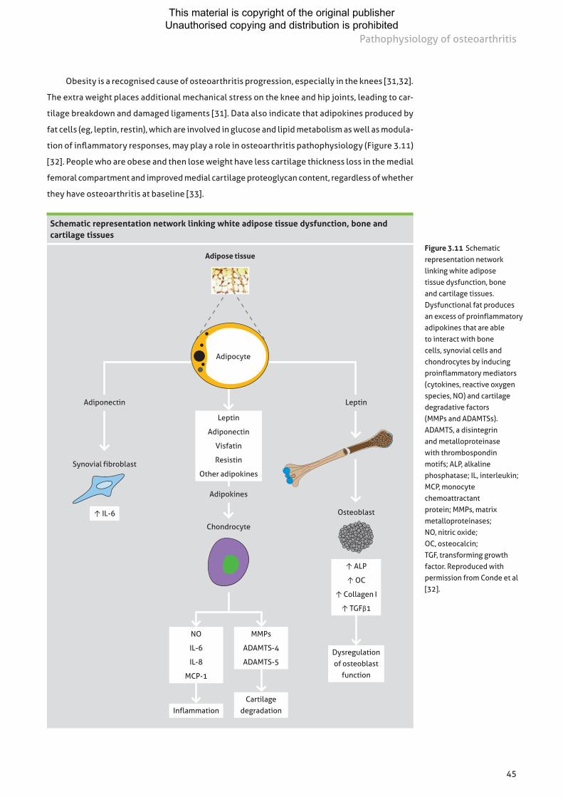
45
Pathophysiology of osteoarthritis
Obesity is a recognised cause of osteoarthritis progression, especially in the knees [31,32].
The extra weight places additional mechanical stress on the knee and hip joints, leading to car-
tilage breakdown and damaged ligaments [31]. Data also indicate that adipokines produced by
fat cells (eg, leptin, restin), which are involved in glucose and lipid metabolism as well as modula-
tion of inflammatory responses, may play a role in osteoarthritis pathophysiology (Figure 3.11)
[32]. People who are obese and then lose weight have less cartilage thickness loss in the medial
femoral compartment and improved medial cartilage proteoglycan content, regardless of whether
they have osteoarthritis at baseline [33].
Figure 3.11 Schematic representation network linking white adipose tissue dysfunction, bone and cartilage tissues. Dysfunctional fat produces an excess of proinflammatory adipokines that are able to interact with bone cells, synovial cells and chondrocytes by inducing proinflammatory mediators (cytokines, reactive oxygen species, NO) and cartilage degradative factors (MMPs and ADAMTSs). ADAMTS, a disintegrin and metalloproteinase with thrombospondin motifs; ALP, alkaline phosphatase; IL, interleukin; MCP, monocyte chemoattractant protein; MMPs, matrix metalloproteinases; NO, nitric oxide; OC, osteocalcin; TGF, transforming growth factor. Reproduced with permission from Conde et al [32].
Schematic representation network linking white adipose tissue dysfunction, bone and cartilage tissues
Adipose tissue
Leptin
Adiponectin
Visfatin
Resistin
Other adipokines
Adipokines
Chondrocyte
InflammationCartilage
degradation
Adiponectin Leptin
Synovial fibroblast
↑ IL-6 Osteoblast
↑ ALP
↑ OC
↑ Collagen I
↑ TGFβ1
Adipocyte
Dysregulation of osteoblast
function
NO
IL-6
IL-8
MCP-1
MMPs
ADAMTS-4
ADAMTS-5
This material is copyright of the original publisher Unauthorised copying and distribution is prohibited
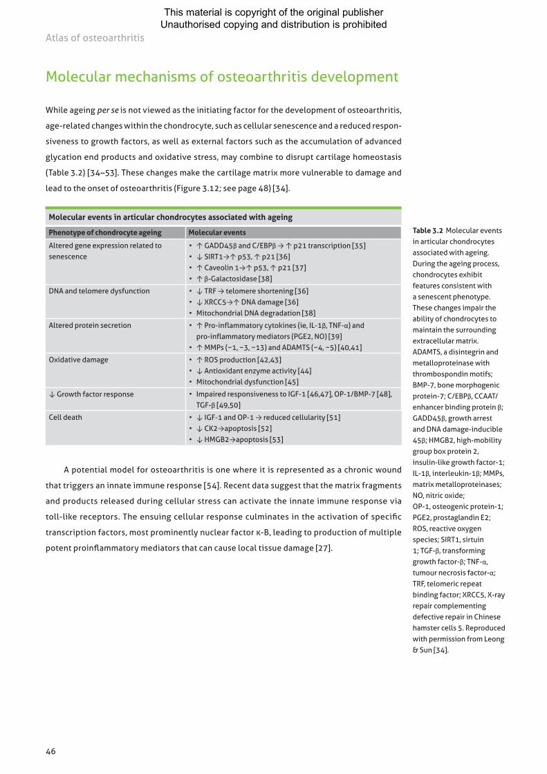
46
Atlas of osteoarthritis
Molecular mechanisms of osteoarthritis development
While ageing per se is not viewed as the initiating factor for the development of osteoarthritis,
age-related changes within the chondrocyte, such as cellular senescence and a reduced respon-
siveness to growth factors, as well as external factors such as the accumulation of advanced
glycation end products and oxidative stress, may combine to disrupt cartilage homeostasis
(Table 3.2) [34–53]. These changes make the cartilage matrix more vulnerable to damage and
lead to the onset of osteoarthritis (Figure 3.12; see page 48) [34].
Molecular events in articular chondrocytes associated with ageing
Phenotype of chondrocyte ageing Molecular events
Altered gene expression related to senescence
• ↑ GADD45β and C/EBPβ → ↑ p21 transcription [35]• ↓ SIRT1→↑ p53, ↑ p21 [36]• ↑ Caveolin 1→↑ p53, ↑ p21 [37]• ↑ β-Galactosidase [38]
DNA and telomere dysfunction • ↓ TRF → telomere shortening [36]• ↓ XRCC5→↑ DNA damage [36]• Mitochondrial DNA degradation [38]
Altered protein secretion • ↑ Pro-inflammatory cytokines (ie, IL-1β, TNF-α) and pro-inflammatory mediators (PGE2, NO) [39]
• ↑ MMPs (−1, −3, −13) and ADAMTS (−4, −5) [40,41]
Oxidative damage • ↑ ROS production [42,43]• ↓ Antioxidant enzyme activity [44]• Mitochondrial dysfunction [45]
↓ Growth factor response • Impaired responsiveness to IGF-1 [46,47], OP-1/BMP-7 [48], TGF-β [49,50]
Cell death • ↓ IGF-1 and OP-1 → reduced cellularity [51]• ↓ CK2→apoptosis [52]• ↓ HMGB2→apoptosis [53]
Table 3.2 Molecular events in articular chondrocytes associated with ageing. During the ageing process, chondrocytes exhibit features consistent with a senescent phenotype. These changes impair the ability of chondrocytes to maintain the surrounding extracellular matrix. ADAMTS, a disintegrin and metalloproteinase with thrombospondin motifs; BMP-7, bone morphogenic protein-7; C/EBPβ, CCAAT/enhancer binding protein β; GADD45β, growth arrest and DNA damage-inducible 45β; HMGB2, high-mobility group box protein 2, insulin-like growth factor-1; IL-1β, interleukin-1β; MMPs, matrix metalloproteinases; NO, nitric oxide; OP-1, osteogenic protein-1; PGE2, prostaglandin E2; ROS, reactive oxygen species; SIRT1, sirtuin 1; TGF-β, transforming growth factor-β; TNF-α, tumour necrosis factor-α; TRF, telomeric repeat binding factor; XRCC5, X-ray repair complementing defective repair in Chinese hamster cells 5. Reproduced with permission from Leong & Sun [34].
A potential model for osteoarthritis is one where it is represented as a chronic wound
that triggers an innate immune response [54]. Recent data suggest that the matrix fragments
and products released during cellular stress can activate the innate immune response via
toll-like receptors. The ensuing cellular response culminates in the activation of specific
transcription factors, most prominently nuclear factor -B, leading to production of multiple
potent proinflammatory mediators that can cause local tissue damage [27].
This material is copyright of the original publisher Unauthorised copying and distribution is prohibited

47
Pathophysiology of osteoarthritis
Chondrocyte ageing and cartilage destruction
↓ Antioxidants
Figure 3.12 Chondrocyte ageing and cartilage destruction. Age-related changes in the cartilage extracellular matrix and surrounding joint tissues initiate a cascade of events within the articular chondrocyte that lead to cartilage destruction and potential development of osteoarth ritis. ADAMTS, a disintegrin and metallopro-teinase with thrombospondin motifs; MMPs, matrix metalloproteinases. Reproduced with permission from Leong & Sun [34].
Reactive oxygen species
Ageing skeleton
Apoptosis↓ Anabolic
activityDNA damage
Telomere shorteningProinflammatory
cytokinesCollagen
crosslinkingSubchondral
bone softeningJoint laxity
Chondrocyte senescence
Proinflammatory mediators
Cartilage stiffness
Altered chondrocyte
loading
MMPs/ADAMTS
Cartilage destruction
Susceptibility for osteoarthritis
Advanced glycation end products
?
Cytokines (primarily interleukins and tumour necrosis factor-α), proteinases (primar-
ily MMPs), lipid mediators and reactive oxygen species all stimulate chondrocytes to release
cartilage-degrading enzymes [6,55]. An analysis of osteoblasts derived from osteophytes
demonstrates that IL-6, IL-8 and MMP-13 levels are greatly increased in patients with osteoarthritis
(Figure 3.13) [24]. Applying nonphysical mechanical stress loads to osteoblasts also increases the
gene expression of IL-6 and IL-8 in a stress magnitude-dependent manner, further demonstrating
the significance of inflammatory factors in osteophytes. Moreover, IL-6 directly induces MMP-13
expression and production in osteoarthritis osteoblasts from osteophytes and subchondral
bone osteoblasts without osteoarthritis [24]. The increased expression of IL-8 and MMP-13 may
promote cartilage degeneration via chondrocyte hypertrophy [24].
Growth factors involved in the synthesis of the physiological matrix, such as insulin-like
growth factor-1, bone morphogenic proteins, platelet-derived growth factor and transforming
growth factor-β can inhibit the effects of proinflammatory cytokines and help to repair the carti-
lage damage associated with osteoarthritis [6,55]. They stimulate chondrocyte anabolic activity
and proteoglycan synthesis and may also inhibit catabolic activity [55].
Currently, there is no reliable biomarker that can be considered a valid tool for the diagnosis
and prognosis of osteoarthritis in routine clinical practice. However, fibulin-3 peptides and
This material is copyright of the original publisher Unauthorised copying and distribution is prohibited

48
Atlas of osteoarthritis
follistatin-like protein-1 (FSTL1), both extracellular proteins, have potential as osteoarthritis bio-
markers [56]. Fibulin 3 is widely distributed in various tissue types and blood vessels of different
sizes and is capable of inhibiting vessel development and angiogenesis. Furthermore, it is also
elevated in osteoarthritis cartilage. In a recent study, Henrotin et al found greater levels of two
fibulin 3 fragments (Fib3-1 and Fib3-2) in the urine and serum of patients with osteoarthritis than
in controls. The increased levels of Fib3-1 were associated with ageing and hormonal status, but
Fib3-2 levels were not modified by gender, age or menopause [57]. FSTL1 is expressed in human
tissues and is induced by ischaemic stress and proinflammatory mediators [58]. It is thought to
play a role in arthritis pathogenesis and has been found to be a biomarker for rheumatoid arthritis
and other autoimmune diseases, as serum FSTL1 levels correlate with inflammatory status [58,59].
Serum FSTL1 levels have been found to be much higher in patients with osteoarthritis than in
healthy controls, and in women were correlated with disease grade and joint space widening [58].
Osteoarthritis pain
The best radiological predictor of knee pain is the presence of osteophytes [60,61], with the
strongest association observed in the skyline view compared with the lateral or anteroposte-
rior views [61]. The presence of osteophytes on any view is a better predictor of knee pain than
joint space width [60,61]. It has been suggested that the induction of synovitis due to greater
expression of IL-6 and IL-8 may also be a factor in the pain associated with osteoarthritis [24].
Concentrations of IL-6, IL-8 and MMP-13 in culture supernatant of osteoblasts isolated from osteophytes of patients with osteoarthritis
•
Figure 3.13 Concentrations of IL-6, IL-8 and MMP-13 in culture supernatant of osteoblasts isolated from osteophytes of patients with osteoarthritis. IL-6 (A), IL-8 (B) and MMP-13 (C) levels in osteoarthritis osteoblasts (OPH) were significantly higher than those of osteoblasts from subchondral bone without osteoarthritis (SBO; P<0.05, <0.05 and <0.01, respectively). The IL-8 and MMP-13 levels in the cell culture supernatant of osteoblasts from subchondral bone without osteoarthritis were below the limits of detection. *P<0.05, †P<0.01 (SBO, n=3; OPH, n=7). IL, Interleukin; MMPs, matrix metalloproteinases. Data from Sakao et al [24]. © 2009, reproduced with permission from The Japanese Society for Bone and Mineral Research and Springer.
600
500
400
300
200
100
0
SBO OPH
IL-6
pro
duct
ion
(pg/
ml p
rote
in)
A
* 70
60
50
40
30
20
10
0
SBO OPH
IL-8
pro
duct
ion
(pg/
ml p
rote
in)
B
* 12000
10000
8000
6000
4000
2000
0
SBO OPH
MM
P-13
pro
duct
ion
(pg/
ml p
rote
in)
C†
• •
••
This material is copyright of the original publisher Unauthorised copying and distribution is prohibited

49
Pathophysiology of osteoarthritis
Subchondral bone marrow oedema
Bone contains pain fibres, and subchondral bone marrow oedema-like lesions (BMLs) have been
frequently noted in osteoarthritis (Figure 3.14) [62,63]. Several trials have noted a cross-sectional
positive association between BMLs, cartilage damage and ligament damage [63].
In a pivotal study involving 351 patients with osteoarthritis and knee pain and 50 patients
with osteoarthritis but no knee pain, 78% of patients with knee pain had MRI evidence of bone
marrow lesions, compared with only 30% of patients without knee pain (P<0.001) [62]. These
results show that BMLs in the knee are associated with pain, the most important symptom of
osteoarthritis. In addition, bone marrow lesions are correlated with the severity of radiographic
disease. In this study, the prevalence of BMLs ranged from 48% in knees with Kellgren and
Lawrence (KL) grades of 0 to 100% in knees with KL grades of 4 [62].
References
1 Gardner DL. Problems and paradigms in joint pathology. J Anat. 1994;184:465-476.2 McLeod WD, Hunter S. Biomechanical analysis of the knee: primary functions as elucidated by anatomy.
Phys Ther. 1980;60:1561-1564.3 Kishner S, Courseault J, Authement A. Knee joint anatomy. Available at:
http://emedicine.medscape.com/article/1898986-overview. Last accessed December 7, 2012.4 Abdul-Jabar HB, Walsh U, Rashid A, Rajkumar S. Primary meningococcal osteoarthritis of the knee—
case report and review of the literature. Eur Orthop Traumatol. 2011;2:149-152.5 Niitsu M. Cystic and cyst-like lesions of the knee. In: Niitsu M, ed. Magnetic Resonance Imaging of
the Knee. Springer-Verlag Berlin Heidelberg; 2013: 181-198.6 Berenbaum F. Osteoarthritis. B. Pathology and pathogenesis. In: Klippel JH, Stone JH, Crofford LJ,
White PH, eds. Primer on the Rheumatic Diseases. New York, NY: Springer Science+Business Media, LLC; 2008:229-234.
7 Symmons D, Mathers C, Pfleger B. Global burden of osteoarthritis in the year 2000. World Health Organization Web site. Available at: www.who.int/healthinfo/statistics/bod_osteoarthritis.pdf. Accessed December 7, 2012.
8 Loeser RF, Goldring SR, Scanzello CR, Goldring MB. Osteoarthritis: a disease of the joint as an organ. Arthritis Rheum. 2012;64:1697-1707.
9 Goldring MB, Goldring SR. Osteoarthritis. J Cell Physiol. 2007;213:626-634.10 Horton WE Jr, Bennion P, Yang L. Cellular, molecular, and matrix changes in cartilage during aging and
osteoarthritis. J Musculoskelet Neuronal Interact. 2006;6:379-381.
Bone marrow lesion in knee osteoarthritis
Figure 3.14 Bone marrow lesion in knee osteoarthritis. This sagittal fat-suppressed proton density-weighted magnetic resonance image (long repetition time/long echo time = 3500/20) shows a large bone marrow lesion (BML; arrows) involving the central and posterior subregions of the lateral tibia. Subchondral BMLs are a common imaging feature of osteoarthritis and several trials have noted a cross-sectional positive association between BMLs, cartilage damage and ligament damage. Importantly, BMLs in the knee have also been associated with pain. Image from Xu et al [63]. © 2012, reproduced with permission from Elsevier.
This material is copyright of the original publisher Unauthorised copying and distribution is prohibited

50
Atlas of osteoarthritis
11 Bahk Y-W. Degenerative joint diseases. In: Bahk Y-W, ed. Combined Scintigraphic and Radiographic Diagnosis of Bone and Joint Diseases, Including Gamma Correction Interpretation. 4th ed. Berlin, Germany: Springer-Verlag Berlin Heidelberg; 2013:141-183.
12 Bijlsma JWJ, Berenbaum F, Lafeber FPJG. Osteoarthritis: an update with relevance for clinical practice. Lancet. 2011;377:2115-2126.
13 Altman RD. Osteoarthritis in the elderly population. In: Nakasato Y, Yung RL, eds. Geriatric Rheumatology. A Comprehensive Approach. New York, NY: Springer Science+Business Media, LLC; 2011:187-196.
14 Myers SL. Osteoarthritis and crystal-associated synovitis. In: Hunder GG, ed. Atlas of Rheumatology, 4th ed. Philadelphia, PA: Current Medicine LLC; 2005:54-81.
15 Castañeda S, Roman-Blas JA, Largo R, Herrero-Beaumont G. Subchondral bone as a key target for osteoarthritis treatment. Biochem Pharmacol. 2012;83:315-323.
16 Burr DB, Gallant MA. Bone remodelling in osteoarthritis. Nat Rev Rheumatol. 2012;8:665-673.17 Sniekers YH, Intema F, Lafeber FPJG, et al. A role for subchondral bone changes in the process of
osteoarthritis; a micro-CT study of two canine models. BMC Musculoskelet Disord. 2008;9:20.18 Tat SK, Pelletier J-P, Lajeunesse D, Fahmi H, Duval N, Martel-Pelletier J. Differential modulation of
RANKL isoforms by human osteoarthritic subchondral bone osteoblasts: influence of osteotropic factors. Bone. 2008;43:284-291.
19 Conaghan PG, Vanharanta H, Dieppe PA. Is progressive osteoarthritis an atheromatous vascular disease? Ann Rheum Dis. 2005;64:1539-1541.
20 Walsh D. Neurogenic factors in the etiopathogenesis of osteoarthritis. Paper presented at: 10th World Congress of the International Cartilage Repair Society; May 12-15, 2012; Montreal, Quebec, Canada.
21 Botter SM, van Osch GJVM, Clockaerts S, Waarsing JH, Weinans H, van Leeuwen JPTM. Osteoarthritis induction leads to early and temporal subchondral plate porosity in the tibial plateau of mice: an in vivo microfocal computed tomography study. Arthritis Rheum. 2011;63:2690-2699.
22 Weinans H, Siebelt M, Agricola R, Botter SM, Piscaer TM, Waarsing JH. Pathophysiology of peri-articular bone changes in osteoarthritis. Bone. 2012;51:190-196.
23 Kumarasinghe DD, Perilli E, Tsangari H, et al. Critical molecular regulators, histomorphometric indices and their correlations in the trabecular bone in primary hip osteoarthritis. Osteoarthritis Cartilage. 2010;18:1337-1344.
24 Sakao K, Takahashi KA, Arai Y, et al. Osteoblasts derived from osteophytes produce interleukin-6, interleukin-8, and matrix metalloproteinase-13 in osteoarthritis. J Bone Miner Metab. 2009;27:412-423.
25 Menkes C-J, Lane NE. Are osteophytes good or bad? Osteoarthritis Cartilage. 2004;12(suppl A):S53-S54.26 Sellam J, Berenbaum F. The role of synovitis in pathophysiology and clinical symptoms of
osteoarthritis. Nat Rev Rheumatol. 2010;6:625-635.27 Scanzello CR, Goldring SR. The role of synovitis in osteoarthritis pathogenesis. Bone. 2012;51:249-257.28 Hill CL, Seo GS, Gale D, Totterman S, Gale ME, Felson DT. Cruciate ligament integrity in osteoarthritis of
the knee. Arthritis Rheum. 2005;52:794-799.29 Hasegawa A, Otsuki S, Pauli C, et al. Anterior cruciate ligament changes in the human knee joint in aging
and osteoarthritis. Arthritis Rheum. 2012;64:696-704.30 Sharma L, Chmiel JS, Almagor O, et al. The role of varus and valgus alignment in the initial development of
knee cartilage damage by MRI: the MOST Study. Ann Rheum Dis. 2012;epub ahead of print.31 Arden N, Nevitt MC. Osteoarthritis: epidemiology. Best Pract Res Clin Rheumatol. 2006;20:3-25.32 Conde J, Scotece M, Gómez R, Lopez V, Gómez-Reino JJ, Gualillo O. Adipokines and osteoarthritis:
novel molecules involved in the pathogenesis and progression of disease. Arthritis. 2011; epub doi: 10.1155/2011/203901.
33 Anandacoomarasamy A, Leibman S, Smith G, et al. Weight loss in obese people has structure-modifying effects on medial but not on lateral knee articular cartilage. Ann Rheum Dis. 2012;71:26-32.
34 Leong DJ, Sun HB. Events in articular chondrocytes with aging. Curr Osteoporos Rep. 2011;9:196-201.35 Shimada H, Sakakima H, Tsuchimochi K, et al. Senescence of chondrocytes in aging articular cartilage:
GADD45β mediates p21 expression in association with C/EBPβ in senescence-accelerated mice. Pathol Res Pract. 2011;207:225-231.
36 Brandl A, Hartmann A, Bechmann V, Graf B, Nerlich M, Angele P. Oxidative stress induces senescence in chondrocytes. J Orthop Res. 2011;29:1114-1120.
37 Dai S-M, Shan Z-Z, Nakamura H, et al. Catabolic stress induces features of chondrocyte senescence through overexpression of caveolin 1: possible involvement of caveolin 1–induced down-regulation of articular chondrocytes in the pathogenesis of osteoarthritis. Arthritis Rheum. 2006;54:818-831.
38 Martin JA, Buckwalter JA. The role of chondrocyte senescence in the pathogenesis of osteoarthritis and in limiting cartilage repair. J Bone Joint Surg Am. 2003;85(suppl 2):106-110.
This material is copyright of the original publisher Unauthorised copying and distribution is prohibited

51
Pathophysiology of osteoarthritis
39 Nah S-S, Choi I-Y, Lee CK, et al. Effects of advanced glycation end products on the expression of COX-2, PGE2 and NO in human osteoarthritic chondrocytes. Rheumatology (Oxford). 2008;47:425-431.
40 Nah S-S, Choi I-Y, Yoo B, Kim YG, Moon H-B, Lee C-K. Advanced glycation end products increases matrix metalloproteinase-1, -3, and -13, and TNF-α in human osteoarthritic chondrocytes. FEBS Lett. 2007;581:1928-1932.
41 Huang C-Y, Lai K-Y, Hung L-F, Wu W-L, Liu F-C, Ho L-J. Advanced glycation end products cause collagen II reduction by activating Janus kinase/signal transducer and activator of transcription 3 pathway in porcine chondrocytes. Rheumatology (Oxford). 2011;50:1379-1389.
42 Hiran TS, Moulton PJ, Hancock JT. Detection of superoxide and NADPH oxidase in porcine articular chondrocytes. Free Radic Biol Med. 1997;23:736-743.
43 Tiku ML, Shah R, Allison GT. Evidence linking chondrocyte lipid peroxidation to cartilage matrix protein degradation. Possible role in cartilage aging and the pathogenesis of osteoarthritis. J Biol Chem. 2000;275:20069-20076.
44 Jallali N, Ridha H, Thrasivoulou C, Underwood C, Butler PEM, Cowen T. Vulnerability to ROS-induced cell death in ageing articular cartilage: the role of antioxidant enzyme activity. Osteoarthritis Cartilage. 2005;13:614-622.
45 Blanco F, Rego I, Ruiz-Romero C. The role of mitochondria in osteoarthritis. Nat Rev Rheumatol. 2011;7:161-169.
46 Loeser RF, Shanker G, Carlson CS, Gardin JF, Shelton BJ, Sonntag WE. Reduction in the chondrocyte response to insulin-like growth factor 1 in aging and osteoarthritis: studies in a non-human primate model of naturally occurring disease. Arthritis Rheum. 2000;43:2110-2120.
47 Martin JA, Ellerbroek SM, Buckwalter JA. Age-related decline in chondrocyte response to insulin-like growth factor-I: the role of growth factor binding proteins. J Orthop Res. 1997;15:491-498.
48 Chubinskaya S, Kumar B, Merrihew C, Heretis K, Rueger DC, Kuettner KE. Age-related changes in cartilage endogenous osteogenic protein-1 (OP-1). Biochim Biophys Acta. 2002;1588:126-134.
49 Blaney Davidson EN, Scharstuhl A, Vitters EL, van der Kraan PM, van den Berg WB. Reduced transforming growth factor-beta signaling in cartilage of old mice: role in impaired repair capacity. Arthritis Res Ther. 2005;7:R1338-R1347.
50 Scharstuhl A, van Beuningen HM, Vitters EL, van der Kraan PM, van den Berg WB. Loss of transforming growth factor counteraction on interleukin 1 mediated effects in cartilage of old mice. Ann Rheum Dis. 2002;61:1095-1098.
51 Loeser RF, Pacione CA, Chubinskaya S. The combination of insulin-like growth factor 1 and osteogenic protein 1 promotes increased survival of and matrix synthesis by normal and osteoarthritic human articular chondrocytes. Arthritis Rheum. 2003;48:2188-2196.
52 Lee SW, Song YS, Lee SY, et al. Downregulation of protein kinase CK2 activity facilitates tumor necrosis factor-α-mediated chondrocyte death through apoptosis and autophagy. PLoS One. 2011;6:e19163.
53 Taniguchi N, Caramés B, Ronfani L, et al. Aging-related loss of the chromatin protein HMGB2 in articular cartilage is linked to reduced cellularity and osteoarthritis. Proc Natl Acad Sci USA. 2009;106:1181-1186.
54 Scanzello CR, Plaas A, Crow MK. Innate immune system activation in osteoarthritis: is osteoarthritis a chronic wound? Curr Opin Rheumatol. 2008;20:565-572.
55 Goldring MB. Update on the biology of the chondrocyte and new approaches to treating cartilage diseases. Best Pract Res Clin Rheumatol. 2006;20:1003-1025.
56 Mobasheri A. Osteoarthritis 2012 year in review: biomarkers. Osteoarthritis Cartilage. 2012;20:1451-1464.
57 Henrotin Y, Gharbi M, Mazzucchelli G, Dubuc J-E, De Pauw E, Deberg M. Fibulin 3 peptides Fib3-1 and Fib3-2 are potential biomarkers of osteoarthritis. Arthritis Rheum. 2012;64:2260-2267.
58 Wang Y, Li D, Xu N, et al. Follistatin-like protein 1: a serum biochemical marker reflecting the severity of joint damage in patients with osteoarthritis. Arthritis Res Ther. 2011;13:R193.
59 Li D, Wang Y, Xu N, et al. Follistatin-like protein 1 is elevated in systemic autoimmune diseases and correlated with disease activity in patients with rheumatoid arthritis. Arthritis Res Ther. 2011;13:R17.
60 Lanyon P, O’Reilly S, Jones A, Doherty M. Radiographic assessment of symptomatic knee osteoarthritis in the community: definitions and normal joint space. Ann Rheum Dis. 1998;57:595-601.
61 Cicuttini FM, Baker J, Hart DJ, Spector TD. Association of pain with radiological changes in different compartments and views of the knee joint. Osteoarthritis Cartilage. 1996;4:143-147.
62 Felson DT, Chaisson CE, Hill CL, et al. The association of bone marrow lesions with pain in knee osteoarthritis. Ann Intern Med. 2001;134:541-549.
63 Xu L, Hayashi D, Roemer FW, Felson DT, Guermazi A. Magnetic resonance imaging of subchondral bone marrow lesions in association with osteoarthritis. Semin Arthritis Rheum. 2012;42:105-118.
This material is copyright of the original publisher Unauthorised copying and distribution is prohibited

52
Francisco J. Blanco
Clinical criteria for osteoarthritis
Clinical criteria will continue to play an important role in the diagnosis of osteoarthritis until a
diagnostic method that integrates clinical findings with aetiological, biochemical and histological
abnormalities is developed [1]. One of the most enduring clinical criteria for osteoarthritis of the
knee is the classification system developed for the American Rheumatism Association in 1986 [1].
The aim was to standardise and clarify the clinical definition of idiopathic osteoarthritis, using
commonly available diagnostic techniques. This resulted in three sets of criteria, depending on
whether the physician is able to draw on clinical examination and laboratory findings, clinical
examination and radiographic results or clinical examination only (Table 4.1) [1].
Chapter 4Clinical features and diagnosis of osteoarthritis
Criteria for classification of idiopathic osteoarthritis of the knee
Clinical and laboratory Clinical and radiographic Clinical*
Knee pain + at least 5 of the following:• Age >50 years• Stiffness <30 minutes• Crepitus• Bony tenderness• Bony enlargement• No palpable warmth• ESR <40 mm/hour• RF <1:40• SF OA
Knee pain + at least 1 of the following:• Age >50 years• Stiffness <30 minutes• Crepitus• Plus osteophytes
Knee pain + at least 3 of the following:• Age >50 years• Stiffness <30 minutes• Crepitus• Bony tenderness• Bony enlargement• No palpable warmth
92% sensitive, 75% specific 91% sensitive, 86% specific 95% sensitive, 69% specific
Table 4.1 Criteria for classification of idiopathic osteoarthritis of the knee. ESR, erythrocyte sedimentation rate (Westergren); RF, rheumatoid factor; SF OA, synovial fluid signs of osteoarthritis (clear, viscous or white blood cell count <2000/mm3). *An alternative for the clinical category would be the presence of 4 of the 6 findings, which is 84% sensitive and 89% specific. Data from Altman et al [1]. Reproduced with permission from John Wiley and Sons.
A set of clinical definitions for knee osteoarthritis were also developed by Zhang et al for
the European League Against Rheumatism (EULAR). The authors noted that, while radiography
is often used as the ‘gold standard’ for diagnosis, it is not the only marker and the definition of
knee osteoarthritis may change depending on the levels of care and clinical requirements [2].
They stated that a confident diagnosis can be made, without recourse to radiographic examination
and even if radiographs appear normal, in adults aged >40 years with [2]:
• usage-related knee pain;
• only short-lived morning stiffness;
• functional limitation; and
• one or more typical examination findings (crepitus, restricted movement, bony enlargement).
The EULAR clinical criteria also emphasised that all patients with knee pain should be examined
for possible osteoarthritis [2].
This material is copyright of the original publisher Unauthorised copying and distribution is prohibited

53
Clinical features and diagnosis of osteoarthritis
Symptoms of osteoarthritis
The onset of osteoarthritis symptoms is often insidious (Table 4.2) and there is often asymmetry
of symptoms [3].
Symptoms and signs of osteoarthritis
Symptoms Signs
Pain Weakness Joint (hard tissue) enlargement Limitation of motion
Altered function Deformity Altered gait Deformity
Stiffness Grinding/clicking Tenderness Instability
Swelling Instability/buckling Crepitus
Table 4.2 Symptoms and signs of osteoarthritis. The onset of symptoms of osteoarthritis is most often insidious, usually beginning in one or a few joints. Reproduced with permission from Altman [3].
Pain in osteoarthritis: potential sites of origin
Synovial inflammation Outer one-third of menisci
Subchondral bone ischaemia (‘bone angina’) Stress at ligamentous insertion
Distension of the joint capsule Inflammation of bursae with/without calcification
Periarticular muscle spasm (eg, nocturnal myoclonus)
Osteophyte distension of periosteum or impingement of spinal canal/foramina
Table 4.3 Pain in osteoarthritis: potential sites of origin. The origin of pain in osteoarthritis is rarely clear, but sometimes can be attributed to anatomical changes in the joint. It is worth noting that there are no nerves in cartilage, the inner two-thirds of the menisci or synovial cavity. Hence, pain from these anatomic sites are induced indirectly through the above anatomical sites. Reproduced with permission from Altman [3]
Pain
Pain is the first and most predominant symptom of osteoarthritis [3–5] and is sometimes described
as a deep ache [3]. The pain in weight-bearing joints is usually worsened by standing and walking
is and relieved by rest. Although it is typically intermittent, pain can become constant [2,3]. The
potential sites of origin for osteoarthritis pain are shown in Table 4.3 [3].
In knee osteoarthritis, localised pain is often identified along the medial joint line or distal
to the patellofemoral attachment. Medial pain is usually correlated with anatomic changes, as
the medial compartment is involved in 70% of knee osteoarthritis cases [3]. In patients who have
lateral compartment osteoarthritis, pain and grinding is localised to the lateral part of the knee
and arthritic destruction is manifested as a valgus deformity [6].
Stiffness
Stiffness in osteoarthritis usually occurs in the morning, after periods of inactivity or especially
in the evening [4]. The stiffness typically resolves within minutes and is relieved by motion of
the joint [3], which distinguishes it from the prolonged stiffness (usually lasting over 30 minutes)
experienced by rheumatoid arthritis sufferers [4].
Loss of movement or function
As osteoarthritis progresses, joint motion becomes restricted [3]. This results in loss of move-
ment and function, which, alongside pain, is a major reason that patients visit their family doctor
[4]. Loss of movement can lead to difficulties with certain daily activities, such as stair climbing,
walking and doing household chores [4].
This material is copyright of the original publisher Unauthorised copying and distribution is prohibited

54
Atlas of osteoarthritis
Other symptoms
Other signs and symptoms associated with osteoarthritis include joint enlargement due to joint
effusion, bony swelling or both. Crepitus, defined as a sensation of crackling or crunching, is also
commonly felt on passive or active movement of an affected joint [4].
Soft tissue contractures can result in varus (inward) or valgus (outward) knee deformity
in osteoarthritis (Figure 4.1) and lead to joint instability [5,7,8]. Patients may also experience
what is described as ‘buckling’, or spontaneous yielding of the quadriceps with knee flexion and
giving way. This may be due to pain, fixed flexion contracture of the knee, quadriceps weakness
and patellar problems such pain and dislocation [8].
Although not common in knee osteoarthritis, synovial effusions may be found along the
medial joint margin and in the suprapatellar bursa. Distension due to synovial effusion can lead
to knee flexion. Late signs include tenderness on palpation and pain on passive motion [3].
Valgus and varus knee deformities in osteoarthritis
A Valgus B Varus
Figure 4.1 Valgus and varus knee deformities in osteoarthritis. A, This patient has a severe valgus deformity of the right knee and normal alignment of the left knee. Standing radiographs of his right knee showed changes indicative of osteoarthritis in the medial, lateral and patellofemoral compartments. B, This patient has a severe varus deformity of both knees. Standing radiographs of her knees showed changes indicative of osteoarthritis in the medidal lateral and patellofemoral compartments. There is no cutaneous erythema to indicate the presence of acute inflammation in both knees, but the majority of specimens of synovial fluid aspirated from osteoarthritic knees contain crystals of either calcium pyrophosphate or apatite. Image courtesy of Dr FJ Blanco.
This material is copyright of the original publisher Unauthorised copying and distribution is prohibited

55
Clinical features and diagnosis of osteoarthritis
Comparison of quality-of-life mean scores between patients with knee osteoarthritis and controls
Knee osteoarthritis (mean ± SD) N=140
Control (mean ± SD) N=40 P value
Pain 74.66 ± 20.12 10.31 ± 10.16 <0.001
Energy level 51.38 ± 38.20 19.16 ± 22.50 <0.001
Emotional reaction 42.45 ± 31.31 9.68 ± 9.59 <0.001
Sleep 36.61 ± 26.72 15.50 ± 16.00 <0.001
Social isolation 19.14 ± 24.56 9.00 ± 10.07 <0.001
Physical mobility 42.72 ± 18.04 14.68 ± 8.43 <0.001
Table 4.4 Comparison of quality-of-life mean scores between patients with knee osteoarthritis and controls. A comparison between subgroups of Nottingham Health Profile subgroups in patients with knee osteoarthritis and healthy controls showed that patients with osteoarthritis had statistically significant higher scores in all subgroups than controls. SD, standard deviation. Reproduced with permission from Yildiz et al [9].
Figure 4.2 Scores and pain scales indicating marked pain and worse functional status. This figure shows Knee Society score, knee functional score and visual analogue pain scale in patients with proven osteoarthritis and radiographically negative cases. The patients with osteoarthritis had less favourable values than those who were radiographically negative. Reproduced with permission from Horváth et al [10].
Effects on patient quality of life
Individuals with knee osteoarthritis have significantly poorer quality of life than healthy indi-
viduals, and pain affects all aspects of health-related quality of life (eg, sleep, mobility, energy)
(Table 4.4) [9]. Furthermore, patients with proven osteoarthritis have lower function and pain
scale scores than radiographically negative cases, indicating marked pain and worse functional
status (Figure 4.2) [10].
Scores and pain scales indicating marked pain and worse functional status
Osteoarthritis Negative
100
75
50
25
0
Knee Society score
Knee functional score
Visual analogue pain scale
Kne
e sc
ores
10
7.5
5
2.5
0
Visual analogue scale
64.1
74.169.8
67.0
5.7
4.2
Studies have shown that patients with osteoarthritis also have a greater risk of mortality,
particularly due to cardiovascular- and gastrointestinal-related causes. The decreased level of
physical activity in those with walking disability probably contributes to the increased rate of
cardiovascular death [11]. These findings were confirmed in a population-based cohort study of
1163 adults with osteoarthritis, which found higher rates of deaths in those studied than in the
general population, especially for cardiovascular- and dementia-related mortality [12].
Diagnosis of osteoarthritis
The primary goal of diagnostic evaluation is to either demonstrate the presence of osteoarthri-
tis or to rule it out [13]. Osteoarthritis should always be suspected in patients who have joint-
specific pain (typically usage-related) and loss of function [2,3], especially in the elderly [3].
This material is copyright of the original publisher Unauthorised copying and distribution is prohibited

56
Atlas of osteoarthritis
Specific historical features of osteoarthritis
Pain Pain at the beginning of movement Permanent/nocturnal pain
Pain during movement Need for analgesics
Loss of function Stiffness Impairment in everyday activities
Limitation of range of movement Need for orthopaedic aids
Other symptoms Crepitation Stepwise progression
Elevated sensitivity to cold and/or damp
Table 4.5 Specific historical features of osteoarthritis. These historical criteria for osteoarthritis are those used at the Department of Orthopaedic and Trauma Surgery, University of Cologne. Reproduced with permission from Michael et al [13].
Risk factors, including age >50 years, female gender, high body mass index, previous knee injury
or malalignment, joint laxity, occupational or recreational usage, family history and the presence
of Heberden’s nodes can help to identify patients in whom knee osteoarthritis is the most likely
diagnosis [2].
Though not everyone with the signs and symptoms of osteoarthritis requires imaging
studies, findings on plain radiograph can confirm the clinical findings. Nevertheless, only about
50% of patients with pathological or radiographic changes have symptoms [3].
While all patients with knee pain should be examined, the current ‘gold standard’ for mor-
phological assessment of knee osteoarthritis is plain radiography [2]. The historical criteria for
osteoarthritis that are relatively specific to the disorder are shown in Table 4.5, although they
can be found in other joint diseases [13].
Differential diagnosis
While diagnosing osteoarthritis is easy, the primary difficulty is in knowing whether joint pain
and disability are indeed due to the joint pathology that is characteristic of the disease [14]. Many
patients with advanced pathology are asymptomatic and osteoarthritis pathology is extremely
common in the elderly. Consequently, it cannot be assumed that symptomatic pain is due to
osteoarthritis pathology in all individuals [14].
Pain may be referred, caused by periarticular problems (eg, bursitis due to ligamentous
and meniscal lesions) or the result of pain sensitisation that leads to abnormal sensations with
normal activities [2,14]. The involvement of other joints may suggest a range of alternative diag-
noses, while severe local inflammation, erythema and progressive pain unrelated to usage may
indicate crystals, sepsis or serious bone pathology [2]. Psychological factors such as depression
and anxiety and social problems such as isolation can also play a role in pain development [14].
This material is copyright of the original publisher Unauthorised copying and distribution is prohibited

57
Clinical features and diagnosis of osteoarthritis
Physical examination
The physical findings of osteoarthritis are characteristic to each stage of the disorder [13].
Physical examinations should include all relevant tests, including inspection and palpation
(Figure 4.3) [5], range of movement (Figure 4.4) [15] and special functional tests when required,
such as meniscus tests, ligament stability and gait analysis [13]. Physical examination of the knee
ligaments consists of [13]:
• testing of the lateral ligaments with varus or valgus stress; and
• testing of the anterior and posterior cruciate ligaments with the drawer test.
Knee examination and palpation
A B C
Figure 4.3 Knee examination and palpation. A, Examination of the osteoarthritic knee should include palpation along and proximal to the joint line, indicated by the dashed line beneath the examiner’s thumb. Crepitus can be elicited by passive flexion and extension of the joint. Palpation may reveal osteophytes that arise at the osteochondral margins or the joint or loose bodies. Tenderness in the gutters along the medial and lateral aspects of femoral condyles or in the suprapatellar bursa suggests underlying synovial inflammation. An estimate of the degree of medial-lateral laxity in the joint can be obtained by applying a valgus and then varus stress to the joint. B, Palpation of the margins of the patella, outlined here by the dashed circle below the examiner’s fingers, may reveal osteophytes. The ‘shrug sign’, or knee pain produced by pressing above the patella (as illustrated), while the patient contracts the quadriceps muscle suggests that cartilage pathology is present in the patellofemoral portion of the knee. C, Bursa palpation. The examiner’s right thumb palpates the anserine bursa, which is below the knee and between the tibia and the pes anserine, a conjoint tendon of the sartorius and gracilis muscles that inserts on the proximal tibia. Pain that arises in the anserine bursa can mimic or exacerbate the pain of knee osteoarthritis and can be reproduced by deep palpation in this area. Local measures, such as hot packs or injection of the bursa with a mixture of bupivacaine and corticosteroids, usually are effective. Reproduced with permission from Myers [5].
The menisci should also be tested manually and the femoropatellar joint assessed for normal
patellar mobility and indications of irritation [13].
Physical examination typically reveals evidence of mild-to-moderate tender swelling around
the joint line, crepitus and restricted range of motion, with pain at the end of the range [14]. There
may be tenderness over the joint line itself. Some patients can have evidence of mild inflamma-
tion, with warmth over the joint line and effusion. Joint deformities and instability may be seen
in advanced cases [14].
Radiological methods in diagnosis
The most commonly used radiological method to confirm the clinical diagnosis of osteoarthritis
is the plain radiograph [14], which can be used to establish the severity of joint damage and
This material is copyright of the original publisher Unauthorised copying and distribution is prohibited

58
Atlas of osteoarthritis
Figure 4.4 Assessment for patellofemoral joint crepitation during active range of motion. A, Extension. B, Flexion. Reproduced with permission from Griffith et al [15].
Assessment for patellofemoral joint crepitation during active range of motion
A
B
Figure 4.5 Plain radiographs of a typical patient with severe osteoarthritis of the knee joint. A, Note the loss of joint space, particularly marked in the medial compartment, caused by loss of articular cartilage, as well as the sclerosis of the underlying subchondral bone and osteophyte formation at the joint margin. B, A lateral radiograph of the knee shows osteoarthritis in the patellofemoral compartment with large osteophytes. Image courtesy of Dr FJ Blanco.
Plain radiographs of a typical patient with severe osteoarthritis of the knee joint
A
B
monitor disease progression [4,13]. Plain films should be obtained in a standardised manner in
at least two planes: anteroposterior and lateral [13]. The main radiographic features associated
with osteoarthritis are osteophytes, narrowing of the joint space due to articular cartilage loss
and several changes in the subchondral bone, such as sclerosis, cysts, shape changes and loss of
bone volume (Figure 4.5) [14].
This material is copyright of the original publisher Unauthorised copying and distribution is prohibited

59
Clinical features and diagnosis of osteoarthritis
Summary of studies investigating radiographic findings and clinical symptoms in knee osteoarthritis
Study N RadiographsRadiographic assessment Clinical scales Findings
Lanyon et al [21]
452 Standing AP, skyline Osteophyte, JSN, subchondral sclerosis, cyst
Pain Present correlation between osteophyte and knee pain, osteophyte as the best predictor for pain
Link et al [22]
50 AP, lateral, sunrise KL grade WOMAC No correlation
McAlindon et al [23]
159 Standing AP, lateral KL grade Pain, disability (Stanford Health Assessment Questionnaire)
No correlation
Ozdemir et al [24]
84 Standing AP Osteophyte, JSN
Range of motion Correlation
Szebenyi et al [25]
167 Standing AP, lateral KL grade VAS pain score, WOMAC function
Structural changes in both compartments are correlated with pain and loss of function and subchondral sclerosis is associated with pain
Zhai et al [26]
500 Standing AP (semiflexed)
Osteophyte, JSN, subchondral sclerosis
WOMAC pain No correlation
Cho et al [16]
600 Standing AP, 45° flexion PA, merchant
KL grade WOMAC, SF-36 Correlation (+), women had more substantial symptomatic progression with increasing grades of knee osteoarthritis than men
Table 4.6 Summary of studies investigating radiographic findings and clinical symptoms in knee osteoarthritis. Although the relationship between radiographic findings and clinical symptoms in knee osteoarthritis has been examined, the worsening of symptoms by radiographic grade has not been well documented, especially in the general population. Many conflicting assertions have been made about the relationship between radiographic findings and clinical symptoms. AP, anteroposterior; JSN, joint space narrowing; KL, Kellgren and Lawrence; PA, posteroanterior; SF-36, short-form health survey (36 questions); VAS, visual analogue scale; WOMAC, Western Ontario and McMaster Universities Osteoarthritis Index. Data from [16–26]. Reproduced with permission from The Association of Bone and Joint Surgeons®.
There is a great deal of conflicting evidence about the relationship between radiographic
findings and clinical symptoms (Table 4.6) [16–26]. However, the Kellgren and Lawrence grading
system, which is based on radiographic findings, does reflect symptom severity, with grade 2
reflecting clinically important osteoarthritis [16]. One study noted a worsening of symptom
severity between grades 1 and 2, with only a slight increase in severity between grades 0 and 1.
It has been suggested that the worsening of symptoms between grades 2 and 3 is due to joint
space narrowing, which is an important indicator of disease progression [16].
This material is copyright of the original publisher Unauthorised copying and distribution is prohibited

60
Atlas of osteoarthritis
Arthrocentesis
If palpable effusion is present, arthrocentesis, or the aspiration of synovial fluid, should be
performed and the fluid analysed in order to rule out inflammatory disease and identify urate and
calcium pyrophosphate crystals [2]. The fluid is typically viscous and translucent in comparison
to aspirated fluid from a patient with rheumatoid arthritis, which is usually thinner and more
opaque due to the greater number of inflammatory cells [14]. Osteoarthritis synovial fluid is
usually non-inflammatory (<2000 leucocytes/mm3). Basic calcium phosphate crystals are also
often present in synovial fluid [2].
Imaging techniques for assessment of tissue-structure changes in osteoarthritis
Imaging technique Primary use Analyses Advantages Disadvantages
Plain radiograph* Cartilage thickness (Semi)quantitative
Low cost; easily applicable
Indirect, two-dimensional image of a three-dimensional problem
CT
Standard* Bone characteristics Semiquantitative 3-dimensional Radiation exposure, only bone
CECT As standard plus cartilage volume
Semiquantitative 3-dimensional; information on cartilage
As standard plus contrast agent needed
MRI
Standard SPGR* Cartilage morphology Quantitative 3-dimensional; quantitative
Time-consuming analyses
T2 MRI relaxation Collagen distribution Semiquantitative Information on cartilage quality
Complex interpretation
T1ρ Proteoglycan distribution
Semiquantitative Information on cartilage quality
Complex interpretation
23Na MRI FCD/proteoglycan content
Semiquantitative Information on cartilage quality
Field strength ≥3T
dGEMRIC FCD/proteoglycan content
Semiquantitative Information on cartilage quality; early changes
Contrast agent needed
MRI whole-organ scoring
KOSS – Semiquantitative Whole-organ score
Time-consuming; observer variance
WORMS – Semiquantitative Whole-organ score
Time-consuming; observer variance
BLOKS – Semiquantitative Whole-organ score
Time-consuming; observer variance
Table 4.7 Imaging techniques for assessment of tissue-structure changes in osteoarthritis. *Techniques that have more common clinical and research applications for the assessment of cartilage (and bone), bone and synovial inflammation, as well as quantitative cartilage morphology (at present the most used magnetic resonance imaging [MRI] modality in clinical trials). BLOKS, Boston Leeds Osteoarthritis Knee score; CECT, contrast-enhanced computed tomography; CT, computed tomography; dGEMRIC, delayed gadolinium-enhanced MRI of cartilage; FCD, fixed charge density; KOSS, knee osteoarthritis scoring system; SPGR, spoiled gradient echo; WORMS, whole-organ MRI score. Reproduced with permission from Bijlsma et al [4].
Other imaging techniques are used to confirm an osteoarthritis diagnosis, such
as computed tomography (CT), ultrasound and magnetic resonance imaging (MRI)
(Table 4.7) [4]. While these modalities do not yield much additional diagnostic informa-
tion, they can be used to assess the soft tissues and fluid-filled spaces (in the case of
ultrasound) [10] or to exclude other diseases and conditions, including osteonecrosis
(avascular necrosis), complex regional pain syndrome, Paget’s disease, inflammatory
arthropathies and stress fractures [4].
This material is copyright of the original publisher Unauthorised copying and distribution is prohibited

61
Clinical features and diagnosis of osteoarthritis
Kellgren and Lawrence staging system of knee osteoarthritis
Stage Description
0 No abnormality
1 Incipient osteoarthritis, beginning of osteophyte formation on eminences
2 Moderate joint space narrowing, moderate subchondral sclerosis
3 >50% joint space narrowing, rounded femoral condyle, extensive subchondral sclerosis, extensive osteophyte formation
4 Joint destruction, obliterated joint space, subchondral cysts in the tibial head and femoral condyle, subluxed position
Table 4.9 Kellgren and Lawrence staging system of knee osteoarthritis. The typical radiological signs of knee osteoarthritis that can be seen on plain films are incorporated into the Kellgren and Lawrence staging system. Adapted from Kellgren & Lawrence [32]. Reproduced with permission from BMJ Publishing Group Ltd and the European League Against Rheumatism.
Arthroscopic features of synovial tissue
Synovial stage Arthroscopic features
Normal synovium Few translucent, slender villi with a fine vascular network can be clearly seen
Proliferation of opaque villi
Reactive synovium Villi have normal morphology or somewhat thicker and squat (‘cut grass’) appearance
Vascular network not seen due to loss of translucence
Inflammatory synovium
Hypervascularisation of synovial membrane and/or proliferation of hypertrophic and hyperaemic villi are apparent
Table 4.8 Arthroscopic features of synovial tissue. Standardised macroscopic description established by Ayral et al [28,30] for the arthroscopic evaluation of the medial perimeniscal synovium. Data from Sellam & Berenbaum [31]. Reproduced with permission from Nature Publishing Group.
Staging of osteoarthritis
As osteoarthritis progresses, the clinical symptoms and signs and their radiological correlates
follow a typical course, which can be incorporated into a clinically useful staging system. Several
staging systems have been developed that vary in their weighting of subjective and objective cri-
teria [13]. The Kellgren and Lawrence system, which has become the de facto standard for assess-
ing osteoarthritis, is based on the typical signs of knee osteoarthritis seen on plain radiological
films (Table 4.9) [32]. However, the Western Ontario and McMaster Universities Osteoarthritis
Index (WOMAC) reflects the severity of the disease and allows a valid, reproducible assessment
of the impairment caused by pain and loss of function [13,33,34]. While WOMAC is not commonly
used in clinical practice [13], it is extensively used in clinical trials [35].
Arthroscopy
Studies using arthroscopy, which is a minimally invasive technique [27], have found that approxi-
mately 50% of patients with osteoarthritis have localised proliferative changes and inflammatory
changes of the synovium [28]. Moreover, macroscopic arthroscopy of the synovium appears to
be more sensitive than weight-bearing radiographs in the detection of disease progression and
may predict structural and clinical changes more accurately [28,29]. Arthroscopy can also be
used to differentiate normal from reactive and inflammatory synovia in osteoarthritis (Table 4.8)
[28,30,31], which may be an important distinction as synovial inflammation appears to have
a direct effect on adjacent cartilage [31].
This material is copyright of the original publisher Unauthorised copying and distribution is prohibited

62
Atlas of osteoarthritis
Figure 4.6 Plain radiograph of an osteoarthritic hip joint. Narrowing of the joint space, subchondral sclerosis and visible osteophythes can be seen in right hip. Image courtesy of Dr FJ Blanco.
Plain radiograph of osteoarthritic hip joints
Osteoarthritis in other joints
While osteoarthritis is commonly manifested in the knee, it is also often seen in other joints,
primarily in the hip and hand.
Hip osteoarthritis
Mechanical stresses to the hip over time, combined with biochemical alterations of cartilage
can result in cartilage disruption. Eventually, this can lead to associated changes in subchondral
bone, synovium, joint margins and para-articular structures that are the manifestations of hip
osteoarthritis [36]. Figure 4.6 shows some of the joint changes seen with hip osteoarthritis. The
superior pole is the most common area affected by hip osteoarthritis [14]. Pain may also be felt in
the inguinal area, trochanter or along the tensor fascia lata [3]. Hip osteoarthritis is often closely
linked with knee osteoarthritis [10].
Combined clinical and radiographical classification for osteoarthritis of the hip
Hip pain + at least 2 of the following:
Erythrocyte sedimentation rate <20 mm/hour
Radiographic femoral or acetabular osteophytes
Radiographic joint space narrowing (superior, axial and/or medial)
Table 4.10 Combined clinical and radiographical classification for osteoarthritis of the hip. This classification method yields a sensitivity of 89% and a specificity of 91%. Data from Altman et al [36]. Reproduced with permission from John Wiley and Sons.
Patients with hip osteoarthritis experience a gradual loss of range of motion, particularly
internal and extension rotation [3]. This leads to a change in gait, which in the elderly contributes
to an increase in falls. One study found that 45% of people aged ≥65 years with hip osteoar-
thritis had fallen at least once during a 12-month period, compared with the estimated general
prevalence rate of 30% [37].
Classification criteria for hip osteoarthritis are given in Table 4.10 and can be used to rule
out other causes of hip pain, such as spondyloarthropathy or rheumatoid arthritis [36].
This material is copyright of the original publisher Unauthorised copying and distribution is prohibited

63
Clinical features and diagnosis of osteoarthritis
Hand osteoarthritis
Hand osteoarthritis is primarily manifested as pain and swelling in the distal interphalangeal
joints (Heberden’s nodes), proximal interphalangeal joints (Bouchard’s nodes) and thumb
base joints [14,38]. There is often bony enlargement with or without deformity. Patients with
polyarticular hand osteoarthritis are at greater risk of developing osteoarthritis in other sites [38].
Table 4.11 lists classification criteria for hand osteoarthritis [39]. Zhang et al have also
developed clinical definitions for hand osteoarthritis for EULAR, as they felt that it merited its
own recommendations. Similar to their recommendations for knee osteoarthritis diagnosis, they
stated that a confident clinical diagnosis can be made in adults aged >40 years with [38]:
• pain on usage;
• intermittent symptoms; and
• only mild morning or inactivity stiffness affecting one or a few joints at any given time.
Combined clinical and radiographical classification for osteoarthritis of the hand
Hand pain, aching or stiffness + 3 or 4 of the following:
Hard tissue enlargement of 2 or more of 10 selected joints
Hard tissue enlargement of 2 or more DIP joints
Fewer than 3 swollen MCP joints
Deformity of at least 1 of 10 selected joints
Table 4.11 Combined clinical and radiographical classification for osteoarthritis of the hand. The 10 selected joints are the second and third distal interphalangeal (DIP), the second and third proximal interphalangeal and the first carpometacarpal joints of both hands. This classification method yields a sensitivity of 94% and a specificity of 87%. MCP, metacarpophalangeal. Data from Altman et al [39]. Reproduced with permission from John Wiley and Sons.
All of these criteria can be used to distinguish hand osteoarthritis from similar conditions such
as rheumatoid arthritis, psoriatic arthritis or gout [38].
As with knee osteoarthritis, plain radiographs are the main method for conducting radiologi-
cal assessments of hand osteoarthritis. Usually, posteroanterior radiographs of both hands are
sufficient to make a diagnosis; features seen include joint space narrowing, subchondral bone
sclerosis and subchondral cysts [38].
References
1 Altman R, Asch E, Bloch D, et al. Development of criteria for the classification and reporting of osteoarthritis. Classification of osteoarthritis of the knee. Diagnostic and Therapeutic Criteria Committee of the American Rheumatism Association. Arthritis Rheum. 1986;29:1039-1049.
2 Zhang W, Doherty M, Peat G, et al. EULAR evidence-based recommendations for the diagnosis of knee osteoarthritis. Ann Rheum Dis. 2010;69:483-489.
3 Altman RD. Osteoarthritis in the elderly population. In: Nakasato Y, Yung RL, eds. Geriatric Rheumatology: A Comprehensive Approach. New York, NY: Springer Science+Business Media, LLC; 2011:187-196.
4 Bijlsma JWJ, Berenbaum F, Lafeber FPJG. Osteoarthritis: an update with relevance for clinical practice. Lancet. 2011;377:2115-2126.
5 Myers SL. Osteoarthritis and crystal-associated synovitis. In: Hunder GG, ed. Atlas of Rheumatology. 4th ed. Philadelphia, PA: Current Medicine LLC; 2005:54-81.
6 Bal BS, Lavernia CJ. Surgical treatment of lateral compartment arthritis. Available at: emedicine.medscape.com/article/1251688-overview. Accessed December 5, 2012.
7 Engh GA. The difficult knee: severe varus and valgus. Clin Orthop Relat Res. 2003;416:58-63.
This material is copyright of the original publisher Unauthorised copying and distribution is prohibited

64
Atlas of osteoarthritis
8 Vince KG, Abdeen A, Sugimori T. The unstable total knee arthroplasty: causes and cures. J Arthroplasty. 2006;21(4 suppl 1):44-49.
9 Yildiz N, Topuz O, Gungen GO, Deniz S, Alkan H, Ardic F. Health-related quality of life (Nottingham Health Profile) in knee osteoarthritis: correlation with clinical variables and self-reported disability. Rheumatol Int. 2010;30:1595-1600.
10 Horváth G, Koroknai G, Ács B, Than P, Bellyei Á, Illés T. Prevalence of radiographic primary hip and knee osteoarthritis in a representative central European population. Int Orthop. 2011;35:971-975.
11 Hochberg MC. Mortality in osteoarthritis. Clin Exp Rheumatol. 2008;26(suppl 51):S120-S124.12 Nüesch E, Dieppe P, Reichenbach S, Williams S, Iff S, Jüni P. All cause and disease specific mortality in
patients with knee or hip osteoarthritis: population based cohort study. BMJ. 2011;342:d1165.13 Michael JW-P, Schlüter-Brust KU, Eysel P. The epidemiology, etiology, diagnosis, and treatment of
osteoarthritis of the knee. Dtsch Arztebl Int. 2010;107:152-162.14 Dieppe P. Osteoarthritis. A. Clinical features In: Klippel JH, Stone JH, Crofford LJ, White PH, eds.
Primer on the Rheumatic Diseases. 13th ed. New York, NY: Springer Science+Business Media, LLC; 2008:224-228.
15 Griffith CJ, LaPrade RF. Medial plica irritation: diagnosis and treatment. Curr Rev Musculoskelet Med. 2008;1:53-60.
16 Cho HJ, Chang CB, Yoo JH, Kim SJ, Kim TK. Gender differences in the correlation between symptom and radiographic severity in patients with knee osteoarthritis. Clin Orthop Relat Res. 2010;468:1749-1758.
17 Barker K, Lamb SE, Toye F, Jackson S, Barrington S. Association between radiographic joint space narrowing, function, pain and muscle power in severe osteoarthritis of the knee. Clin Rehabil. 2004;18:793-800.
18 Bruyere O, Honore A, Rovati LC, et al. Radiologic features poorly predict clinical outcomes in knee osteoarthritis. Scand J Rheumatol. 2002;31:13-16.
19 Cicuttini FM, Baker J, Hart DJ, Spector TD. Association of pain with radiological changes in different compartments and views of the knee joint. Osteoarthritis Cartilage. 1996;4:143-147.
20 Dieppe PA, Cushnaghan J, Shepstone L. The Bristol ‘OA500’ study: progression of osteoarthritis (OA) over 3 years and the relationship between clinical and radiographic changes at the knee joint. Osteoarthritis Cartilage. 1997;5:87-97.
21 Lanyon P, O’Reilly S, Jones A, Doherty M. Radiographic assessment of symptomatic knee osteoarthritis in the community: definitions and normal joint space. Ann Rheum Dis. 1998;57:595-601.
22 Link TM, Steinbach LS, Ghosh S, et al. Osteoarthritis: MR imaging findings in different stages of disease and correlation with clinical findings. Radiology. 2003;226:373-381.
23 McAlindon TE, Cooper C, Kirwan JR, Dieppe PA. Determinants of disability in osteoarthritis of the knee. Ann Rheum Dis. 1993;52:258-262.
24 Ozdemir F, Tukenmez O, Kokino S, Turan FN. How do marginal osteophytes, joint space narrowing and range of motion affect each other in patients with knee osteoarthritis. Rheumatol Int. 2006;26:516-522.
25 Szebenyi B, Hollander AP, Dieppe P, et al. Associations between pain, function, and radiographic features in osteoarthritis of the knee. Arthritis Rheum. 2006;54:230-235.
26 Zhai G, Blizzard L, Srikanth V, et al. Correlates of knee pain in older adults: Tasmanian Older Adult Cohort Study. Arthritis Rheum. 2006;55:264-271.
27 af Klint E, Catrina AI, Matt P, et al. Evaluation of arthroscopy and macroscopic scoring. Arthritis Res Ther. 2009;11:R81.
28 Ayral X, Pickering EH, Woodworth TG, Mackillop N, Dougados M. Synovitis: a potential predictive factor of structural progression of medial tibiofemoral knee osteoarthritis – results of a 1 year longitudinal arthroscopic study in 422 patients. Osteoarthritis Cartilage. 2005;13:361-367.
29 Ayral X. Efficacy and role of topical treatment of gonarthrosis. Presse Med. 1999;28:1195-1200.30 Ayral X, Mayoux-Benhamou A, Dougados M. Proposed scoring system for assessing synovial
membrane abnormalities at arthroscopy in knee osteoarthritis. Br J Rheumatol. 1996;35 (suppl 3):14-17.
31 Sellam J, Berenbaum F. The role of synovitis in pathophysiology and clinical symptoms of osteoarthritis. Nat Rev Rheumatol. 2010;6:625-635.
32 Kellgren JH, Lawrence JS. Radiological assessment of osteo-arthrosis. Ann Rheum Dis. 1957;16:494-502.
33 Bellamy N, Buchanan WW, Goldsmith CH, Campbell J, Stitt LW. Validation study of WOMAC: a health status instrument for measuring clinically important patient relevant outcomes to antirheumatic drug therapy in patients with osteoarthritis of the hip or knee. J Rheumatol. 1988;15:1833-1840.
This material is copyright of the original publisher Unauthorised copying and distribution is prohibited

65
Clinical features and diagnosis of osteoarthritis
34 Bellamy N. WOMAC Osteoarthritis Index. WOMAC® 3.1 Index. Available at: www.womac.com. Accessed March 5, 2013.
35 American College of Rheumatology. Western Ontario and McMaster Universities Osteoarthritis Index (WOMAC). Available at: www.rheumatology.org/practice/clinical/clinicianresearchers/outcomes-instrumentation/WOMAC.asp. Accessed December 4, 2012.
36 Altman R, Alarcón G, Appelrouth D, et al. The American College of Rheumatology criteria for the classification and reporting of osteoarthritis of the hip. Arthritis Rheum. 1991;34:505-514.
37 Arnold CM, Faulkner RA. The history of falls and the association of the timed up and go test to falls and near-falls in older adults with hip osteoarthritis. BMC Geriatr. 2007;7:17.
38 Zhang W, Doherty M, Leeb BF, et al. EULAR evidence-based recommendations for the diagnosis of hand osteoarthritis: report of a task force of ESCISIT. Ann Rheum Dis. 2009;68:8-17.
39 Altman R, Alarcón G, Appelrouth D, et al. The American College of Rheumatology criteria for the classification and reporting of osteoarthritis of the hand. Arthritis Rheum. 1990;33:1601-1610.
This material is copyright of the original publisher Unauthorised copying and distribution is prohibited

66
Introduction
Osteoarthritis is a highly prevalent joint disease that primarily affects the elderly (see Figures 2.4
and 2.5). The increasing importance of imaging in osteoarthritis for diagnosis, prognostication
and follow-up is well recognised by clinicians and osteoarthritis researchers. While conventional
radiography is the gold standard imaging technique for the evaluation of known or suspected
osteoarthritis in clinical practice and research, it has limitations that have become apparent in the
course of recent magnetic resonance imaging (MRI)-based knee osteoarthritis studies [1]. Of the
common imaging techniques, only MRI can assess all of the structures of the joint (ie, cartilage,
meniscus, subarticular bone marrow and synovium) (Table 5.1), and thus can show the knee as a
whole organ three-dimensionally and directly help in the assessment of cartilage morphology and
composition. This imaging modality, therefore, plays a crucial role in increasing our understanding
of the natural history of osteoarthritis and in the development of new therapies. The uses and the
limitations of conventional radiography, MRI and other techniques such as ultrasound, nuclear
medicine, computed tomography (CT) and CT arthrography in the imaging of osteoarthritis in
both clinical practice and research are described in this chapter.
Chapter 5Assessing joint damage in osteoarthritis
Daichi Hayashi, Frank W. Roemer and Ali Guermazi
Pathological features that can be visualised by radiography and magnetic resonance imaging in osteoarthritis-affected joints
Radiography MRI
Osteophytes Osteophytes
Subchondral cysts Subchondral, intraarticular and periarticular cysts
Sclerosis Cartilage loss
Joint space narrowing/loss Bone marrow oedema pattern (bone marrow lesion)
Malalignment of the joint Attrition
Effusion
Synovitis (with contrast-enhanced magnetic resonance imaging)
Ligamentous lesions
Meniscal damage/extrusion (knee)
Labral lesions (shoulder and hip)
Intervertebral disc pathology (spine)
Table 5.1 Pathological features that can be visualised by radiography and magnetic resonance imaging in osteoarthritis-affected joints.
This material is copyright of the original publisher Unauthorised copying and distribution is prohibited

67
Assessing joint damage in osteoarthritis
Figure 5.1 Radiographic manifestations of osteoarthritis. A, Anteroposterior (AP) radiograph of the knee shows large marginal osteophytes of the medial (arrows) and lateral (arrowheads) tibiofemoral compartments. Note additional joint space narrowing (JSN) of the medial compartment. Image represents the hypertrophic phenotype of tibiofemoral osteoarthritis with severe osteophyte formation and comparatively discrete JSN. B, Atrophic phenotype of osteoarthritis. AP radiograph of the knee shows severe JSN of the medial compartment (arrowheads). Only tiny osteophytes are seen at the medial (white arrow) and lateral (green arrow) joint margins. Figure courtesy of Drs D Hayashi, FW Roemer and A Guermazi.
Radiographic manifestations of osteoarthritis
A
B
Conventional radiography
It is common to acquire knee radiographs in the posteroanterior fixed-flexion view using the
SynaFlexer™ (Synarc Inc., Boston, MA, USA) positioning frame. This method permits highly fairly
precise and reproducible measurements of joint space width. Radiographically, osteoarthritis
is defined as the presence of definite osteophytes [2]. An increase in joint space narrowing (JSN)
is the most commonly used criterion for assessing the progression of osteoarthritis, and the
complete loss of joint space width characterised by bone-on-bone contact is an indicator for
joint replacement. Currently, radiographically detected JSN is the only US Food and Drug Admin-
istrator- and European Medicines Agency-recommended imaging-based end point for phase III
osteoarthritis clinical trials. The low cost and wide availability makes radiography the first choice
of imaging for routine clinical management of osteoarthritis patients.
Radiography enables the detection of bony features associated with osteoarthritis, includ-
ing marginal osteophytes, subchondral sclerosis, attrition and subchondral cysts (Figure 5.1).
The presence of these features can be observed in any joint affected by osteoarthritis. Loss of
joint space is an indirect marker of articular cartilage loss because it is not possible to directly
This material is copyright of the original publisher Unauthorised copying and distribution is prohibited

68
Atlas of osteoarthritis
Figure 5.2 Insensitivity of semiquantitative assessment of radiographic joint space narrowing. A, Anteroposterior radiograph of the knee shows definite joint space narrowing (JSN) of the medial tibiofemoral compartment (arrowhead). This represents grade 3 tibiofemoral osteoarthritis according to the Kellgren and Lawrence (KL) grading scheme. B, Two years later, there is definite worsening of JSN, but still no bone-to-bone contact. This will still be scored as grade 3 according to KL scheme. Semiquantitative scoring has only a limited capacity for assessing progression in KL grade 3 osteoarthritis. Figure courtesy of Drs D Hayashi, FW Roemer and A Guermazi.
Insensitivity of semiquantitative assessment of radiographic joint space narrowing
A
B
visualise cartilage on radiographs. In the knee joint, radiographic joint space width reflects both
cartilage thickness and meniscal integrity, but precise measurement of these articular structures
is impossible with radiography.
The severity of osteoarthritis can be semiquantitatively assessed using published scoring
systems. In the most widely utilised system, the Kellgren and Lawrence (KL) grade, the presence
of radiographic osteoarthritis is defined as KL grade 2 or above [3]. However, KL grading has limita-
tions; for example, KL grade 3 includes all degrees of JSN, regardless of actual extent (Figure 5.2).
By contrast, the Osteoarthritis Research Society International classification grades tibiofemoral
joint space width and osteophytes separately for each compartment of the knee [4]. Compartmental
scoring appears to be more sensitive to longitudinal radiographic changes than KL grading [1]. Joint
space width can also be assessed quantitatively using a ruler, either a physical device or a software
application, to measure the joint space width as the distance between the projected femoral and
tibial margins on the image. Joint space width is highly dependent on the angulation and positioning
of the knee joint at the time of radiographic acquisition, and thus the use of a positioning frame (such
as the SynaFlexer™) is important in regard to the reproducibility of measurements. Progression of
JSN is a known predictor of knee replacement surgery at a later stage of life.
This material is copyright of the original publisher Unauthorised copying and distribution is prohibited

69
Assessing joint damage in osteoarthritis
Magnetic resonance imaging
Magnetic resonance imaging can depict all components of the joint (Table 5.1, Figures 5.3 and
5.4), allowing for the joint to be evaluated as a whole organ. In general, fluid-sensitive fat-sup-
pressed sequences (eg, T2-weighted, proton density-weighted or intermediate-weighted fast
spin echo sequences) are useful for evaluating cartilage, bone marrow, ligaments, menisci and
tendons [5]. These sequences are essential to assess focal cartilage defects and bone marrow
Superiority of magnetic resonance imaging for depicting osteoarthritis as a whole-joint disease
Figure 5.3 Superiority of magnetic resonance imaging for depicting osteoarthritis as a whole-joint disease. A, Baseline anteroposterior radiograph shows normal medial tibiofemoral joint space width (arrows). B, At the 3-year follow-up, definite joint space narrowing (JSN) is detected. Soft tissues are not visible on the radiograph. C, Baseline magnetic resonance image (MRI) of the same knee shows multiple tissues relevant to osteoarthritis that are not depicted by the radiograph. Cartilage is visualised indirectly as a structure of intermediate signal intensity in this proton density-weighted coronal MRI (white arrows). The anterior (white arrowhead) and posterior (black arrowhead) cruciate ligaments are clearly depicted as hypointense structures. In addition, the menisci are visualised as hypointense triangular structures in the medial and lateral joint space (black arrows). Note that the medial meniscus is aligned with the medial joint margin (white line). D, At the 3-year follow-up, the MRI shows incident meniscal extrusion of the medial meniscal body, which is responsible for the radiographic JSN (arrowheads and white line). No cartilage loss is observed during the follow-up interval. Figure courtesy of Drs D Hayashi, FW Roemer and A Guermazi.
A B
C D
This material is copyright of the original publisher Unauthorised copying and distribution is prohibited

70
Atlas of osteoarthritis
lesions. Gradient recalled echo-type sequences (eg, 3-dimensional spoiled gradient echo at
steady state and double echo steady state) are not suitable for assessing marrow or focal defects
as they are prone to susceptibility artefacts, which hinder accurate interpretation (Figures 5.5
and 5.6; see pages 72 and 73) [6]. However, these sequences provide high spatial resolution and
excellent contrast of cartilage to subchondral bone and are well suited for quantitative measure-
ment of volume and thickness based on segmentation [7]. For the assessment of synovitis, only
contrast-enhanced MRI can depict the true extent of synovial thickening and thus is preferable
to noncontrast-enhanced MRI [8].
Spine osteoarthritis
Figure 5.4 Spine osteoarthritis. A, Sagittal T2-weighted magnetic resonance image shows severe degenerative changes of the lumbar spine. There is marked narrowing of the intervertebral spaces L2-S1 with adjacent bone marrow alterations reflecting lipomatous endplate conversion (arrowheads). In addition, severe disc bulging is observed, leading to spinal canal stenosis (arrows). B, Corresponding coronal T2-weighted image shows severe left-convex scoliosis and marginal osteophyte formation (arrowheads). Circumscribed fatty peridiscal endplate changes in the L3/4 segment are seen (arrows). C, Corresponding T2-weighted axial image shows severe hypertrophic facet joint osteoarthritis (arrowheads), causing spinal canal stenosis in conjunction with ligamentum flavum hypertrophy and disc bulging. Figure courtesy of Drs D Hayashi, FW Roemer and A Guermazi.
A
C
B
This material is copyright of the original publisher Unauthorised copying and distribution is prohibited

71
Assessing joint damage in osteoarthritis
Relevance of sequence selection for magnetic resonance imaging assessment of different osteoarthritis features
A
B
C
D
Figure 5.5 Relevance of sequence selection for magnetic resonance imaging assessment of different osteoarthritis features. A, This coronal fat-suppressed, intermediate-weighted, turbo-spin echo magnetic resonance image (MRI) shows subchondral bone marrow lesions in the medial femur (arrows) and tibia (asterisk). B, Corresponding coronal fast low angle shot (FLASH) image, commonly used for cartilage segmentation, barely depicts the femoral bone marrow lesion (arrows) and shows tibial bone marrow lesion only minimally. Note also the marked femoral and tibial cartilage loss, marginal osteophytes and severe meniscal extrusion. C, At baseline, a very discrete surface indentation of the cartilaginous surface is observed (arrowhead). D, At the 2-year follow-up, a definite fissure-like, full-thickness defect has developed that undermines the chondral coverage representing partial delamination. The chondral fragment is at high risk of detaching. C and D show development of a small focal cartilage defect over 2 years as visualised by intermediate-weighted MRI, which is ideally suited to depicting these early focal cartilage surface changes. Figure courtesy of Drs D Hayashi, FW Roemer and A Guermazi.
This material is copyright of the original publisher Unauthorised copying and distribution is prohibited
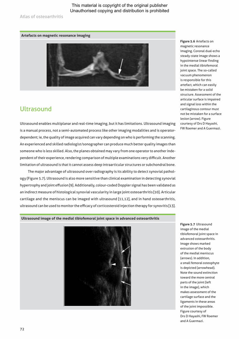
72
Atlas of osteoarthritis
Ultrasound
Ultrasound enables multiplanar and real-time imaging, but it has limitations. Ultrasound imaging
is a manual process, not a semi-automated process like other imaging modalities and is operator-
dependent; ie, the quality of image acquired can vary depending on who is performing the scanning.
An experienced and skilled radiologist/sonographer can produce much better quality images than
someone who is less skilled. Also, the planes obtained may vary from one operator to another inde-
pendent of their experience, rendering comparison of multiple examinations very difficult. Another
limitation of ultrasound is that it cannot assess deep intraarticular structures or subchondral bone.
The major advantage of ultrasound over radiography is its ability to detect synovial pathol-
ogy (Figure 5.7). Ultrasound is also more sensitive than clinical examination in detecting synovial
hypertrophy and joint effusion [9]. Additionally, colour-coded Doppler signal has been validated as
an indirect measure of histological synovial vascularity in large joint osteoarthritis [10]. Articular
cartilage and the meniscus can be imaged with ultrasound [11,12], and in hand osteoarthritis,
ultrasound can be used to monitor the efficacy of corticosteroid injection therapy for synovitis [13].
Figure 5.6 Artefacts on magnetic resonance imaging. Coronal dual-echo steady-state image shows a hypointense linear finding in the medial tibiofemoral joint space. The so-called vacuum phenomenon is responsible for this artefact, which can easily be mistaken for a solid structure. Assessment of the articular surface is impaired and signal loss within the cartilaginous contour must not be mistaken for a surface lesion (arrow). Figure courtesy of Drs D Hayashi, FW Roemer and A Guermazi.
Artefacts on magnetic resonance imaging
Ultrasound image of the medial tibiofemoral joint space in advanced osteoarthritis
Figure 5.7 Ultrasound image of the medial tibiofemoral joint space in advanced osteoarthritis. Image shows marked extrusion of the body of the medial meniscus (arrows). In addition, a small femoral osteophyte is depicted (arrowhead). Note the sound extinction toward the more central parts of the joint (left in the image), which makes assessment of the cartilage surface and the ligaments in these areas of the joint impossible. Figure courtesy of Drs D Hayashi, FW Roemer and A Guermazi.
This material is copyright of the original publisher Unauthorised copying and distribution is prohibited

73
Assessing joint damage in osteoarthritis
Computed tomography and computed tomography arthrography
Computed tomography is a valuable tool when imaging of osseous changes or detailed pre-
surgical planning is required. It depicts cortical bone and soft tissue calcifications better than
MRI (Figure 5.8) and has an established clinical role in assessing facet-joint osteoarthritis of the
spine [14]. CT arthrography is an alternative method for indirect visualisation of cartilage and
other intrinsic joint structures, especially in the knee and shoulder joints, and it enables imaging
of focal cartilage defects (Figure 5.9) [15]. Penetration of contrast medium within deeper layers
of the cartilage surface indicates an articular-sided defect of the chondral surface.
Figure 5.8 Use of computed tomography for evaluation of osteoarthritis. A, Coronal computed tomography (CT) image of the shoulder in advanced post-traumatic instability osteoarthritis. There is severe joint space narrowing (JSN) of the glenohumeral joint (short arrows). In addition, a large inferior humeral osteophyte is depicted (arrowhead) and a small subchondral cyst in the inferior glenoid is shown (long arrow). B, Sagittal CT image of post-traumatic ankle osteoarthritis. Large anterior osteophytes are depicted at the tibiotalar joint margin (large arrows). In addition, anterior JSN and small subchondral cysts are observed (small arrows). Note the intraarticular vacuum phenomenon, visualised as a hypodense intraarticular line, a common finding in osteoarthritic joints (see Figure 5.6). Figure courtesy of Drs D Hayashi, FW Roemer and A Guermazi.
Use of computed tomography for evaluation of osteoarthritis
B
A
This material is copyright of the original publisher Unauthorised copying and distribution is prohibited

74
Atlas of osteoarthritis
Figure 5.9 Superior delineation of small focal cartilage defects by arthrography. A, Coronal computed tomography image of the wrist shows a focal full-thickness defect of the scaphoid at the radial articular surface (arrow). In addition, note the full thickness tear of the scapholunate ligament (arrowhead). B, Corresponding T1- weighted nonarthrographic magnetic resonance imaging (MRI) shows a normal cartilaginous surface and ligament is inferiorly visualised. C, Proton density-weighted MRI with fat suppression depicts subtle bone marrow oedema in the scaphoid but no articular surface damage of the scaphoid. Figure courtesy of Drs D Hayashi, FW Roemer and A Guermazi.
Superior delineation of small focal cartilage defects by arthrography
A
B
C
This material is copyright of the original publisher Unauthorised copying and distribution is prohibited

75
Assessing joint damage in osteoarthritis
Nuclear medicine
Positron emission tomography can be used to detect metabolic changes in target tissues.
Increased uptake of 2-18F-fluoro-2-deoxy–d-glucose can be seen in periarticular regions, inflamed
synovium, the intercondylar notch and in areas of subchondral bone marrow corresponding to
MRI-detected bone marrow lesions (Figure 5.10) [16,17].
Figure 5.10 2-18F-fluoro-2-deoxy–D-glucose positron emission tomography. A, Axial positron emission tomography (PET) image of both knees shows marked glucose uptake in the intercondylar notch of the right knee. B, Reconstructed low-resolution coronal computed tomography (CT) image depicts joint space narrowing of the medial tibiofemoral joint (arrows) and subchondral tibial sclerosis (*). In addition, there is a small medial tibial osteophyte (arrowhead). C, Coronal fusion image of PET and CT localises the pathologic glucose accumulation clearly to the intercondylar notch around the posterior cruciate ligament, the most common site of synovitis in knee osteoarthritis. Hypermetabolic findings represent peri-ligamentous synovitis. Note the high sensitivity of PET for hypermetabolism but the low specificity and poor spatial localisation without the support of additional cross-sectional imaging with CT or magnetic resonance imaging. Figure courtesy of Drs D Hayashi, FW Roemer and A Guermazi.
2-18F-fluoro-2-deoxy–D-glucose positron emission tomography
A
B
C
This material is copyright of the original publisher Unauthorised copying and distribution is prohibited

76
Atlas of osteoarthritis
Typical imaging findings of various osteoarthritis-affected joints
Joint Imaging features
Hand • JSN/loss, subchondral eburnation, marginal osteophyte formation, small ossicles in distal and proximal interphalangeal (DIP and PIP) joints− Osteophytosis at DIP is called Heberden’s nodes− Osteophytosis at PIP is called Bouchard nodes
• Radial subluxation of first metacarpal base• JSN and eburnation of trapezioscaphoid area
Shoulder • Rotator cuff pathology• Labral tears• Osseous changes reflecting previous dislocation
Hip • Acetabular and femoral osteophytes, sclerosis and subchondral cysts• Thickening/buttressing of medial femoral cortex• Superolateral subluxation of femoral head• Medial/axial subluxation with or without protrusio acetabuli• Signs of femoroacetabular impingement
Knee • Medial tibiofemoral compartment is more commonly affected than lateral compartment
• Tibiofemoral joint is more commonly affected than patellofemoral joint• Varus malalignment
Spine • Sclerosis with narrowing of intervertebral apophyseal joints• Osteophytosis associated with disc pathology• Peridiscal endplate changes Modic type I–III• Spinal canal stenosis• Ligamentum flavum hypertrophy• Narrowing of neural foramina
Sacroiliac • Can be bilateral or unilateral• If unilateral, the affected side is contralateral to the bad hip• Diffuse loss of joint space• Well-defined line of sclerosis, particularly on the iliac side of the joint• Bridging osteophytes at superior and inferior limits of joint
Table 5.2 Typical imaging findings of various osteoarthritis-affected joints. DIP, distal interphalangeal; JSN, joint space narrowing; PIP, proximal interphalangeal.
Summary of imaging findings in various osteoarthritis-affected joints
Osteoarthritis can affect various joints of the body, including the hand, shoulder, hip, knee, spine
and sacroiliac joints. Typical imaging findings that affect these joints are summarised in Table 5.2.
An illustration of multimodality imaging assessment with pathological correlation is presented
in Figure 5.11 (see page 78), depicting end stage hip osteoarthritis using MRI and radiography
as the primary assessment tools prior to total hip replacement.
This material is copyright of the original publisher Unauthorised copying and distribution is prohibited

77
Assessing joint damage in osteoarthritis
Multimodality imaging of severe hip osteoarthritis with pathologic correlation
A
B
C
Figure 5.11 Multimodality imaging of severe hip osteoarthritis with pathologic correlationA, Anteroposterior (AP) radiograph shows marked joint space narrowing and an acetabular osteophyte. There are also distinct subchondral cystic lesions in the femoral head (arrows) and acetabulum (arrowheads). B, Coronal fat-suppressed proton density-weighted magnetic resonance imaging (MRI) depicts these subchondral cysts as hyperintense, fluid-equivalent lesions in the acetabulum (arrowheads) and femoral head (arrows). Note the marked diffuse bone marrow oedema visualised as areas of hyperintensity in the femoral head (*). C, Macroscopic specimen shows diffuse cartilage loss of the articular surface of the femoral head (depicted on photograph as haemorrhagic areas). Subchondral changes cannot be visualised by surface photography. (continues opposite).
This material is copyright of the original publisher Unauthorised copying and distribution is prohibited

78
Atlas of osteoarthritis
Multimodality imaging of severe hip osteoarthritis with pathologic correlation (continued)
Figure 5.11 Multimodality imaging of severe hip osteoarthritis with pathologic correlation (continued). D, Macroscopic section through femoral head confirms MRI-depicted cystic lesions (arrows). E, Haematoxylin-eosin stain of histologic specimen of the femoral head (corresponding to MRI B in this figure) confirms large subchondral cysts of the femoral head (arrows). Eosinophilic changes of the femoral head in the subchondral bone represent a mixture of oedema, subchondral sclerosis and fibrosis (asterisks). F, Postsurgical AP radiograph of the same left hip after total joint replacement. Figure courtesy of Drs D Hayashi, FW Roemer and A Guermazi.
D
E
F
This material is copyright of the original publisher Unauthorised copying and distribution is prohibited

79
Assessing joint damage in osteoarthritis
Future directions
Conventional radiography is still the first and most widely used imaging technique for evaluation
of people with osteoarthritis. However, the ability of MRI to image the knee as a whole organ and
to directly visualise lesions that are not detected with radiography is crucial to understanding
the natural history of the disease, and ensures that MRI will play an important role in guiding
future therapies. Ultrasound and contrast-enhanced MRI are particularly useful for imaging
osteoarthritis-related synovitis.
References
1 Guermazi A, Hunter DJ, Roemer FW. Plain radiography and magnetic resonance imaging diagnostics in osteoarthritis: validated staging and scoring. J Bone Joint Surg Am. 2009;91(suppl 1):54-62.
2 Altman R, Asch E, Bloch D, et al. Development of criteria for the classification and reporting of osteoarthritis. Classification of osteoarthritis of the knee. Diagnostic and Therapeutic Criteria Committee of the American Rheumatism Association. Arthritis Rheum. 1986;29:1039-1049.
3 Kellgren JH, Lawrence JS. Radiological assessment of osteo-arthrosis. Ann Rheum Dis. 1957;16:494-502.4 Altman RD, Gold GE. Atlas of individual radiographic features in osteoarthritis, revised. Osteoarthritis
Cartilage. 2007;15(suppl A):A1-A56.5 Peterfy CG, Gold G, Eckstein F, Cicuttini F, Dardzinski B, Stevens R. MRI protocols for whole-organ
assessment of the knee in osteoarthritis. Osteoarthritis Cartilage. 2006;14(suppl A):A95-A111.6 Roemer FW, Guermazi A. MR imaging-based semiquantitative assessment in osteoarthritis.
Radiol Clin North Am. 2009;47:633-654.7 Link TM. MR imaging in osteoarthritis: hardware, coils, and sequences. Radiol Clin North Am.
2009;47:617-632.8 Hayashi D, Roemer FW, Katur A, et al. Imaging of synovitis in osteoarthritis: current status and outlook.
Semin Arthritis Rheum. 2011;41:116-130.9 Karim Z, Wakefield RJ, Quinn M, et al. Validation and reproducibility of ultrasonography in the detection
of synovitis in the knee: a comparison with arthroscopy and clinical examination. Arthritis Rheum. 2004;50:387-394.
10 Walther M, Harms H, Krenn V, Radke S, Faehndrich T-P, Gohlke F. Correlation of power Doppler sonography with vascularity of the synovial tissue of the knee joint in patients with osteoarthritis and rheumatoid arthritis. Arthritis Rheum. 2001;44:331-338.
11 Saarakkala S, Waris P, Waris V, et al. Diagnostic performance of knee ultrasonography for detecting degenerative changes of articular cartilage. Osteoarthritis Cartilage. 2012;20:376-381.
12 Kawaguchi K, Enokida M, Otsuki R, Teshima R. Ultrasonographic evaluation of medial radial displacement of the medial meniscus in knee osteoarthritis. Arthritis Rheum. 2012;64:173-180.
13 Keen HI, Wakefield RJ, Hensor EMA, Emery P, Conaghan PG. Response of symptoms and synovitis to intra-muscular methylprednisolone in osteoarthritis of the hand: an ultrasonographic study. Rheumatology (Oxford). 2010;49:1093-1100.
14 Hechelhammer L, Pfirmann CWA, Zanetti M, Hodler J, Boos N, Schmid MR. Imaging findings predicting the outcome of cervical facet joint blocks. Eur Radiol. 2007;17:959-964.
15 Vande Berg BC, Lecouvet FE, Poilvache P, et al. Assessment of knee cartilage in cadavers with dual-detector spiral CT arthrography and MR imaging. Radiology. 2002;222:430-436.
16 Nakamura H, Masuko K, Yudoh K, et al. Positron emission tomography with 18F-FDG in osteoarthritic knee. Osteoarthritis Cartilage. 2007;15:673-681.
17 Elzinga EH, van der Laken CJ, Comans EFI, Lammertsma AA, Dijkmans BAC, Voskuyl AE. 2-Deoxy-2-[F-18]fluoro-d-glucose joint uptake on positron emission tomography images: rheumatoid arthritis versus osteoarthritis. Mol Imaging Biol. 2007;9:357-360.
This material is copyright of the original publisher Unauthorised copying and distribution is prohibited

80
In the absence of a cure for osteoarthritis, current therapeutic modalities are primarily aimed
at reducing pain and improving joint function by targeting symptom relief, without facilitating
any improvement in the joint structure itself [1]. The management of osteoarthritis should be
individualised so that it conforms to the specific findings of the clinical examination [2,3]. This is
especially the case for patients with obesity, depression, malalignment and/or muscle weakness.
Comprehensive management should always include a combination of treatment options that are
directed towards the common goal of improving the patient’s pain and tolerance for functional
activity. Treatment plans should never be defined rigidly based on the X-ray appearance of the
joint, but instead remain flexible so that they can be altered in line with the functional and symp-
tomatic responses obtained [2]. Guidelines recommend that the hierarchy of management should
consist of nonpharmacological modalities first, then drugs and then surgery [3–6].
Nonpharmacological treatments
Self-management and education
All patients should be encouraged to participate in self-management programs (such as those
conducted by the Arthritis Foundation [7]) that offer information on the natural history of osteo-
arthritis and provide resources for social support and instructions on coping skills [8,9].
The majority of individuals with osteoarthritis are either overweight or obese [10]. There is good
evidence for the efficacy of weight management [11], and this is advocated by most osteoarthri-
tis guidelines [4,5]. However, in practice, weight management is not frequently implemented
[12–15]. Another pivotal yet often ignored aspect of the conservative management of osteoar-
thritis is exercise. Although guidelines routinely advocate exercise [2, 16–20], clinical practice
does not reflect this recommendation [12–15].
Osteoarthritis and other obesity-related diseases and conditions place an enormous physi-
cal and financial burden on healthcare systems [21]. Weight loss has been universally recom-
mended as a treatment for knee osteoarthritis [4,5], but is not always attainable due to patient
and physician challenges in adhering to guidelines [13,22]. Recent data indicate that intensive
dietary restriction plus exercise safely achieved a mean long-term (18 months) weight loss of
11.4% and yielded a 50% improvement in osteoarthritis symptoms (Figure 6.1) [23]. The wider
adoption of dietary restrictions combined with exercise has a marked potential for reducing the
burden of disability related to the increasing prevalence of osteoarthritis.
Chapter 6Treatment of osteoarthritis
David Hunter
This material is copyright of the original publisher Unauthorised copying and distribution is prohibited

81
Treatment of osteoarthritis
Exercise
Exercise is essential for all people with knee osteoarthritis, irrespective of disease severity, age,
comorbidity, pain severity or disability. Meta-analyses have found small-to-moderate effects in
pain and function with exercise [24], similar to those achieved with analgesics and nonsteroidal
anti-inflammatory drugs (NSAIDs).
High-intensity, home-based strength training can produce substantial improvements in
strength, pain, physical function and quality of life in people with knee osteoarthritis (Figure 6.2)
[25–31]. Given the deficits in muscle function present with knee osteoarthritis, muscle rehabili-
tation plays an important role in disease management in general and in reducing symptoms and
improving function in particular. Table 6.1 lists the practical aspects of prescribing exercise for
patients and reviews the current evidence on the optimum mode of delivery, type of exercise
and dosage [32].
While exercise is a core treatment for knee osteoarthritis, adherence to exercise regimens
is difficult to maintain, with research indicating that lack of adherence limits long-term effective-
ness (Table 6.2) [33].
The effect of intensive weight loss on symptoms
Diet (D) Exercise (E) Diet + exercise (D+E)
Figure 6.1 The effect of intensive weight loss on symptoms. Mean (±SE) Western Ontario and McMaster Universities Osteoarthritis Index (WOMAC) pain scores (adjusted for gender, body mass index [BMI] and baseline values) across the 18-month intervention period. Baseline mean is overall for the three groups. Combined intensive dietary restriction and exercise led to a mean long-term weight loss of 11.4%, with 50% symptom improvement.At 18 months, 40% of participants in the diet plus exercise group reported little or no pain, with pain scores of 0 or 1 on a scale of 0–20, compared to around 20% of the diet-only and exercise-only groups. Data from Messier et al [23].
8
7
6
5
4
3
2
1
0
Baseline 6 months 12 months 18 months
Time
WO
MA
C p
ain
scor
es (r
ange
0–2
0)
D+E 38%
D 20%
E 22%
*P=0.001
This material is copyright of the original publisher Unauthorised copying and distribution is prohibited

82
Atlas of osteoarthritis
The effect of quadriceps strengthening on physical function in knee osteoarthritis trials
50
45
40
35
30
25
20
15
10
5
0
Phys
ical
func
tion
(% c
hang
e)
Baker et al 2000 [27]
RØgind et al 1998 [25]
O’Reilly et al 1999 [24]
Fisher et al 1993 [26]
Fisher et al 1997 [28]
Ettinger et al 1997 [23]
Figure 6.2 The effect of quadriceps strengthening on physical functionin knee osteoarthritis trials. Improvements in knee extension strength are directly related to improvements in physical function. This figure shows the association between change in strengthand change in physical function in published studies of exercise in knee osteoarthritis.Physical function is self-reported. r=0.877;P=0.02. Data from Baker et al. [25]. Reproduced with permission fromDr K Baker and the Journal of Rheumatology.
0 10 20 30 40 50 60 70 80
Knee extenstion strength (% change)
Summary of exercise prescription for muscle rehabilitation
• Refer the patient to a healthcare professional for appropriate exercise prescription
• Supervised group or individual treatments are superior to independent home exercise for pain reduction
• Supplement home exercise with initial group exercise
• Exercise handouts or audiovisual material alone are ineffective
• Target quadriceps, hamstrings and hip abductors for strengthening
• Minimise compressive joint forces
• Clinical outcome is not influenced by the type of strengthening exercise
• Use a combined program of strengthening, flexibility and functional exercises
• Use strategies to maximise long-term patient compliance with exercise
Table 6.1 Summary of exercise prescription for muscle rehabilitation. Data from Bennell et al. [32]. Reproduced with permission from Elsevier.
This material is copyright of the original publisher Unauthorised copying and distribution is prohibited

83
Treatment of osteoarthritis
Assistive devices
The shared goal of many noninvasive devices for knee osteoarthritis is to alter the lower limb
biomechanics in such a way as to limit the exposure of one or more knee compartments to poten-
tially damaging and provocative mechanical stresses. While the magnitude and direction of the
ground reaction forces determine how much overall compressive load the tibiofemoral (TF)
joint routinely sustains, the relative bony alignment of the tibia and femur also has an enormous
impact on the manner in which this compressive load is distributed across the medial and lateral
compartments. Improved frontal plane knee alignment and mediolateral stability against thrust
are commonly cited reasons for prescribing either a valgus-inducing unloader brace to patients
with medial TF osteoarthritis or, less commonly, a varus-inducing unloader brace to patients with
lateral TF osteoarthritis (Figure 6.3) [34,35]. Meta-analysis of randomised trials suggests valgus
bracing for medial knee OA results in small-to-moderate improvements in pain [36].
Strategies to facilitate long-term adherence to exercise and physical activity in people with knee osteoarthritis
Educate the patient about the disease and the benefits of exercise
Develop the exercise/physical activity plan with the patient and vary to maintain interest and enthusiasm
Use a graded progressive exercise/physical activity prescription and ensure pain/discomfort is not excessive during or after exercise
Initiate exercise under expert instruction and supervise exercise sessions if possible
Supplement face-to-face instruction with other materials; eg, written handouts, video/DVD/online demonstrations
Incorporate behavioural techniques to increase self efficacy; eg, positive reinforcement, goal setting, use of an exercise contract, self-monitoring via diary, pedometer
Include spouse/family in exercise program and garner support from family and friends
Monitor over the long-term via periodic reassessment by a health professional
Table 6.2 Strategies to facilitate long-termadherence to exercise and physical activity in people with knee osteoarthritis. Data from Bennell et al. [33]. Reproduced with permission from the British Medical Journal Publishing Group.
This material is copyright of the original publisher Unauthorised copying and distribution is prohibited

84
Atlas of osteoarthritis
Use of brace device for symptomatic knee osteoarthritis
B
Genu varum malalignment Genu varum alignment corrected
Valgus unloader braceAnterior view
A Genu varum malalignmentAnterior view in walking
GRF
Madd
Right leg
Figure 6.3 Use of brace device for symptomatic knee osteoarthritis.A, Loading of the knee with genu varum, or bowlegged, malalignment. Genu varum increases the adduction moment (Madd) at the knee and the magnitude of the compressive load on the medial TF compartment.B, Correction of genu varum malalignment using a valgus unloader brace. Data from Gross [35]. GRF, ground force reaction.© 2010, reproduced with permission from Elsevier.
This material is copyright of the original publisher Unauthorised copying and distribution is prohibited

85
Treatment of osteoarthritis
Pharmacological treatments
The pharmacological management of osteoarthritis includes [37]:
• Simple analgesics;
• NSAIDs;
• Intra-articular therapies (corticosteroids, hyaluronic acid);
• Supplements or alternative therapy;
• Disease modification therapy; and
• Symptomatic slow-acting drugs in osteoarthritis (SYSADOAs).
Current drug treatment options reduce osteoarthritis symptoms, but their efficacy is limited
[38], leaving patients with a substantial pain burden. In addition, many of these agents, par-
ticularly cyclooxygenase-2 (COX-2) specific inhibitors, have adverse-event profiles that raise a
number of legitimate concerns about their long-term safety [39,40] . The judicious use of topical
NSAIDs has been demonstrated to be effective for the relief of pain in both hand and knee
osteoarthritis compared with placebo [41,42]. This route of administration possibly reduces
gastrointestinal (GI) adverse reactions by maximising local delivery and minimising systemic
toxicity [43]. Table 6.3 provides an overview of the recommendations, based on the most com-
monly used guidelines, for different pharmacological agents in the management of knee and
hip osteoarthritis [4,5].
This material is copyright of the original publisher Unauthorised copying and distribution is prohibited

86
Atlas of osteoarthritis
Pharmacologic recommendations
Organisation*A
ceta
min
ophe
n or
pa
race
tam
ol (<
4g/d
ay)
Ora
l NSA
ID–N
on-
sele
ctiv
e
Ora
l NSA
ID (C
OX
-2
inhi
bito
rs)
Topi
cal N
SAID
Gas
trop
rote
ctio
n fo
r hi
gh-r
isk
pati
ents
∏
Tram
adol
Caps
aici
n
Opi
oids
§
Dul
oxet
ine
Intr
a-ar
ticu
lar
cort
icos
tero
ids
Intr
a-ar
ticu
lar
hyal
uron
ic a
cid
Her
bal r
emed
ies
Ris
edro
nate
OARSI-Knee**
App (7), (6), (7), (6)
App (7), App (7.5)
App (7), App (7)
App (8), App (7), Unc (6), Unc (6)
App (7), Unc
(6), (6), (6)
Unc (5), (4), (5), (6)
App (7), Unc (6), App
(7), (7)
App (7), (7), (7), (7)
Unc (5), (4),
Not app
(3), (3)
Unc, (5), (5), (5), (5)
Not App
(3), (3), (3), (3)
OARSI Moderate Comorbidity Risk***
Unc (5), Unc (4)
Unc (6), Not
App (7),
OARSI High Comorbidity Risk***
Not app
(2), Not App (2)
Not App
(3), Not App (3)
App, App
AAOS I S S S I I NR-S
ACR (Hand) CR& CR CR CR CR CNR CNR
ACR (Knee) CR CR& CR CR CNR SR CR§ CR
ACR (Hip) CR CR& CR SR CR
MOVE
NICE R R R R R R R R R NR
ESCEO R R R R R R R R R R
EULAR-Hand IV (87) Ia (81) Ia (75) Ia (81) Ia (75) Ib (60) IIb (63)
APTA-OS R (I) R (I)
Dutch
Ottawa
SOFMER (Gelis)
SOFMER (Mazieres)
ACCP R R& R
ACPMAB R R
MQIC R R R R (D) R R R R R
ItalianIa, IV (86)
IIb; IV (71)
III (82) Ib-IV (72)
Table 6.3 Pharmacologic recommendations [3,5]** Appropriateness scores () listed in order of knee OA without comorbidities, with comorbidities, multisite OA without comorbidities and with comorbidities. *** Appropriateness scores () listed in order of knee OA with comorbidities and multisite OA with comorbidities. *Grading systems described in Table 1. ∏COX-2, topical over oral NSAID, or add PPI or other agent; §for cases refractory to other modalities; & after acetaminophen. AAOS, American Academy of Orthopaedic Surgeons; ACCP, American College of Clinical Pharmacy; ACPMAB, Asian Chronic Pain Management Advisory Board; ACR, American College of Radiology; App, Appropriate; APTA-OS, American Physical Therapy Association - Organizational Scorecard; CNR, Conditionally not recommended; CR, Conditional recommendation; ESCEO, European Society for Clinical and Economic Aspects of Osteoporosis, Osteoarthritis and Musculoskeletal Diseases; EULAR, European League Against Rheumatism; I, Inconclusive; Ia, Meta-analysis of RCTs; Ib, Randomised controlled trial; IIb, Quasi-experimental study; III, Non-experimental/descriptive; IV, Expert committee report/opinion/clinical experience; MQIC, Michigan Quality Improvement Consortium; NICE, National Institute For Health and Clinical Excellence; Not App, Not appropriate; NR, Not recommended; NS, Not stated; OARSI, Osteoarthritis Research Society International; R, Recommended; S, Strong; SOFMER, French Society of Physical Medicine and Rehabilitation; Unc, Uncertain. Reproduced from Meneses et al. [5]Supplementary Table 7. © 2016, reproduced with permission from Elsevier.
This material is copyright of the original publisher Unauthorised copying and distribution is prohibited

87
Treatment of osteoarthritis
Figure 6.4 COX-1 versus COX-2. Representative biosynthetic pathwayof prostaglandin (PG) biosynthesis from arachidonic acid (AA) via cyclooxygenase (COX)-1/COX-2 isoform catalysis. The NSAIDs, aspirin, indomethacin and ibuprofen, arenonselective inhibitors of COX isozymes, whereas celecoxib exhibits selective COX-2 inhibition. LOX, lipoxygenase;LT, leukotriene. Reproduced from Rao PNP and Knaus EE: J Pharm Pharmaceut Sci. 11(2): 81s-110s; 2008. ©The Authors, 2008. (This article is published with open access at www.cspsCanada.org.) [47]
COX-1 versus COX-2
Inhibitors• NSAIDS (non–COX-2)• Aspirin• Indomethacin• Ibuprofen
Cytoprotective prostaglandins• Protect gastric mucosa• Aid platelet aggregation
Inflammatory prostaglandins• Recruit inflammatory cells• Sensitise skin pain receptors• Regulate hypothalamic
temperature control
Membrane phospholipids
Tissue injury
Phospholipids
Phospholipase A2
Leukotrienes• Bronchoconstriction
InhibitorsAspirinIndomethacinIbuprofenCelecoxib
InducersLTB4, LTC4, LTD4, LTE4
PGD synthase Tx synthase
5-LOX5-HPETE LTA4
12/15-LOX12/15 HPETE
COOH
COX-1 COX-2
(Constitutional) (Inducible)
Arachidonic acid
PGG2
O
O
COOH
OOH
O
O
COOH
OH
PGD2 TxA2
PGF synthase
PGI synthase
PGE2 PGF2PGI2
PGH2
PGE synthase
Nonsteroidal anti-inflammatory drugs
Nonsteroidal anti-inflammatory drugs, including both traditional and specific COX-2 inhibitors,
provide significant health benefits in the treatment of pain and inflammation [44]. However, they
are associated with an increased risk of serious GI [45] and cardiovascular adverse events [46].
Both beneficial and adverse effects are due to the same mechanism of action—the inhibition of
COX-dependent prostanoids (Figures 6.4–6.7, Table 6.4; see pages 90–92) [47–50].
This material is copyright of the original publisher Unauthorised copying and distribution is prohibited
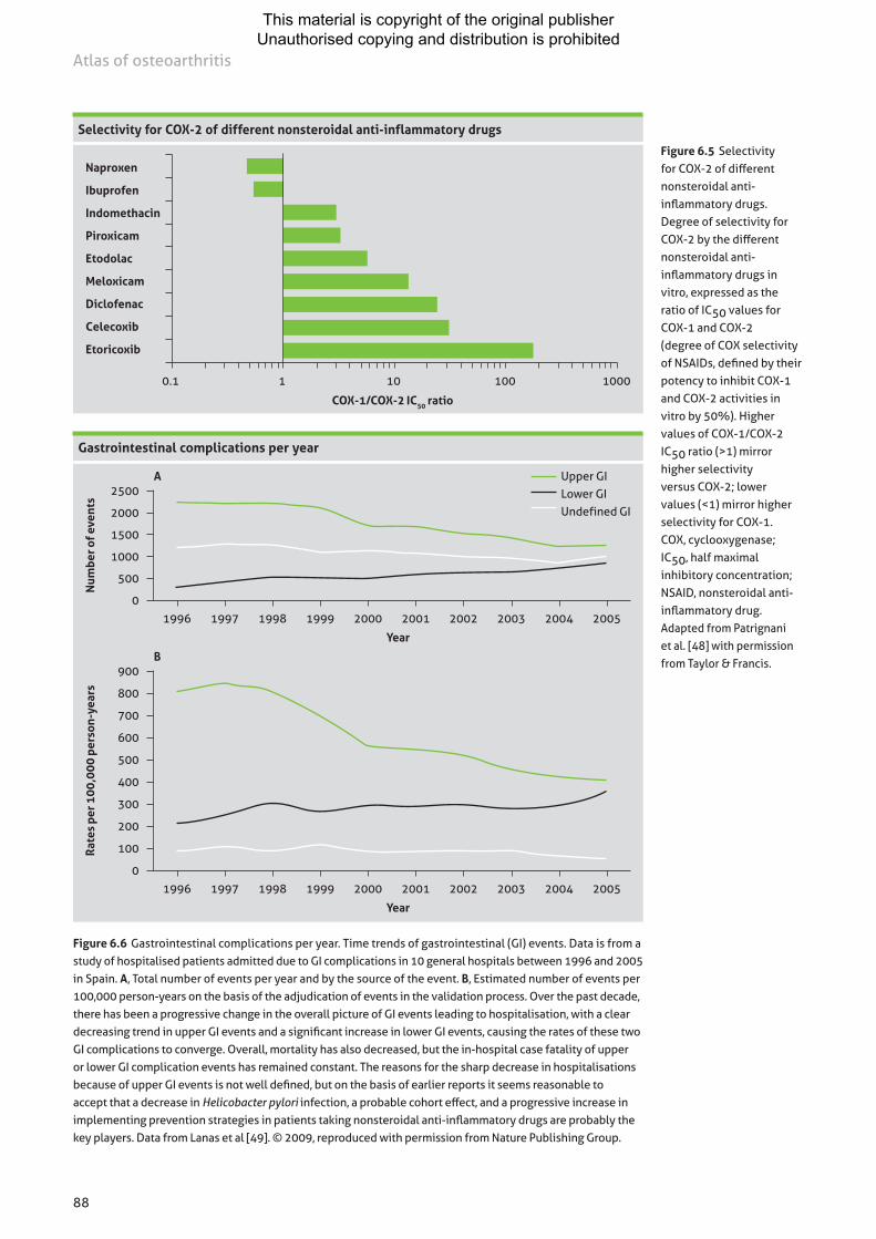
88
Atlas of osteoarthritis
Figure 6.5 Selectivity for COX-2 of different nonsteroidal anti-inflammatory drugs. Degree of selectivity for COX-2 by the different nonsteroidal anti-inflammatory drugs in vitro, expressed as the ratio of IC50 values for COX-1 and COX-2(degree of COX selectivity of NSAIDs, defined by their potency to inhibit COX-1 and COX-2 activities in vitro by 50%). Higher values of COX-1/COX-2 IC50 ratio (>1) mirror higher selectivityversus COX-2; lower values (<1) mirror higher selectivity for COX-1.COX, cyclooxygenase; IC50, half maximal inhibitory concentration;NSAID, nonsteroidal anti-inflammatory drug.Adapted from Patrignani et al. [48] with permission from Taylor & Francis.
Selectivity for COX-2 of different nonsteroidal anti-inflammatory drugs
0.1 1 10 100 1000
COX-1/COX-2 IC50 ratio
Naproxen
Ibuprofen
Indomethacin
Piroxicam
Etodolac
Meloxicam
Diclofenac
Celecoxib
Etoricoxib
Gastrointestinal complications per year
Upper GI Lower GI Undefined GI
A2500
2000
1500
1000
500
0
Num
ber o
f eve
nts
1996 1997 1998 1999 2000 2001 2002 2003 2004 2005
Year
B900
800
700
600
500
400
300
200
100
0
Rat
es p
er 1
00,0
00 p
erso
n-ye
ars
1996 1997 1998 1999 2000 2001 2002 2003 2004 2005
Year
Figure 6.6 Gastrointestinal complications per year. Time trends of gastrointestinal (GI) events. Data is from a study of hospitalised patients admitted due to GI complications in 10 general hospitals between 1996 and 2005 in Spain. A, Total number of events per year and by the source of the event. B, Estimated number of events per 100,000 person-years on the basis of the adjudication of events in the validation process. Over the past decade, there has been a progressive change in the overall picture of GI events leading to hospitalisation, with a clear decreasing trend in upper GI events and a significant increase in lower GI events, causing the rates of these two GI complications to converge. Overall, mortality has also decreased, but the in-hospital case fatality of upper or lower GI complication events has remained constant. The reasons for the sharp decrease in hospitalisations because of upper GI events is not well defined, but on the basis of earlier reports it seems reasonable to accept that a decrease in Helicobacter pylori infection, a probable cohort effect, and a progressive increase in implementing prevention strategies in patients taking nonsteroidal anti-inflammatory drugs are probably the key players. Data from Lanas et al [49]. © 2009, reproduced with permission from Nature Publishing Group.
This material is copyright of the original publisher Unauthorised copying and distribution is prohibited

89
Treatment of osteoarthritis
Figure 6.7 Morbidity rate ratios for nonsteroidal anti-inflammatories compared with placebo. Although uncertainty remains, little evidence exists to suggest that any of the nonsteroidal anti-inflammatory drugs (NSAIDs) are safe in terms of cardiovascular events. Naproxen appears to be the least harmful. Cardiovascular risk needs to be taken into account when prescribing any NSAID.Data from Trelle et al. [50].© 2011, reproduced with permission fromthe British Medical Journal Publishing Group.
Morbidity rate ratios for nonsteroidal anti-inflammatories compared with placebo
Rate ratio (95% credibility interval) Rate ratio (95% credibility interval)
Myocardial infarction
Naproxen 0.82 (0.37–1.67)
Ibuprofen 1.61 (0.50–5.77)
Diclofenac 0.82 (0.29–2.20)
Celecoxib 1.35 (0.71–2.72)
Etoricoxib 0.75 (0.23–2.39)
Lumiracoxib 2.00 (0.71–6.21)
Stroke
Naproxen 1.76 (0.91–3.33)
Ibuprofen 3.36 (1.00–11.60)
Diclofenac 2.86 (1.09–8.36)
Celecoxib 1.12 (0.60–2.06)
Etoricoxib 2.67 (0.82–8.72)
Lumiracoxib 2.81 (1.05–7.48)
Cardiovascular death
Naproxen 0.98 (0.41–2.37)
Ibuprofen 2.39 (0.69–8.64)
Diclofenac 3.98 (1.48–12.70)
Celecoxib 2.07 (0.98–4.55)
Etoricoxib 4.07 (1.23–15.70)
Lumiracoxib 1.89 (0.64–7.09)
Death from any cause
Naproxen 1.23 (0.71–2.12)
Ibuprofen 1.77 (0.73–4.30)
Diclofenac 2.31 (1.00–4.95)
Celecoxib 1.50 (0.96–2.54)
Etoricoxib 2.29 (0.94–5.71)
Lumiracoxib 1.75 (0.78–4.17)
Favours NSAID Favours placebo
0.2 0.5 1 2 5
This material is copyright of the original publisher Unauthorised copying and distribution is prohibited

90
Atlas of osteoarthritis
The American Academy of Orthopaedic Surgeons (AAOS) recommends that patients with
symptomatic knee osteoarthritis and increased GI risk (age ≥60 years, comorbid medical condi-
tions, history of peptic ulcer disease, history of GI bleeding, concurrent corticosteroid use and/
or the concomitant use of anticoagulants) receive one of the following analgesics for pain [51]:
• Paracetamol/acetaminophen (not to exceed 4 g/day);
• Topical NSAIDs;
• Nonselective oral NSAIDs plus a gastroprotective agent; or
• COX-2 specific inhibitors.
Simple analgesics
The effect size (ES) for pain relief with paracetamol/acetaminophen is very small, at 0.14 (95%
confidence interval [CI]: 0.05, 0.22), and is no longer significant when the analysis is restricted
to high-quality trials (ES=0.10, 95% CI: 0.0, 0.23) (Figure 6.8). In the light of concerns over GI
toxicity, mortality, and cardiovascular and renal AEs the role of paracetamol/acetaminophen in
the treatment of osteoarthritis has been revised [52,53].
Inhibition data, selectivity for NSAIDs and COX-1/2 clinical dose for treating rheumatoid arthritis and osteoarthritis
Whole blood assay IC50 (µM) Clinical dose
Drug Trade name COX-1 COX-2Selectivity
indexRheumatoid
arthritis Osteoarthritis
Aspirin Aspirin®
Ecotrin®
1.7 >100 0.017 2600–3900 –
Diclofenac Voltaren® 0.075 0.038 1.97 150–200 150–200
Ibuprofen Advil®
Motren®
7.6 7.2 1.05 1200–3200 1200–3200
Indomethacin Indocin® 0.013 1.0 0.013 150–200 150–200
Ketoprofen Orudis® 0.047 2.9 0.016 200–300 200–300
Fluorbiprofen Fluorbiprofen®
Ansaid®
0.075 5.5 0.013 200–300 200–300
Naproxen Naprosyn®
Aleve®
9.3 28 0.33 500–1000 500–1000
Nimesulide Mesulid® 10 1.9 5.26 – 200
Meloxicam Mobic® 5.7 2.1 2.7 7.5–15 7.5–15
Paracetamol Tylenol® >100 49 >2.04 2600–4000 2600–4000
Celecoxib Celebrex® 6.7 0.87 7.7 200–400 200
Etoricoxib Arcoxia® 116 1.1 105.4 90 60
Lumiracoxib Prexige® 67 0.13 515 – 200–400
Table 6.4 Inhibition data, selectivity for NSAIDs and COX-1/2 clinical dosefor treating rheumatoid arthritis and osteoarthritis. COX, cyclooxygenase;IC50, half maximalinhibitory concentration; NSAID, nonsteroidal anti-inflammatory drug. Reproduced from Rao PNP and Knaus EE: J Pharm Pharmaceut Sci. 11(2): 81s-110s; 2008. ©The Authors, 2008. (This article is published with open access at www.cspsCanada.org.) [47]
This material is copyright of the original publisher Unauthorised copying and distribution is prohibited

91
Treatment of osteoarthritis
Intra-articular therapies
Intra-articular corticosteroids are used widely in the management of knee osteoarthritis.
However, a Cochrane review found that the reduction in pain lasts for only 1–2 weeks [59]. Given
this short duration of benefit, high cost and potential adverse effects, corticosteroid use may
not be merited in a chronic disease such as osteoarthritis (Table 6.5). Despite the temptation to
use these agents in patients with the features of clinical inflammation (such as a large effusion),
the evidence to support this is limited. Furthermore, repeat administration every three months
does not appear to provide a clinical benefit at two years with a suggestion of increased MRI
cartilage loss [60].
The use of intra-articular injections of viscosupplements (e.g., hyaluronic acid), usually given
weekly for 3–5 weeks, has been extensively researched, but a recent meta-analysis found that
the trials are generally of low quality and that viscosupplementation is associated with a small
and clinically irrelevant reduction in pain but an increased risk of serious adverse events [61].
Figure 6.8 Pain relief effect of paracetamol. CI, confidence interval. Data from [44,54–58].Data from Zhang et al. [18]. Reproduced with permission from Elsevier.
Pain relief effect of paracetamol
Case et al 2003 [42] -0.03 (-0.55, 0.49)
Golden et al 2004 [43] 0.20 (-0.03, 0.43)
Miceli-Richard et al 2004 [44] 0.08 (-0.07, 0.22)
Pincus et al 2004 [45] 0.13 (-0.09, 0.34)
Pincus et al 2000 [46] 0.23 (0.03, 0.44)
Herrero-Beaumont et al 2007 [47] 0.16 (-0.011, 0.43)
Combined 0.14 (0.05, 0.23)
-0.60 -0.35 -0.10 0.15 0.40 0.65
Effect size (95% CI)
Overview of intra-articular steroids
AgentRelative
anti-inflammatory potency Solubility Dose
Hydrocortisone acetate 1 High 10–25 mg
Methylprednisolone acetate 5 Medium 20–80 mg
Triamcinolone acetonide
Triamcinolone hexacetonide5 Medium
10–40 mg
10–20 mg
Betamethasone sodium phosphate and acetate
20 Low 0.25–2 mL
Table 6.5 Overview of intra-articular steroids. Injectable corticosteroids can be classified in terms of solubility and duration of action. High-solubility preparations have a short duration of action andlow-solubility compounds have a long duration.This table shows the relative anti-inflammatory potencies of various injectable corticosteroids, with hydrocortisone used as the standard with a value of 1.0 [59].
This material is copyright of the original publisher Unauthorised copying and distribution is prohibited

92
Atlas of osteoarthritis
Antidepressants
Because some people with knee osteoarthritis also have depression and symptoms of neu-
ropathic pain (shooting or burning pain, pins and needles), the role of centrally active agents,
including selective serotonin and noradrenaline (norepinephrine) reuptake inhibitors, has been
investigated. In a randomised controlled trial (RCT) of duloxetine versus placebo, 65% of par-
ticipants in the duloxetine group reported a reduction in pain of more than 30%, compared with
just 44% in the placebo group [62]. This was the result of a primary analgesic effect and not an
elevation in mood or changes in anxiety or depression [42]. These agents may be useful in sub-
groups of patients with knee osteoarthritis.
Disease modification therapy
Once a popular area for drug development, with a multitude of discovery and preclinical programs
at major pharmaceutical and biotech companies and dozens of compounds moving through
pharmaceutical pipelines toward pivotal clinical trials, research on slowing or stopping the pro-
gression of cartilage loss and other structural changes in the joint has been significantly scaled
back. This is due in large part to challenges over target identification and clinical development
(Table 6.6, Figure 6.9; see pages 96–97) [63,64]. The cited references provide a detailed review of
this complicated area including the lessons learned from prior disease-modifying osteoarthritis
drug trials and the challenges that lie therein in trial conduct.
This material is copyright of the original publisher Unauthorised copying and distribution is prohibited
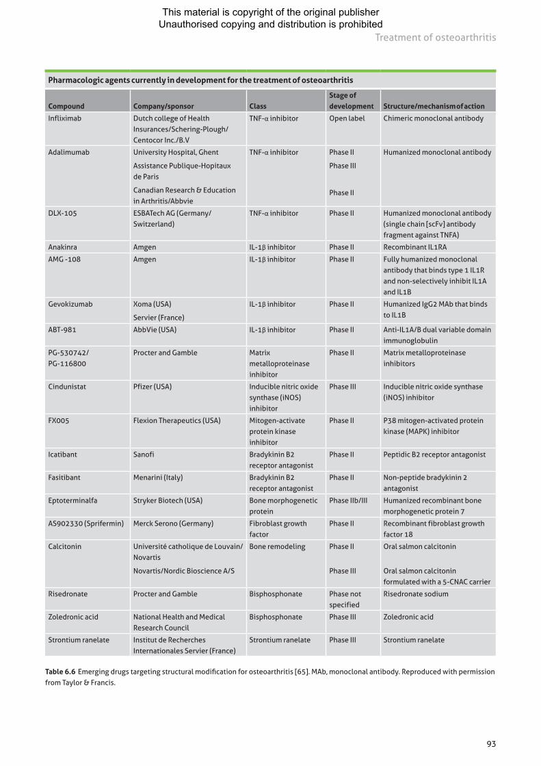
93
Treatment of osteoarthritis
Pharmacologic agents currently in development for the treatment of osteoarthritis
Compound Company/sponsor Class Stage of development Structure/mechanism of action
Infliximab Dutch college of Health Insurances/Schering-Plough/Centocor Inc./B.V
TNF-α inhibitor Open label Chimeric monoclonal antibody
Adalimumab University Hospital, Ghent
Assistance Publique-Hopitaux de Paris
Canadian Research & Education in Arthritis/Abbvie
TNF-α inhibitor Phase II
Phase III
Phase II
Humanized monoclonal antibody
DLX-105 ESBATech AG (Germany/Switzerland)
TNF-α inhibitor Phase II Humanized monoclonal antibody (single chain [scFv] antibody fragment against TNFA)
Anakinra Amgen IL-1β inhibitor Phase II Recombinant IL1RA
AMG -108 Amgen IL-1β inhibitor Phase II Fully humanized monoclonal antibody that binds type 1 IL1R and non-selectively inhibit IL1A and IL1B
Gevokizumab Xoma (USA)
Servier (France)
IL-1β inhibitor Phase II Humanized IgG2 MAb that binds to IL1B
ABT-981 AbbVie (USA) IL-1β inhibitor Phase II Anti-IL1A/B dual variable domain immunoglobulin
PG-530742/ PG-116800
Procter and Gamble Matrix metalloproteinase inhibitor
Phase II Matrix metalloproteinase inhibitors
Cindunistat Pfizer (USA) Inducible nitric oxide synthase (iNOS) inhibitor
Phase III Inducible nitric oxide synthase (iNOS) inhibitor
FX005 Flexion Therapeutics (USA) Mitogen-activate protein kinase inhibitor
Phase II P38 mitogen-activated protein kinase (MAPK) inhibitor
Icatibant Sanofi Bradykinin B2 receptor antagonist
Phase II Peptidic B2 receptor antagonist
Fasitibant Menarini (Italy) Bradykinin B2 receptor antagonist
Phase II Non-peptide bradykinin 2 antagonist
Eptoterminalfa Stryker Biotech (USA) Bone morphogenetic protein
Phase IIb/III Humanized recombinant bone morphogenetic protein 7
AS902330 (Sprifermin) Merck Serono (Germany) Fibroblast growth factor
Phase II Recombinant fibroblast growth factor 18
Calcitonin Université catholique de Louvain/Novartis
Novartis/Nordic Bioscience A/S
Bone remodeling Phase II
Phase III
Oral salmon calcitonin
Oral salmon calcitonin formulated with a 5-CNAC carrier
Risedronate Procter and Gamble Bisphosphonate Phase not specified
Risedronate sodium
Zoledronic acid National Health and Medical Research Council
Bisphosphonate Phase III Zoledronic acid
Strontium ranelate Institut de Recherches Internationales Servier (France)
Strontium ranelate Phase III Strontium ranelate
Table 6.6 Emerging drugs targeting structural modification for osteoarthritis [65]. MAb, monoclonal antibody. Reproduced with permission from Taylor & Francis.
This material is copyright of the original publisher Unauthorised copying and distribution is prohibited

94
Atlas of osteoarthritis
Schematic of the knee joint depicting the synovial joint tissues affected in osteoarthritis
Figure 6.9 Schematic of the knee joint depicting the synovial joint tissues affected in osteoarthritis. Consistent with the theory that osteoarthritis is a disease of the whole synovial joint, current disease- modifying osteoarthritis drug development is now targeting synovial-joint tissue structures, including bone, cartilage and synovium. Some of the agents that target these relevant tissues are listed here. BMP, bone morphogenetic protein; FGF, fibroblast growth factor; IL, interleukin; MMP, matrix metalloproteinase;TNF, tumour necrosis factor. Adapted from Hunter [64]. © 2010, rights managed by Nature Publishing Group.
BisphosphonatesBMP7Calcitonin
Aggrecanase inhibitorsBMP7Cathepsin K inhibitorsFGF18IL-1β inhibitionMMP inhibition
Bragykinin antagonistsIL-1 inhibitorsTNF inhibitors
Leptin
Normal knee Osteoarthritic knee
Muscle atrophy
Bone remodeling and sclerosis
Cartilage breaking down
Meniscval damage
Synovial hypertrophy
Osteophytes
Ligament dysfunction
Altered fat metabolism
Surgical treatments
Surgery should be considered only when symptoms cannot be managed by other more conserva-
tive treatment modalities [66].
Arthroscopy
The AAOS recommends that arthroscopic lavage or debridement (or both) and meniscal resec-
tion be performed only in patients with mechanical symptoms, such as the sudden onset of the
inability to fully extend the knee or disabling, repeated catching or locking of the joint [51].
Arthroscopic debridement and meniscal resection remain the most frequently performed pro-
cedure by orthopaedic surgeons in most developed countries [67,68], with up to 1 million knee
arthroscopies performed annually in the US alone (Figures 6.10 and 6.11; see page 98) [69].
This operation has no demonstrable effect on pain in knee osteoarthritis compared with more
conservative modes of care [70–72].
This material is copyright of the original publisher Unauthorised copying and distribution is prohibited

95
Treatment of osteoarthritis
Knee-pain assessments after arthroscopy
Figure 6.10 Knee-pain assessments after arthroscopy. Mean values (and 95% CI) on theKnee-Specific Pain Scale. Assessments were made before the procedure and 2 weeks, 6 weeks,3 months, 6 months,12 months, 18 months and 24 months after the procedure. Higher scores indicate moresevere pain. Reproduced with permissionfrom Moseley [69].© 2002, Massachusetts Medical Society.
Number at risk
Debridement 58 59 59 58 56 50 51 53
Lavage 61 59 57 59 59 57 56 55
Placebo 60 59 57 56 57 53 52 55
Before procedure
2 weeks 2 weeks 6 weeks 3 months 1 year 18 months 2 years
After procedure
100
90
80
70
60
50
40
30
20
10
0
Mea
n kn
ee-s
peci
fic
pain
sca
le s
core
Arthroscopic procedure
Figure 6.11 Arthroscopic procedure. Image courtesy of Dr D Hunter.
Debridement Lavage Placebo
This material is copyright of the original publisher Unauthorised copying and distribution is prohibited

96
Atlas of osteoarthritis
Osteotomy
A systematic review of valgus high tibial osteotomy suggested that this intervention leads to
improvements in pain and function [73]. Recovery is typically prolonged, but osteotomy may
delay the need for total joint replacement for 5–10 years [74]. Currently, there is a debate as to
the relative merits of osteotomy versus unicompartmental knee replacement, which warrants
further investigation in well-designed clinical trials [75]. It is important to note that no trials to
date have compared osteotomy with conservative treatment.
Joint replacement
Joint arthroplasty is reserved for patients with severe disease (Figure 6.12) [73], defined as per-
sistent moderate-to-severe pain, functional limitation and reduced quality of life despite optimal
conservative treatment, combined with radiologic findings [76]. Patients should be referred
to an orthopaedic surgeon when joint replacement is required, preferably before substantive
functional decline has occurred as this may not be regained following surgery [77].
Bilateral total knee arthroplasties
Figure 6.12 Bilateral total knee arthroplasties. Arthroplasty is an extremely cost-effective treatment for end-stage knee osteoarthritis. It is noted for high durability and excellent long-term survival. Image from Richmond [66].© 2008, reproduced with permission from Elsevier.
References
1 Hunter DJ, Lo GH. The management of osteoarthritis: an overview and call to appropriate conservative treatment. Rheum Dis Clin North Am. 2008;34:689-712.
2 Hunter DJ, Felson DT. Osteoarthritis. BMJ. 2006;332:639-642.3 Bruyère O, Cooper C, Pelletier JP, et al. A consensus statement on the European Society for Clinical
and Economic Aspects of Osteoporosis and Osteoarthritis (ESCEO) algorithm for the management of knee osteoarthritis-From evidence-based medicine to the real-life setting. Semin Arthritis Rheum. 2016;45:S3-11.
4 Nelson AE, Allen KD, Golightly YM, et al. A systematic review of recommendations and guidelines for the management of osteoarthritis: The chronic osteoarthritis management initiative of the U.S. bone and joint initiative.Semin Arthritis Rheum. 2014;43:701-712.
5 Meneses SR, Goode AP, Nelson AE, et al. Clinical algorithms to aid osteoarthritis guideline dissemination.Osteoarthritis Cartilage 2016; 24: 1487-1499.
This material is copyright of the original publisher Unauthorised copying and distribution is prohibited

97
Treatment of osteoarthritis
6 Bruyère O, Cooper C, Pelletier JP, et al. An algorithm recommendation for the management of knee osteoarthritis in Europe and internationally: a report from a task force of the European Society for Clinical and Economic Aspects of Osteoporosis and Osteoarthritis (ESCEO). Semin Arthritis Rheum. 2014;44:253-263.
7 Superio-Cabuslay E, Ward MM, Lorig KR. Patient education interventions in osteoarthritis and rheumatoid arthritis: a meta-analytic comparison with nonsteroidal antiinflammatory drug treatment. Arthritis Care Res. 1996;9:292-301.
8 Arthritis Foundation website. Disease Center. Available at: www.arthritis.org/disease-center.php Accessed 8 October 2012.
9 Marks R, Allegrante JP, Lorig K. A review and synthesis of research evidence for self-efficacy-enhancing interventions for reducing chronic disability: implications for health education practice (part I). Health Promot Pract. 2005;6:37-43.
10 Felson DT, Lawrence RC, Dieppe PA, et al. Osteoarthritis: new insights. Part 1: the disease and its risk factors. Ann Intern Med. 2000;133:635-646.
11 Messier SP, Loeser RF, Miller GD, et al. Exercise and dietary weight loss in overweight and obese older adults with knee osteoarthritis: the Arthritis, Diet, and Activity Promotion Trial. Arthritis Rheum. 2004;50:1501-1510.
12 Hutchings A, Calloway M, Choy E, et al. The Longitudinal Examination of Arthritis Pain (LEAP) study: relationships between weekly fluctuations in patient-rated joint pain and other health outcomes. J Rheumatol. 2007;34:2291-2300.
13 DeHaan MN, Guzman J, Bayley MT, Bell MJ. Knee osteoarthritis clinical practice guidelines – how are we doing? J Rheumatol. 2007;34:2099-2105.
14 Jordan KM, Sawyer S, Coakley P, Smith HE, Cooper C, Arden NK. The use of conventional and complementary treatments for knee osteoarthritis in the community. Rheumatology (Oxford). 2004;43:381-384.
15 Basedow M and Esterman A. Assessing appropriateness of osteoarthritis care using quality indicators: a systematic review. J Eval Clin Pract. 2015; 21:782-789.
16 Jordan KM, Arden NK, Doherty M, et al; Standing Committee for International Clinical Studies Including Therapeutic Trials. EULAR recommendations 2003: an evidence based approach to the management of knee osteoarthritis: report of a task force of the Standing Committee for International Clinical Studies Including Therapeutic Trials (ESCISIT). Ann Rheum Dis. 2003;62:1145-1155.
17 Zhang W, Doherty M, Arden N, et al; EULAR Standing Committee for International Clinical Studies Including Therapeutics. EULAR evidence based recommendations for the management of hip osteoarthritis: report of a task force of the EULAR Standing Committee for International Clinical Studies Including Therapeutics (ESCISIT). Ann Rheum Dis. 2005;64:669-681.
18 Zhang W, Moskowitz RW, Nuki G, et al. OARSI recommendations for the management of hip and knee osteoarthritis, Part II: OARSI evidence-based, expert consensus guidelines. Osteoarthritis Cartilage. 2008;16:137-162.
19 American College of Rheumatology Subcommittee on Osteoarthritis Guidelines. Recommendations for the medical management of osteoarthritis of the hip and knee: 2000 update. Arthritis Rheum. 2000;43:1905-1915.
20 Hunter DJ. In the clinic. Osteoarthritis. Ann Intern Med 2007;147:ITC8-1-ITC8-16.21 Hunter DJ, Scofield D, Callander E. The individual and socioeconomic impact of osteoarthritis. Nat Rev
Rheumatol. 2014; 10: 437-441.22 Glazier RH, Dalby DM, Badley EM, et al. Management of common musculoskeletal problems: a survey
of Ontario primary care physicians. CMAJ. 1998;158:1037-1040.23 Messier S, Nicklas B, Legault C, et al. The Intensive Diet and Exercise for Arthritis (IDEA) trial:
18-month clinical outcomes. Paper presented at: 2011 American College of Rheumatology/ Association of Rheumatology Health Professionals Annual Meeting; November 5–9, 2011; Chicago, IL.
24 Fransen M, McConnell S, Harmer AR, et al. Exercise for osteoarthritis of the knee: a Cochrane systematic review. Br J Sports Med. 2016; 49: 1554-1557.
25 Baker KR, Nelson ME, Felson DT, Layne JE, Sarno R, Roubenoff R. The efficacy of home based progressive strength training in older adults with knee osteoarthritis: a randomized controlled trial. J Rheumatol. 2001; 28:1655-1665.
This material is copyright of the original publisher Unauthorised copying and distribution is prohibited

98
Atlas of osteoarthritis
26 Ettinger WH Jr, Burns R, Messier SP, et al. A randomized trial comparing aerobic exercise and resistance exercise with a health education program in older adults with knee osteoarthritis. The Fitness Arthritis and Seniors Trial (FAST). JAMA. 1997; 277: 25-31.
27 O’Reilly SC, Muir KR, Doherty M. Effectiveness of home exercise on pain and disability from osteoarthritis of the knee: a randomised controlled trial. Ann Rheum Dis. 1999; 58:15-19.
28 Røgind H, Bibow-Nielsen B, Jensen B, Møller HC, Frimodt-Møller H, Bliddal H. The effects of a physical training program on patients with osteoarthritis of the knees. Arch Phys Med Rehabil. 1998; 79: 1421-1427.
29 Fisher NM, Gresham GE, Abrams M, Hicks J, Horrigan D, Pendergast DR. Quantitative effects of physical therapy on muscular and functional performance in subjects with osteoarthritis of the knees. Arch Phys Med Rehabil. 1993;74: 840-847.
30 Baker K, McAlindon T. Exercise for knee osteoarthritis. Curr Opin Rheumatol. 2000;12:456-463.31 Fisher NM, White SC, Yack HJ, Smolinski RJ, Pendergast DR. Muscle function and gait in patients with
knee osteoarthritis before and after muscle rehabilitation. Disabil Rehabil. 1997; 19: 47-55.32 Bennell KL, Hunt MA, Wrigley TV, Lim BW, Hinman RS. Role of muscle in the genesis and management
of knee osteoarthritis. Rheum Dis Clin North Am. 2008; 34: 731-754.33 Bennell KL, Hunter DJ, Hinman RS. Management of osteoarthritis of the knee. BMJ. 2012;345: e4934.34 Gross KD, Hillstrom HJ. Noninvasive devices targeting the mechanics of osteoarthritis. Rheum Dis Clin
North Am. 2008; 34: 755-776.35 Gross KD. Device use: walking aids, braces, and orthoses for symptomatic knee osteoarthritis. Clin
Geriatr Med. 2010; 26: 479-502.36 Moyer RF, Birmingham TB, Bryant DM, et al. Valgus bracing for knee osteoarthritis: a meta-analysis of
randomized trials. Arthritis Care Res (Hoboken) 2015; 67: 493-501. 37 Harvey WF, Hunter DJ. The role of analgesics and intra-articular injections in disease management.
Rheum Dis Clin North Am. 2008; 34: 777-788.38 McAlindon TE, Bannuru RR, Sullivan MC, et al. OARSI guidelines for the non-surgical management of
knee osteoarthritis. Osteoarthritis Cartilage 2014; 22: 363-388. 39 Clegg DO, Reda DJ, Harris CL, et al. Glucosamine, chondroitin sulfate, and the two in combination for
painful knee osteoarthritis. N Engl J Med. 2006; 354: 795-808.40 Ortiz E. Market withdrawal of Vioxx: is it time to rethink the use of COX-2 inhibitors? J Manag Care
Pharm. 2004; 10: 551-554.41 Bookman AA, Williams KS, Shainhouse JZ. Effect of a topical diclofenac solution for relieving symptoms
of primary osteoarthritis of the knee: a randomized controlled trial. CMAJ. 2004; 171: 333-338.42 Lin J, Zhang W, Jones A, Doherty M. Efficacy of topical non-steroidal anti-inflammatory drugs in the
treatment of osteoarthritis: meta-analysis of randomised controlled trials. BMJ. 2004; 329: 324.43 Altman R, Barkin RL. Topical therapy for osteoarthritis: clinical and pharmacologic perspectives.
Postgrad Med. 2009; 121: 139-147.44 Pincus T, Swearingen C, Cummins P, Callahan LF. Preference for nonsteroidal antiinflammatory drugs
versus acetaminophen and concomitant use of both types of drugs in patients with osteoarthritis. J Rheumatol 2000; 27: 1020-1027.
45 Massó González EL, Patrignani P, Tacconelli S, García Rodríguez LA. Variability among nonsteroidal antiinflammatory drugs in risk of upper gastrointestinal bleeding. Arthritis Rheum. 2010; 62: 1592-1601.
46 McGettigan P, Henry D. Cardiovascular risk and inhibition of cyclooxygenase: a systematic review of the observational studies of selective and nonselective inhibitors of cyclooxygenase 2. JAMA. 2006; 296: 1633-1644.
47 Rao PNP, Knaus EE. Evolution of nonsteroidal anti-inflammatory drugs (NSAIDs): cyclooxygenase (COX) inhibition and beyond. J Pharm Pharm Sci. 2008;11:81s-110s.
48 Patrignani P, Tacconelli S, Bruno A, Sostres C, Lanas A. Managing the adverse effects of nonsteroidal anti-inflammatory drugs. Expert Rev Clin Pharmacol. 2011;4:605-621.
49 Lanas A, Garcia-Rodriguez LA, Polo-Tomás M, et al. Time trends and impact of upper and lower gastro-intestinal bleeding and perforation in clinical practice. Am J Gastroenterol. 2009;104:1633-1641.
50 Trelle S, Reichenbach S, Wandel S, et al. Cardiovascular safety of non-steroidal anti-inflammatory drugs: network meta-analysis. BMJ. 2011; 342: c7086.
51 Richmond J, Hunter D, Irrgang JJ, Jet al; American Academy of Orthopaedic Surgeons Workgroup. Treatment of osteoarthritis of the knee (nonarthroplasty): full guideline. Rosemont, IL: American Academy of Orthopaedic Surgeons; 2008.
This material is copyright of the original publisher Unauthorised copying and distribution is prohibited

99
Treatment of osteoarthritis
52 Roberts E, Delgado Nunes V, Buckner S, et al. Paracetamol: not as safe as we thought? A systematic literature review of observational studies. Ann Rheum Dis. 2016; 75: 552-559.
53 Machado GC, Maher CG, Ferreira PH, et al. Efficacy and safety of paracetamol for spinal pain and osteoarthritis: systematic review and meta-analysis of randomised placebo controlled trials. BMJ 2015; 350: h1225.
54 Case JP, Baliunas AJ, Block JA. Lack of efficacy of acetaminophen in treating symptomatic knee osteoarthritis: a randomized, double-blind, placebo-controlled comparison trial with diclofenac sodium. Arch Intern Med. 2003;163:169-178.
55 Golden HE, Moskowitz RW, Minic M. Analgesic efficacy and safety of nonprescription doses of naproxen sodium compared with acetaminophen in the treatment of osteoarthritis of the knee. Am J Ther. 2004;11:85-94.
56 Miceli-Richard C, Le Bars M, Schmidely N, Dougados M. Paracetamol in osteoarthritis of the knee. Ann Rheum Dis. 2004;63:923-930.
57 Pincus T, Koch G, Lei H, et al. Patient Preference for Placebo, Acetaminophen (paracetamol) or Celecoxib Efficacy Studies (PACES): two randomised, double blind, placebo controlled, crossover clinical trials in patients with knee or hip osteoarthritis. Ann Rheum Dis. 2004;63:931-939.
58 Herrero-Beaumont G, Ivorra JA, Del Carmen Trabado M, et al. Glucosamine sulfate in the treatment of knee osteoarthritis symptoms: a randomized, double-blind, placebo-controlled study using acetaminophen as a side comparator. Arthritis Rheum. 2007;56:555-567.
59 Bellamy N, Campbell J, Robinson V, Gee T, Bourne R, Wells G. Intraarticular corticosteroid for treatment of osteoarthritis of the knee. Cochrane Database Syst Rev. 2005; CD005328.
60 McAlindon TE, LaValley MP, Harvey WF, et al. Effect of Intra-articular Triamcinolone vs Saline on Knee Cartilage Volume and Pain in Patients With Knee Osteoarthritis: A Randomized Clinical Trial. JAMA 2017; 317: 1967-1975.
61 Hunter DJ. Viscosupplementation for osteoarthritis of the knee. N Engl J Med. 2015; 372: 1040-1047.62 Chappell AS, Desaiah D, Liu-Seifert H, et al. A double-blind, randomized, placebo-controlled study
of the efficacy and safety of duloxetine for the treatment of chronic pain due to osteoarthritis of the knee. Pain Pract. 2011;11:33-41.
63 Matthews GL, Hunter DJ. Emerging drugs for osteoarthritis. Expert Opin Emerg Drugs. 2011;16:479-491.
64 Hunter DJ. Pharmacologic therapy for osteoarthritis – the era of disease modification. Nat Rev Rheumatol. 2011;7:13-22.
65 Yu SP and Hunter DJ. Emerging drugs for the treatment of knee osteoarthritis. Expert Opin Emerg Drugs. 2015; 20: 361-378.
66 Richmond JC. Surgery for osteoarthritis of the knee. Rheum Dis Clin North Am. 2008;34:815-825.67 Hall MJ, Lawrence L. Ambulatory surgery in the United States, 1996. Adv Data. 1998;300:1-16.68 Brinker MR, O’Connor DP, Pierce P, Woods GW, Elliott MN. Utilization of orthopaedic services in a
capitated population. J Bone Joint Surg Am. 2002;84-A:1926-1932.69 Moseley JB, O’Malley K, Petersen NJ, et al. A controlled trial of arthroscopic surgery for osteoarthritis
of the knee. N Engl J Med. 2002;347:81-88.70 Thorlund JB, Juhl CB, Roos EM, et al. Arthroscopic surgery for degenerative knee: systematic review
and meta-analysis of benefits and harms. BMJ. 2015; 350: h2747.71 Brignardello-Petersen R, Guyatt GH, Buchbinder R, et al. Knee arthroscopy versus conservative
management in patients with degenerative knee disease: a systematic review. BMJ Open. 2017; 7(5): e016114.
72 Siemieniuk RAC, Harris IA, Agoritsas T, et al. Arthroscopic surgery for degenerative knee arthritis and meniscal tears: a clinical practice guideline. BMJ. 2017; 357: j1982.
73 Brouwer RW, Bierma-Zeinstra SMA, Verhagen AP, Jakma TSC, Verhaar JAN. Osteotomy for treating knee osteoarthritis. Cochrane Database Syst Rev. 2007;(3):CD004019.
74 Naudie D, Bourne RB, Rorabeck CH, Bourne TJ. The Install Award. Survivorship of the high tibial valgus osteotomy. A 10- to -22-year follow-up study. Clin Orthop Relat Res. 1999;367:18-27.
75 Stukenborg-Colsman C, Wirth CJ, Lazovic D, Wefer A. High tibial osteotomy versus unicompartmental joint replacement in unicompartmental knee joint osteoarthritis: 7–10-year follow-up prospective randomised study. Knee. 2001;8:187-194.
76 Mancuso CA, Ranawat CS, Esdaile JM, Johanson NA, Charlson ME. Indications for total hip and total knee arthroplasties. Results of orthopaedic surveys. J Arthroplasty. 1996;11:34-46.
77 Fortin PR, Clarke AE, Joseph L, et al. Outcomes of total hip and knee replacement: preoperative functional status predicts outcomes at six months after surgery. Arthritis Rheum. 1999;42:1722-1728.
This material is copyright of the original publisher Unauthorised copying and distribution is prohibited

100
Olivier Bruyère, Cyrus Cooper and Jean-Yves Reginster
Chapter 7SYSADOAs
SYSADOAs in osteoarthritis
Symptomatic slow-acting drugs for osteoarthritis (SYSADOAs) are an important class in the
pharmacological treatment armamentarium for osteoarthritis (OA) that are demonstrated to
alleviate the symptoms of pain and functional impairment, with additional evidence of a disease-
modifying effect in the long term [1–5]. There are many different agents in the class of SYSADOAs
including glucosamine, chondroitin, diacerein and avocado soybean unsaponifiables (ASU), which
are supported by varying degrees of clinical efficacy data. The European Society for Clinical and
Economic Aspects of Osteoporosis, Osteoarthritis and Musculoskeletal Diseases (ESCEO) recom-
mends the use of SYSADOAs as Step 1 pharmacological background therapy, with paracetamol
as add-on rescue analgesia when needed [6]. However, the level of recommendation afforded
to SYSADOAs by other international and national guidelines is less favourable, likely due to the
multiple products available in various countries (both on prescription, over-the-counter and
as nutritional supplements) that contain the active ingredients included in this class but for
which the pharmaceutical quality and strength of the supporting evidence base is considerably
reduced [7–10]. Among the SYSADOA products available, the ESCEO recommends specifically
the use of prescription, pharmaceutical-grade glucosamine and chondroitin products, for which
the evidence base is unequivocal [6].
Glucosamine
Glucosamine occurs naturally in the body as a principal substrate in the biosynthesis of pro-
teoglycan, a compound essential for maintaining cartilage integrity. Although the mechanisms
underlying the favourable actions of glucosamine are not fully known, glucosamine is shown to
induce reversal of the pro-inflammatory and joint-degenerating effects of interleukin-1 (IL-1)
on osteoarthritic cartilage and chondrocytes [11–13].
Multiple studies investigating the effect of glucosamine preparations on OA symptoms
have resulted in a wide heterogeneity of results, largely due to the variety of formulations of
glucosamine sulphate and hydrochloride employed (Figure 7.1). A Cochrane review of 25 ran-
domised controlled trials (RCTs) of all glucosamine formulations in 4,963 OA patients overall
failed to show any benefit of glucosamine for pain [14]. However, analysis of only the trials using
the prescription patented crystalline glucosamine sulphate (pCGS) formulation found pCGS
was superior to placebo for pain (standardised mean difference [SMD] -1.11; 95% confidence
interval [CI] -1.66 to -0.57) and function (Lequesne index SMD -0.47; 95% CI -0.82 to -0.12),
This material is copyright of the original publisher Unauthorised copying and distribution is prohibited
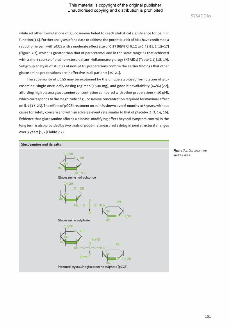
101
SYSADOAs
while all other formulations of glucosamine failed to reach statistical significance for pain or
function [14]. Further analyses of the data to address the potential risk of bias have confirmed a
reduction in pain with pCGS with a moderate effect size of 0.27 (95% CI 0.12 to 0.43) [1, 2, 15–17]
(Figure 7.2), which is greater than that of paracetamol and in the same range as that achieved
with a short course of oral non-steroidal anti-inflammatory drugs (NSAIDs) (Table 7.1) [18, 19].
Subgroup analysis of studies of non-pCGS preparations confirm the earlier findings that other
glucosamine preparations are ineffective in all patients [20, 21].
The superiority of pCGS may be explained by the unique stabilised formulation of glu-
cosamine, single once-daily dosing regimen (1500 mg), and good bioavailability (44%) [22],
affording high plasma glucosamine concentration compared with other preparations (~10 µM),
which corresponds to the magnitude of glucosamine concentration required for maximal effect
on IL-1 [13, 23]. The effect of pCGS treatment on pain is shown over 6 months to 3 years, without
cause for safety concern and with an adverse event rate similar to that of placebo [1, 2, 14, 16].
Evidence that glucosamine affords a disease-modifying effect beyond symptom control in the
long term is also provided by two trials of pCGS that measured a delay in joint structural changes
over 3 years [1, 2] (Table 7.2).
Glucosamine and its salts
Glucosamine hydrochloride
OH
HO
OHO
O
O
=
HO
OH
OOH
Na+Cl–
_ O__S__O_
CH2OH
NH3+Cl–
HOOH
O OH
Patented crystalline glucosamine sulphate (pCGS)
OH
HO
OHO
O
O
=
HO
OH
OOH
_ O__S __O_
Glucosamine sulphate
CH2OH
CH2OH
NH3+ +H3N
CH2OH
Cl–Na+CH2OH
NH3+ +H3N
Figure 7.1 Glucosamine and its salts.
This material is copyright of the original publisher Unauthorised copying and distribution is prohibited

102
Atlas of osteoarthritis
Effect of SYSADOAs versus analgesics on pain outcomes in knee and hip osteoarthritis References Treatment Effect size 95% confidence interval
Zhang et al. 2010 [18] Paracetamol 0.14 0.05–0.23
Bjordal et al. 2004 [19] NSAIDs† 0.32 0.24–0.39
Reginster et al. 2007 [17] and Eriksen et al. 2014 [15]
pCGS 0.27 0.12–0.43
Hochberg et al. 2010 [24] Chondroitin sulphate 0.23 0.11–0.35
Bartels et al. 2010 [5] Diacerein 0.24 0.08–0.39
Table 7.1 Effect of SYSADOAs versus analgesics on pain outcomes in knee and hip osteoarthritis †Short course of treatment for 2–13 weeks; NSAID, non-steroidal anti-inflammatory drug; pCGS, patented crystalline glucosamine sulphate.
Symptom outcomes for patented crystalline glucosamine sulphate (pCGS) in knee osteoarthritis
0.25 0 0.25 0.5 0.75
Favours placebo Favours pCGS
Outcome Effect size (95% CI)†
WOMAC scale
Total 0.33 (0.17–0.49)
Pain 0.27 (0.12–0.43)
Function 0.33 (0.17–0.48)
Lequesne index‡ 0.38 (0.19–0.57)
Test for heterogeneity, I2=0.00
Figure 7.2 Symptom outcomes for patented crystalline glucosamine sulphate (pCGS) in knee osteoarthritis: pooled fixed-model effect size from three pivotal trials. WOMAC, Western Ontario and McMaster Universities Osteoarthritis Index. Adapted from Reginster 2007 [17]. Reproduced from Kucharz et al. 2016 [25] with permission from Taylor & Francis. †Estimates and 95% confidence intervals (CIs) from fixed-model meta-analysis method using the pooled standard deviation in each study/outcome: the data in the table have been depicted as a forest plot in the right-hand panel. ‡Not assessed in one study.
Delay in joint structural changes
TreatmentJSW at enrolment,
mm (mean ± SD)3-year JSN, mm (mean, 95% CI)
Difference between treatment P value
Reginster et al. 2001 [1]
Placebo (n=106) 3.95 ± 1.24 -0.40 (-0.56 to -0.24) 0.33 (0.12 to 0.54) 0.003
pCGS (n=106) 3.82 ± 1.32 -0.07 (-0.22 to 0.07)
Pavelka et al. 2002 [2]
Placebo (n=101) 3.63 ± 1.57 -0.19 (-0.29 to -0.09) 0.23 (0.09 to 0.37) 0.001
pCGS (n=101) 3.89 ± 1.48 +0.04 (-0.06 to 0.14)
Table 7.2 Delay in joint structural changes with patented crystalline glucosamine sulphate (pCGS) treatment over 3 years. CI, confidence interval; JSN, joint space narrowing; JSW, joint space width; SD, standard deviation.
This material is copyright of the original publisher Unauthorised copying and distribution is prohibited

103
SYSADOAs
Chondroitin sulphate
Chondroitin sulphate (CS) is a complex polysaccharide extracted from various animal cartilages,
and thus has a range of molecular weights and different patterns of sulphation (Figure 7.3), which
can affect its chemical properties and biological/pharmacological activities [26]. CS is reported to
elicit anti-inflammatory effects, an increase in type II collagen and proteoglycans, a reduction in
bone resorption and better anabolic/catabolic balance in chondrocytes [26]. Trials conducted with
CS have reported mixed findings, due in part to inclusion of studies using non-pharmaceutical
grade CS [27–29]. Only the pharmaceutical-grade CS has been evaluated for purity, content and
physio-chemical parameters [30]. A Cochrane review including 43 RCTs of 4,962 participants
treated with CS found a small to moderate benefit with an 8-point greater improvement in visual
analogue score (VAS) pain (range 0–100) and a 2-point greater improvement in Lequesne index
(range 0–24) compared with placebo in studies up to 6 months, although most studies were of
low quality, with high heterogeneity between trials (I2 = 70%) [31]. Conversely, in studies that
employed pharmaceutical-grade CS (800 mg), significant improvement in pain and function were
measured after 3–6 months as compared with placebo (p = 0.05) [3, 32]. In addition, pharmaceu-
tical-grade CS (800 mg) has shown similar efficacy to the selective NSAID celecoxib (200 mg/day)
for improvement in pain and function when given daily for 6 months (Figure 7.4) [33].
CS may also offer benefits on joint structure changes in patients with mild to moderate disease.
From pooled results of three clinical trials of 2-year duration, CS was shown to have a small
but significant effect on the rate of decline in minimum joint space width (JSW) of 0.13 mm
(95% CI 0.06 to 0.19; p = 0.0002), corresponding to an effect size of 0.23 (95% CI 0.11 to 0.35;
p = 0.0001) [24]. CS has a good safety profile at doses up to 1200 mg per day [34, 35].
The chemical structure of chondroitin sulphate
COOH
OR3
OHO H2COR1
HN
OO
O
R2O O
CH3
O n
Figure 7.3 The chemical structure of chondroitin sulphate. The chemical structure identifies one unit in a chondroitin sulphate chain. A chondroitin chain can have over 100 individual sugars, each of which can be sulphated in variable positions and quantities. For example, chondroitin-4-sulphate: R1 = H; R2 = SO3H; R3 = H; chondroitin-6-sulphate: R1 = SO3H; R2, R3 = H. [Source: structure of chondroitin sulphate from Wikipedia].
This material is copyright of the original publisher Unauthorised copying and distribution is prohibited

104
Atlas of osteoarthritis
Glucosamine plus chondroitin combination
Glucosamine and CS are often used in combination as dietary supplements even though there
are no published trials of the combination of the two pharmaceutical-grade prescription prepa-
rations. Some studies have described a positive trend for a symptomatic effect of the combina-
tion of CS with glucosamine hydrochloride, with efficacy comparable to that of celecoxib after
6 months in knee OA [34, 36]. There is limited evidence for a reduction in disease progression
with non-pharmaceutical-grade glucosamine sulphate and CS with a reduced loss of cartilage and
reduction in joint space narrowing (JSN) over 2 years [37, 38]. Since both prescription pCGS and
CS are safe and effective medications, it could be advantageous to perform placebo-controlled
RCTs to confirm the clinical benefit of the combination of the two prescription-grade agents
beyond the single agents alone.
Improvement in pain and function with chondroitin sulphate versus celecoxib and placebo
70
60
50
40
30
20
VAS
(adj
uste
d m
ean
± SE
)
Months
0 1 2 3 4 5 6
CS
Celecoxib
Placebop=0.869p=0.429
p=0.159p=0.213 p=0.001
p=0.009
*
*
*
*
CS vs placebo comparison – mixed model p-valueCelecoxib vs placebo comparison – mixed model p-valueMixed model p-value <0.001 for the 3 treatment groups – comparison vs baseline
12
11
10
9
8
7
6
LI (a
djus
ted
mea
n ±
SE)
Months
0 1 2 3 4 5 6
CS
Celecoxib
Placebop=0.714p=0.045 p=0.050
p=0.027p=0.023p=0.015
*
CS vs placebo comparison – mixed model p-valueCelecoxib vs placebo comparison – mixed model p-valueMixed model p-value <0.001 for the 3 treatment groups – comparison vs baseline
*
*
*
A
B
Figure 7.4 Improvement in A) pain (visual analogue scale [VAS]) and B) function (Lequesne index) with chondroitin sulphate (CS) versus celecoxib and placebo.Reproduced from Reginster et al. 2017 [4] with permission from BMJ Publishing Group Ltd.
This material is copyright of the original publisher Unauthorised copying and distribution is prohibited

105
SYSADOAs
Diacerein
Diacerein is an anthraquinone derivative (Figure 7.5) with anti-inflammatory activity against IL-1,
anti-catabolic and pro-anabolic effects on cartilage and the synovial membrane [39]. A Cochrane
review of six RCTs of 1,283 participants indicates that diacerein has a small beneficial effect on
overall pain at 3–36 months, equivalent to a 9% reduction in pain (95% CI -16% to -2%) [40].
In a meta-analysis of 19 studies including 2,637 patients, diacerein was shown to be superior
to placebo and similar to standard treatments (mostly NSAIDs) for effect on pain reduction and
physical function improvement, with a carry-over effect after stopping treatment (Figure 7.6) [41].
From RCTs, diacerein is estimated to have an effect size on pain of 0.24 (95% CI 0.08–0.39) [5].
Limited benefit in delay of joint progression has been reported in hip OA [42], but significant
long-term effects in knee OA are yet to be shown [43]. The safety of diacerein was called into ques-
tion following reports of severe diarrhoea and rare cases of potentially serious hepatotoxicity.
However, a report from the European Medicines Agency (EMA) concluded that the benefit–risk
balance of diacerein remains positive for hip and knee OA in patients aged <65 years [44]. It is
advised that patients start treatment on half the normal dose (i.e. 50 mg daily instead of 100 mg
daily), and should stop taking diacerein if diarrhoea occurs. The use of diacerein is further sup-
ported by a recent ESCEO report, which positions diacerein as a possible background treatment
of OA, of particular benefit in patients with a contraindication to NSAIDs or paracetamol [45].
The chemical structure of diacerein
O O O
O
OHO
O OFigure 7.5 The chemical structure of diacerein [source: structure of diacerein from Wikipedia].
This material is copyright of the original publisher Unauthorised copying and distribution is prohibited

106
Atlas of osteoarthritis
Avocado soybean unsaponifiables (ASU)
ASU is a complex mixture of many natural vegetable extracts taken from avocado and soybean
oils, including fat-soluble vitamins, sterols, triterpene alcohols and furan fatty acids. The identity
of the active component(s) is not known and analysis of commercially-available ASU supplements
demonstrates variation in the sterol content (Figure 7.7) [46]. The sterols contained within ASU
have been shown to have chondroprotective, anabolic and anti-catabolic properties in animal
models [46].
In clinical studies over 3–6 months, some improvement in pain, stiffness and physical func-
tion has been demonstrated with ASU (300 mg/day), leading to a reduced need for analgesics
[47–49]. Mixed results for the effect of ASU on disease progression were found in studies of
2–3 years’ treatment in patients with hip or knee OA [50, 51]. Adverse events affecting the skin,
liver, gastrointestinal system and platelet aggregation have been reported, which raise concerns
about the content and purity of ASU supplements that require further investigation [52].
Effect of diacerein versus placebo and active comparator on pain and physical function
favours comparator
-5 -4 -3 -2 -1 0 1 2 3 4 5
favours diacerein
Jadad-weighted Glass’ standardised mean difference
A
B
diacerein vs placebo, treatment period, pain
diacerein vs placebo, treatment period, function
diacerein vs placebo, dechallenge period, pain
diacerein vs active comparator, treatment period, pain
diacerein vs active comparator, treatment period, function
diacerein vs active comparator, dechallenge period, pain
diacerein vs active comparator, dechallenge period, function
Figure 7.6 Effect of diacerein versus A) placebo and B) active comparator (mostly NSAIDs) on pain and physical function. Standardised mean difference measured at the end of the active treatment period as well as after the treatment-free follow-up period (dechallenge). Error bars indicate 95% confidence intervals, glass’ standardised mean differences >0.8 are commonly regarded as clinically relevant. Adapted from Rintelen 2006 [41]; Reproduced from Pavelka et al. 2016 [45] with permission from Springer Nature.
This material is copyright of the original publisher Unauthorised copying and distribution is prohibited
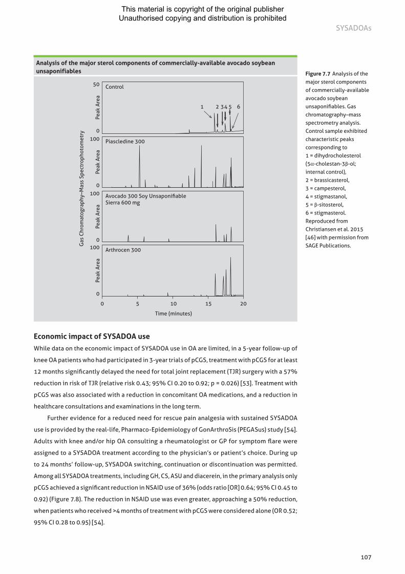
107
SYSADOAs
Economic impact of SYSADOA use
While data on the economic impact of SYSADOA use in OA are limited, in a 5-year follow-up of
knee OA patients who had participated in 3-year trials of pCGS, treatment with pCGS for at least
12 months significantly delayed the need for total joint replacement (TJR) surgery with a 57%
reduction in risk of TJR (relative risk 0.43; 95% CI 0.20 to 0.92; p = 0.026) [53]. Treatment with
pCGS was also associated with a reduction in concomitant OA medications, and a reduction in
healthcare consultations and examinations in the long term.
Further evidence for a reduced need for rescue pain analgesia with sustained SYSADOA
use is provided by the real-life, Pharmaco-Epidemiology of GonArthroSis (PEGASus) study [54].
Adults with knee and/or hip OA consulting a rheumatologist or GP for symptom flare were
assigned to a SYSADOA treatment according to the physician’s or patient’s choice. During up
to 24 months’ follow-up, SYSADOA switching, continuation or discontinuation was permitted.
Among all SYSADOA treatments, including GH, CS, ASU and diacerein, in the primary analysis only
pCGS achieved a significant reduction in NSAID use of 36% (odds ratio [OR] 0.64; 95% CI 0.45 to
0.92) (Figure 7.8). The reduction in NSAID use was even greater, approaching a 50% reduction,
when patients who received >4 months of treatment with pCGS were considered alone (OR 0.52;
95% CI 0.28 to 0.95) [54].
Analysis of the major sterol components of commercially-available avocado soybean unsaponifiables
Control50
0
Peak
Are
a
Piascledine 300100
0
Peak
Are
a
Avocado 300 Soy Unsaponifiable Sierra 600 mg
100
0
Peak
Are
a
Arthrocen 300100
0
Peak
Are
a
Time (minutes)
0 5 10 15 20
1 2 34 5 6
Gas
Chr
omat
ogra
phy–
Mas
s Sp
ectr
opho
tom
etry
Figure 7.7 Analysis of the major sterol components of commercially-available avocado soybean unsaponifiables. Gas chromatography–mass spectrometry analysis. Control sample exhibited characteristic peaks corresponding to 1 = dihydrocholesterol (5α-cholestan-3β-ol; internal control), 2 = brassicasterol, 3 = campesterol, 4 = stigmastanol, 5 = β-sitosterol, 6 = stigmasterol. Reproduced from Christiansen et al. 2015 [46] with permission from SAGE Publications.
This material is copyright of the original publisher Unauthorised copying and distribution is prohibited

108
Atlas of osteoarthritis
References
1 Reginster JY, Deroisy R, Rovati LC, et al. Long-term effects of glucosamine sulphate on osteoarthritis progression: a randomised, placebo-controlled clinical trial. Lancet 2001;357(9252):251-256.
2 Pavelka K, Gatterova J, Olejarova M, et al. Glucosamine sulfate use and delay of progression of knee osteoarthritis: a 3-year, randomized, placebo-controlled, double-blind study. Arch Intern Med 2002;162(18):2113-2123.
3 Kahan A, Uebelhart D, De Vathaire F, Delmas PD, Reginster JY. Long-term effects of chondroitins 4 and 6 sulfate on knee osteoarthritis: the study on osteoarthritis progression prevention, a two-year, randomized, double-blind, placebo-controlled trial. Arthritis Rheum 2009;60(2):524-533.
4 Reginster JL, CONCEPT Investigation Group. CONCEPT provides robust evidence that chondroitin sulfate is superior to placebo and similar to celecoxib in the symptomatic management of osteoarthritis. Ann Rheum Dis 2017.
5 Bartels EM, Bliddal H, Schondorff PK, et al. Symptomatic efficacy and safety of diacerein in the treatment of osteoarthritis: a meta-analysis of randomized placebo-controlled trials. Osteoarthritis Cartilage 2010;18(3):289-296.
6 Bruyere O, Cooper C, Pelletier JP, et al. An algorithm recommendation for the management of knee osteoarthritis in Europe and internationally: a report from a task force of the European Society for Clinical and Economic Aspects of Osteoporosis and Osteoarthritis (ESCEO). Semin Arthritis Rheum 2014;44(3):253-263.
7 McAlindon TE, Bannuru RR, Sullivan MC, et al. OARSI guidelines for the non-surgical management of knee osteoarthritis. Osteoarthritis Cartilage 2014;22(3):363-388.
8 Hochberg MC, Altman RD, April KT, et al. American College of Rheumatology 2012 recommendations for the use of nonpharmacologic and pharmacologic therapies in osteoarthritis of the hand, hip, and knee. Arthritis Care & Research 2012;64(4):465-474.
9 Jordan KM, Arden NK, Doherty M, et al. EULAR Recommendations 2003: an evidence based approach to the management of knee osteoarthritis: Report of a Task Force of the Standing Committee for International Clinical Studies Including Therapeutic Trials (ESCISIT). Ann Rheum Dis 2003;62(12):1145-1155.
10 Reginster JY, Cooper C, Hochberg M, et al. Comments on the discordant recommendations for the use of symptomatic slow-acting drugs in knee osteoarthritis. Curr Med Res Opin 2015;31(5):1041-1045.
11 Hamerman D. The biology of osteoarthritis. N Engl J Med 1989;320(20):1322-1330.
Reduction in the need for NSAID analgesia with SYSADOAs in the Pharmaco-Epidemiology of GonArthroSis (PEGASus) study
Crystalline glucosamine sulphate (primary analysis)
Crystalline glucosamine sulphate (sensitivity analysis)
Glucosamine hydrochloride(primary analysis)
Glucosamine hydrochloride(sensitivity analysis)
Avocado soybean unsaponifiables
Diacerein
Odds ratio (and 95% CI) use
0.45 0.64 0.92
0.54 0.74 1.01
0.81 0.98 1.19
0.91 1.09 1.29
0.77 0.94 1.14
0.82 0.98 1.17
0.87 1.08 1.33
0.4 0.6 0.8 1.0 1.2 1.4
Chondroitin sulphate(all products)
Figure 7.8 Reduction in need for NSAID analgesia with SYSADOAs in the Pharmaco-Epidemiology of GonArthroSis (PEGASus) study. Reproduced from Rovati et al. 2016 [54] with permission from Elsevier.
This material is copyright of the original publisher Unauthorised copying and distribution is prohibited

109
SYSADOAs
12 Rovati LC, Girolami F, Persiani S. Crystalline glucosamine sulfate in the management of knee osteoarthritis: efficacy, safety, and pharmacokinetic properties. Ther Adv Musculoskelet Dis 2012;4(3):167-180.
13 Chiusaroli R, Piepoli T, Zanelli T, et al. Experimental pharmacology of glucosamine sulfate. Int J Rheumatol 2011;2011:939265.
14 Towheed TE, Maxwell L, Anastassiades TP, et al. Glucosamine therapy for treating osteoarthritis. Cochrane Database Syst Rev 2009;(2):CD002946.
15 Eriksen P, Bartels EM, Altman RD, et al. Risk of bias and brand explain the observed inconsistency in trials on glucosamine for symptomatic relief of osteoarthritis: a meta-analysis of placebo-controlled trials. Arthritis Care Res (Hoboken) 2014;66(12):1844-1855.
16 Herrero-Beaumont G, Ivorra JA, Del Carmen Trabado M, et al. Glucosamine sulfate in the treatment of knee osteoarthritis symptoms: a randomized, double-blind, placebo-controlled study using acetaminophen as a side comparator. Arthritis Rheum 2007;56(2):555-567.
17 Reginster JY. The efficacy of glucosamine sulfate in osteoarthritis: financial and nonfinancial conflict of interest. Arthritis Rheum 2007;56(7):2105-2110.
18 Zhang W, Nuki G, Moskowitz RW, et al. OARSI recommendations for the management of hip and knee osteoarthritis: part III: Changes in evidence following systematic cumulative update of research published through January 2009. Osteoarthritis Cartilage 2010;18(4):476-499.
19 Bjordal JM, Ljunggren AE, Klovning A, Slordal L. Non-steroidal anti-inflammatory drugs, including cyclo-oxygenase-2 inhibitors, in osteoarthritic knee pain: meta-analysis of randomised placebo controlled trials. BMJ 2004;329(7478):1317.
20 Runhaar J, Rozendaal RM, Middelkoop MV, et al. Subgroup analyses of the effectiveness of oral glucosamine for knee and hip osteoarthritis: a systematic review and individual patient data meta-analysis from the OA trail bank. Ann Rheum Dis 2017.
21 Reginster JL, Bruyere O, Cooper C. Different glucosamine sulfate products generate different outcomes on osteoarthritis symptoms. Ann Rheum Dis 2017.
22 Setnikar I, Rovati LC. Absorption, distribution, metabolism and excretion of glucosamine sulfate. A review. Arzneimittelforschung 2001;51(9):699-725.
23 Altman RD. Glucosamine therapy for knee osteoarthritis: pharmacokinetic consideratio ns. Expert Rev Clin Pharmacol 2009;2(4):359-371.
24 Hochberg MC. Structure-modifying effects of chondroitin sulfate in knee osteoarthritis: an updated meta-analysis of randomized placebo-controlled trials of 2-year duration. Osteoarthritis Cartilage 2010;18 [Suppl 1]:S28-31.
25 Kucharz EJ, Kovalenko V, Szanto S, et al. A review of glucosamine for knee osteoarthritis: why patented crystalline glucosamine sulfate should be differentiated from other glucosamines to maximize clinical outcomes. Curr Med Res Opin 2016;32(6):997-1004.
26 Martel-Pelletier J, Farran A, Montell E, Verges J, Pelletier JP. Discrepancies in composition and biological effects of different formulations of chondroitin sulfate. Molecules 2015,20(3):4277-4289.
27 Schneider H, Maheu E, Cucherat M. Symptom-modifying effect of chondroitin sulfate in knee osteoarthritis: a meta-analysis of randomized placebo-controlled trials performed with structum®. Open Rheumatol J 2012;6:183-189.
28 Wandel S, Juni P, Tendal B, et al. Effects of glucosamine, chondroitin, or placebo in patients with osteoarthritis of hip or knee: network meta-analysis. BMJ 2010;341:c4675.
29 Rainsford KD. Importance of pharmaceutical composition and evidence from clinical trials and pharmacological studies in determining effectiveness of chondroitin sulphate and other glycosaminoglycans: a critique. J Pharm Pharmacol 2009;61(10):1263-1270.
30 Volpi N. Quality of different chondroitin sulfate preparations in relation to their therapeutic activity. J Pharm Pharmacol 2009;61(10):1271-1280.
31 Singh JA, Noorbaloochi S, MacDonald R, Maxwell LJ. Chondroitin for osteoarthritis. Cochrane Database Syst Rev 2015;1:CD005614.
32 Zegels B, Crozes P, Uebelhart D, Bruyere O, Reginster JY. Equivalence of a single dose (1200 mg) compared to a three-time a day dose (400 mg) of chondroitin 4&6 sulfate in patients with knee osteoarthritis. Results of a randomized double blind placebo controlled study. Osteoarthritis Cartilage 2013;21(1):22-27.
33 Reginster JY, Dudler J, Blicharski T, Pavelka K. Pharmaceutical-grade Chondroitin sulfate is as effective as celecoxib and superior to placebo in symptomatic knee osteoarthritis: the ChONdroitin versus CElecoxib versus Placebo Trial (CONCEPT). Ann Rheum Dis 2017;76(9):1537-1543.
34 Clegg DO, Reda DJ, Harris CL, et al. Glucosamine, chondroitin sulfate, and the two in combination for painful knee osteoarthritis. N Engl J Med 2006;354(8):795-808.
This material is copyright of the original publisher Unauthorised copying and distribution is prohibited

110
Atlas of osteoarthritis
35 Hathcock JN, Shao A. Risk assessment for glucosamine and chondroitin sulfate. Regul Toxicol Pharmacol 2007;47(1):78-83.
36 Hochberg MC, Martel-Pelletier J, Monfort J, et al. Combined chondroitin sulfate and glucosamine for painful knee osteoarthritis: a multicentre, randomised, double-blind, non-inferiority trial versus celecoxib. Ann Rheum Dis 2016;75(1):37-44.
37 Martel-Pelletier J, Roubille C, Abram F, et al. First-line analysis of the effects of treatment on progression of structural changes in knee osteoarthritis over 24 months: data from the osteoarthritis initiative progression cohort. Ann Rheum Dis 2015;74(3):547-556.
38 Fransen M, Agaliotis M, Nairn L, et al. Glucosamine and chondroitin for knee osteoarthritis: a double-blind randomised placebo-controlled clinical trial evaluating single and combination regimens. Ann Rheum Dis 2015;74(5):851-858.
39 Martel-Pelletier J, Pelletier JP. Effects of diacerein at the molecular level in the osteoarthritis disease process. Ther Adv Musculoskelet Dis 2010;2(2):95-104.
40 Fidelix TS, Macedo CR, Maxwell LJ, Fernandes Moca Trevisani V. Diacerein for osteoarthritis. Cochrane Database Syst Rev 2014;2:CD005117.
41 Rintelen B, Neumann K, Leeb BF. A meta-analysis of controlled clinical studies with diacerein in the treatment of osteoarthritis. Arch Intern Med 2006;166(17):1899-1906.
42 Dougados M, Nguyen M, Berdah L, et al. Evaluation of the structure-modifying effects of diacerein in hip osteoarthritis: ECHODIAH, a three-year, placebo-controlled trial. Evaluation of the Chondromodulating Effect of Diacerein in OA of the Hip. Arthritis Rheum 2001;44(11):2539-2547.
43 Pham T, Le Henanff A, Ravaud P, et al. Evaluation of the symptomatic and structural efficacy of a new hyaluronic acid compound, NRD101, in comparison with diacerein and placebo in a 1 year randomised controlled study in symptomatic knee osteoarthritis. Ann Rheum Dis 2004;63(12):1611-1617.
44 European Medicines Agency. Assessment report for diacerein containing medicinal products. EMA/527347/2014. Available from: http://www.ema.europa.eu/docs/en_GB/document_library/Referrals_document/Diacerein/European_Commission_final_decision/WC500173145.pdf [Accessed 25 Sept 2017].
45 Pavelka K, Bruyere O, Cooper C, et al. Diacerein: Benefits, Risks and Place in the Management of Osteoarthritis. An Opinion-Based Report from the ESCEO. Drugs Aging 2016;33(2):75-85.
46 Christiansen BA, Bhatti S, Goudarzi R, Emami S. Management of Osteoarthritis with Avocado/Soybean Unsaponifiables. Cartilage 2015;6(1):30-44.
47 Blotman F, Maheu E, Wulwik A, Caspard H, Lopez A. Efficacy and safety of avocado/soybean unsaponifiables in the treatment of symptomatic osteoarthritis of the knee and hip. A prospective, multicenter, three-month, randomized, double-blind, placebo-controlled trial. Rev Rhum Engl Ed 1997;64(12):825-834.
48 Maheu E, Mazieres B, Valat JP, et al. Symptomatic efficacy of avocado/soybean unsaponifiables in the treatment of osteoarthritis of the knee and hip: a prospective, randomized, double-blind, placebo-controlled, multicenter clinical trial with a six-month treatment period and a two-month followup demonstrating a persistent effect. Arthritis Rheum 1998;41(1):81-91.
49 Appelboom T, Schuermans J, Verbruggen G, Henrotin Y, Reginster JY. Symptoms modifying effect of avocado/soybean unsaponifiables (ASU) in knee osteoarthritis. A double blind, prospective, placebo-controlled study. Scand J Rheumatol 2001;30(4):242-247.
50 Lequesne M, Maheu E, Cadet C, Dreiser RL. Structural effect of avocado/soybean unsaponifiables on joint space loss in osteoarthritis of the hip. Arthritis Rheum 2002;47(1):50-58.
51 Maheu E, Cadet C, Marty M, et al. Randomised, controlled trial of avocado-soybean unsaponifiable (Piascledine) effect on structure modification in hip osteoarthritis: the ERADIAS study. Ann Rheum Dis 2014;73(2):376-384.
52 Olivier P, Montastruc JL. Reseau français des centres regionaux de p; [Post-marketing safety profile of avocado-soybean unsaponifiables]. Presse Med 2010;39(10):e211-216.
53 Bruyere O, Pavelka K, Rovati LC, et al. Total joint replacement after glucosamine sulphate treatment in knee osteoarthritis: results of a mean 8-year observation of patients from two previous 3-year, randomised, placebo-controlled trials. Osteoarthritis Cartilage 2008;16(2):254-260.
54 Rovati LC, Girolami F, D’Amato M, Giacovelli G. Effects of glucosamine sulfate on the use of rescue non-steroidal anti-inflammatory drugs in knee osteoarthritis: results from the Pharmaco-Epidemiology of GonArthroSis (PEGASus) study. Semin Arthritis Rheum 2016;45(Suppl 4S):S34-S41.
This material is copyright of the original publisher Unauthorised copying and distribution is prohibited

This material is copyright of the original publisher Unauthorised copying and distribution is prohibited