atlas of union chapel mine fossil...
TRANSCRIPT

339Buta, R. J., Rindsberg, A. K., and Kopaska-Merkel, D. C., eds., 2005, Pennsylvanian Footprints in the Black Warrior Basin of Alabama.Alabama Paleontological Society Monograph no. 1
ATLAS OF UNION CHAPEL MINE FOSSIL PLANTS
DAVID L. DILCHER and TERRY A. LOTTFlorida Museum of Natural History, University of Florida, Gainesville, Florida 32611-7800, USA
This Plant Atlas is a compilation of photographs of the fossil plants that have been made available to us fromthe Union Chapel Mine, now the Steven C. Minkin Paleozoic Footprint Site. These specimens include those thatwere donated to the Paleobotanical Collection at the Florida Museum of Natural History (UF specimen numbers)and those photographed at “Track Meet 3 and Plant Fest”, a meeting held in Anniston, Alabama on May 12,2001. The UCM-P numbers indicate specimens shown to DLD by private collectors that were photographed atthis meeting and retained by the collectors. This Plant Atlas is an addition to the chapter on the Fossil Plants fromthe Union Chapel Mine, Alabama by Dilcher, Lott and Axsmith published in this monograph. Here we illustratemany more specimens than shown in the chapter on fossil plants including some plant fossils that are not illus-trated or mentioned in the chapter.
Often a pictorial atlas is more useful and more appealing to both amateur and professional paleontologists.We present 112 pictures of the Union Coal Mine plants in 23 plates. A scale or scale bar (millimeter units) isincluded for each individual figure. A legend is prepared for each of the plates so that the specimens can be easilyreferenced. However the text and references given in Dilcher et al., (2005) should be consulted for more completeinformation about any specific specimen or species illustrated. This Plant Atlas should not be considered as adefinitive list or compendium of plants from the Union Coal Mine, but as a working document illustrating thosespecies known thus far. What is illustrated here should represent the more common species that occur in the roofshales of the mine. If in the future new species, not illustrated here, were found, it would be helpful if these couldbe included in the collection.
REFERENCE
Dilcher, D., Lott, T. A., and Axsmith, B. J., 2005, Fossil plantsfrom the Union Chapel Mine, Alabama; in Buta, R. J.,Rindsberg, A. K. and Kopaska-Merkel, D. C., eds., Pennsyl-vanian Footprints in the Black Warrior Basin of Alabama:Alabama Paleontological Society Monograph no. 1, p. 153-168.
AUTHORS’ E-MAIL ADDRESSES
David L. Dilcher: [email protected] A. Lott: [email protected]

340

341
Plate 117. Figure 1: Lepidodendron obovatum UF 34008, leaf cushions on branch or young trunk of the tree.Figures 2-4: Lepidodendron aculeatum Fig. 2 and 4, UCM-P 173; Fig. 4 is an enlargement of Fig. 2 showing thedetails of the leaf cushions and leaf attachment area often still containing carbon residue; Fig.3, UF 34014. Allspecimens represent Lepidodendron branches or trunks not yet decorticated.

342
Plate 118. Figure 1, 2, 3 Lepidophloios laricinus Fig. 1, UCM-P 155; Fig. 2, UCM-P 159; Fig 3, UCM-P 160.Fig. 2 shows the typical wide triangular-shape of Lepidophloios leaf cushion with the leaf scar in the lower half ofthe cushion. Immediately above the leaf scar is a ligule scar. Fig. 3 is orientated sideways. Figure 4: Lepidodendronobovatum UCM-P 015 larger axis not yet decorticated showing leaf cushions.

343
Plate 119. Figures 1, 3, 4: Lepidophloios laricinus Fig. 1, UF 34371; Fig. 3, UCM-P 172; Fig. 4, UCM-P 180.Figs. 1 and 3 show the typical laterally elongated diamond-shape of the leaf cushions of Lepidophloios. Fig. 3shows the nature of the overlapping leaf cushions. Figure 4 illustrates this species in a slightly decorticatedcondition in which only the outer most bark layer is lost. This might be considered by the name Aspidaria. Figure2: Sigillaria elegans UCM-P 165. More or less isodiametric leaf scars. In several scars the central vascular leaftrace, bordered on either side by cleft-shaped parichnos scars, can be seen. Rarely the ligule scar at the top of theleaf scar can be seen.

344
Plate 120. Figure 1-4: Lepidodendron lycopodioides Fig. 1 and 2, UCM-P 221, Fig. 2 is an enlargement of Fig.1; Fig. 3, UCM-P 163; Fig. 4, UCM-P 224. Terminal branches of a Lepidodendron tree showing their elongatenature, dichotomous branching and attached leaves.

345
Plate 121. Figure 1: Aspidiopsis sp. UF 34013. Deeply decortified layer or internal cast of the innermost layer ofthe tough and thick outer cortex of a Lycopod tree or large branch. The scars are produced by the steeply archingvascular tissue for the numerous leaves that transverse this tissue from the inner vascular cylinder to the outersurface of the tree. Figure 2: Calamites sp UF 33985. Poorly preserved pith cast of a Calamites stem or branch.Figure 3: Cyclopteris sp. UF 34046´. A vegetative specialized leaf that often occurs at the base of a larger frondor leaf of seed ferns. This is some what like a stipule in flowering plants. Figure 4: Branch UCM-P 200. A stemor branch cast of undetermined affinities. Perhaps fern or seed fern in nature.

346
Plate 122. Figure 1, 3: Lepidostrobus sp. B Fig. 1, UF 34375; Fig. 3, UF 34042. Casts of lycopod cones showingthe leaf-like overlapping sporophylls in broken, face on position and sometimes the hollow areas between themwhere the sporangia were located can be seen (esp. in Fig 3). Figure 2, 4: Lepidostrobus sp. A Fig. 2, UF 33933’;Fig. 4, UF 33993. Part and counterpart of a lycopod cone compression showing the overlapping sporophylls andtheir leaf-like terminal tips along the cone margins.

347
Plate 123. Figure 1, 5: Lepidostrobus sp. A Fig. 1, UF 34007; Fig. 5, UF 34372. Compressions of lycopod conesshowing overlapping sporophylls. Figure 2: Sigillariostrobus quadrangulatus UF 34367. Conpression of a coneof Sigillaria. It has the typical angular pattern. Figure 3: Lepidostrobus sp. B UF 34365. Compression of thebase of a cone or portion of a broken cone showing the broad nature of the sporophyll lamina as it extends past thesporangia and how they taper to a leaf-like tip. This is very much like the compression of what is named Lepidocarponwhen it is found petrified. This means the cone was female and produced megaspores. Figure 4, 6: Lepidodendronlycopodioides Fig. 4, UCM-P; Fig. 6, UCM-P 219. Near terminal branches of lycopod trees.

348
Plate 124. Figure 1-6: Lepidostrobophyllum cf majus Fig. 1, UF 34374; Fig. 2, UF 34369a; Fig. 3, UCM-P 154,an enlargement of Figure 6; Fig. 4, UF 34374; Fig. 5, UF 34377; Fig. 6, UCM-P 154; Fig. 7, UCM-P 153.Compressions of the typical dispersed sporophylls.

349
Plate 125. Figure 1-4: Syringodendron sp. Fig. 1, UF 34379; Fig. 2, UF34016; Fig. 3, UCM-P 162; Fig. 4,UCM-P. Fig. 1 is an internal cast of a small Sigillaria stem or branch. Figs. 2 and 3 clearly show the parichnosscars that formed from the thin walled tissue important in allowing the transport of oxygen through the thickperiderm layers to the inner tissues of the stems and few branches of the sigillarian trees. Fig. 4, decorticated stemof Sigillaria.

350
Plate 126. Figure 1-4: Asterophyllites charaeformis Fig. 1, 2, UF 34373a, Fig. 2 is an enlargement of upperportion of the branch shown in Figure 1.; Fig. 3, UF34010; Fig. 4, UMC-P. Typical compressions of axes andbranching axis (Figs.1 and 2) of the Calamites tree. Note that at each node there are whorled leaves arranged atnearly right angles to the stems. These are latteral branches to the calamitean tree. Figure 5: Asterophyllites sp.UCM-P 218. Compression of a different species of a leafy branch of a calamitean tree. Note the numerouswhorled leaves and the narrow elongate nature of the individual leaves.

351
Plate 127. Figure 1: Calamites undulatus UF 34047. Pith cast of Calamites showing one node. The longitudinalgrooves may be formed from the deep extensions of the woody tissue into the pith area of the stem. Figures 2-4, 6:Calamites suckowii Fig. 2, UF 34043; Fig. 3, UF 34019; Fig. 4 is an enlargement of one node of Fig. 3. Fig. 6,UF 34366. Pith casts and portions of pith casts showing typical nodes and ribbing of the casts. Fig. 4 shows thebranch scars located just above the node while the node shows the alternating pattern of the primary xylem (firstformed wood) that occurs at each node (also seen in Fig. 6). There are 14 nodes shown in the internal pith castshown in Fig. 2. Figure 5: Calamites goepperti UF 33994. Compression of a calamitean stem or branch showingnumerous nodes at which leaves (perhaps Asterophyllites) are attached and large branch scars located just abovethe nodes but occupying most of the internodal area of the stem.
352
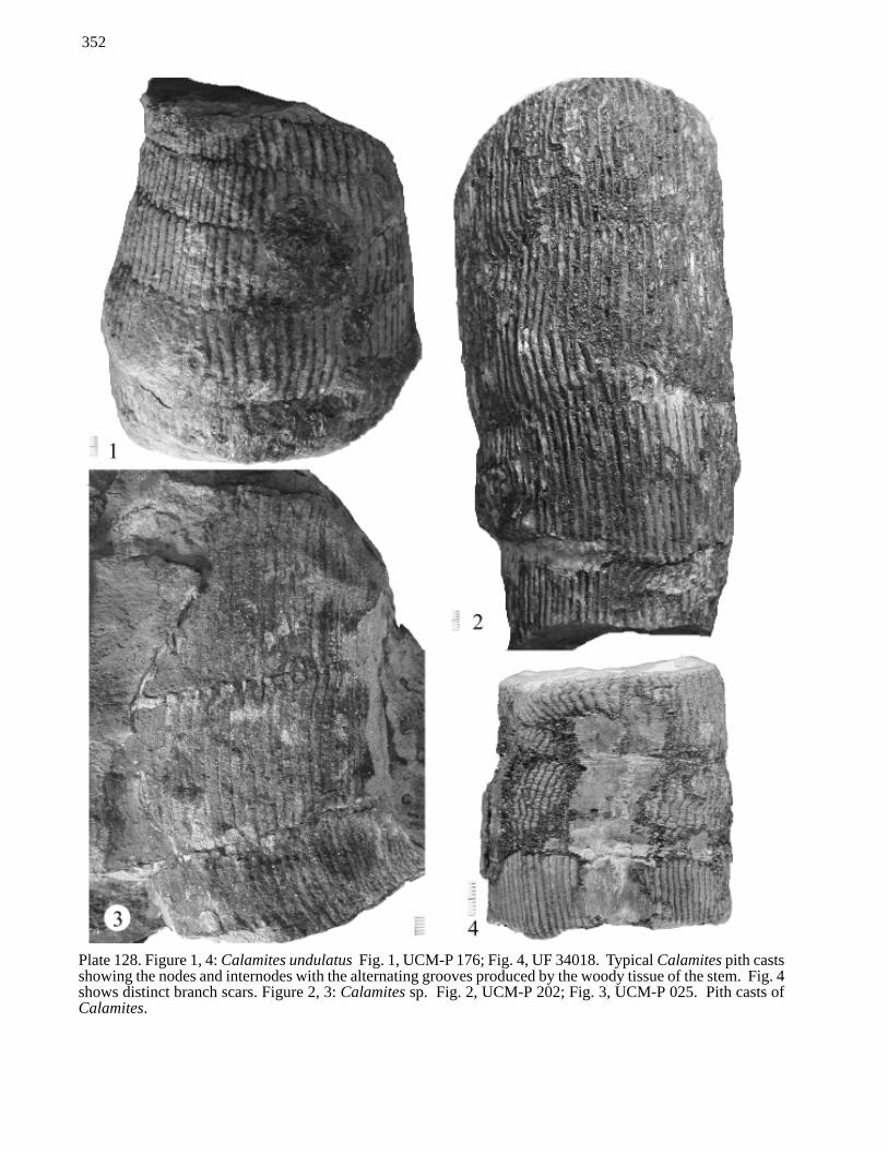
Plate 128. Figure 1, 4: Calamites undulatus Fig. 1, UCM-P 176; Fig. 4, UF 34018. Typical Calamites pith castsshowing the nodes and internodes with the alternating grooves produced by the woody tissue of the stem. Fig. 4shows distinct branch scars. Figure 2, 3: Calamites sp. Fig. 2, UCM-P 202; Fig. 3, UCM-P 025. Pith casts ofCalamites.

353
Plate 129. Figure 1-4: Calamites goepperti Fig. 1, UF 36866; Fig. 2, UF 48553; Fig. 3, UCM-P 201; Fig. 4, UF33992. Compression fossils of the stems or branches of Calamites showing leaf scars at every node and numerousbranch scars located above the node at only at a few select nodes. It is interesting to note that some species ofCalamites have been characterized by their branching patterns and the number of nodes between the sets ofbranches. Fig. 3 clearly show that branching is either rare or spaced at every 12 node along the stem or branch.
354
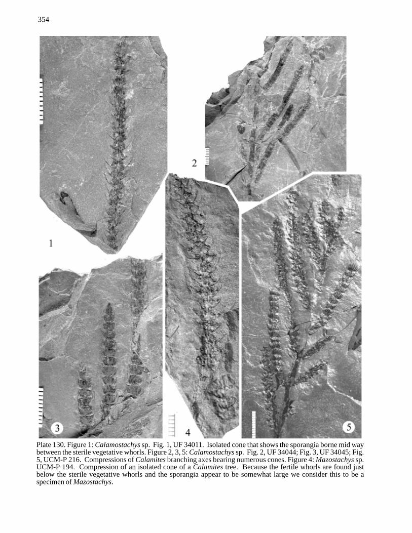
Plate 130. Figure 1: Calamostachys sp. Fig. 1, UF 34011. Isolated cone that shows the sporangia borne mid waybetween the sterile vegetative whorls. Figure 2, 3, 5: Calamostachys sp. Fig. 2, UF 34044; Fig. 3, UF 34045; Fig.5, UCM-P 216. Compressions of Calamites branching axes bearing numerous cones. Figure 4: Mazostachys sp.UCM-P 194. Compression of an isolated cone of a Calamites tree. Because the fertile whorls are found justbelow the sterile vegetative whorls and the sporangia appear to be somewhat large we consider this to be aspecimen of Mazostachys.

355
Plate 131. Figure 1-3: Alethopteris valida Fig. 1, UF 34037; Fig. 2, UF 34037´; Fig. 3, UF 34036. These figuresshow compression material of partial leaves or fragments of multiply compounded leaves of seed ferns. Thepinnae are opposite. The ultimate pinnules are broadly attached to the rachis and opposite, typical of Alethopterisvalida. This may have been the foliage of a Medullosa tree that bore the seeds and pollen organs shown on thefollowing plates. Figure 4: Neuralethopteris biformis UCM-P 184. Compression specimen of a portion of acompound seed fern leaf. This leaf probably was 2 or 3 times compound and here are three pinnae that representparts of the same leaf. These pinnae have characteristic pinnules of Neuralethopteris alternating along them.This may have been the foliage of a Medullosa tree that bore the seeds and pollen organs shown on the followingplates.

356
Plate 132. Figure 1: Sphenopteris elegans UCM-P 169. Compression of the mid section of a seed fern leaf of atleast a fourth order (as shown here) compound leaf. The leaf may have had one or two more orders of compound-ing not shown here. Figure 2, 3: Sphenopteris pottsvillea Fig. 2, UF 34033; Fig. 3, UF 36875. Compressionspecimens of fern leaf fragments. These may have belonged to a fern-like plant or to an extinct seed fern-typeplant.
357
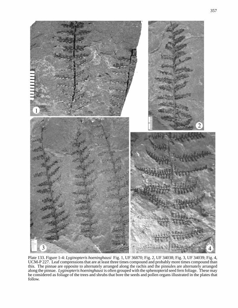
Plate 133. Figure 1-4: Lyginopteris hoeninghausi Fig. 1, UF 36870; Fig. 2, UF 34038; Fig. 3, UF 34039; Fig. 4,UCM-P 227. Leaf compressions that are at least three times compound and probably more times compound thanthis. The pinnae are opposite to alternately arranged along the rachis and the pinnules are alternately arrangedalong the pinnae. Lyginopteris hoeninghausi is often grouped with the sphenopterid seed fern foliage. These maybe considered as foliage of the trees and shrubs that bore the seeds and pollen organs illustrated in the plates thatfollow.

358
Plate 134. Figure 1, 3, 4-7: Neuralethopteris biformis Fig. 1, UF 34024; Fig. 3, UF 34027; Fig. 4, UCM-P; Fig.5, UF 34028; Fig. 6, UF 34029; Fig. 7, UF 34023. Compressions of the ultimate portions and isolated pinnulesof seed fern foliage. This species is characterized by the distinctive pinnule venation and large lateral pinnuleswith rounded distinct bases, each attached to the pinnae independently. The ultimate terminal pinnule is elongate.Figure 2: Neuralethopteris pocahontas UF 34025. Compression of a seed fern pinnae bearing alternate, ovatepinnules that narrow at their base and the terminal pinnule is narrow, oblong with basal lobes.

359
Plate 135. Figure 1: Neuralethopteris biformis Fig. 1, UCM-P 158. Compression of whole or partial, 4 pinnaewith numerous pinnules. Foliage of seed fern plants such as Medullosa. Figure 2: Neuralethopteris sp. UF34350. Compression of a single pinnae with several pinnules. Probably of seed fern origin. Figure 3:Neuralethopteris pocahontas UF 34022. Compression of 4 partial pinnae with numerous pinnules. Foliage ofseed fern plants such as Medullosa.

360
Plate 136. Figure 1: Aphlebia sp. UCM-P 220. Compression of ornate foliage-like material often associated withseed fern foliage. Probably attached directly to the main rachis of a large leaf. Figure 2: Palmatopteris furcataUF 34031. Impression of an ultimate pinnule of a fern or seed fern leaf showing a distinctive branching pattern.Figure 3: Neuralethopteris pocahontas UF 34035. Compression/ impression of numerous pinnae attached to arachis. Each bearing numerous pinnules. Folliage probably at least 3 X compound and belonging to seed fernplants such as Medullosa. Figure 4 Trunk UCM-P. Cast of a trunk or large rachis perhaps of fern or seed fernorigin.

361
Plate 137. Figure 1-9: Whittleseya elegans Fig. 1, UF 36901; Fig. 2, UF 36890; Fig. 3, UF 36873; Fig. 4, UF36896; Fig. 5, 34364; Fig. 6, UF 36891; Fig. 7, UF 34364´; Fig. 8, UF 36883´; Fig. 9, UF 36908. Compressionsand impressions of pollen organs of a seed fern such as Medullosa. Note the long narrow tubes that give a linearstriation appearance to the pollen organs. Each of these tubes was filled with pollen and it was shed in greatabundance as these hung from the leaves of a Medullosa plant. Fig. 7 shows a short attachment stock.

362
Plate 138
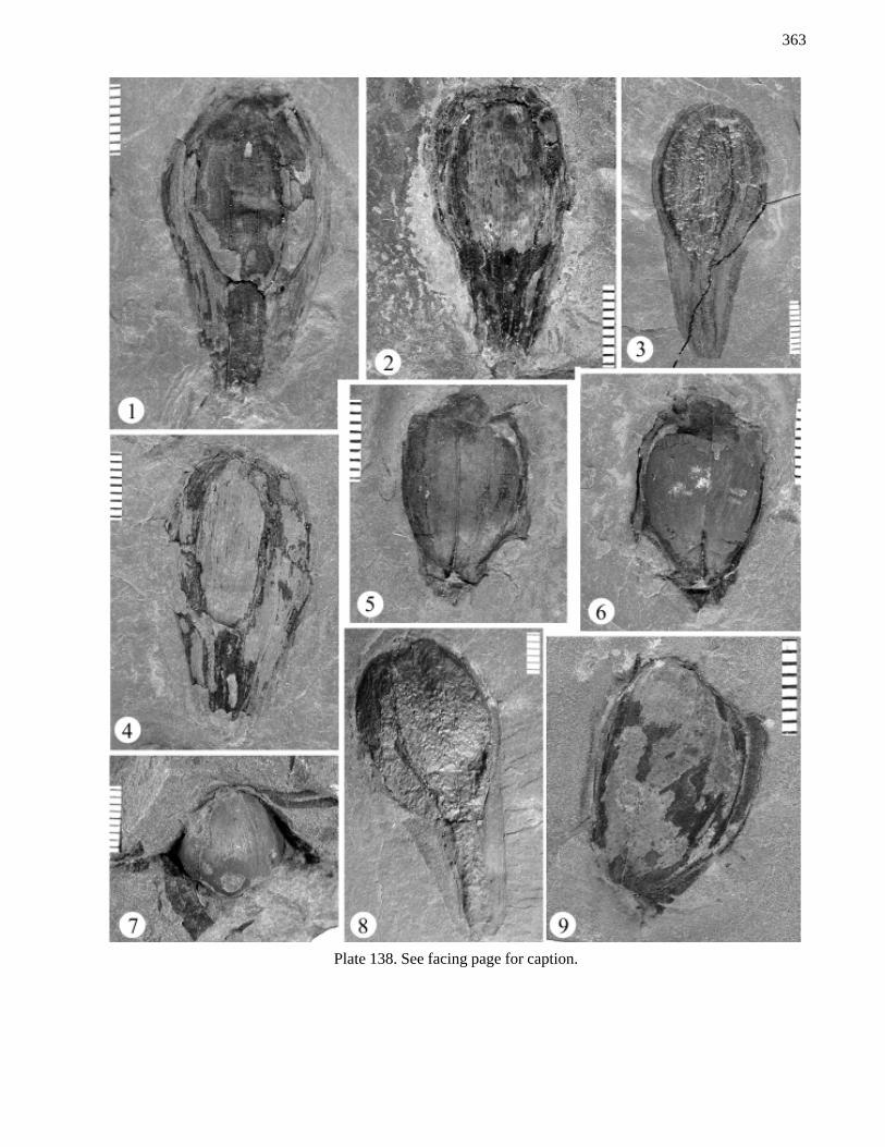
Figure 1-4, 8: Trigonocarpus ampulliforme Fig. 1, UF36879; Fig. 2, UF 36877; Fig. 3, UF 34362; Fig. 4, UF36903; Fig. 8, UCM-P. Compression - impression mixed of the seeds and Medullosa and Medullosa - like plants.These fossils have preserved some aspects of the softer tissues of the outer parts of this large seed. The long“neck” of tissue surrounding the micropylar area can be seen. In Fig. 3 the nature of the outer and inner tissues inthis micropylar extension can be seen. The center is almost a compressed cast of the chamber where the living partof the seed was contained. Fig. 8 shows a cast of this inner part of the seed also showing one of the ribs and theinner area of the micropylar opening as well as a compression of the tissues surrounding it. These seeds representsome of the larger seeds known from the Pennsylvanian.
Figure 5, 6, 9: Trigonocarpus sp. Fig. 5, UF 34040´; Fig. 6, UF 34040; Fig. 9, UF 34376. Internal casts of theseeds of Medullosa. The name comes from the three ribbed aspect of casts isolated from the matrix. These ribscome from the “ribs” associated with the position of the strands of vascular tissue. These casts reflect the size andshape of the internal chambers of the seeds occupied by the female gametophyte tissue (stored food) and embryo.These seeds represent some of the larger seeds known from the Pennsylvanian. Figure 8 and 9 are mixtures of castand compression preservation.
Figure 7: Carpolithes sp. UF 34041. A partial cast of a seed perhaps coming from a seed fern. It is an internalcast of the proximal 1/3 of the seed. It appears to have 2 or the 3 ribs showing and is perhaps a Trigonocarpussimilar to the casts shown in figs 5,6, and 9. It is especially interesting to note that Neuralethopteris foliageappears attached to one end and is closely associated with the cast along the left side of it. This might be showingattachment of this seed to a leaf with this type of foliage, but is not exposed sufficiently to prove this possibility.
363
Plate 138. See facing page for caption.

364
Plate 139
Figure 1: Cordaites sp. UF 33989, bar = 3 cm. Compression of a partial leaf of the Cordaites tree. This showsmultiple veins with a central vein more prominent.
Figure 2: Lepidophylloides intermedium UF 34378. Compression of a lycopod leaf that clearly shows the thincentral vein with 2 conspicuous lateral stomatal grooves on either side. The width of this leaf suggests thepossibility that it could have affinities with Sigillaria (thus be a Sigillariophyllum) or it could represent a niceexample of a Lepidodendron or Lepidophloios leaf.
Figure 3, 5: Holcospermum sp. Fig. 3, UCM-P; Fig. 5, UF 34370b. Compressions of seeds from a seed fern.Note the numerous ribs that can be seen in these seeds.
Figure 4: Cordaicarpon sp. UF 34368. Heart-shaped impression somewhat similar to Cordaicarpon which is theseed of a Cordaites. Not all of the details of the seed are evident and this structure could be a pair of cone scalesor bracts of a seed plant.
Figure 6: Sphenophyllum sp. UCM-P 215. Compression of a stem with whorls of wedge-shaped leaves.Sphenophyllum was a common vein-like plant that covered the damp forest floor of the Pennsylvanian swamps. Itis a Sphenopsid and produced complex cones bearing numerous spores.

365
Plate 139. See facing page for caption.