atomic force microscopy investigation of vaccinia virus...
TRANSCRIPT

JOURNAL OF VIROLOGY, Aug. 2008, p. 7551–7566 Vol. 82, No. 150022-538X/08/$08.00�0 doi:10.1128/JVI.00016-08Copyright © 2008, American Society for Microbiology. All Rights Reserved.
Atomic Force Microscopy Investigation of Vaccinia Virus Structure�
Y. Kuznetsov, P. D. Gershon,† and A. McPherson†*Department of Molecular Biology and Biochemistry, University of California—Irvine, Irvine, California 92697
Received 3 January 2008/Accepted 21 May 2008
Vaccinia virus was treated in a controlled manner with various combinations of nonionic detergents,reducing agents, and proteolytic enzymes, and successive products of the reactions were visualized usingatomic force microscopy (AFM). Following removal of the outer lipid/protein membrane, a layer 20 to 40 nmin thickness was encountered that was composed of fibrous elements which, under reducing conditions, rapidlydecomposed into individual monomers on the substrate. Beneath this layer was the virus core and itsprominent lateral bodies, which could be dissociated or degraded with proteases. The core, in addition to thelateral bodies, was composed of a thick, multilayered shell of proteins of diverse sizes and shapes. The shell,which was readily etched with proteases, was thoroughly permeated with pores, or channels. Prolongedexposure to proteases and reductants produced disgorgement of the viral DNA from the remainders of thecores and also left residual, flattened, protease-resistant sacs on the imaging substrate. The DNA was readilyvisualized by AFM, which revealed some regions to be “soldered” by proteins, others to be heavily complexedwith protein, and yet other parts to apparently exist as bundled, naked DNA. Prolonged exposure to proteasesdeproteinized the DNA, leaving masses of extended, free DNA. Estimates of the interior core volume suggestmoderate but not extreme compaction of the genome.
Vaccinia virus, the best-characterized member of the Poxviri-dae family, is unusual among most double-stranded DNA vi-ruses in that both transcription and replication occur in thecytoplasm of the host cell (5, 8, 24). The 191-kbp genome ofthe Copenhagen strain contains up to 266 open reading framesencoding proteins �65 amino acids in length (9, 15), with 63 to80 vaccinia virus gene products being packaged in the virion (4,53, 57, 60). Replication takes place within discrete cytoplasmicviral factories that are thought to be devoid of cellular or-ganelles (3) but which contain ribosomes (16). Infectious viri-ons can be isolated in several forms, depending on their degreeof maturation, including mature virus (MV), wrapped virus,and enveloped virus (25).
Many general features of vaccinia virion architecture arereadily apparent from a variety of studies (3), notably a ge-nome/enzyme-containing proteinaceous core, proteinaceouslateral bodies (LBs), and surrounding surface tubular elementsand lipid membrane. Nonetheless, despite �60 years of studyby conventional electron microscopy (EM) (11, 14, 15, 45, 48,54, 58), cryoelectron microscopy (1, 9, 54), and more recentlycryoelectron tomography (4) freeze etch (31), deep etch (11),and atomic force (22) microscopies, specific controversies re-main in issues such as the intrinsic rigidity of the core, thedegree of order in genome packaging, the degree and affinityof the association of the genome with protein in the packagedstate, the presence/absence of an “inner membrane” or “pali-sade” enwrapping the core, and the number of lipid bilayersthat enwrap the intact virion. Structural details of the virion arerelevant to several stages of the vaccinia life cycle, including
mechanisms of fusion and entry, extrusion of early mRNAfrom the infecting virion, disassembly of the infecting virion,genome packaging into nascent virions during assembly, andvirion maturation/membrane enwrapment.
A persistent drawback with EM images, irrespective of thetechnique applied, is that they generally provide two-dimen-sional projections of specimens onto a single plane. Three-dimensional information is usually absent, and approximationscan be recovered only via reconstruction. Thus, ambiguitiesand uncertainties arise in the assignment of features to internalstructures and surfaces. In addition, various artifacts may beintroduced, particularly via the fixing and staining proceduresgenerally employed to enhance contrast. Atomic force micros-copy (AFM) complements EM in a number of ways. Althoughthe resolution of AFM is theoretically lower than that of EM,it often reveals details of soft, biological specimens, includingviruses (20), that cannot otherwise be obtained. The three-dimensionality of specimens can easily be preserved duringimaging, and instead of the specimen being projected onto aplane, topographical features of a specimen’s surface are re-vealed. This might appear to limit the utility of the techniquefor delineating the internal structures of specimens such ascomplex viruses, but these drawbacks can be largely overcome.Viruses, as we have now shown in several investigations (32, 35,38–40, 42–44, 52), can be broken down or disassembled incontrolled ways to reveal, sequentially, internal layers. Bychemical and enzymatic dissection of virions accompanied byAFM visualization of the products, information regarding in-ternal structures can be revealed. AFM is a single-particleanalytical method that does not rely on a homogeneous pop-ulation (as would, for example, cryoelectron microscopy recon-struction). It records not only the structures and states com-mon within a population but also, with equivalent accuracy,features found in just a few individuals. AFM can be appliedwith equal ease to dried specimens or specimens fully hydrated
* Corresponding author. Mailing address: University of California,Irvine, Department of Molecular Biology and Biochemistry, 560 Stein-haus Hall, Irvine, CA 92697-3900. Phone: (949) 824-1931. Fax: (949)824-1954. E-mail: [email protected].
† These two authors contributed equally to this work.� Published ahead of print on 28 May 2008.
7551
on June 1, 2018 by guesthttp://jvi.asm
.org/D
ownloaded from

in physiological media. AFM records topography irrespectiveof the electron density.
We have previously established, as a proof of principle, theamenability of vaccinia virus to AFM analysis (22). Here wereport a more in-depth AFM analysis of vaccinia virus that webelieve clarifies some significant structural questions and pro-vides additional, general details. The images presented hereare, we believe, a significant improvement over those shown inthe previous study (22) in terms of resolution and populationcoverage within individual preparations. We have focused inparticular upon the virion core and LBs, along with aspects ofthe packaged genome, such as its degree of order and associ-ation with protein and its appearance when released fromvirion cores. We focus exclusively on MV released from mam-malian cells using standard protocols. We have imaged MV instandard buffers akin to those used for storage and infectionand have also treated it with a number of vigorous reagents andreagent combinations.
MATERIALS AND METHODS
Sample preparation and AFM analysis. The virus preparation described else-where (22) was used throughout the current study, at the titer described previ-ously. Intact and treated viruses were imaged on glass coverslips or freshlycleaved mica coated with poly-L-lysine. Genomic DNA was imaged on freshlycleaved, uncoated mica. For virus imaging, 1.5 �l of 50-fold-diluted freshlyresuspended MV in 10 mM Tris (pH 7.8) was deposited on the substrate andallowed to sediment for 30 min at room temperature. For imaging in air, thespecimen, on the substrate, was rinsed multiple times with distilled water andthen dried under a stream of N2 gas. Prior to imaging under liquid, specimenswere treated with a solution of 5% glutaraldehyde in phosphate-buffered saline(PBS) for 15 min and then rinsed with distilled water. Fixation with glutaralde-hyde has been shown in previous investigations to not perturb the surface struc-tures of virus particles within the resolution of the AFM technique (32, 34, 35,39, 40).
For enzyme digestion experiments, after deposition of the intact specimenonto the imaging substrate, the substrate was rinsed multiple times with PBS andexcess PBS was wicked away with torn filter paper. After addition of 15 �l ofenzyme solution, the substrate was sealed in a plastic cell (to minimize evapo-ration), followed by incubation either in an incubator at 37°C or at room tem-perature. For sequential treatments with different reagents, the specimen on thesubstrate was rinsed with PBS after removal of the first reagent before additionof the second.
Imaging procedures were essentially as previously described for other viruses(32, 34, 40, 42, 44) and RNA (33, 37), with use of a Nanoscope III multimodeAFM instrument (Veeco Instruments, Santa Barbara, CA) calibrated to smalllateral distances by imaging the 111 face of a thaumatin protein crystal as astandard, based on the known lattice spacings (19, 21). After the sample sub-strates were placed in a 75-�l fluid cell, images were acquired at 26°C in tappingmode (17, 18) using commercially available silicon tips (imaging under air) oroxide-sharpened silicon nitride tips (imaging under fluid) at a scan frequency of1 Hz and an oscillation frequency of 9.2 kHz (under fluid) or 300 kHz (in air). Inthe AFM images presented here, increasing height above the substrate is indi-cated by an increasingly lighter color. Due to the distortion of lateral distancesdue to convolution of the cantilever tip shape and the surface features scanned,quantitative measures of size were based either on heights above the substrate oron center-to-center distances between particles.
RESULTS
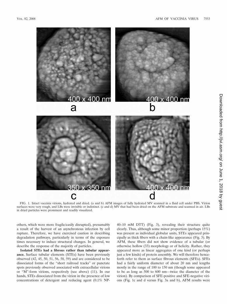
The intact virion. The shape, dimensions, and general sur-face characteristics of hydrated MV imaged by AFM (Fig. 1aand b) were as previously reported (22), with the virus assum-ing a familiar rectangular (“brick”) shape upon dehydration(Fig. 1c and d). Particles were pleomorphic, with an outersurface design varying from the more regular, short, haphaz-ardly intersecting “railroad tracks” topography of naturally
cell-extruded virus (see reference11 and references therein) toa more punctate topography, referred to in the early literatureas the “M” or “mulberry” form of the virion (7, 25, 47, 58),which may be more diagnostic of intracellular virus released bycell rupture (10, 11). Parenthetically, the virion surface alsoclosely resembled the external surfaces of various animal cellsas imaged by AFM (34, 36, 39, 40), consistent with a compa-rable array of membrane proteins anchored in a lipid bilayer,perhaps with an underlying membrane skeleton.
One LB (or very occasionally both) was discernible attachedto hydrated specimens (Fig. 1 and 2). As observed previously(22), the profile of the LBs visibly associated with every particlebecame greatly accentuated upon virion dehydration (Fig. 1and 2), and a deep trench formed around the visible LBs whichclearly delineated them with respect to the background viralsurface. LBs were about one-third the overall dimensions ofdried intact MV (albeit the height could not be measured) butless rectangular in shape.
Virions in both hydrated and dehydrated preparations werefrequently found to be aggregated with one another (a well-known property of vaccinia virus [17]), albeit isolated virionswere also frequently observed. The isolated virions permittedobservation of structural changes arising from chemical orenzymatic treatment (below). Insofar as individual particleswere readily visualized against a very clean substrate back-ground (Fig. 1), the MV preparation appeared almost com-pletely devoid of large cellular assemblies or subviral materials(such as partially degraded particles, smaller viral fragments,or protein complexes).
In the dehydrated samples, a corona was frequently dis-cerned surrounding the particle (Fig. 2). We interpreted this asa partial retraction of outer membrane(s) and associated pro-teins from the upper part of the virion surface toward theimaging substrate surface, leading to the exposure of a some-what smoother underlying surface that may be diagnostic of aform previously referred to as the “C” form (6, 32). Brieftreatment of virions with nonionic detergent-containing solu-tion (e.g., NP-40 plus dithiothreitol [DTT]) dissolved the mem-brane quickly but left the ring of proteins at the peripheryintact.
Interestingly, attempts to induce osmotic phenomena in hy-drated particles by exposing them to pure water had no dis-cernible effect on the integrity of virions, indicating a stronginsulation from ion flux, consistent with the virions’ toleranceto low-ionic-strength buffers, such as the standard storagebuffer (10 mM Tris-HCl). Nonetheless, the severe change involume upon dehydration/rehydration (Fig. 1) (22) suggeststhat water can be exchanged.
Chemical and enzymatic dissection of the vaccinia virusvirion. Before discussing the treatment of particles with vari-ous reagents, we should emphasize that the population oftreated particles was an ensemble of individuals having a va-riety of structural strengths and weaknesses. Not all particlesresponded to a given treatment or became degraded in anidentical or synchronous manner. Different particles, exposedto the same reagents, were observed in a variety of states ofdisassembly as a function of time. Since it seemed unlikely thatthe glutaraldehyde-fixed particles were not equivalently ex-posed to reagents, they were presumably structurally noniden-tical (some being stronger/more resistant to degradation than
7552 KUZNETSOV ET AL. J. VIROL.
on June 1, 2018 by guesthttp://jvi.asm
.org/D
ownloaded from
others, which were more fragile/easily disrupted), presumablya result of the harvest of an asynchronous infection by cellrupture. Therefore, we have exercised caution in describingdegradation pathways, particularly in terms of the exposuretimes necessary to induce structural changes. In general, wedescribe the response of the majority of particles.
Isolated STEs had a fibrous rather than tubular appear-ance. Surface tubular elements (STEs) have been previouslyobserved (42, 45, 50, 51, 56, 58, 59) and are considered to bedissociated forms of the “short railroad tracks” or punctatespots previously observed associated with extracellular virionsor “M”-form virions, respectively (see above) (11). In ourhands, STEs dissociated from the virion in the presence of lowconcentrations of detergent and reducing agent (0.1% NP-
40–10 mM DTT) (Fig. 3), revealing their structure quiteclearly. Thus, although some minor proportion (perhaps 15%)was present as individual globular units, STEs appeared prin-cipally as thick fibers with a chain-like appearance (Fig. 3). ByAFM, these fibers did not show evidence of a tubular (orotherwise hollow (33) morphology or of helicity. Rather, theyappeared more as linear aggregates of one kind (or perhapsjust a few kinds) of protein assembly. We will therefore hence-forth refer to them as surface fibrous elements (SFEs). SFEshad a fairly uniform diameter of about 20 nm and lengthsmostly in the range of 100 to 150 nm (though some appearedto be as long as 500 to 600 nm—twice the diameter of thevirion). By comparison of SFE-positive and SFE-negative viri-ons (Fig. 1c and d versus Fig. 3a and b), AFM results were
FIG. 1. Intact vaccinia virions, hydrated and dried. (a and b) AFM images of fully hydrated MV scanned in a fluid cell under PBS. Virionsurfaces were very rough, and LBs were invisible or indistinct. (c and d) MV that had been dried on the AFM substrate and scanned in air. LBsin dried particles were prominent and readily visualized.
VOL. 82, 2008 AFM OF VACCINIA VIRUS 7553
on June 1, 2018 by guesthttp://jvi.asm
.org/D
ownloaded from

consistent with the deep-etch EM (11, 14) observations thatwhen attached to the virus, SFEs form a dense network orarray oriented more or less parallel to the virion surface(“short railroad tracks”). However, by AFM, some individualSFEs, when packed around the virion core, appeared laterallytwisted and/or bent and sometimes doubled back on them-selves (Fig. 1c and d). The observed SFE diameter of 20 nmwas consistent with the thickness of the virion “outer mem-brane” in virion thin sections (6, 9, 12, 21).
Upon exposure to higher concentrations of NP-40–DTT(0.5%–50 mM, respectively), SFEs appeared to depolymerize/disaggregate, prior to loss from virions (data not shown). Un-der these conditions, shedding and disaggregation into globu-lar units was so rapid that it could scarcely be visualized. Bycomparison, after treatment in other ways, EM studies haveshown partially disaggregated forms still attached to the virus(see references 14, 15, and 20 and references therein). Aftershedding, the individual globular units observed by AFMshowed remarkable uniformity in size and shape, coating thesubstrate in the vicinity of the shedding particle (Fig. 3a and b).After 60 min of exposure, even at lower concentrations ofNP-40–DTT (0.1%–10 mM, respectively), the fibrous layers ofmost particles were entirely stripped and monomerized andthe underlying virion cores were exposed. SFEs appeared to bethe principal material filling the space between the virion outermembrane(s) and the virion core and therefore presumablycomprise some of the more abundant of the structural proteinsin the virion, arguably virion protein p4b (33). Other possibil-ities might be actin or a residual fragment of the A-type inclu-sion body protein or inclusion body embedding factor VO.
MV cores and sacs. Vaccinia virion cores could be repro-ducibly generated by 60 min of exposure of MV to 0.5% NP-40plus 50 mM DTT at 37°C. Examples of vaccinia cores areshown in Fig. 4. Those in panels a and b were dried on thesubstrate, while those in panels c and d were imaged in their
fully hydrated state. Dried cores appeared complex and pleo-morphic in their structural detail. Most, however, shared somecommon features and exhibited similar behavior upon furtherexposure to reagents. The typical hydrated core, as revealed byAFM (Fig. 4c and d), was an oblong body of dimensions 220nm by 200 nm and a height above the substrate of �120 nm.These dimensions shrank by approximately 15 to 20% whenthe cores were dried and then imaged in air. However, becauseof their apparent softness, the cores’ surface structures wereresolved in considerably greater detail when dry.
The surfaces of dried cores appeared as dense networks ofproteins, having a variety of shapes and sizes, and were exten-sively perforated with holes or channels. The outer surfaces ofhydrated cores, by contrast, generally appeared quite smooth.Cores from most particles could be progressively etched byseveral hours of exposure to NP-40 and DTT (though a fewindividuals appeared to degrade much more rapidly). Thisprocess was noticeably accelerated if the incubation containedprotease or was followed by protease treatment. In the pres-ence of proteinase K, for example (after first washing thor-oughly with water to remove NP-40), or in the presence oftrypsin plus 0.5% NP-40, a progressive etching of the coreouter surface could be observed, with the core surface becomingrougher in appearance with increased duration of proteolysis(data not shown).
Two interpretations of the “etching” observations were con-sidered: (i) removal of a smoother outer protein layer reveal-ing a rougher inner one or (ii) deepening of preexisting etchpits within a unitary wall. Our observations seemed consistentwith the core wall being identical to the “palisade layer” (alsoreferred to as the “core membrane”) identified in EM thinsections, since the deeply ridged or etched structure imaged byAFM could be expected to appear striated in section. Ourobservations are therefore consistent with the core wall havinga structurally asymmetric unitary structure, without the need to
FIG. 2. Two MV particles in panels a and b were dried in air on the substrate and imaged by AFM. Many dried particles showed a corona ofmembrane with embedded protein (indicated by arrows). This membrane appears to have withdrawn and disengaged from the particle. Theprominence of LBs was also notable.
7554 KUZNETSOV ET AL. J. VIROL.
on June 1, 2018 by guesthttp://jvi.asm
.org/D
ownloaded from

invoke the greater structural complexity of a double wall or amembrane surrounding the core. It should be noted, however,that the particles had been treated with NP-40, which coulddisrupt any lipid membrane. A multiwalled architecture, how-ever, remains a possibility.
Cores degraded in the presence of proteinase K finally lostor expelled their internal DNA. Although the remaining pro-tein superstructure of the cores remained largely intact, thecore superstructure collapsed on the substrate as flat, round“pancakes” of 12 to 15 nm in height (Fig. 5), as one wouldexpect of deflated balloons or empty sacks (“ghosts” [14]). InFig. 5d and e, the surfaces of the ghosts appeared to be very
rough in texture, which was apparent whether or not they werehydrated. In the process of proteolytic etching of the outerlayer of the cores, LBs were also degraded and lost. In somecases we observed what appeared to be intact LBs separatedfrom cores, suggesting a particular susceptibility of the LB-corelinkage. Most LBs, however, were progressively degraded untilonly remnants could be detected still associated with the cores.
LBs. As illustrated in Fig. 1 and 2, LBs were sometimesapparent even in fully hydrated, intact MV but became moredistinct upon drying. The profound trench developing aroundthe LBs upon drying showed them to be distinct entities (“or-ganelles”) of the virion. As expected, the LBs were associated
FIG. 3. Particles in the process of SFE shedding. (a) MV were treated for 5 min at 22°C with 0.1% NP-40 and 10 mM DTT, dried in air, andthen imaged by AFM. (b) The particles underwent the same treatment, but for 15 min. The SFE layer lying immediately beneath the outer lipidmembranes became exposed, with SFEs being rapidly shed onto the imaging substrate as linear aggregates (see the text). Note the substantialincrease in density with time of the SFE on the substrate surrounding the particles. SFEs apparently occupy most if not all of the space betweenthe outer membrane and the core. (c and d) Higher-magnification images of shed SFEs. After shedding, SFEs rapidly disassembled into individualglobular units.
VOL. 82, 2008 AFM OF VACCINIA VIRUS 7555
on June 1, 2018 by guesthttp://jvi.asm
.org/D
ownloaded from

with opposing sides of the virion core (Fig. 6a and b). In manyimages, only one lateral surface of a particle was visible to theAFM tip, and therefore, only one core-associated LB could beverifiably ascertained. In virtually all of those cases in whichthe orientation of a particle was such that both LBs would bevisible to the AFM tip, two symmetrically placed LBs were infact observed. Thus, we feel that most particles did indeedpossess two LBs. In Fig. 4d, the features of a hydrated LBattached to a virion core were particularly well resolved. TheLB was significantly rougher and more irregular in texture thanthe remainder of the core and appeared to contain aggregates
or assemblies of proteins having a range of sizes and shapes (asalso indicated in Fig. 4c).
The LBs were firmly attached to (and apparently covalentlycontinuous by other than disulfide bonds with) the core, asindicated by their resistance to prolonged treatment withNP-40 plus DTT (though for some cores, an LB appeared tohave become completely sheared away even in the absence ofadded protease, consistent with some heterogeneity in thepreparation of intracellular vaccinia virus). LBs could, how-ever, be consistently degraded by treatment with proteinase K(Fig. 7). As shown in Fig. 6c and d, LBs could occasionally be
FIG. 4. Longer-duration treatment produced fully stripped virion cores. (a and b) Two examples of dried virion cores prepared by 60-minexposure of MV to 0.5% NP-40–50 mM DTT at 22°C, followed by drying in air. SFEs have been lost, revealing the DNA-containing cores withtheir associated LBs. In these dried particles, the wall enclosing the nucleic acid appears to be heavily perforated. (c and d) Fully hydrated cores,treated in the same way. Dimensions of the hydrated cores were approximately 220 nm by 200 nm by 120 nm. In panel d, the proteins making upthe LBs were particularly well defined.
7556 KUZNETSOV ET AL. J. VIROL.
on June 1, 2018 by guesthttp://jvi.asm
.org/D
ownloaded from

lost even from intact MV, and their removal left a deep pit orindentation in the host particle. It was difficult to approximatethe depth of the pit for various reasons, including the radius ofcurvature of the AFM tip. However, insofar as the genome didnot spill out and DNA (see below) was not imaged within thepit, we do not believe the pit to represent a perforation in thecore wall. The pit was even more clearly revealed in additionalimages of cores immediately following LB removal by prote-olysis with trypsin (Fig. 7a and b) (11). While many coresexhibited a profound central depression resulting from loss ofLBs (as shown in Fig. 7a and b), occasionally trypsin treatedcores did not. Interestingly, those that did not appeared toincrease in number with time of exposure to protease. It ispossible that cores lacking pits were simply the occasionalproducts of a somewhat arbitrary proteolytic process. On theother hand, we suspected the progressive assumption of a“relaxed” shape from the initially pitted cores. That is, follow-ing loss of the LBs, the core wall may be prone to relax or “pop
out” into a fuller, more loaf-like shape (as exhibited by theparticles in Fig. 7c and d).
Nucleic acid bundles within the cores. As described above,once the DNA had exited the core, the vacant protein saccollapsed like a plastic bag or a deflated ball onto the imagingsubstrate. The genomic DNA, visualized by AFM at varioustimes both within and after leaving the cores and in differentstates of unraveling, displayed a variety of interesting features.In some cases, after NP-40–DTT treatment, the DNA withinthe virion cores was relatively unperturbed even when thesurrounding sac was damaged. This is shown in Fig. 8a and b,where the sac was “peeled back” without disturbing its con-tents. In other instances after prolonged NP-40/DTT treat-ment, the entire sac was separated from the nucleic acid, leav-ing the DNA as a condensed packet or bundle with someunraveling at its edges (Fig. 8c). This bundle was reminiscentof the virion nucleoids observed by EM after sodium dodecylsulfate treatment of particles (13). The dried nucleic acid bun-
FIG. 5. (a to c) Virion cores prepared by treatment of MV with 0.5% NP-40 and 50 mM DTT followed by exposure to proteinase K in PBSfor 16 h at 37°C, drying in air, and imaging by AFM. Virion DNA has been lost from the cores along with the LBs. Core walls have beensubstantially degraded and appear as flattened sacs on the imaging substrate. These sacs are 12 to 15 nm in height. Proteins and protein fragmentscan be seen on the substrate surrounding the sacs. (d and e) Higher-magnification images of the flattened sacs showing their rough and irregularsurfaces, presumably maintained by residual protein-protein linkages. Note the absence of any LBs. (f) Residual sac of a core which had not beenexposed to proteinase K at all but had nonetheless lost its DNA spontaneously. Its thickness is somewhat greater, 15 to 20 nm. Note the residualremnants of an LB (arrow).
VOL. 82, 2008 AFM OF VACCINIA VIRUS 7557
on June 1, 2018 by guesthttp://jvi.asm
.org/D
ownloaded from

dle had a loaf-like shape, �100 to 120 nm in length, reachingup to �80 nm in height. The observation of this “nucleoid” asan almost entirely intact entity in the absence of the sac im-plied an absence of strong internal pressure within the “re-laxed” core (after a loss of LB indentation, albeit there couldhave been slightly greater pressure with LBs in place). Al-though the nucleoid showed no inclination to spontaneouslyburst or spread, it was able to unravel, starting to assume themore recognizable features of DNA strands, mostly decoratedwith proteins (Fig. 8C and 9).
Vaccinia DNA. After protease treatment, the virion DNA wasnot visualized as a sac-free packet, as shown in Fig. 8c, but rather
as nucleoprotein flowing out of a perforated or protease-damagedcore onto the substrate surface in the immediate vicinity of thedisintegrating core (Fig. 9). During the time course of proteasedigestion (which is relatively short), DNA seemed to be mostheavily studded with protein at the earliest times after its emer-gence from the cores (e.g., see Fig. 9), with some strands beingcompletely coated with protein (particularly evident in Fig. 9c andd). However, some protein-free strands could also be seen even atearly times. Later in the proteolytic time course, the DNA ap-peared more as shown in Fig. 10, with significantly smalleramounts of associated protein. Most of the protein units associ-ated with the DNA appeared fairly uniform in size, as though
FIG. 6. Additional virion cores. (a) After exposure of MV to 0.5% NP-40 and 50 mM DTT for 60 min at 22°C and imaging under water.Associated with the core were two LBs disposed on opposite sides. Dissociated proteins cluttered the substrate surface. (b) Again, a pair of LBscan be seen associated with some core particles. (c and d) Low- and high-magnification images of a native, untreated MV particle that had beenair dried and imaged by AFM. An LB has been lost from the particle (the cause is unknown), and a deep pit remains in the otherwise still-intactvirion.
7558 KUZNETSOV ET AL. J. VIROL.
on June 1, 2018 by guesthttp://jvi.asm
.org/D
ownloaded from

there was a predominant nucleoprotein unit (consistent with thefour-protein “subnucleoid” structure described in reference 12),albeit DNA-free protein aggregates of other sizes and shapeswere also observed (Fig. 9 and 10). Presumably a variety of pro-teins (e.g., enzymes) were included in the mass of protein andDNA. After prolonged exposure to proteinase K (for severalhours or overnight), protein was almost entirely stripped from theDNA, and the DNA appeared ultimately as complex networks offolded, tangled, and looped, mostly naked, strands (Fig. 11). Of-ten the most stubbornly remaining proteins appeared to be “sol-dering” together various strands and loops in the expelled DNA.
When condensed in packets, as in the intact virion core, the
strands did not appear to be arranged or packed in an orderedmanner. It seemed as though the DNA or nucleoprotein wassimply stuffed inside the sac-like core in a disordered manner,as one might stuff a down parka or sleeping bag inside a nylonsac. Furthermore, when the nucleic acid unraveled at the edgesof the DNA packets, its arrangement appeared arbitrary withno obvious systematic mode of packing. It appeared like a bagfull of wet yarn. We saw no evidence for an ordered, tube-likenucleoprotein structure (27).
Radically ruptured MV. The chemical and enzymatic dissec-tion of vaccinia virions described above was a result of slow,reasonably controlled digestion with specific reagents and may
FIG. 7. Cores produced by treatment of MV with a mixture of 0.5% NP-40 plus 50 mM DTT plus 0.1 mg/ml trypsin at 37°C for 60 min andthen imaged by AFM in a fluid cell (thus, the particles represent fully hydrated cores). (a and b) LB loss left a deep, central depression. (c andd) Similarly treated cores, though these had more of a loaf shape and failed to exhibit the pits left by degraded LBs. In panel d, one of the threecores (indicated by an arrow) had lost its DNA and collapsed to a flattened sac, while its two nucleic acid-containing neighbors retained their fullshape.
VOL. 82, 2008 AFM OF VACCINIA VIRUS 7559
on June 1, 2018 by guesthttp://jvi.asm
.org/D
ownloaded from

bear some resemblance to what occurs during the early stagesof vaccinia virus infection. We also identified reagents leadingto a more radical rupture kinetic. “Radical rupture” led to thedisplay of nucleic acid on the substrate directly from intact,native virions, even in the absence of any detergent or pro-tease. For example, exposure to high concentrations of DTTalone (0.25 M) (Fig. 12a and b) or brief exposure to a high pH(pH 12) (Fig. 12c and d) led some particles to spontaneouslyrupture, followed by an apparently sudden release of nucleicacid to the exterior. When nucleic acid was spilled by a nativeparticle undergoing chemical or physical “radical rupture,” amassive egress of what appeared to be almost entirely nakedDNA, with little or no accompanying protein, was apparent.Our impression was that only the naked portions of the DNAexited the particle, with the more protein-rich portions perhapsremaining attached to the interior surface of the protein sac.This can be seen, to some extent, in Fig. 12b and c.
mRNA synthesis. We made extensive efforts to treat virioncores with buffer/reagent mixtures known to support mRNAsynthesis in the hope of visualizing newly made mRNA on thesubstrate or on the surfaces of particles. In this way we mighthave characterized the production of early mRNA from theencapsidated DNA. However, no evidence was ever detectedof mRNA emerging from the particles, as might have beenobservable by AFM (33, 37). It must be emphasized, however,that because of protein loss induced by the transcription buffer(which also contained NP-40 and DTT), the substrate aroundthe particles became very rough and cluttered with shed anddamaged proteins. Similarly, the surfaces of the virion coresthemselves became very uneven because of their associatedprotein. This surface irregularity may have precluded observa-tion of mRNA by AFM, since a clean, flat surface wouldnormally be required.
Anomalous particles. We made a final, curious observation.In one or two images, we observed what appeared to be aparticle that was not recognizable as vaccinia virus. An exam-
ple can be seen in Fig. 13. It was nonenveloped, had a diameterof about 160 nm, and exhibited what appeared to be an or-dered or partially ordered arrangement of protein clusters,much like capsomers, on its surface, as one might expect of aconventional icosahedral virus. However, we were not able tounambiguously verify the symmetry of the particles from AFMimages. Presumably this was a contaminating virus of minorabundance or some aberrant vaccinia virus.
DISCUSSION
There have been a significant number of structural studies ofvaccinia over the decades, principally via various forms of EM(see the introduction) but also by AFM (22). This body of workis extensive and has recently been reviewed (3). From thecurrent AFM work, we have been able to confirm and lendweight to specific interpretations of EM observations, which,along with some additional findings, suggest a consensus modelof vaccinia virion structure. In particular, we have been able toaddress SFEs, LBs, the nature of the core wall, and the pack-aged genome.
The fibers observed surrounding the vaccinia core (likelyconsistent with the “coats” observed in a previous study [22]),which were named SFEs, appeared to be composed predomi-nantly of a multimerized protein complex, which reverted ef-ficiently to a monomer form upon treatment with NP-40 andDTT. The fibers appeared to be oriented parallel to the virionsurface (Fig. 1 and 2), an observation consistent with both the“short railroad tracks” surface topography described byHeuser (20) and the very notable ropelike helical surface fibersof parapoxvirus (46, 48, 49, 55). Although somewhat cytoskel-etal in appearance, the mode of depolymerization of theseSFEs, i.e., primarily under reducing conditions, would seematypical for common cellular cytoskeletal proteins. Overall, ourobservations would support a view that the inner portion of thevirion double outer layer, visible in some EM thin sections,
FIG. 8. Core contents after treatment with 0.5% NP-40 and 50 mM DTT for 20 min at 22°C. (a) The wall/sac of a core has been partiallysheared away (arrow), revealing an inner packet of nucleic acid, some of which was apparently associated with protein. (b) A higher-resolutionimage of the sheared sac and the core contents. Note that the nucleic acid and its associated protein had not burst from the opened core butremained intact. (c) Another unusual observation: the internal contents of a virion core, which has been entirely shorn of its sac, are shown on theAFM substrate. At the top, the nucleic acid with associated protein can be seen initially unraveling and spreading on the substrate around it. Thepackets of DNA inside the core were present as unique entities that in general unraveled only slowly into recognizable strands of nucleic acid.
7560 KUZNETSOV ET AL. J. VIROL.
on June 1, 2018 by guesthttp://jvi.asm
.org/D
ownloaded from

may comprise these fibers in sections covered with a virionouter lipid bilayer, itself studded with proteins.
LBs were clearly imaged by AFM. Other than to say that theLBs surely do exist, we can only speculate as to their compo-sition and function. The sizes and gross appearances of the twoLBs associated with a core were equivalent (or roughly so).The LBs appeared similar to large protein complexes found incells and may have comparable functions, such as the anchor-ing of DNA telomeres, orientation or anchoring of membraneproteins at the virion surface, or compression of the internalvolume of the core (clamping the core wall down over thegenome by taking up the slack in the core wall). The attach-
ment sites for the LBs appeared symmetrically placed, indicat-ing that they do not adhere arbitrarily to the core but insteadhave specific attachment or binding sites. This evokes twopossible models for LB/virion morphogenesis. In one, the pro-tomers of the core sac are constructed prior to LB attachment,and yet instead of being randomly distributed, some (at least)occupy discrete locations in the shell to provide the LB an-choring sites. In an alternative view, the LBs might appearearly in the virion morphogenic pathway, acting as nucleationpoints for core wall growth so that once the LBs are saturatedwith core wall proteins, the latter would continue to assembleby interacting only with one another. Indeed, the collapsibility
FIG. 9. Nucleoprotein loss from sac. (a) Cores were produced by treatment of MV with 0.5% NP-40 and 50 mM DTT at 22°C, washed withPBS, and subsequently treated with 0.1 mg/ml proteinase K for 30 min at 37°C. The core has begun disgorging its contents via a rupture in thecore sac/wall. Spilled nucleic acid appears to be almost entirely associated with protein. (b) Nucleoprotein complex imaged at higher magnification.(c and d) the same nucleoprotein after a 60-min exposure; strands of naked DNA are beginning to appear (indicated by arrows). Note, however,that most of the DNA is still protein coated.
VOL. 82, 2008 AFM OF VACCINIA VIRUS 7561
on June 1, 2018 by guesthttp://jvi.asm
.org/D
ownloaded from

of the empty sac comprising the core wall (Fig. 5) (14) might beconsistent with such a model. The collapsibility of the core wallwould be consistent with the presence of an apparent externalbracing during virion assembly (provided by the vaccinia virusD13 protein’s clathrin-like networks) (11, 31). The core wallmay be built within such a template, or mold, within the cres-cents that are characteristic of the early stages of virion mor-phogenesis.
With regard to genome packaging, we should first point outthat in a previous AFM study (22), images of degraded vacciniavirus particles were presented that contained, or so it wasbelieved, segmented tubules of about 16 nm in diameter whichappeared on the exterior to have a helical character. These
were visualized in association with thin fibers thought to beextended strands of DNA. The suggestion was advanced thatthe DNA might be enclosed in the cores within the tubules andmight be superhelically stressed. These observations and con-clusions were almost certainly incorrect. We have subsequentlyobserved segmented 16-nm-diameter tubules, virtually identi-cal to those previously shown, in media contaminated withbacteria. We are now convinced that these tubules were in factof bacterial origin and were in no way a part of the vacciniavirus structure. We have taken some additional care in theexperiments reported here to avoid any repetition of theirappearance. Moreover, we have found no evidence in the cur-rent study to suggest that the packaged vaccinia virus genome
FIG. 10. Nucleoprotein. (a) A cluster of vaccinia cores at the edge of the image (indicated by arrow) had emptied its nucleic acid entirely, whichis seen expanded about it on the AFM substrate. The remains of the sacs are still present. (b to d) Higher-magnification images of various regionsof disgorged DNA from the cores in panel a and others (of which there were many). DNA was also frequently exuded from cores after exposureto a mixture of 0.5% NP-40 plus 50 mM DTT plus 0.1 mg/ml proteinase K for 30 min.
7562 KUZNETSOV ET AL. J. VIROL.
on June 1, 2018 by guesthttp://jvi.asm
.org/D
ownloaded from

is superhelically stressed, at least not in the quiescent state ofthe virion.
By contrast, our AFM images of DNA “nucleoid” structuresindicated that a large portion of the packaged genome may bemoderately tightly packed and perhaps not extensively com-plexed with protein. With regard to intravirion pressure, if onecalculates the volume occupied by the DNA in the condensedpackets seen in Fig. 8 or calculates an approximate volume forthe interiors of the hydrated cores, a value of about 4 � 106
nm3 is obtained. From this, along with a genome size of �191kbp, a DNA density of about 0.05 bp/nm3 may be computed.Similar calculations for bacteriophages lambda (28) and P22(2), which are fairly representative of double-stranded DNAphages, give a value of around 0.60 bp/nm3. In the cases oflambda and P22, it is known that the encapsidated DNA is not
accompanied by any protein and that it is packed with about ashigh a density as is believed to be theoretically possible (28)(about equivalent to that of DNA in a crystal [1]). Comparisonof the packing density of the DNA in the vaccinia virus coreswith corresponding densities for the bacteriophages (29, 41)clearly shows there to be at least 10 times more space in thevaccinia virus cores than in the heads of bacteriophages inproportion to the amount of nucleic acid.
The absence of extreme packaging pressures notwithstand-ing, our AFM images do suggest that the interior bulk of thevaccinia virus DNA is likely to consist primarily of naked DNAstrands that are relatively closely packed. This is supported bylow-angle X-ray scattering evidence, with �25-Å spacing beingobserved for both vaccinia virus (23) and the bacteriophages(7). A relatively low packing density for vaccinia virus DNA
FIG. 11. Naked genomic DNA. (a to d) Images of vaccinia DNA which has emerged from ruptured core particles after exposure to proteinaseK for 12 h. The protein was almost entirely degraded, leaving only naked DNA on the AFM substrate.
VOL. 82, 2008 AFM OF VACCINIA VIRUS 7563
on June 1, 2018 by guesthttp://jvi.asm
.org/D
ownloaded from

would be consistent with the observation that when the core isruptured by proteases, the DNA does not burst out of the sacbut pours out like a thick fluid and unravels gradually. It isinteresting to speculate that stable interstrand connectionsmay have constrained the DNA from radical ejection in termsof flexibility/degrees of freedom. The apparent protein-based“soldering” of DNA strands and its apparent stability duringvirion degradation (Fig. 11) may be consistent with this andwith the stability of DNA “nucleoids” observed previously un-der extractive conditions (12, 13) and may serve to mitigate theinternal pressure arising during moderate genome compaction.
In light of the evidence presented here that the packing ofthe DNA chiefly determines the core shape, which in turn isreflected in the virion shape, then ultimately the distribution orpacking of the nucleic acid determines virion shape character-
istics. In such a case, how is the genome packed that it imposesa rectangular shape on the core? One possibility, consistentwith all of the observations, is that the DNA is simply a bundleof long loops, sometimes referred to as a “climber’s rope,”equivalent to a coil collapsed about an axis in its plane. Such afolded bundle of rope, soldered predominantly at positionstoward its equator, would likely appear relatively disorderedwhen imaged from one end (“loop-on”) or in a partially dis-assembled state. An attraction of such an arrangement is thatit has two distinct directions, one parallel and one perpendic-ular to the direction of the strands in the loops, yieldingroughly rectilinear and biconcave morphologies, respectively.Moreover, it would provide consistency with prior observationsof an internal tube-like structure (27). It has biological virtuetoo, in that it may be possible to bundle and unbundle in a
FIG. 12. (a and b) MV treated with 0.25 M DTT only at 37°C for about 15 min and imaged in air by AFM. (c and d) MV exposed briefly topH 12. Under either condition, a few MV ruptured precipitously and DNA poured from the damaged virions onto the substrate.
7564 KUZNETSOV ET AL. J. VIROL.
on June 1, 2018 by guesthttp://jvi.asm
.org/D
ownloaded from

relatively straightforward and uncomplicated way when its ge-netic content is required.
The sac containing the DNA we know to be flexible and ableto assume multiple shapes (rectangular and dumbbell shapedin intact cores, loaf shaped in cores lacking LBs, or round andflat following a loss of DNA). If, as we suspect, there is achange in the overall shape of partially degraded cores whichstill contain their DNA, upon loss of LBs, from particles havingprofound central depressions to more loaf-like shapes (Fig. 7),then there would almost certainly have to be some rearrange-ment of the nucleic acid and its associated proteins within thecore that either produces or accompanies the change in coreshape. Conversely, the DNA may be more compressed with theLBs present.
In numerous cases (e.g., see Fig. 9 and 10), the DNA ob-served pouring from ruptured cores appeared to be partiallyassociated with protein in addition to the “soldering sites,”with some strands appearing to be even more fully proteincoated. One possibility (though not unequivocally demon-strated) is that while much of the nucleic acid may be packagedas bundles of naked or “soldered” DNA, portions of the DNAnear the inside surface of the core wall (i.e., the outer surfaceof the DNA bundle) may be more extensively protein associ-ated. If so, then a possibly related observation might be theaction of the vaccinia virion as a transcriptosome early duringinfection, prior to virion disassembly. A key criterion might bethe rate at which mRNA could (directionally) exit the bundlegiven the speed, after the initiation of transcription, at whichmRNA starts to be extruded from the virion cores into theexternal milieu (30). As a result of this efficient extrusion in thecontext of a moderately compact genome, mRNA may not be
synthesized throughout the bulk of the DNA bundle within thesac but only near the DNA bundle surface. Channels in theshell may then be more immediately available for mRNA es-cape from the core.
This is speculative, we concede, though it would have anumber of implications for the packaging of the DNA and theinternal structure/protein composition of the core. We willmake no effort to address such a model here, other than topoint to one obvious implication. For the purposes of intra-virion transcription of early genes, the packaging of the DNAcould not be an entirely random stuffing of pliable DNA intothe sac as some of the AFM images might suggest. The genomewould need to follow a path in which the early genes interfacedmore closely with the interior of the sac. In this regard, earlytranscription complexes may be poised at early promoters dur-ing the late gene expression and/or the packaging phase.Clearly the question of genome packaging requires furtherstudy before clear conclusions can be drawn. Moreover, nothaving imaged DNA within the core under transcription con-ditions, i.e., in the context of changes known to occur underconditions that would support transcription (18, 26, 29), it isunclear how DNA morphology might be affected.
ACKNOWLEDGMENTS
This research was supported by a grant from the NIH (GM58868-02).We thank Aaron Greenwood for assistance in preparation of figures.
REFERENCES
1. Baker, T. S., N. H. Olson, and S. D. Fuller. 1999. Adding the third dimensionto virus life cycles: three-dimensional reconstruction of icosahedral virusesfrom cryo-electron micrographs. Microbol. Mol. Biol. Rev. 63:862–922.
2. Casjens, S., L. Sampson, S. Randall, K. Eppler, H. Wu, J. B. Petri, and H.Schmieger. 1992. Molecular genetic analysis of bacteriophage P22 gene 3product, a protein involved in the initiation of headful DNA packaging. J.Mol. Biol. 227:1086–1099.
3. Casjens, S., and P. Weigele. 2005. Headfull DNA packaging by bacterio-phage P 22, p. 80–88. In C. Calalano (ed.), Viral genome packaging ma-chines. Genetics, structure and mechanisms. Landis Publishing, Georgetown,TX.
4. Chung, C. S., C. H. Chen, M. Y. Ho, C. Y. Huang, C. L. Liao, and W. Chang.2006. Vaccinia virus proteome: identification of proteins in vaccinia virusintracellular mature virion particles. J. Virol. 80:2127–2140.
5. Condit, R. C., N. Moussatche, and P. Traktman. 2006. In a nutshell: struc-ture and assembly of the vaccinia virion. Adv. Virus Res. 66:31–124.
6. Cyrklaff, M., C. Risco, J. J. Fernandez, M. V. Jimenez, M. Esteba, W.Baumeister, and J. L. Carrascosa. 2005. Cryo-electron tomography of vac-cinia virus. Proc. Natl. Acad. Sci. USA 102:2772–2777.
7. Dales, S. 1962. An electron microscope study of the early associationbetween two mammalian viruses and their hosts. J. Cell Biol. 13:303–322.
8. Dales, S., and B. G. T. Pogo (ed.). 1981. Biology of poxviruses. Springer-Verlag, Vienna, Austria.
9. Dubochet, J., M. Adrian, K. Richter, J. Garces, and R. Wittek. 1994. Struc-ture of intracellular mature vaccinia virus observed by cryoelectron micros-copy. J. Virol. 68:1935–1941.
10. Earnshaw, W. C., and S. C. Harrison. 1977. DNA arrangement in isometricphage heads. Nature 268:598–602.
11. Easterbrook, K. B. 1966. Controlled degradation of vaccinia virions in vitro:an electron microscopic study. J. Ultrastruct. Res. 14:484–496.
12. Fenner, F., R. Wittek, and K. R. Dumbell. 1989. The orthopoxviruses. Aca-demic Press, Inc., New York, NY.
13. Goebel, S. J., G. P. Johnson, M. E. Perkus, S. W. Davis, J. P. Winslow, andE. Paoletti. 1990. The complete DNA sequence of vaccinia virus. Virology179:247–266.
14. Griffiths, G., N. Roos, S. Schleich, and J. Krijne-Locker. 2001. Structure andassembly of intracellular mature vaccinia virus: thin-section analysis. J. Virol.75:11056–11070.
15. Griffiths, G., R. Wepf, R. Wendt, J. Krijnse-Locker, M. Cyrklaff, and N.Roos. 2001. Structure and assembly of intracellular mature vaccinia virus:isolated-particle analysis. J. Virol. 75:11034–11055.
16. Grubisha, O., and P. Traktman. 2003. Genetic analysis of the vaccinia virusI6 telomere-binding protein uncovers a key role in genome encapsidation.J. Virol. 77:10929–10942.
FIG. 13. Particle X: occasionally, particles like this were observedamong untreated MV. These particles were larger in diameter thanMV, almost completely spherical (even after drying), and appeared tohave an ordered arrangement of protein clusters (or capsomers) ontheir surfaces.
VOL. 82, 2008 AFM OF VACCINIA VIRUS 7565
on June 1, 2018 by guesthttp://jvi.asm
.org/D
ownloaded from

17. Hansma, H. G., and J. H. Hoh. 1994. Biomolecular imaging with the atomicforce microscope. Annu. Rev. Biophys. Biomol. Struct. 23:115–139.
18. Hansma, H. G., and L. Pietrasanta. 1998. Atomic force microscopy andother scanning probe microscopies. Curr. Opin. Chem. Biol. 2:579–584.
19. Harris, W. J., and J. C. Westwood. 1964. Phosphotungstate staining ofvaccinia virus. J. Gen. Microbiol. 34:491–495.
20. Heuser, J. 2005. Deep-etch EM reveals that the early poxvirus envelope is asingle membrane bilayer stabilized by a geodetic “honeycomb” surface coat.J. Cell Biol. 169:269–283.
21. Hollinshead, M., A. Vanderplassen, G. L. Smith, and D. J. Vaux. 1999.Vaccinia virus intracellular virions contain only one lipid membrane. J. Virol.73:1503–1517.
22. Holowczak, J. A. 1982. Poxvirus DNA. Curr. Top. Microbiol. Immunol.97:27–79.
23. Holowczak, J. A., V. L. Thomas, and L. Flores. 1975. Isolation and charac-terization of vaccinia virus “nucleoids.” Virology 67:506–519.
24. Ichihashi, Y., M. Oie, and T. Tsuruhara. 1984. Location of protein-bindingproteins and disulfide-linked proteins in vaccinia virus structural elements.J. Virol. 50:929–938.
25. Ikoma, K., Y. Hiramatsu, F. Uno, M. Yoshida, and S. Nii. 1992. Ultra-high-resolution scanning electron microscopy of vaccinia virus and its recombi-nant carrying the gag gene of human immunodeficiency virus type 1. J.Electron Microsc. (Tokyo) 41:167–173.
26. Johnson, G. P., S. J. Goebel, and E. Paoletti. 1993. An update on the vacciniavirus genome. Virology 196:381–401.
27. Katsafanas, G. C., and B. Moss. 2007. Colocalization of transcription andtranslation within cytoplasmic poxvirus factories coordinates viral expressionand subjugates host functions. Cell Host Microbe 2:221–228.
28. Kim, K. S., and D. G. Sharp. 1966. Electron microscopic observations on thenature of vaccinia virus particle aggregation. J. Immunol. 97:197–202.
29. Kindt, J., S. Tzlil, A. Ben-Shaul, and W. M. Gelbart. 2001. DNA packagingand ejection forces in bacteriophage. Proc. Natl. Acad. Sci. USA 98:13671–13674.
30. Kleiman, J. H., and B. Moss. 1975. Characterization of a protein kinase andtwo phosphate acceptor proteins from vaccinia virions. J. Biol. Chem. 250:2430–2437.
31. Ko, T. P., J. Day, A. Greenwood, and A. McPherson. 1994. Structures of threecrystal forms of the sweet protein thaumatin. Acta Crystallogr. D Biol.Crystallogr. 50:813–825.
32. Kuznetsov, Y., A. Malkin, R. Lucas, M. Plomp, and A. McPherson. 2001.Imaging of viruses by atomic force microscopy. J. Gen. Virol. 82:2025–2034.
33. Kuznetsov, Y. G., S. Daijogo, J. Zhou, B. L. Semler, and A. McPherson. 2005.Atomic force microscopy analysis of icosahedral virus RNA. J. Mol. Biol.347:41–52.
34. Kuznetsov, Y. G., S. Datta, N. H. Kothari, A. Greenwood, H. Fan, and A.McPherson. 2002. Atomic force microscopy investigation of fibroblasts in-fected with wild-type and mutant murine leukemia virus (MuLV). Biophys.J. 83:3665–3674.
35. Kuznetsov, Y. G., J. R. Gurnon, J. L. Van Etten, and A. McPherson. 2005.Atomic force microscopy investigation of a chlorella virus, PBCV-1. J. Struct.Biol. 149:256–263.
36. Kuznetsov, Y. G., A. J. Malkin, and A. McPherson. 1997. Atomic forcemicroscopy studies of living cells: visualization of motility, division, aggrega-tion, transformation, and apoptosis. J. Struct. Biol. 120:180–191.
37. Kuznetsov, Y. G., and A. McPherson. 2006. Atomic force microscopy inves-tigation of Turnip Yellow Mosaic Virus capsid disruption and RNA extru-sion. Virology 352:329–337.
38. Kuznetsov, Y. G., P. Ulbrich, S. Haubova, T. Ruml, and A. McPherson. 2007.Atomic force microscopy investigation of Mason-Pfizer monkey virus andhuman immunodeficiency virus type 1 reassembled particles. Virology 360:434–446.
39. Kuznetsov, Y. G., J. G. Victoria, A. Low, W. E. Robinson, Jr., H. Fan, and A.
McPherson. 2004. Atomic force microscopy imaging of retroviruses: humanimmunodeficiency virus and murine leukemia virus. Scanning 26:209–216.
40. Kuznetsov, Y. G., J. G. Victoria, W. E. J. Robinson, and A. McPherson. 2003.Atomic force microscopy investigation of HIV and HIV-infected lympho-cytes. J. Virol. 77:11896–11909.
41. LaMarque, J., and S. Harvey. 2004. Packaging double helical DNA into viralcapsids. Biopolymers 73:348–360.
42. Malkin, A. J., A. McPherson, and P. D. Gershon. 2003. Structure of intra-cellular mature vaccinia virus visualized by in situ atomic force microscopy.J. Virol. 77:6332–6340.
43. Malkin, A. J., M. Plomp, T. J. Leighton, A. McPherson, and K. E. Wheeler.2006. Unraveling the architecture and structural dynamics of pathogens byhigh resolution in vitro atomic force microscopy, p. 32–85. In Microscopy andmicroanalysis, vol. 11. Cambridge University Press, Cambridge, United King-dom.
44. Malkin, A. J., M. Plomp, and A. McPherson. 2004. Unraveling the architec-ture of viruses by high-resolution atomic force microscopy, p. 85–108. InP. M. Lieberman (ed.), Virus structure and imaging, DNA viruses, methodsand protocols. Humana Press, Totowa, NJ.
45. Medzon, E. L., and H. Bauer. 1970. Structural features of vaccinia virusrevealed by negative staining, sectioning, and freeze-etching. Virology 40:860–867.
46. Mitchiner, M. B. 1969. The envelope of vaccinia and orf viruses: an electron-cytochemical investigation. J. Gen. Virol. 5:211–220.
47. Muller, G., and J. D. Williamson. 1987. Poxviridae, p. 421–433. In M. V. Nermutand A. C. Steven (ed.), Animal virus structure. Elsevier, New York, NY.
48. Nagington, J., and R. W. Horne. 1962. Morphological studies of orf andvaccinia viruses. Virology 16:248–260.
49. Nagington, J., A. A. Newton, and R. W. Horne. 1964. The structure of Orfvirus. Virology 23:461–472.
50. Noyes, W. F. 1962. Further studies on the structure of vaccinia virus. Virol-ogy 18:511–516.
51. Noyes, W. F. 1962. The surface fine structure of vaccinia virus. Virology17:282–287.
52. Plomp, M., M. K. Rice, E. K. Wagner, A. McPherson, and A. J. Malkin. 2002.Rapid visualization at high resolution of pathogens by atomic force micros-copy: structural studies of herpes simplex virus-1. Am. J. Pathol. 160:1959–1966.
53. Resch, W., K. K. Hixson, R. J. Moore, M. S. Lipton, and B. Moss. 2007.Protein composition of the vaccinia virus mature virion. Virology 358:233–247.
54. Roos, N., M. Cyrklaff, S. Cudmore, R. Blasco, J. Krijnse-Locker, and G.Griffiths. 1996. A novel immunogold cryoelectron microscopic approach toinvestigate the structure of the intracellular and extracellular forms of vac-cinia virus. EMBO J. 15:2345–2355.
55. Spehner, D., S. De Carlo, R. Drillien, F. Weiland, K. Mildner, D. Hanau, andH. J. Rziha. 2004. Appearance of the bona fide spiral tubule of ORF virus isdependent on an intact 10-kilodalton viral protein. J. Virol. 78:8085–8093.
56. Stern, W., and S. Dales. 1976. Biogenesis of vaccinia: isolation and charac-terization of a surface component that elicits antibody suppressing infectivityand cell-cell fusion. Virology 75:232–241.
57. Takahashi, T., M. Oie, and Y. Ichihashi. 1994. N-terminal amino acid se-quences of vaccinia virus structural proteins. Virology 202:844–852.
58. Westwood, J. C. M., W. J. Harris, H. T. Zwartouw, D. H. J. Titmuss, and G.Appleyard. 1964. Studies on the structure of vaccinia virus. J. Gen. Micro-biol. 34:67–78.
59. Wilton, S., A. R. Mohandas, and S. Dales. 1995. Organization of vacciniaenvelope and relationship to the structure of intracellular mature virions.Virology 214:503–511.
60. Yoder, J. D., T. S. Chen, C. R. Gagnier, S. Vemulapalli, C. S. Maier, andD. E. Hruby. 2006. Pox proteomics: mass spectrometry analysis and identi-fication of vaccinia virion proteins. Virol. J. 3:10.
7566 KUZNETSOV ET AL. J. VIROL.
on June 1, 2018 by guesthttp://jvi.asm
.org/D
ownloaded from