atp-induced cytoplasmic calcium mobilization in bergmann
TRANSCRIPT

The Journal of Neuroscience, December 1995, 15(12): 7861-7871
ATP-Induced Cytoplasmic Calcium Mobilization in Bergmann Glial Cells
S. Kirischuk,’ T. M6ller,2 N. Voitenko,’ H. Kettenmann,’ and A. Verkhratskyl
lE3ogomoletz Institute of Physiology, Kiev-24, GSP 252601, Ukraine and 2Max-Delbriick-Center of Molecular Medicine, 13122 Berlin-Buch, Germany
ATP receptor mediated Ca2+ signalling was recorded from Bergmann glial cells in cerebellar slices obtained from mice of different ages (postnatal days 6 to 45). To measure the cytoplasmic concentration of Ca2+ ([Ca2+]J, either in- dividual cells were loaded with the Ca*+-sensitive probes using the whole cell patch clamp technique or slices were incubated with the dye and the microfluorimetric system was focused on individual cells. Signals were recorded ei- ther with single-detector microfluorimetry of the dye fura- or by confocal laser scanning microfluorimetry (fluo-3- based recordings). Extracellular application of 100 PM ATP caused a transient elevation of [Ca2+lin, which amplitude was significantly higher in Bergmann glial cell processes as compared with their soma. The rank order of potency for the purinoreceptor agonists was: ADP 2 ATP > UTP >> AMP = adenosine = cY&methylene-ATP. ATP-trig- gered Ca*+ transients were reversibly inhibited by the P, purinoreceptor agonist suramin (100 PM). The involvement of P, metabotropic receptors is inferred by the observation that ATP mediated cytoplasmic Ca*+ transients were not associated with a measurable change in membrane con- ductance. The [Ca2+lin increase was due to release from inositol-1,4,Wrisphosphate (InsPJ-sensitive intracellular stores since responses were still observed in Ca2+-free ex- tracellular solutions and were irreversibly blocked by the inhibitor of the sarco(endo)plasmic reticulum Ca2+ ATPase, thapsigargin, and by the competitive inhibitior of the InsP,- gated intracellular Ca2+ channels heparin. Intracellular di- alysis altered the refilling process of the InsP,-sensitive stores, suggesting that cytoplasmic factors control ATP- mediated Ca*+ signalling.
[Key words: cerebellum, glial cells, confocal microfluor- imetry, calcium signalling, purinoreceptors, thapsigargin, InsPJnduced calcium release]
Bergmann glial cells are important elements of the cerebellar cytoarchitecture being in intimate contact with Purkinje neurons and in particular with synaptic structures. While they play a
Received Apr. 24, 1995; revised July 7, 1995; accepted July 13, 1995. This research was supported by Bundesministerium fiir Forschung und Tech-
nologie (H.K.), by Sandoz Gerontological foundation (A.V.), and by Deutsche Forschungsgemeinschaft (Grant UKR 436 to A.V.). We are grateful to Professor Haberkorn, Bayer AC, Leverkusen, Germany, for providing suramin and to Dr. S. Wray for helpful suggestions on the manuscript. We thank R. KrauR for excellent technical assistance.
Correspondence should be addressed to H. Kettenmann, Max-Delbrtick-Cen- ter of Molecular Medicine, Robert-RRssle-Strasse 10, 13122 Berlin-Buch, Ger- many.
Copyright 0 1995 Society for Neuroscience 0270-6474/95/157861-l 1$05.00/O
defined role for the development of the cerebellum by forming a pathway for granule cell migration (Hatten et al., 1990) their function in the adult is not known. Recent experiments indicate that Bergmann glial cells express transmitter receptors that may enable these cells to sense synaptic activity. These receptors in- clude those for the inhibitory neurotransmitter GABA (Mtiller et al., 1994) and for the excitatory glutamate (Burnashev et al., 1992; Mtiller et al., 1992). The glutamate-induced response is due to the activation of AMPA/kainate-ionotropic receptors; moreover, NMDA responses have also been detected (Mtiller et al., 1993). All these receptors are ionotropic thus mediating elec- trical signalling. The AMPA/kainate receptor provides a path- way for Ca2+ mflux, participating thus in the generation of cy- toplasmic CaZ+ signals (Burnashev et al., 1992; Mtiller et al., 1992).
While the presence of ligand-gated channels in Bergmann glial cells is well documented, metabotropic receptors have not been described so far. These receptors could provide an alter- native pathway for cytoplasmic Ca2’ signalling in Bergmann glial cells. Activation of metabotropic receptors controls the ac- tivity of phospholipase C (PLC), which hydrolyses the mem- brane-bound phospholipid phosphatidylinositol 4,5biphosphate (PIP,), forming diacylglycerol (DAG-an activator of protein- kinase C) and inositol-1,4,5- trisphosphate (InsP,); InsP,, in turn, activates CaZ+ release from internal stores (see Ferris and Sny- der, 1992; Berridge, 1993, for review). A number of metabo- tropic receptors coupled with InsP, turnover have been discov- ered in astrocytes and neurons (Fisher and Agranof, 1987; Ki- melberg, 1988).
Recent studies demonstrated that the family of CNS neuro- transmitters now includes ATP (Stone, 1981; Burnstock, 1990). ATP was reported to act via two classes of purinoreceptors, namely ionotropic (ATP-gated ionic channels; Bean, 1992; Ket- tenmann et al., 1993) or metabotropic, coupled with InsP, turn- over and subsequent cytoplasmic Ca*+ release (Harden et al., 1990; Illes and Nbrenberg, 1993). While cultured astrocytes and microglial cells possess the ionotropic (P& receptor, the meta- botropic (P,,,,) receptor is present in oligodendrocytes (Ma- gowski and Walz, 1992; Walz et al., 1993; Kirischuk et al., 1994). In the present article, we demonstrate the presence of metabotropic purinoreceptors in Bergmann glial cells; the acti- vation of these receptors by adenine nucleotides triggers the gen- eration of a cytoplasmic calcium elevation.
Materials and Methods Cerebellar slices preparation Experiments were performed on cerebellar slices acutely isolated from mice of various age groups (namely, postnatal day 6, 16-20, and 40-

7862 Kirischuk et al. * ATP-Induced Ca2* Signailing in Bergmann Glia
45). The experimental technique for electrophvsiological recordings from cells in cerebellar slices has been described elsewhere (Edwards et al., 1989; Kano et al.. 1992: Mtiller et al.. 1992). Brieflv. mice were decapitated and 150-2dO brn thick sagittal slices were &t from the cerebellum. Cerebellar slices were transferred to the experimental cham- ber, mounted on the stage of an upright microscope (Axioscope, Zeiss, Oberkochen, Germany), and continuously superfused with a salt solu- tion. Cells were visualized by a long-distance water-immersion objec- tive (40X, NA 0.75).
Current recording Membrane currents and transmembrane potential were recorded using the standard whole-cell patch-clamp technique (Hamill et al., 1981) in the voltage- or current-clamp mode. Seal resistances greater than 10 GR were obtained using patch pipettes with resistances of 3-5 MR Morphologically identified Bergmann glial cells were aooroached with patch pipettes under visual con&o1 with positive pressurk’applied to the patch pipette solution; no cleaning of the cell somata was performed prior to the experiment. In all experiments, compensation of the series resistance was performed using the standard procedure of either RK- 300 (BioLogic, France) or EPC-7 amplifier (List Electronics, Darmstadt, Germany). Current signals were filtered at 3 kHz and sampled at 3-5 kHz by an interface (TIDA, Battelle, Germany) connected to an AT- compatible computer system, which also served as a stimulus generator.
[Caz+],, measurement We measured [Ca*+],, fluorometrically using either a fura-2-based sin- gle-detector technique or fluo-3-based laser confocal scanning micros- copy.
Furu-2-based [Ca>+],, measurements. Bergmann glial cells were loaded with fura- via either incubation of cerebellar slices with fura- acetoxymethylester (bulk loading procedure) or by dialyzing the cell through the patch pipette with a solution containing fura- pentapotas- sium salt (fura-2KJ. For bulk loading, cerebellar slices were incubated in a physiological salt solution supplemented with fura-2/AM (5 FM,
diluted in DMSO) for 20 min at 35°C. Subsequently, slices were in- cubated in physiological solution for an additional 60 min to ensure fura-2/AM deesterification.
For fura- excitation, cells were illuminated with two alternating wavelength at 360 t 5 nm and 390 t 5 nm. Excitation filters were mounted in a filter wheel set at 5 revolutions per second. The emitted light was collected at 5 10 + 10 nm by a photomultiplier. Both the filter wheel and photomultiplier output were controlled by the Fura- system (by Luigs and Neumann, Rattingen, Germany). Signals corresponding to both excitation wavelengths were fed to an IBM compatible PC via the TIDA interface. [Ca*+],” values wefe calculated off line.
To reduce the background fluorescence and select the region of in- terest, the UV illumination was attenuated by an adjustable diaphragm installed in the light path. The 1 mm pinhole was inserted in front of the photomultiplier housing such that fluorescence collection was equiv- alent to a 40 km spot at the object plane. Dye-loaded Bergmann glial cells were positioned in such a way that the fluorescent signal was collected from their soma and proximal dendrites (see Fig. 1). After completion of [Ca*+],, recordings from bulk-loaded cells, they were ap- proached with a patch pipette to measure their resting potential. Only cells with RP more negative than -65 mV were considered for further analysis. After 10 min of cell dialysis with dye-free intrapipette solution, the background fluorescence (composed of cell autofluorescence, fluo- rescence from compartmentalized dye and from neighboring tissue) was determined. These values of background fluorescence were used to cor- rect the [Cal+],, recordings.
In patch-clamp experiments, background fluorescence at both wave- lengths was measured after seal formation in a cell-attached mode and subtracted. After the establishment of the whole-cell mode the loading of the cell with fura- took lo-15 min; therefore, we started [Ca*+],, measurements dialyzing the cell for 15 min. The [Ca2+],, was calculated from the ratio (R) of fluorescence recorded at 360 and 390 excitation wavelengths using the equation of Grynkiewicz, Poenie, and Tsien (1985):
D2+1,, = K&W - R,,,YK,,, - RI, where R,,, is the fluorescence ratio of Ca2+-free fura- and R,,, is the ratio of Ca2+-bound fura-2, while the constant K,B was determined em- pirically. The system was calibrated in viva by dialyzing Bergmann cells with various pipette solutions. R,,, was measured in cells loaded with
(in mM): KC1 130, EGTA 10, fura-2K, 0.1, HEPES 50, pH 7.3 (adjusted with KOH); R,,, was measured in cells loaded with KC1 130, CaCl, 10, fura-2K, 0.1, HEPES 50, pH (KOH) 7.3. The constant KdB was calculated using R values obtained from cells loaded with (in 111~) KC1 50; EGTA 20, CaCl, 15, fura-2K, 0.1, HEPES 50, pH 7.4 (with KOH); which had a calculated free Ca*+ concentration of 212 nM at 24°C. The parameters K,,B, R,,, and R,,, characterizing the system were 3225 nM, 0.9 and 9.7, respectively.
Measurements of spatial [Ca”+],, distribution. For measuring the spa- tial distribution of [Ca*+],, we have used a confocal laser scanning mi- croscope Sarastro 2000 (Molecular Dynamics, Sunnyvale, CA). The scanner was mounted on an upright microscope (Axioscope FS from Zeiss, Germany) equipped with 40X water immersion objective; nu- merical aperture was 0.75. Optical excitation was at the 488 nm line of an argon laser. Bergmann glial cells were loaded with the [Cal+],, probe fluo-3 (Minta et al., 1989) either using a bulk loading procedure (with 5 FM of fluo-3/AM; see above) or cell dialysis with 200 PM fluo-3 pentapotassium salt containing intrapipette solution. Fluorescence of fluo-3 was measured at an emission wavelength of 530 + 15 nm se- lected with a narrow bandpass filter. The power of the laser was adjusted to levels between 9 and 10 mW (the actual energy applied to the cell was less than 1 mW) to avoid fluo-3 bleaching. The confocal system was used in the image scan mode; typically the image was constructed from 256 X 256 pixels; these images were acquired every 5 set; in some cases 128 X 128 pixel resolution was used, so the images could be collected every 2.5 sec. At the beginning of each experiment a series of sections of the stained cell was collected in the vertical direction to find an optimal focal plane. Fluo-3 is a one-wavelength Caz+-sensitive dye (i.e., there is no spectral shift upon calcium binding) and therefore fluorescence data could not be accurately calibrated as absolute values of [Ca*+],,. In order to obtain the fluorescence intensity ratio, the resting fluorescence value was determined at the beginning of each experiment. This resting fluorescence was obtained by averaging 10 images. All images obtained during the experiment were divided by the average resting fluorescence values. Subcellular topology of Ca*+-dependent fluo-3 fluorescence could be analyzed by selecting regions from the stored images and the pixels in these regions were averaged and ex- trapolated to continuous traces. For constructing images of Lucifer yel- low-loaded cells, fluorescence was excited at 457 nm and recorded at wavelength 530 2 15 nm selected with an appropriate bandpass filter. Acquisition of the fluorescence data and image analysis was performed using the software provided by Molecular Dynamics for the Silicon Graphics workstation. To analyze the fluorescent signals, a series of programs for IBM PC connected with the Silicon Graphics workstation were developed (S.K.).
Solutions and reagents The physiological salt solution contained (in mu): NaCl 135; KC1 5.4; CaCl, 2.5; MgCl, 1; NaHCO, 25, NaH,PO, 1.6; glucose 10; pH 7.4, when continuously gassed by 5% CO, i 95% O,.-To obtain calcium- free solution, CaCl, was omitted, M&l, was increased to 2 mM. and 0.5 mM EGTA was-added, yielding an estimated Ca*+ concentration of about 30 nM. The intrapipette solution was (in mM): KC1 130; MgCl, 1.1; fura-2K 0.1 (or fluo-3K, 0.2); HEPES/KOH 10; pH 7.4. Fura-2K,, Fluo-3K,, Fura-2/AM, and Fluo-3/AM were obtained from Molecular Probes, Eugene, OR, suramin was the generous gift from Prof. Haber- corn, Bayer AG, Leverkusen, Germany, and all other chemicals were from Sigma Chemical Co. (St. Louis, MO).
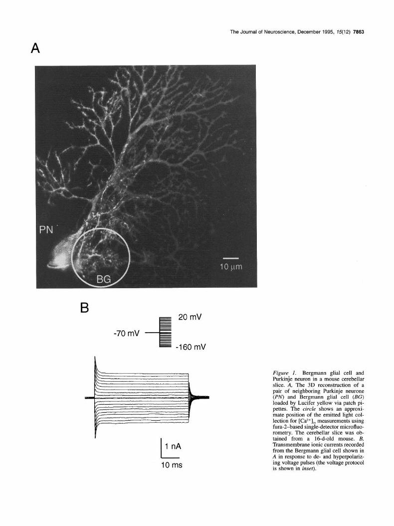
Results Identification of Bergmann glial cells and experimental paradigm Bergmann glial cells in cerebellar slices could be easily identi- fied based on their characteristic morphology (small cell bodies, approximately 10 pm in diameter, and several processes termi- nating at the pia) and their location in the Purkinje cell layer. Figure 1 illustrates that morphological appearance; an individual Bergmann glial cell and, for comparison, a Purkinje cell were consecutively injected with the fluorescent dye Lucifer Yellow via a patch pipette. The image was obtained with a confocal microscope. We used two approaches to study Ca2+ transients in Bergmann glial cells:
The Journal of Neuroscience, December 1995, 75(12) 7883
- E 20 mV
-70 mV --ii -160 mV
I 1 nA
10ms
Figure 1. Bergmann glial cell and Purkinje neuron in a mouse cerebellar slice. A, The 3D reconstruction of a pair of neighboring Purkinje neurone (PN) and Bergmann glial cell (BG) loaded by Lucifer yellow via patch pi- pettes. The circle shows an approxi- mate position of the emitted light col- lection for [Caz+],, measurements using fura-2-based single-detector microfluo- rometry. The cerebellar slice was ob- tained from a 16-d-old mouse. B, Transmembrane ionic currents recorded from the Bergmann glial cell shown in A in response to de- and hyperpolariz- ing voltage pulses (the voltage protocol is shown in inset).

7884 Kirischuk et al. l ATP-Induced Ca*+ Signalling in Bergmann Glia
Figure 2. ATP-induced Ca2+ eleva- tion and dependence on resting ICaZ+L A. Examoles of ATP-mediated jCa2+j:i transients’ recorded from three fura-2/AM bulk-loaded Bergmann glial cells. ATP (100 PM) was applied as in- dicated by bars. The resting membrane potential determined after the [Caz-I,, measurements by means of whole-cell current clamp was -68 mV, -77 mV, and -70 mV (respective for the re- sponses from left to right). B, [Ca2+],, transients in response to increasing concentrations of ATP measured from two different fura-2/AM bulk-loaded Bergmann glial cells with different lev- els of basal [Ca2+],, concentration. C, The peak values of ATP-induced (100 pM) [Ca*+],, transients measured from 112 fura-21AM bulk loaded Bergmann glial cells cfilled circles) and resting [Ca*+],, (open circles) are plotted against the corresponding resting [Ca*+],, level. D, ATP-induced [Ca2+], responses evoked at different levels of [Ca2+],,. [Caz+lm transients evoked by ATP (100 FM) were recorded from fura-2/AM bulk-loaded Bergmann glial cell in control conditions and during the recovery phase of a kainate (100 pM)-triggered [Ca*‘],, elevation. Re- cordings were separated by 3 min in- tervals as indicated on the graph.
A
ATP 0
1000 MM ATP
ATP _
ATP
m resting [Ca2+lin (nM)
1000 PM ATP D ATP ATP ATP ATP
(1) Individual cells were loaded via the patch pipette, and this procedure confined the dye to one single cell. The whole-cell patch-clamp technique allowed us to discriminate Bergmann glial cell from neuronal elements by their distinct electrophysi- ological characteristics (Fig. 1B). As shown previously, the dom- inating conductance of the Bergmann glial cell membrane is associated with potassium channels (Miiller et al., 1992). To ex- clude Ca*+ recordings from damaged cells, we used the resting potential (RP) as an indicator of the cell’s viability. Only the cells with RP more negative than -65 mV were considered for further analysis. Among these cells the resting potential aver- aged at -73 ? 6 mV (mean -C SD, II = 112).
However, while loading the cell via internal dialysis, one re- striction has to be taken into account: since the cell cytoplasm was dialyzed with the patch pipette solution, the intracellular machinery involved in [Ca*+], homeostasis might be affected.
(2) Bergmann glial cells could also be labeled by incubating the slice with the membrane-permeant forms of Ca*+-sensitive dyes. This was feasible, since Bergmann glial cells accumulated both fura- and flue-3 considerably faster than the neighboring Purkinje neurons. Thus, by varying the incubation time we pref- erentially labeled Bergmann glial cells. After completion of the [Ca2+lin measurements the cells were dialyzed with fura-Zfree internal solution to (1) measure their RP, and to (2) determine the background fluorescence for correction of [Ca*+&. record- ings. Only cells with RP more negative than -65 mV were considered for further analysis. The resting [Ca*+],, determined in bulk loaded Bergmann glial cells was in the range of 30-200 nM.
ATP triggers [Ca2+li, elevation, but no membrane currents
ATP (100 FM) produced an elevation of [Ca2+lin in the majority of Bergmann glial cells (110 out of 112). The rise in [Ca*+],,
KA
reached a peak within 2-4 set and [Ca2+Jn recovered towards the basal level in the continued presence of ATP Washout of ATP caused a drop in [Ca2+Jn to the resting level. Representative examples of ATP-evoked [Ca*+],, transients are shown in Figure 2A. The amplitude of ATP-induced [Ca*+],, responses varied be- tween 10 and 250 nM; this amplitude appeared to correlate with the basal [Ca*+],,: at higher basal [Ca*+],“, we recorded the smaller amplitudes of ATP-mediated [Ca”],, responses (Fig. 2AD).
To study the developmental regulation of the ATP-mediated Ca*+ signalling, we used Bergmann glial cells obtained from mice of three age groups, namely, postnatal day 6, 16-20, and 40-45. There was no significant difference with respect to the amplitude and shape of ATP-induced [Ca*+],” transients. The ac- tion of ATP on [Ca*+],, in Bergmann glial cells appeared to be concentration dependent: examples of [Ca*+],, transients evoked by increasing ATP concentrations are shown in Figure 2B. Sim- ilarly, as described above, the amplitude of ATP-induced [Ca”],, transients was strongly modulated by the basal [Ca*+],, level: in cells with resting [Ca2*],, in the range of 30-50 nM, 1 mu of ATP elevated [Caz+],, to 400-500 nM (Fig. 2B, top trace); in cells with basal [Ca*+],, of 150 nM the response to 1 mM ATP did not exceed 200 nM.
The absolute value for the [CazL],, elevation induced by 100 p,M ATP peaked at about 150-250 nM. We observed a strong variation in the resting Ca2’ levels among different Bergmann glial cells ranging between 30 to 200 nM. As a consequence, ATP-induced Ca2’ transients from cells with a low basal [Ca*+],, level were larger and varied, while responses of cells with an increased level were smaller. The relationship between the rest- ing [Ca”],, level and the ATP-induced increase is summarized in Figure 2C.
Moreover, under experimentally increased [Ca2-I,, levels, the

The Journal of Neuroscience, December 1995, 75(12) 7865
B
1 180
50s
Figure 3. [Ca*+],, transients and trans- membrane ionic currents recorded from fura-2K,-loaded Bergmann glial cell in response to stimulation of the ATP-sen- sitive pathway and glutamate ionotrop- ic receptors. A, Extracellular applica- tion of kainate (KA, 100 FM) induced the generation of an inward current (bottom) and a [Ca*+],” elevation (top).
KA
amplitudes of the ATP responses were strongly attenuated. An example of such an experiment is shown in Figure 2C. To ex- perimentally increase [Ca2+lln we used externally applied 100 pM kainate, which is known to induce a transmembrane Ca*+ influx (Miiller et al., 1992). After the kainate application and thus during the recovery phase of kainate-triggered [Caz+], re- sponse, ATP was given at variable intervals. At higher levels of [CaZ+],, the amplitudes of ATP-induced [Ca*+],,, transients were smaller.
To test for the presence of ionotropic (P,,) receptors, we si- multaneously recorded membrane currents in the whole-cell re- cording configuration of the patch-clamp technique. To verify our ability to record ligand-gated membrane currents, we com- pared ATP responses with those,‘activated by kainate. The latter involves transmembrane Caz+ influx via ionotropic glutamate re- ceptors of the AMPA/kainate family (Burnashev et al., 1992; Mtiller et al., 1992). Figure 3 illustrates the simultaneous re- cording of transmembrane currents and [Ca*+],, obtained from the same Bergmann glial cell in response to 100 pM ATP and 100 pM kainate. ATP triggers an increase in [Caz+lln only, whereas kainate generated inward current in concert with a [Ca”],, transient. Moreover, while recording in the current- clamp mode we never observed a measurable change in the membrane potential upon ATP application.
To test whether the ATP response in the Bergmann glial cell is mediated by indirect effects involving neuronal spiking or the GABA, or kainate/AMPA receptors, slices were incubated with the Na+ channel blocker tetrodotoxin (1 FM), and the receptor antagonists CNQX (10 FM) and bicuculline (1 FM); as compared to controls, these substances did not influence the ATP-induced [Ca*+],, elevation (n = 9).
Subcellular heterogeneity of ATP-induced [Ca2+],, responses
To investigate the spatial distribution of the ATP-evoked [Caz+lln responses we used the high-resolution laser scanning confocal microscopy. Cells were loaded with either fluo-3 pentapotassium salt (fluo-3K,) or fluo-3/AM using the bulk-loading procedure, and confocal images of flue-3 fluorescence were taken every 5 sec. The [Caz+],, increased much more rapidly and reached high-
B, Application of ATP (100 FM) gen- erated a [Ca*+],, transient (top) that was not accompanied by measurable
- ATP
100 pA changes in ihe transmembrane ionic current (bottom). The holding potential was -75 mV throughout the experi- ment.
er levels in distal processes as compared to the soma (Fig. 4). The amplitude of the [Ca*+],, increase was significantly larger in the processes, namely 2.1 + 0.8 times (n = 12) as compared to the soma. Figure 4 shows the distribution of [Ca2+]ln in a fluo- 3K,-loaded Bergmann glial cell during ATP application.
ATP-induced [Ca”],,, elevation is due to Ca2+ release from InsP,-sensitive internal stores
As shown above, ATP-induced [Caz+],. transients were not ac- companied by the generation of a detectable membrane current,
2+ suggesting that transmembrane Ca influx is not involved in the generation of the ATP-mediated [Ca2+lin response. To substan- tiate the finding that the [Ca’+],, elevation produced by ATP originates not from transmembrane Ca2+ influx, but from Ca*+ release from internal stores, we compared ATP-mediated [Ca2+],n responses in Ca*+-containing versus Ca*+-free extracellular so- lution. After incubating the slice for 5 min in a Ca*+-free solu- tion, ATP (100 p,M) produced a [Ca*+],, elevation similar to the one recorded in normal bathing solution (Fig. 5A; n = 12). In contrast, Ca*+-free superfusion (for 5 min) completely abolished kainate-induced [Ca2+Jn responses (n = 9), indicating that ex- tracellular Ca*+ in the slice could-be lowered to levels preventing influx via plasma membrane pathways.
Prolonged incubation in Ca *+-free solution, however, caused a reduction in the ATP response amplitudes: superfusion of slic- es with Ca*+-free solution for 15 min reduced the amplitude of ATP-mediated [Ca2+lln transients to about 20% of control levels (n = 7). This was reversible, since the reintroduction of Ca2+ ions into the external medium completely restored the ampli- tudes of ATP-triggered [Ca”],, transients.
To characterize the intracellular pool involved in ATP-trig- gered [Ca”],, signalling, we used the tumor promoter thapsi- gargin, a specific blocker of sarco(endo)plasmic reticulum cal- cium ATPase (SERCA) pump (Thastrup, 1990; Lytton et al., 1991). Thapsigargin treatment prevented accumulation of Ca2+ ions by intracellular pools, thereby decreasing the amount of releasable calcium. As illustrated in Figure 5B, extracellular ad- ministration of thapsigargin (500 nM) did not change the resting [Caz+],,. However, 5 min after thapsigargin application, ATP-

7886 Kirischuk et al. * ATP-Induced Ca*+ SIgnaIling In Bergmann Glia
A
E I c:’
&&$ .-:_ . : . . .t
..‘. . .: *
. . . . .f,rl&!‘. : ., ‘Z
:.. ; .%
3-.., .
m I . 1. .-. :. : :
: -
_’ . .
3
. .
. ; - . , ‘.d
: . ,_.
.+ 1,: . , - ,7,’
c : : . ..-I’ .
25 s
ATP
Figure 4. Spatial distribution of the ATP-induced [Ca*+lin signal. A, Pseudocolor images of a fluo-3K,-loaded Bergmann glial cell during ATP application (100 PM). Images were aquired using a laser scanning confocal microscope and then normalized (with pixel by pixel ratioing) to the images taken from the resting state of the cell (see Experimental Procedures). The increases in [Ca*+],, were color coded; low [Ca*+],, levels correspond to blue, high [Ca*+],,, levels to red. B, Top: the relative increase of fluo-3 fluorescence (corresponding to the [Caz+lin elevation) in response to ATP application (100 PM) measured separately from distal (a) and proximal (b) processes and from the soma (c) of the Bergmann glial cell shown in A. The selected regions for the measurements are indicated by white boxes in A. Images shown in A were taken at points indicated by arrows. Bottom: simultaneous recording of transmembrane current (holding potential -75 mV) shows no changes in membrane permeability upon ATP application.

65-
B ATP ATP
ca2+-free 140 -
+ “m 0
80-
ATP ATP ATP
C Thapsigargin
180
z
+
2
50
Control 50s :L 70
‘. 50 - ATP
25 s
1 k 1 .j&PL.+/. .k..
ATP
Figure 5. ATP induces Ca*+ release from internal stores in Bergmann g&l cells. A, The [CaZ+],, was recorded from a fura-2/AM bulk- ioaded Bergmann glial cell. Applications of ATP (100 FM) induced a [Ca*+],,, elevation in control conditions and after 5 min slice superfusion with Ca*+-free, EGTA-containing extracellular solution. The RP of the cell determined after the end of [Ca*+],, recording was ~74 mV. B, Thap- sigargin inhibits ATP-induced [CaZ+],, transients in a fura-2/AM bulk- loaded Bergmann glial cell. The ATP (100 PM)-induced [Ca*+],, re- sponses are compared in control solution and after slice superfusion with 500 nM thapsigargin-containing solution. The RP of the cell de- termined at the end of [Caz+],, recordings was -70 mV. C, Intracellu- larly applied heparin (1 FM) blocks ATP-induced [Ca2+],,, signals in a Bergmann glial cell. The control [Ca2+],. transient was recorded from the fura-2/AM bulk-loaded cell; then the cell was dialyzed with heparin- containing pipette solution and 5 min later the second record (shown on the right) was taken. The ATP-induced [Ca*+],, transient activated slower and the peak amplitude was considerably smaller.
evoked [Ca*+],, transients were considerably smaller as com- pared to controls. Moreover, a consecutive application of ATP failed to elicit any changes in [Ca2+],“. The action of thapsigargin was almost irreversible: 30 to 45 min washout with control so- lution restored the amplitude of ATP-induced [Ca2+lin transients to only lo-15% as compared to the control levels at the begin- ning of the experiment. Thapsigargin effectively inhibited ATP- mediated [Ca2+],, transients in all cells studied (n = 10).
To examine the intracellular mechanism responsible for ATP- induced [CaZ+],, signalling we used the specific blocker of InsP,- gated Ca2+ channels of the endoplasmic reticulum, heparin (Hill et al., 1987). As heparin is a membrane impermeant agent, we delivered it by intracellular dialysis via patch pipette. Control responses were measured from fura-2/AM-loaded Bergmann
The Journal of Neuroscience, December 1995, 75(12) 7867
glial cells; then these cells were approached with patch pipette containing intracellular solution supplemented with 100 pM
fura- pentapotassium salt and 1 pM heparin. Five minutes of internal dialysis with heparin strongly reduced the amplitude (in heparin treated cells the amplitude of ATP-induced [Ca*+],” el- evation was only 11 ? 3% (n = 7) of the control) and attenuated the rising phase of the ATP-mediated [Ca*+],, signal. In contrast, 5 min of internal dialysis with heparin-free intrapipette solution reduced the amplitude of ATP-evoked Ca2+ transient to only 93 -C 3% of the control (Fig. 5C, IZ = 8).
Rejilling of ATP-sensitive internal Ca2+ stores is metabolically dependent
To test for the ability of the intracellular Ca*+ pool for repetitive signalling, ATP was applied twice separated by a 60 set interval. As shown in Figure 6A, the second ATP application induced a smaller Ca*+ signal (58 2 4% from the control; IZ = 6). Further applications separated by 60 set led to a progressive decrease, and the [Ca2+],, response was abolished after four to six se- quential ATP applications. Such a decrease presumably reflects the depletion of intracellular stores. After a recovery period of 10 min, ATP again induced Ca*+ signals comparable to the first control, indicating that the stores were refilled.
This recovery was irreversibly blocked by thapsigargin, sug- gesting that the SERCA pumps are responsible for the restora- tion (n = 4). Moreover, the recovery process also required the presence of extracellular Ca*+ ions: changing to Ca2+-free bath- ing solution after a series of successive ATP applications did not allow for the recovery of the ATP-induced [Ca*+],, increase (n = 5).
The intracellular dialysis via the patch pipette also affected the recovery of the ATP-induced [Caz+],, increase. In dialyzed cells, ATP induced [Ca2+],, elevations not more than two or three times, and even after long recording times a response could no longer be elicited (n = 11). Figure 6B shows an example of ATP-induced [Ca’+],, transients measured from an internally di- alyzed Bergmann glial cell. It is evident that two successive applications of ATP depleted internal CaZ+ stores, and responses could not be elicited even after long recovery times.
Characterization of the type of ATP receptor
To characterize the type of purinergic receptor linked to the lib- eration of Ca*+ from internal stores, we tested the effect of dif- ferent purine and pyrimidine nucleotides that have been de- scribed to discriminate between different types of purinergic re- ceptors (O’Connor et al., 1991; Illes and Norenberg, 1993). Ag- onists, at a concentration of 100 p,M, were successively tested on one cell separated by 10 min intervals. For this series of experiments, bulk-loaded cerebellar Bergmann glial cells were tested to avoid a rundown of the response as observed with the patch-clamp experiments. Figure 7A shows the result of repre- sentative experiments in which the amplitude of the fluorescence signal in response to adenosine, AMP, ADP, ATP, a$-methylene ATP (Me-ATP) and UTP were compared. The rank order of potency for the purinoreceptor agonists was: ADP 2 ATP > UTP >> AMP = adenosine = Me-ATP (n = 9), thus sug- gesting the involvement of P, purinoreceptor subtype (Illes and Norenberg, 1993).
The involvement of P, purinoreceptors was further confirmed by the blockade of the ATP-induced response by the selective PZ receptors antagonist, suramin (Dunn and Blakeley, 1988). In- cubation of cerebellar slices with suramin significantly inhibited

7888 Kirischuk et al. *ATP-Induced Ca2+ Signalling in Bergmann Glia
Figure 6. Recovery of the ATP-in- duced CaZ+ transients in bulk-loaded and dialyzed Bergmann glial cells. A, ATP-induced [Ca*+],, transients record- ed from a fura-2/AM bulk-loaded Bergmann glial cell. Subsequent appli- cations of 100 PM ATP separated with 50 set intervals caused a progressive decrease of the amplitude of [Ca2+],, responses, presumabely due to the de- pletion of internal Ca2+ stores. The am- plitude of the ATP-induced [Ca*+],, transient recovered to the control value after 10 min, reflecting the replenish- ment of the Ca2+ pool. The RP of the cell determined at the end of [Ca2+],” recordings was -80 mV. B, A similar experiment as in A was performed on an internally dialyzed Bergmann glial cell loaded with fura-2K5. ATP (100 (LM) applications caused an irreversible depletion of the internal stores. A si- multaneous membrane current record- ing is shown below the [CaZ+],. trace. The holding potential was -75 mV.
- - B - - -
ATP ATP ATP ATP ATP ATP
60 s
B
150,
g + “a 0 -I h 100 "' 10 min /&&g+Q#WN
50 s
-0 min /iWWwWW
ATP ATP ATP
25 pA
ATP-triggered [Ca”],, elevation in Bergmann glial cells (Fig. 7B). In the presence of suramin (100 FM, 30 set preapplication) the amplitudes of ATP-induced [Ca*+],, responses were 21 ? 7% (n = 6) of the control level. The suramin-induced blockade of the ATP-mediated [Ca2+lln mobilization was reversible: wash- out with standard bathing solution completely restored [Ca2+],” responses within lo-15 min. In comparison, concentrations of up to 500 pM suramin failed to attenuate the kainate-induced [Ca*+],, mobilization (n = 4; data not shown).
A
2.5
- - 100 PM ATP 100 PM ADP
ATP
Discussion Bergmann glial cells express P, metabotropic purinergic receptors In the present study we have demonstrated that ATP increased [Ca2+lln in Bergmann glial cells indicating the presence of ATP receptive sites. ATP is a ligand for a family of purinergic recep- tors, the P, receptors. These comprise the P,,, P,,, and P,, re- ceptors coupled with plasmalemmal ionic channels (Bean, 1992) and P,, and P,, receptors linked to PLC-driven formation of
100 PM UTP 100 KM AMP
25 s
40 s
.~.. .EizdRm&
-
100 PM Me-ATP 100 PM ADEN.
ATP
100 pM suramin
ATP
Figure 7. Pharmacological properties of ATP-induced [Ca*+],, responses. A, [Ca*+],, transients were recorded as fluorescence ratio (F/F,) from the fluo-3/AM bulk-loaded Bergmann glial cell in response to application of 100 PM adenosine (ADEN), AMP, ADP, ATR c-u,@methylen-ATP (Me- ATP), and UTP Recordings were separated by 10 min intervals to ensure the refilling of ATP-sensitive CaZ+ stores. [Ca*+],, levels were separately measured at the soma (a) and in the processes (b) of the cell. The RP of the cell determined at the end of [Ca2+],, recordings was -68 mV. B, Inhibition of the ATP-induced [Ca*+],, transients by suramin. [Ca*+],, transients (as the fluo-3 fluorescence ratio) were recorded from the soma (a) and processes (b) of the fluo-S/AM bulk-loaded Bergmann glial cell. As a control, 100 FM ATP was applied. After 10 min, 100 FM ATP was applied in the presence of 100 PM suramin. The response was markedly reduced. After a 15 min washout, 100 FM ATP induced a similar response as in the control. The RP of the cell determined at the end of [Ca2+li, recordings was -75 mV.

The Journal of Neuroscience, December 1995, 75(12) 7869
InsP, (Illes and Norenberg, 1993). Alternatively, ATP may act through its metabolites AMP and adenosine via activation of P, purinoreceptors.
In the present study on Bergmann glial cells, we demonstrated that the ATP-induced [Ca’+],, elevation was mimicked by ADP and (to certain extents) UTP, but not by AMP or adenosine, suggesting, therfore, the involvement of P, purinoreceptors. Moreover, the selective blocker of metabotropic P2 receptors, suramin, caused a reversible inhibition of ATP-mediated [Ca”],, responses. A similar action of suramin on P,-metabotropic re- ceptors-driven [C$+],, transients has been found in a number of other systems (Inoue et al., 1991; Kalthof et al., 1993; Salter and Hicks, 1994; Kirischuk et al., 1995). The lack of P?x recep- tors is inferred by the observation that (1) the selective agonist of P,, receptors, a$-methylene-ATP, did not produce a [Ca?+],n elevation and (2) that ATP did not activate membrane currents as expected after P2x receptor activation. We thus conclude that Bergmann glial cells express P, metabotropic purinergic recep- tors.
ATP causes Ca2+ release from InsP.,-sensitive internal pools
Activation of metabotropic P, purinoreceptors is commonly linked to Ca’+ release from cytoplasmic pools (Harden et al., 1990; O’Connor et al., 1991). This is in line with our observa- tions that (1) the ATP-induced signal can still be recorded in the absence of extracellular Ca’+, suggesting the involvement of in- tracellular calcium release; and that (2) we did not observe an ATP activated ionic current across the plasma membrane.
Based on the observation that intracellular administration of heparin antagonized the effects of ATP on [Ca*+],,, we can con- clude that ATP action is mediated by the intracellular second- messenger InsP, and involves the activation of [Caz+],, release from an InsP,-sensitive calcium pool.
Fura- was reported to act as a competitive inhibitor of InsP, binding to its receptor with a K, of -120 pM (Richardson and Taylor, 1993). The estimated cytoplasmic fura- concentration in our experiments on bulk-loaded Bergmann glial cells was in the range of 50-60 PM, while in patch-clamp experiments we used intracellular solutions supplemented with 100 FM fura-2. Thus, our experiments were performed in a fura- concentration range in which the inhibitory action of fura- on InsP,-gated Ca*’ channel might lead to an underestimation of the amplitudes of ATP-triggered [Ca?+],” transients. This may also explain why the EC,, of the ATP dose-response curve was unusually high for P, purinoreceptors (see Fig. 2B).
The intracellular Ca2+ pool responsible for the InsP,-mediated calcium release accumulates Ca*+ by the activity of the SERCA pumps. Several ATP challenges applied within short time inter- vals almost completely depleted internal Ca*+ stores; however, within 10 min, the pool of releasable CaZ+ was restored. Block- ing the SERCA pump with the specific blocker thapsigargin (Thastrup, 1990) did not prevent a first ATP-induced release, but subsequent ATP applications did no longer elicit a Ca’+ signal, since the CaZ+ gradient could no longer be restored. This inhi- bition was almost irreversible: ATP responses recovered only partially during 30-45 min after thapsigargin removal. These findings are consistent with the previously reported irreversibil- ity of the thapsigargin-induced blockade of SERCA pumps, pre- sumably via covalent modification (Lytton et al., 1991). Dialyz- ing the cell interior with the patch pipette impaired the ability of restoring the Ca2+ pools. This suggests that intracellular per-
fusion removes critical (yet unknown) factors controlling Ca?+ uptake into intracellular Ca2+ pools.
[Caz+],, homeostasis and ATP-induced Ca2+ signalling in Bergmann glial cells
In previous studies on nonexcitable cells outside the CNS, the blockade of the SERCA pump by thapsigargin produced a sig- nificant elevation in [Ca2+lin (see Petersen et al., 1994 for re- view); in Bergmann glial cells resting [Ca*+],” levels were not affected. This discrepancy might be due to a different interplay between the various [Ca*+],, homeostasis mechanisms: the rest- ing [Ca”],,, levels are determined by a competition between Ca’+ leakage (from internal stores and the extracellular space), the efficiency of the Ca’+ buffer systems, and by the activity of the Ca*+ pumps (transferring Ca2+ ions into internal pools or to the extracellular compartment). The absence of thapsigargin-in- duced [CaZ+],,, elevation in the Bergmann glial cells makes it likely that the levels of [Ca?+],” are strongly controlled by mech- anisms other than SERCA pumping, such as highly efficient cytosolic Ca2+ buffer mechanisms, Ca2+ uptake into mitochon- dria or Ca*+ extrusion into the extracellular space. The impor- tance of the latter mechanism is inferred by the observation that prolonged exposure of cerebellar slices in Ca*+-free solution markedly depressed ATP-mediated [Ca*+],” responses. These ob- servations indicate that Ca2+ extrusion from the Bergmann cell dominates over the Ca’+ turnover between the cytoplasm and internal pools; the latter might be depleted by removal of extra- cellular calcium. This may also suggest the involvement of trans- membrane Ca*+ transport in supplying stores with releasable Ca*+ (“capacitative Ca2+ entry;” Penner et al., 1993; Fasolato et al., 1994).
We found a significant variation in resting [Caz+],, levels among the individual Bergmann glial cells. It is conceivable that elevated [Ca2+lin might indicate cell damage; however, this seems to be unlikely due to the fact that we analyzed [CaZ+],, signals only in cells with high resting potential (RP more neg- ative than -65 mV). We found that the amplitudes of ATP- induced [Ca*+],, responses of Bergmann glial cells appeared to be controlled by the resting [Ca*+],, level: in cells that possessed higher resting [Ca2+],“, the amplitudes of the ATP-mediated re- sponses were usually smaller. If the [Ca2+lln levels exceeded 170-180 nM, ATP was almost unable to elevate [Ca’+],, even when applied in high doses. The mechanisms controlling this ceiling concentration remain unresolved. One possibility would be that Ca2’ ions serve as coagonists of InsP,-gated Ca2+ release channels, stimulating them at low and moderate concentrations and inhibiting at high concentrations as described for InsP,-gated channels derived from endoplasmic reticulum of cerebellar neu- rons (Bezprozvanny et al., 1991). However, Ca2+ ions block InsP,-gated channels in a micromolar range, whereas we have seen inhibition of ATP-induced responses at [CaZ+],, in a range of 150-200 nM. It is even likely that the resting [Ca?+],” levels may control the responsiveness of the entire ATP-sensitive [Ca2+],,, signalling machinery; interaction with any component of this pathway (including purinoreceptors, Ca*+-release chan- nels, SERCA pumps, etc.) may be responsible for the observed phenomena.
Physiological relevance of ATP-mediated [Caz+],,, signals in Bergmann glial cells
The role of ATP in intracellular metabolism is well established; however, in addition, ATP plays an important role as an inter-
7870 Kirischuk et al. * ATP-Induced Ca*+ Signalling in Bergmann Glia
cellular signalling molecule. It has been shown that ATP acts as an excitatory chemical mediator in both the PNS and CNS (Burnstock, 1990; El-Moatassim et al., 1992). ATP-mediated ex- citatory synaptic transmission was found in neuromuscular junc- tions (Benham, 1989) in neuron-neuron synapses in coeliac gan- glion (Evans et al., 1992), dorsal horn (Salter et al., 1993), and in the medial habenula (Edwards et al., 1992). In addition, ATP was found to increase [Ca’+],, in a variety of excitable and non- excitable cells including osteoblasts and osteoclasts (Reimer and Dixon, 1992; Yu and Ferrier, 1993) cultured renal cells (Cejka et al., 1993), smooth muscle cells (Droogmans et al., 1991; Car- ter and Ogden, 1992; Kalthof et al., 1993), and dorsal horn neu- rons and astrocytes (Salter and Hicks, 1994).
ATP can be released alone or coreleased with other neuro- transmitters from synaptic vesicles (Sawynok et al., 1993), and in addition, it could be liberated from neurites during electrical activity (Maire et al., 1982). In different types of glial cells ATP was found to be the most reliable and widespread agent, which induces cytoplasmic Ca*+ mobilization (see Finkbeiner, 1993; Kastritsis and McCarthy, 1993; Kirischuk et al., 1995). In Berg- mann glial cells we have demonstrated that ATP induced a cy- toplasmic Ca2+ signal with distinct spatial organization: the ATP-mediated [Ca2+lln responses are higher in Bergmann glial processes versus the soma. This subcellular heterogeneity could be functionally important, taking into account the intimate in- teraction of the Bergmann glial processes with the Purkinje neu- ron’s dendritic tree. We can speculate that ATP released from synaptic endings of parallel and climbing fibers, which innervate Purkinje neurons, may generate [Ca*+],, signals in Bergmann glial cells. This signal may trigger or regulate several unknown cytoplasmic events or participate in the regulation of interstitial Ca2+ levels. It is known that neuronal activity may induce a significant drop of extracellular calcium: it was demonstrated that during electrical stimulation of hippocampal slices [CaZ+lo fell from 2 to 1.4 mM (Benninger et al., 1980); and spreading depression caused [Caz+], to fall from 2.2 to 0.8 mM (Kraig and Nicholson, 1978). One possible function of Bergmann glial cells may be to serve as dynamic Ca*+ pools during intense synaptic activity by releasing Ca 2+ from their internal stores into the ex- tracellular compartment involving purinergic receptor activation, cytoplasmic Ca*+ increase, and Ca2+ extrusion.
References
Bean BP (1992) Pharmacology and electrophysiology of ATP-activated ion channels. Trends Pharmacol Sci 13:87-90.
Benham CD (1989) ATP-activated channels gate calcium entry in sin- gle smooth muscle cells dissociated from rabbit ear artery. J Physiol (Lond) 419:686-701.
Benninger C, Kadis J, Prince DA (1980) Extracellular calcium and potassium changes in hippocampal slices. Brain Res 187:165-182.
Berridge MJ (1993) lnositol trisphosphate and calcium signalling. Na- ture 361:315-325.
Bezprozvanny I, Watras J, Ehrlich BE (1991) Bell-shaped calcium- response curves of Ins( 1,4,5)P, and calcium-gated channels from en- doplasmic reticulum of cerebellum. Nature 35 1:75 l-754.
Burnashev N, Khodorova A, Jonas P Helm PJ, Wisden W, Monyer H, Seeburg PH, Sakmann B (1992) Calcium-permeable AMPA-kainate receptors in fusiform cerebellar glial cells. Science 256:1566-1570.
Burnstock G (1990) Overview: Purinergic mechanisms. Ann NY Acad Sci 603:1-17.
Carter TD, Ogden D (1992) Kinetics of intracellular calcium release bv inositol 1.4.5 trisohosohate and extracellular ATP in porcine cul- tured aortic endothehal cells. Proc R Sot Lond [Biol] 250:235-241.
Cejka JC, Bidet M, Taut M, Poujeol P (1993) Nucleotides mobilize intracellular calcium stores of renal proximal cells in primary culture:
existence of a suramin-sensitive mechanisms. Biochim Biophys Acta Mol Cell Res 1176:7-12.
Droogmans G, Callewaert G, Declerk I, Casteels R (1991) ATP-in- duced Ca2+ release and Cl- current in cultured smooth muscle cells from pig aorta. J Physiol (Lond) 440:623-634.
Dunn PM, Blakeley AGH (1988) Suramin: a reversible P,-purinore- ceptor antagonist in the mouse vas deferens. Br J Pharmacol 93:243- 245.
Edwards FA, Gibb AJ, Colquhoun D (1992) ATP receptor-mediated synaptic currents in the central nervous system. Nature 359:144-147.
El-Moatassim C, Dornand J, Mani J-C (1992) Extracellular ATP and cell siunallinn. Biochim Bioohvs Acta Mol Cell Res 1134:31-45 _ . I .
Evans R?, Derkzh V, SuprenatantA (1992) ATP mediates fast synaptic transmission in mammalian neurons. Nature 357:503-505.
Fasolato C, Innocenti B, Pozzan T (1994) Receptor-activated Ca2+ in- flux: how many mechanisms for how many channels? Trends Neu- rosci 15:77-83.
Ferris CD, Snyder SH (1992) Inositol phosphate receptors and calcium disoosition in the brain. J Neurosci 12:1567-1574.
Finkdeiner SM (1993) Glial calcium. Glia 9:83-104. Fisher SK, Agranof BW (1987) Receptor activation and inositol lipid
hydrolysis in neuronal tissues. J Neurochem 48:999-1017. Grynkiewicz G, Poenie M, Tsien RY (1985) A new generation of Ca2+
indicators with greatly improved fluorescent properties. J Biol Chem 260:3440-3450.
Hamill OP, Marty A, Neher E, Sakmann B, Sigworth FJ (1981) Im- proved patch clamp techniques for high-resolution current recording from cell and cell-free membrane patches. Pflugers Arch 391:85-100.
Harden TK, Boyer JL, Brown HA, Cooper CL, Jeffs RA, Martin, MW (1990) Biochemical properties of a P,,-purinergic receptor. Ann NY Acad Sci 603:256-266.
Hatten ME, Fishell G, Stitt TN, Mason CA (1990) Astroglia as a scaf- fold for development of the CNS. Neurosciences 2:455%465.
Hill DT, Berggren PO, Boynton AL (1987) Heparin inhibits inositol trisphosphate induced calcium release in permeabilized rat liver cells. Biochem Biophys Res Commun 149:879-901.
Illes P, Norenberg W (1993) Neuronal ATP receptors and their mech- anism of action. Trends Pharmacol Sci 14:50-54.
Inoue K, Nakazawa K, Ohara-Imaizumi M, Obama T, Fujimori K, Tak- anaka A (1991) Selective and competetive antagonism by suramin of ATP-stimulated catecholamine secretion from PC1 2 phaeochrom- ocytoma cells. Br J Pharmacol 102:581-584.
Kalthof B, Bechem M, Flocke K, Pott L, Schramm M (1993) Kinetics of ATP-induced Ca*+ transients in cultured pig aortic smooth muscle cells depend on ATP concentration and CaZ+ stores. J Physiol (Lond) 4661245-262.
Kano M, Rexhausen U, Dreessen J, Konnerth A (1992) Synaptic ex- citation produces a long-lasting rebound potentiation of inhibitory synaptic signals in cerebellar Purkinje cells. Nature 356:601-604.
Kastritsis CHC, McCarthy KD (1993) Oligodendroglial lineage cells express neuroligand receptors. Glia 8: 106-l 13.
Kettenmann H, Banati R, Walz W (1993) Electrophysiological behav- ior of microglia. Glia 7:93-101.
Kimelberg HK (1988) Glial cell receptors. New York: Raven. Kirischuk S, Scherer J, Kettenmann H, Verkhratsky A (1995) Activa-
tion of P, purinoreceptors triggers Ca*+ release from InsP,-sensitive internal stores in mammalian oligodendrocytes. J Physiol (Land) -\-- , 483.1:41-57.
Kraig RT, Nicholson C (1978) Extracellular ionic variations during soreading depression. Neuroscience 3:1045-1059.
Lytion J, Westlin M, Hanley MR (1991) Thapsigargin inhibits the sar- .coplasmic or endoplasmic reticulum Ca-ATPase family of calcium oums. Biol Chem 266:17067-17071.
Magowski NS, Walz W (1992) Ionic dependence of a P,-purinorecep- tor mediated depolarization of cultured astrocytes. J Neurosci Res 32: 530-538.
Maire JC, Medilanski J, Straub RW (1982) Uptake of adenosine and release of adenine derivates in mammalian non-myelinated nerve fob- ers at rest and during activity. J Physiol (Lond) 323:589-602.
Minta A. Kao J. Tsien R (1989) Fluorescent indicators for cvtosolic -,--I----
calcium based on rhodamine and fluorescein chromophores. J Biol Chem 264:8171-8182.
Miiller T, Mdller T, Berger T, Schnitzer J, Kettenmann H (1992) Cal- cium entry through kainate receptors and resulting potassium-channel blockade in Bergmann glial cells. Science 256:1563-1566.

The Journal of Neuroscience, December 1995, 15(12) 7871
Mtiller T, Grosche .I, Ohlemeyer C, Kettenmann H (1993) NMDA- activated currents in Bergmann glial cells. Neuroreport 4:671-674.
Miller T, Fritschy JM, Grosche J, Pratt GD, Mohler H, Kettenmann H (1994) Developmental regulation of voltage-gated K+ channels and GABA, receptor expression in Bergmann glial cells. J Neurosci 14: 2503-25 14.
O’Connor SE, Dainty IA, Leff P (1991) Further subclassification of ATP receptors based on agonist studies. Trends Pharmacol Sci 12: 137-141.
Penner R, Fasolato C, Hoth M (1993) Calcium influx and its control by calcium release. Curr Opin Neurobiol 3:368-374.
Petersen OH, Petersen CCH, Kasai H (1994) Calcium and hormone action. Annu Rev Physiol 56:297-319.
Reimer WJ, Dixon SJ (1992) Extracellular nucleotides elevate [Ca*+], in rat osteoblastic cells by intercation with two receptor subtypes. Am J Physiol 263:C1040-Cl048
Richardson A, Taylor CW (1993) Effects of Ca2+ chelators on purified inositol 1,4,5-trisphosphate (InsP,) receptors and InsP,-stimulated Ca*+ mobilization. J Biol Chem 268: 11528-l 1533.
Salter MW, Hicks JL (1994) ATP-evoked increases in intracellular cal- cium in neurons and glial from the dorsal spinal cord. J Neurosci 14: 1563-1575.
Salter MW, De Konink Y, Henry JL (1993) Physiological roles for adenosine and ATP in synaptic transmission in the spinal dorsal horn. Prog Neurobiol 41: 125-l 56.
Sawynok J, Downie JW, Reid AR, Cahill CM, White TD (1993) ATP release from dorsal spinal cord synaptosomes: characterization and neuronal origin. Brain Res 610:32-38.
Stone TW (1981) Physiological roles for adenosine and adenosine 5’- triphosphate in the nervous system. Neuroscience 6:523-555.
Thastrup 0 (1990) Role of Ca*+-ATPases in regulation of cellular Ca2+ signalling, as studied with the selective microsomal Ca*+-ATPase in- hibitor, thapsigargin. Agents Actions 29:9-15.
Walz W, Ilschner S, Ohlemeyer C, Banati R, Kettenmann H (1993) Extracellular ATP activates a cation conductance and K+ conductance in cultured microglial cells from mouse brain. J Neurosci 13:4403- 4411.
Yu H, Ferrier J (1993) ATP induces an intracellular calcium pulse in osteoclasts. Biochem Biophys Res Commun 191:357-363.