atp8b1 is essential for maintaining normal · pdf fileatp8b1 is essential for maintaining...
TRANSCRIPT

ATP8B1 is essential for maintaining normal hearingJanneke M. Stapelbroeka,b,1,2, Theo A. Petersc,1, Denis H. A. van Beurdenb, Jo H. A. J. Curfsc, Anneke Joostena,Andy J. Beynonc, Bibian M. van Leeuwena, Lieke M. van der Veldenb, Laura Bulld, Ronald P. Oude Elferinke,Bert A. van Zantenf, Leo W. J. Klompb, and Roderick H. J. Houwena
Departments of aPediatric Gastroenterology and fENT/Audiology, University Medical Center Utrecht, Utrecht, The Netherlands; bDepartment of Metabolicand Endocrine Diseases, University Medical Center Utrecht and Netherlands Metabolomics Center, Utrecht, The Netherlands; cDepartment ofOtorhinolaryngology, Donders Center for Neuroscience, Radboud University Nijmegen Medical Center, Nijmegen, The Netherlands; dThe LiverCenter at the University of California, San Francisco, Rice Liver Center Laboratory, San Francisco General Hospital, San Francisco, CA 94110;and eAMC Liver Center, Academic Medical Center, Amsterdam, The Netherlands
Edited by Jonathan G. Seidman, Harvard Medical School, Boston, MA, and approved April 17, 2009 (received for review August 11, 2008)
ATP8B1 deficiency is caused by autosomal recessive mutationsin ATP8B1, which encodes the putative phospatidylserine flippaseATP8B1 (formerly called FIC1). ATP8B1 deficiency is primarily char-acterized by cholestasis, but extrahepatic symptoms are alsofound. Because patients sometimes report reduced hearing capa-bility, we investigated the role of ATP8B1 in auditory function.Here we show that ATP8B1/Atp8b1 deficiency, both in patients andin Atp8b1G308V/G308V mutant mice, causes hearing loss, associatedwith progressive degeneration of cochlear hair cells. Atp8b1 isspecifically localized in the stereocilia of these hair cells. Thisindicates that the mechanosensory function and integrity of thecochlear hair cells is critically dependent on ATP8B1 activity, pos-sibly through maintaining lipid asymmetry in the cellular mem-branes of stereocilia.
ATP8B1 deficiency � extrahepatic symptoms � hearing impairment
P -type ATPases are essential for normal function of the humanbody (1). In general, this family of transport proteins maintains
a cation gradient across cellular membranes. Of the five differentsubfamilies, the recently discovered P4 P-type ATPases (P4-ATPases) share a distinct function as phospholipid flippases (2–4).By translocation of aminophospholipids from the outer to the innerleaflet of cellular membranes, P4-ATPases are thought to beessential for maintaining membrane lipid asymmetry. This asym-metry is important for fundamental processes such as membranetransport, intracellular signaling and apoptosis (5–7).
ATP8B1 deficiency is a human disease known to be associatedwith mutations in the gene encoding the P4 P-type ATPase,ATP8B1 (formerly called FIC1) (8, 9). This disease presents withintrahepatic cholestasis either as benign recurrent intrahepaticcholestasis type 1 (BRIC type 1; MIM#243300) or progressiveintrahepatic cholestasis type 1 (PFIC type 1; MIM#211600) (9–12).BRIC type 2 and PFIC type 2 are genetically distinct disorderscaused by mutations in ABCB11, which encodes the main bile saltexport pump (BSEP) that is exclusively expressed in the canalicularmembrane of the liver (13, 14). Interestingly, ATP8B1 is similarlyexpressed in canalicular membranes of hepatocytes, but also inother epithelial tissues, which may explain some extrahepaticfeatures exclusively observed in patients with ATP8B1 defi-ciency (9, 10, 15, 16). Pancreatitis, secretory diarrhea and growthretardation are well known extrahepatic features in ATP8B1deficiency, that may persist after liver transplantation in PFICtype 1 patients (17–19).
Sporadically, hearing loss has been mentioned in patients withPFIC or BRIC of unknown genetic subtype (20). Given thewidespread expression of ATP8B1 we hypothesized that thesehearing problems comprise another extrahepatic feature in patientswith ATP8B1 deficiency and consequently that the ATP8B1 pro-tein is important for normal hearing. We therefore examined therole of ATP8B1 protein in auditory function in patients and micewith ATP8B1/Atp8b1 deficiency. We show here that ATP8B1deficiency causes hearing loss, associated with progressive degen-eration of the cochlear hair cells.
ResultsSensorineural Hearing Loss Is an Extrahepatic Feature in BRIC Type 1.To investigate whether hearing loss is an extrahepatic feature inBRIC type 1, we tested hearing in ten BRIC type 1 patients. Tocontrol for secondary effects resulting from the cholestatic episodesin BRIC, we also included BRIC type 2 patients, who have episodiccholestasis, as occurs in BRIC type 1, but no extrahepatic symptoms(13). Patients with primary sclerosing cholangitis (PSC), affected bymild chronic cholestasis, formed a second control group. In total 10patients with BRIC type 1, three BRIC type 2 patients, and sevenpatients with PSC were included. No confounders such as a familyhistory of hearing disorders, noise-induced hearing loss or use ofototoxic medication were noted.
Retrospectively, nine of 10 patients with BRIC type 1 reportedhearing problems, which were first noticed at a mean age of 20 years(range, 17–29 years). More importantly, on age-corrected pure toneaudiometry, BRIC type 1 patients displayed significant hearing lossin both ears at all frequencies tested except for 500 Hz, comparedwith the group of PSC patients who displayed normal hearing. Thishearing loss was more pronounced at higher frequencies (Fig. 1A).The transient evoked otoacoustic emissions (TEOAEs) were ab-normal in all BRIC type 1 patients with hearing loss. In two BRICtype 2 patients, a normal hearing for age in both ears and at allfrequencies tested was found. The third BRIC type 2 patientdisplayed a hearing loss of 65 dB in one ear at 4 and 8 kHz, but nohearing loss was found in the contralateral ear. Tympanometryresults were normal in BRIC type 1 patients and all controls.
Atp8b1G308V/G308V Mutant Mice Show Significant Hearing Loss. Tofurther investigate the role of the ATP8B1 protein in auditoryfunction, Atp8b1G308V/G308V mutant mice were tested for auditorybrainstem responses (ABRs) as described previously (21). Thesemutant mice harbor the mutation G308V (NP�005594), similar tothe mutation in Amish PFIC type 1 patients (10, 22). This mutationcauses a marked decreased expression of Atp8b1 in mice (22).However, in contrast to the Amish patients, these mutant micedisplay only a mild hepatic phenotype when on normal diet, andcholestasis only develops when high amounts of bile acids are addedto the diet (22). In this work al mice were kept on normal diet. UsingABRs, the sensitivity of the auditory system was tested in 16 days,1, 3 and 6-month-old mutant mice versus wild-type mice. Fig. 1Bdisplays the mean auditory brainstem thresholds for three differenttone burst stimuli and the click stimulus. These data revealed that
Author contributions: J.M.S., L.M.v.d.V., L.W.J.K., and R.H.J.H. designed research; J.M.S.,T.A.P., D.H.A.v.B., J.H.A.J.C., A.J., and B.M.v.L. performed research; L.B., R.P.O.E., B.A.v.Z.,and L.W.J.K. contributed new reagents/analytic tools; J.M.S., T.A.P., and A.J.B. analyzeddata; and J.M.S., T.A.P., and R.H.J.H. wrote the paper.
The authors declare no conflict of interest.
This article is a PNAS Direct Submission.
1J.M.S. and T.A.P. contributed equally to this work.
2To whom correspondence should be addressed at: Department of Pediatric Gastroenter-ology [KB.03.023.2], Wilhelmina Children’s Hospital, University Medical Center Utrecht,Postbox 85090, 3508 AB, Utrecht, The Netherlands. E-mail: [email protected].
www.pnas.org�cgi�doi�10.1073�pnas.0807919106 PNAS � June 16, 2009 � vol. 106 � no. 24 � 9709–9714
CELL
BIO
LOG
Y
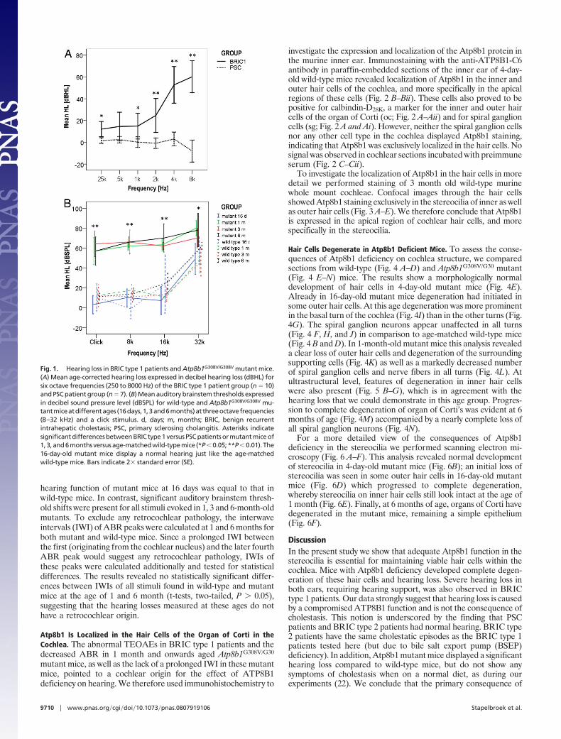
hearing function of mutant mice at 16 days was equal to that inwild-type mice. In contrast, significant auditory brainstem thresh-old shifts were present for all stimuli evoked in 1, 3 and 6-month-oldmutants. To exclude any retrocochlear pathology, the interwaveintervals (IWI) of ABR peaks were calculated at 1 and 6 months forboth mutant and wild-type mice. Since a prolonged IWI betweenthe first (originating from the cochlear nucleus) and the later fourthABR peak would suggest any retrocochlear pathology, IWIs ofthese peaks were calculated additionally and tested for statisticaldifferences. The results revealed no statistically significant differ-ences between IWIs of all stimuli found in wild-type and mutantmice at the age of 1 and 6 month (t-tests, two-tailed, P � 0.05),suggesting that the hearing losses measured at these ages do nothave a retrocochlear origin.
Atp8b1 Is Localized in the Hair Cells of the Organ of Corti in theCochlea. The abnormal TEOAEs in BRIC type 1 patients and thedecreased ABR in 1 month and onwards aged Atp8b1G308V/G30
mutant mice, as well as the lack of a prolonged IWI in these mutantmice, pointed to a cochlear origin for the effect of ATP8B1deficiency on hearing. We therefore used immunohistochemistry to
investigate the expression and localization of the Atp8b1 protein inthe murine inner ear. Immunostaining with the anti-ATP8B1-C6antibody in paraffin-embedded sections of the inner ear of 4-day-old wild-type mice revealed localization of Atp8b1 in the inner andouter hair cells of the cochlea, and more specifically in the apicalregions of these cells (Fig. 2 B–Bii). These cells also proved to bepositive for calbindin-D28K, a marker for the inner and outer haircells of the organ of Corti (oc; Fig. 2 A–Aii) and for spiral ganglioncells (sg; Fig. 2 A and Ai). However, neither the spiral ganglion cellsnor any other cell type in the cochlea displayed Atp8b1 staining,indicating that Atp8b1 was exclusively localized in the hair cells. Nosignal was observed in cochlear sections incubated with preimmuneserum (Fig. 2 C–Cii).
To investigate the localization of Atp8b1 in the hair cells in moredetail we performed staining of 3 month old wild-type murinewhole mount cochleae. Confocal images through the hair cellsshowed Atp8b1 staining exclusively in the stereocilia of inner as wellas outer hair cells (Fig. 3 A–E). We therefore conclude that Atp8b1is expressed in the apical region of cochlear hair cells, and morespecifically in the stereocilia.
Hair Cells Degenerate in Atp8b1 Deficient Mice. To assess the conse-quences of Atp8b1 deficiency on cochlea structure, we comparedsections from wild-type (Fig. 4 A–D) and Atp8b1G308V/G30 mutant(Fig. 4 E–N) mice. The results show a morphologically normaldevelopment of hair cells in 4-day-old mutant mice (Fig. 4E).Already in 16-day-old mutant mice degeneration had initiated insome outer hair cells. At this age degeneration was more prominentin the basal turn of the cochlea (Fig. 4I) than in the other turns (Fig.4G). The spiral ganglion neurons appear unaffected in all turns(Fig. 4 F, H, and J) in comparison to age-matched wild-type mice(Fig. 4 B and D). In 1-month-old mutant mice this analysis revealeda clear loss of outer hair cells and degeneration of the surroundingsupporting cells (Fig. 4K) as well as a markedly decreased numberof spiral ganglion cells and nerve fibers in all turns (Fig. 4L). Atultrastructural level, features of degeneration in inner hair cellswere also present (Fig. 5 B–G), which is in agreement with thehearing loss that we could demonstrate in this age group. Progres-sion to complete degeneration of organ of Corti’s was evident at 6months of age (Fig. 4M) accompanied by a nearly complete loss ofall spiral ganglion neurons (Fig. 4N).
For a more detailed view of the consequences of Atp8b1deficiency in the stereocilia we performed scanning electron mi-croscopy (Fig. 6 A–F). This analysis revealed normal developmentof stereocilia in 4-day-old mutant mice (Fig. 6B); an initial loss ofstereocilia was seen in some outer hair cells in 16-day-old mutantmice (Fig. 6D) which progressed to complete degeneration,whereby stereocilia on inner hair cells still look intact at the age of1 month (Fig. 6E). Finally, at 6 months of age, organs of Corti havedegenerated in the mutant mice, remaining a simple epithelium(Fig. 6F).
DiscussionIn the present study we show that adequate Atp8b1 function in thestereocilia is essential for maintaining viable hair cells within thecochlea. Mice with Atp8b1 deficiency developed complete degen-eration of these hair cells and hearing loss. Severe hearing loss inboth ears, requiring hearing support, was also observed in BRICtype 1 patients. Our data strongly suggest that hearing loss is causedby a compromised ATP8B1 function and is not the consequence ofcholestasis. This notion is underscored by the finding that PSCpatients and BRIC type 2 patients had normal hearing. BRIC type2 patients have the same cholestatic episodes as the BRIC type 1patients tested here (but due to bile salt export pump (BSEP)deficiency). In addition, Atp8b1 mutant mice displayed a significanthearing loss compared to wild-type mice, but do not show anysymptoms of cholestasis when on a normal diet, as during ourexperiments (22). We conclude that the primary consequence of
Fig. 1. Hearing loss in BRIC type 1 patients and Atp8b1G308V/G308V mutant mice.(A) Mean age-corrected hearing loss expressed in decibel hearing loss (dBHL) forsix octave frequencies (250 to 8000 Hz) of the BRIC type 1 patient group (n � 10)and PSC patient group (n � 7). (B) Mean auditory brainstem thresholds expressedin decibel sound pressure level (dBSPL) for wild-type and Atp8b1G308V/G308V mu-tantmiceatdifferentages (16days,1,3and6months)atthreeoctavefrequencies(8–32 kHz) and a click stimulus. d, days; m, months; BRIC, benign recurrentintrahepatic cholestasis; PSC, primary sclerosing cholangitis. Asterisks indicatesignificant differences between BRIC type 1 versus PSC patients or mutant mice of1,3,and6monthsversusage-matchedwild-typemice (*P�0.05;**P�0.01). The16-day-old mutant mice display a normal hearing just like the age-matchedwild-type mice. Bars indicate 2� standard error (SE).
9710 � www.pnas.org�cgi�doi�10.1073�pnas.0807919106 Stapelbroek et al.

the Atp8b1G308V/G308V mutation is cochlear hair cell degenerationduring early postnatal development of the organ of Corti. Theobserved secondary loss of surrounding supporting cells and spiralganglion neurons is a well known consequence of deprivation frominput signals originating from cochlear hair cells, as has beenshowed in other genetically modified animal models with degen-erated cochlear hair cells (23–31).
We propose several possible pathogenic mechanisms that linkATP8B1 deficiency to hearing loss. ATP8B1 is thought to beimportant for maintaining phospholipid asymmetry of the cel-lular membranes, most likely by translocating phosphatidylserinefrom the outer leaflet of the membrane to the inner leaflet (3,4, 32). Asymmetric distribution of phospholipids in the cellmembrane improves the mechanical stability of the red cellmembrane (33). Similarly, the canalicular membrane of patientsand mice affected by ATP8B1/Atp8b1 deficiency is unstable,
causing extraction of lipids and ectoenzymes into the bile as aconsequence of the continuous chemical stress by the detergentaction of bile salts. Eventually this results in the formation ofcoarsely granular bile that contains remnants of extruded cellmembranes (34, 35). We here showed that Atp8b1 is abundantlyexpressed at the stereociliar membrane. Presumably, a defi-ciency of this protein would result in an impaired phosphatidyl-serine translocation, and alter the composition of the stereociliarmembrane, resulting in a reduced mechanical stability, withdevastating effects for hair bundle stereocilia that are underconstant undulating mechanical stress forces (36, 37).
Proper membrane lipid composition has also been found to beessential for ATP-dependent Ca2�-transporter activity (38). Ca2�-ATPase isoform type 2 (PMCA2) is highly expressed in stereociliarmembranes where it is pivotal for normalizing intracellular Ca2�
concentration in the hair cells after mechanical deflection of the
A
B
C
i
i
i
ii
ii
ii
Fig. 2. Localization of Atp8b1 in cochlea. (A–C) Fluorescence images of paraffin sections of 4-day-old wild-type murine inner ear (basal turn) incubated withanti-ATP8B1-C6 or anti-calbindin-D28k antibody (green or white signal) and counterstained with propidium iodide (red signal). (A–Aii) For orientation anti-calbindin-D28K was used as a marker for hair cells in the organ of Corti. (B–Bii) Atp8b1 expression was seen in apical region of these hair cells. (C–Cii) Serial sections were incubatedwith preimmune serum as a negative control. Arrow points to row of inner hair cells; arrowhead points to three rows of outer hair cells. sg, spiral ganglion; oc, organof Corti. Scale bar, (Ai–Ci) 50 �m, (Aii–Cii) 10 �m.
A B C D E
Fig. 3. Localization of Atp8b1 in cochlear hair cell stereocilia. Confocal images of cochlear whole mounts of 3-month-old wild-type mice incubated with anti-ATP8B1.Confocal images overlay (A) or at level 0.0 �m (B), 1.9 �m (C), 3.5 �m (D), and 5.0 �m (E) showing Atp8b1 staining from top to base in the stereocilia of the cochlearbasal turn. The arrow points to the row of inner hair cells, the arrowhead points to the (three) rows of outer hair cells. Scale bar, 6 �m.
Stapelbroek et al. PNAS � June 16, 2009 � vol. 106 � no. 24 � 9711
CELL
BIO
LOG
Y

stereocilia (37, 39, 40). Cochlear hair cells of Pmca2 mutant micedegenerate and cause severe hearing loss early in life, demonstrat-ing that Pmca2 activity is essential for the preservation of thestructural integrity of these cells (25, 30, 41). Consequently ATP8B1
deficiency, with a secondary disturbance of membrane lipid asym-metry, likely inhibits PMCA2 activity and affects the efficiency ofmechanotransduction. Hence, the similarities in histopathologicaland physiological findings between Atp8b1 deficient and Pmca2deficient mice provide a likely explanation for the inadequatehearing associated with Atp8b1 deficiency. Interestingly, compen-satory Pmca1 and 3 isoforms are present in the inner hair cellsbodies, but not in outer hair cells, which is consistent with our obser-vation that outer hair cells degenerate before inner hair cells (39).
Finally, ATP8B1 deficiency might alter the phospholipid com-position of the inner membrane leaflet, thereby impairing thegeneration of phosphoinositides (42, 43) that are important foradequate mechanotransduction and maintenance of the hair bun-dle structure (23, 44). Phosphoinositide signaling was recentlyfound to be involved in age-related hearing loss, raising the possi-bility that ATP8B1 may reflect a modifier locus for presbycusis (45).
Our study included only patients with the recurrent form ofATP8B1 deficiency (BRIC type 1) because, until recently, patientswith progressive ATP8B1 deficiency (PFIC type 1) did not survivebeyond childhood. In addition, this permitted us to study adultpatients from different age groups, and will allow future follow upstudies to monitor the progression of ATP8B1-induced hearingloss. Consequently, all BRIC type 1 patients studied here havemutations in the ATP8B1 gene with relatively mild consequences,and supposedly have some residual function of ATP8B1. In con-trast, the Atp8b1G308V/G308V mutant mice have severely compro-mised Atp8b1 function and expression, thus providing a rationalefor the relatively mild hearing loss in ATP8B1 deficient patients asopposed to mice. Conversely, the liver phenotype of Atp8b1 mutantmice is relatively mild. This phenomenon is generally explained bythe presence of large amounts of hydrophilic bile salts in murinebile. The murine bile salt pool is less detergent and therefore lessefficient in destabilizing canalicular membranes with altered phos-pholipid asymmetry (22, 35). Collectively, these findings indicatethat the severity of hepatic and extrahepatic features of ATP8B1deficiency are both dependent on the extent by which the ATP8B1protein function is compromised, as well as tissue specific additionalfactors that are relevant for the pathogenesis of the disorder (22, 35).
In conclusion, we show that ATP8B1/Atp8b1 deficiency causeshearing loss, associated with progressive degeneration of cochlearhair cells. These data indicate that the preservation of the cochlearhair cells in the inner ear, required for adequate hearing, is criticallydependent on normal ATP8B1 function, analogous to the preser-
A C
B D
E G
F H
I
J
K
L
M
N
Fig. 4. Degeneration of hair cells and spiralganglion neurons in Atp8b1G308V/G308V mu-tant mice. Light micrographs of cochlear mor-phology of the organ of Corti (A, C, E, G, I, K,M) and spiral ganglion (B, D, F, H, J, L, N) inwild-type (A–D) and mutant (E–N) mice at 4days (A, B, E, F), 16 days (C, D, G–J), 1 month (K,L) and 6 months (M, N) of age. All sections arefrom the basal turn except for (G, H), whichrepresent themedial turn.Althoughmorpho-logically intact looking inner hair cells couldbe seen in the mutant mice till the age of 1month (E, G, I, K), the outer hair cells showsigns of degeneration from 16 days on (I, K,M), and the number of neurons in the spiralganglionisdecreasedfrom1monthofage(L).Complete degeneration of the organ of Cortiand nearly all connected spiral ganglion cellsand nerve fibers is observed in 6-month-oldmutant mice (M, N). Scale bar, 24 �m.
A B
F
C
G
D
E
Fig. 5. Ultrastructural features of inner hair cell degeneration inAtp8b1G308V/G308V mutant mice. Transmission electron micrographs of innerhair cells in the upper basal turn of the cochlea of wild-type (A) and mutant(B–G) mice at 1 month of age. The inner hair cells of the mutant mice displaya normal cell shape and nucleus (B) and largely intact stereocilia (B, F).However, signs of degeneration are already observed, such as condensedcytoplasm (B), membrane-bound vesicles enclosing cellular debris (D, arrow),multivesicular bodies (D, double arrow), degenerating mitochondria (E, ar-rowhead) and vacuoles with cellular debris (E, asterisk). More severe degen-eration aspects can also be seen in hair cells of the same turn (C) especiallyusing higher magnification (G). Scale bars: A, B, 2 �m; C–G, 400 nm.
9712 � www.pnas.org�cgi�doi�10.1073�pnas.0807919106 Stapelbroek et al.

vation of structural integrity of the bile canaliculus, necessary foradequate bile salt excretory function.
Materials and MethodsPatients. The 10 adult Dutch BRIC type 1 patients investigated had at least 1episode of cholestasis with normal serum concentration of gamma-glutamyltranspeptidase (GGT) in combination with known mutations on both alleles ofATP8B1. To control for secondary effects of cholestasis on hearing, three BRICtype 2 and seven PSC patients were tested. The BRIC type 2 patients also had atleast one episode of low GGT cholestasis, in combination with known ABCB11mutations on both alleles. All patients with PSC were previously diagnosed withinflammatory bowel disease and did have elevated transaminases and GGT. Thediagnosis was confirmed with magnetic resonance cholangiopancreatographyand/ora liverbiopsy.At thetimeofaudiometry themeanageofthepatientswithBRIC type 1 was 37 years (range, 18–60 years), in the BRIC type 2 this was 36 years(range, 23–43 years), whereas the mean age of the patients with PSC was 47 years(range, 35–57 years). This human study was conducted under approved protocolsand after written informed consent was obtained for all patients, in accordancewith the guidelines of the medical ethical committee of the University MedicalCenter Utrecht.
Audiometry. For detection of hearing loss, all patients were tested with standardpure tone audiometry for six octave frequencies (250–8000 Hz), both for air andbone conduction. To exclude possible effects of age-related hearing loss (pres-bycusis) audiometrical thresholds were individually corrected for age accordingto ISO 7029 (1984) values based on normative data by Robinson and Sutton (46).Although hearing losses were generally symmetrical, in cases of small asymme-tries thresholds of the best ear were used for statistical analysis. Middle earpressure and tympanic compliance was measured by tympanometry. The normalrange for middle ear pressure was taken as between �100 and �100 daPa andnormalcompliancewasdefinedas0.3–1.5mlofequivalentairvolume.Totest thecochlear sensory function, TEOAEs were recorded. The TEOAEs were classified asnormal or abnormal based on the criteria described by Welzl-Muller and Stephan(47). Only with a normal tympanogram do abnormal emissions indicate cochleardysfunction.
Mice. Atp8b1G308V/G308V mutant mice (129/Sv strain) are used as a murine modelfor PFIC1 disease (22). For control experiments, wild-type mice of the same strainas the mutants were used. All mice were maintained on a standard 12-hourlight,12-hour dark cycle and fed ad libitum with standard rodent chow. Animalexperiments were approved by the institutional animal care and use committees.
Electrophysiological Testing: Auditory Brainstem Responses. For ABR compari-son 16 days, 1, 3 and 6-month-old Atp8b1G308V/G308V mutant (n � 5, 12, 5, 5) and
wild-type (n � 5, 7, 5, 5) mice were measured. In both groups auditory brainstemresponses (ABRs) were evoked with four different stimuli: i.e., 8, 16, and 32 kHztone burst with 1 millisecond rise/fall times, 3 millisecond plateau and with amonophasic click stimulus (100 microsecond duration) according to standardaudiometric procedures. Stimuli were presented with a stimulation rate of 30pulses per second in a sound field setup by a high-frequency loudspeaker (Elac JetIII) 6 cm in front of the ear. All stimulus intensity levels were calibrated accordingto IEC 61672-2 standards (2004) using a frequency analyzer (Bruel & Kjaer, type2260) and corrected for the sound field. The EEG recording system was externallytriggered by a stimulus generator implemented in a PC using a National Instru-ments sound interface card using a digital-to-analog converter (type 6062E) totrigger the tone bursts. Click stimuli were internally generated and triggered bythe EEG system. Before ABR measurements, mice were anesthesized by i.p.injections of ketamine (200 mg/kg) and xylazine (14 mg/kg). ABRs were obtainedusing an EEG recording system (Modeled Synergy, Oxford Instruments, UK) withbandpass filter settings set between 300 Hz (HP) and 5000 Hz (LP), auto rejectmode (set at 50 �V) and a 60 Hz notch filter. A minimum of 500 averages wererecorded suprathreshold; at threshold levels the minimum of averages was set at1500 averages. For identification of possible electrical stimulus artifacts, therecording time window was set at 1.5 millisecond prestimulus time, and lasted13.5 milliseconds, comprising all brainstem potentials. Recording needle elec-trodes were placed at Cz and M1 (non-inverting and inverting electrode respec-tively), a ground electrode was placed in the tail. Electrode impedances weremeasured before ABR recording and revealed inter-electrode impedances of lessthan 5 kOhm. The auditory threshold was defined at the level where the brain-stem responses show just noticeable reproducible peaks. Brainstem peaks werevisually determined and the interwave interval (IWI) JI-IV were evaluated for anyprolonged interpeak latencies to exclude retrocochlear pathology.
Immunohistochemistry. Immersion-fixed (5 hours; methanol:acetone:water[2:2:1])paraffin-embeddedsectionsof4-day-oldwild-type(n�6) innerearswereused for immunohistochemistry. Paraffin-embedded sections were dewaxed,rehydrated, rinsed in phosphate-buffered saline (PBS) containing 0.1% Triton-X100 and boiled for 10 minutes in sodium citrate buffer (pH 6.0). The sectionswere blocked in 3.0% blocking reagent (supplied in TSA fluorescein system:Perkin-ElmerLife Science, Boston, MA) in PBS for 1 hour at room temperature andincubated overnight with either 1:200 dilution of rabbit anti-ATP8B1-C6 (15, 16)or 1:200 diluted preimmune serum. After washing, endogenous peroxidaseactivity was blocked with 3% H2O2 in PBS. As secondary antibody horseradishperoxidase (HRP)–conjugated goat anti-Rabbit IgG was used. HRP-based signalamplification was applied by using the Alexa Fluor 488 tyramide fluoresceinsystem procedure according to the manufacturer’s description. Sections werecounterstained with propidium iodide. Immunofluorescent labeling was visual-ized by confocal laser scan microscopy using a Nikon Eclips E600 microscope. As
A B
C D
E F
Fig. 6. Degeneration of hair cells and stereocilia in Atp8b1G308V/G308V mutant mice. Scanning electron microscopy images of the organ of Corti in wild-type (A, C) andmutant mice (B, D–F) at 4 days (A, B), 16 days (C, D), 1 month (E) and 6 months (F) of age, all in the basal turn. There is normal development of stereocilia in the 4-day-oldmutant mice (B). Loss of the stereocilia starts in some outer hair cells in 16-day-old mutant mice (D) and progresses to complete absence, whereas stereocilia on innerhair cells do not show detectable morphological aberrations in 1-month-old mutant mice (E). The total organ of Corti has degenerated at 6 months in mutant mice,resembling merely a simple nonciliated epithelial layer (F). Scale bar, 1 �m.
Stapelbroek et al. PNAS � June 16, 2009 � vol. 106 � no. 24 � 9713
CELL
BIO
LOG
Y

a positive control sections were stained with rabbit anti-calbindin-D28K (1:500;Sigma Chemicals).
Murine Cochlear Whole Mounts. Perfusion-fixed (4% paraformaldehyde in PBS)inner ears of 3-month-old wild-type mice (n � 8) were dissected to expose thecochlear organ of Corti containing the sensory hair cells with stereociliar bundles,by removing the cartilaginous capsule, the lateral part of the cochlear duct, andthe tectorial membrane. The organ of Corti together with the medial part wasthen detached as a single spiral coil from the central modiolus of the cochlea.These cochlear whole mounts were permeabilized with 0.5% Tween-20 in PBSand incubated overnight with either rabbit anti-ATP8B1-C6 (15, 16) (1:200) orpreimmune serum (1:200) diluted in a blocking solution of 0.1% ovalbumin with0.5% fishgelatin in PBS. As secondary antibody goat anti-rabbit conjugated toAlexa 488 (Molecular Probes) was used. After washing, the specimens weremounted in Vectashield and examined with a Leica FCS sp2 AOBS confocalmicroscope using 63� oil immersion lens (numerical aperture 1.4).
Light Microscopy. Perfusion-fixed (2.5% glutaraldehyde in 0.1 mol/l phosphatebuffer) inner ears of 4 days, 16 days, 1 month, and 6-month-old wild-type (n �3/age group) and Atp8b1G308V/G308V mutant (n � 3/age group) mice wereisolated, decalcified for 2 (16-day-old mice), 5 (1-month-old mice), or 7 days(6-month-old mice) in 10% ethylenediaminetetraacetate (EDTA) and embed-ded in the plastic glycol methacrylate (JB4). Sections (2 �m) were stained with2.5% toluidine blue and viewed with a standard Zeiss Axioskop microscope.
Scanning Electron Microscopy. For exposing the cochlear hair cells with theirstereocila, perfusion-fixed (2.5% glutaraldehyde in 0.1 mol/l phosphate buffer)
inner ears of wild-type (n � 2) and Atp8b1G308V/G308V mutant (n � 2) mice 4 days,16 days, 1 month, and 6 months of age were dissected by removing the surround-ing bone, stria vascularis, and tectorial membrane. Tissues were processed in 1%osmium tetroxide, dehydrated, and critical point dried. Hereafter the specimensare sputter-coated with gold and examined at 15 kV with a Jeol 6310 scanningelectron microscope.
Transmission Electron Microscopy. Dissected cochleae from perfusion-fixed(2.5% glutaraldehyde in 0.1 mol/l phosphate buffer) inner ears of 1-month-oldwild-type (n � 3) and Atp8b1G308V/G308V mutant (n � 3) mice were decalcified for5 days in 10% EDTA containing 1.25% glutaraldehyde. The specimens wereembedded in epon, sectioned, contrasted via standard procedures and examinedwith a Jeol 1010 transmission electron microscope.
Statistical Analysis. Individual psycho-acoustical auditory threshold data ofBRIC type 1 and PSC patients as well as all electrophysiological data of micewere analyzed and compared between groups using two-tailed t-tests forindependent samples after groups were tested for equal variances (Levenestatistic). All statistical analyses were performed using SPSS statistical package(V14.0).
ACKNOWLEDGMENTS. We thank I. Otte-Holler, E.L.G.M. Tonnaer, and E.Verbeek-Camps for technical assistance. V. Prijs is acknowledged for criticaldiscussions and K. van Erpecum for referring PSC patients. EuroHear sup-ported the technical training of T.A.P.
1. Kuhlbrandt W, Auer M. Scarborough GA. (1998) Structure of the P-type ATPases. CurrOpin Struct Biol 8:510–516.
2. der-Baerens N, Lisman Q, Luong L, Pomorski T, Holthuis JC (2006) Loss of P4 ATPasesDrs2p and Dnf3p disrupts aminophospholipid transport and asymmetry in yeast post-Golgi secretory vesicles. Mol Biol Cell 17:1632–1642.
3. Paulusma CC, et al. (2008) ATP8B1 requires an accessory protein for endoplasmicreticulum exit and plasma membrane lipid flippase activity. Hepatology 47:268–278.
4. Pomorski T, et al. (2003) Drs2p-related P-type ATPases Dnf1p and Dnf2p are requiredfor phospholipid translocation across the yeast plasma membrane and serve a role inendocytosis. Mol Biol Cell 14:1240–1254.
5. Holthuis JC, Levine TP (2005) Lipid traffic: Floppy drives and a superhighway. Nat RevMol Cell Biol 6:209–220.
6. Ikeda M, Kihara A, Igarashi Y (2006) Lipid asymmetry of the eukaryoticplasma membrane: functions and related enzymes. Biol Pharm Bull 29:1542–1546.
7. Verkleij AJ, Post JA (2000) Membrane phospholipid asymmetry and signal transduc-tion. J Membr Biol 178:1–10.
8. Paulusma CC, Oude Elferink RP (2005) The type 4 subfamily of P-type ATPases, putativeaminophospholipid translocases with a role in human disease. Biochim Biophys Acta1741:11–24.
9. van Mil SW, Klomp LW, Bull LN, Houwen RH (2001) FIC1 disease: A spectrum ofintrahepatic cholestatic disorders. Semin Liver Dis 21:535–544.
10. Bull LN, et al. (1998) A gene encoding a P-type ATPase mutated in two forms ofhereditary cholestasis. Nat Genet 18:219–224.
11. Klomp LW, et al. (2000) A missense mutation in FIC1 is associated with greenlandfamilial cholestasis. Hepatology 32:1337–1341.
12. Klomp LW, et al. (2004) Characterization of mutations in ATP8B1 associated withhereditary cholestasis. Hepatology 40:27–38.
13. van Mil SW, et al. (2004) Benign recurrent intrahepatic cholestasis type 2 is caused bymutations in ABCB11. Gastroenterology 127:379–384.
14. Strautnieks SS, et al. (1998) A gene encoding a liver-specific ABC transporter is mutatedin progressive familial intrahepatic cholestasis. Nat Genet 20:233–238.
15. Eppens EF, et al. (2001) FIC1, the protein affected in two forms of hereditary cholestasis,is localized in the cholangiocyte and the canalicular membrane of the hepatocyte.J Hepatol 35:436–443.
16. van Mil SW, et al. (2004) Fic1 is expressed at apical membranes of different epithelialcells in the digestive tract and is induced in the small intestine during postnataldevelopment of mice. Pediatr Res 56:981–987.
17. Egawa H, et al. (2002) Intractable diarrhea after liver transplantation for Byler’sdisease: Successful treatment with bile adsorptive resin. Liver Transpl 8:714–716.
18. Lykavieris P, et al. (2003) Progressive familial intrahepatic cholestasis type 1 andextrahepatic features: No catch-up of stature growth, exacerbation of diarrhea, andappearance of liver steatosis after liver transplantation. J Hepatol 39:447–452.
19. Tygstrup N, Steig BA, Juijn JA, Bull LN, Houwen RH (1999) Recurrent familial intrahe-patic cholestasis in the Faeroe Islands. Phenotypic heterogeneity but genetic homo-geneity. Hepatology 29:506–508.
20. Oshima T, Ikeda K, Takasaka T (1999) Sensorineural hearing loss associated with Bylerdisease. Tohoku J Exp Med 187:83–88.
21. Shin JB, et al. (2007) Hair bundles are specialized for ATP delivery via creatine kinase.Neuron 53:371–386.
22. Pawlikowska L, et al. (2004) A mouse genetic model for familial cholestasis caused byATP8B1 mutations reveals perturbed bile salt homeostasis but no impairment in bilesecretion. Hum Mol Genet 13:881–892.
23. Goodyear RJ, et al. (2003) A receptor-like inositol lipid phosphatase is required for thematuration of developing cochlear hair bundles. J Neurosci 23:9208–9219.
24. Kurima K, et al. (2002) Dominant and recessive deafness caused by mutations of a novelgene, TMC1, required for cochlear hair-cell function. Nat Genet 30:277–284.
25. Street VA, Kee-Johnson JW, Fonseca RC, Tempel BL, Noben-Trauth K (1998) Mutationsin a plasma membrane Ca2�-ATPase gene cause deafness in deafwaddler mice. NatGenet 19:390–394.
26. Avraham KB, et al. (1995) The mouse Snell’s waltzer deafness gene encodes anunconventional myosin required for structural integrity of inner ear hair cells. NatGenet 11:369–375.
27. Sage C, et al. (2006) Essential role of retinoblastoma protein in mammalian hair celldevelopment and hearing. Proc Natl Acad Sci USA 103:7345–7350.
28. Du X, et al. (2008) A catechol-O-methyltransferase that is essential for auditoryfunction in mice and humans. Proc Natl Acad Sci USA 105:14609–14614.
29. Longo-Guess CM, et al. (2005) A missense mutation in the previously undescribed geneTmhs underlies deafness in hurry-scurry (hscy) mice. Proc Natl Acad Sci USA 102:7894–7899.
30. Kozel PJ, et al. (1998) Balance and hearing deficits in mice with a null mutation in thegene encoding plasma membrane Ca2�-ATPase isoform 2. J Biol Chem 273:18693–18696.
31. Brown SD, Hardisty-Hughes RE, Mburu P (2008) Quiet as a mouse: Dissecting themolecular and genetic basis of hearing. Nat Rev Genet 9:277–290.
32. Ujhazy P, et al. (2001) Familial intrahepatic cholestasis 1: Studies of localization andfunction. Hepatology 34:768–775.
33. Manno S, Takakuwa Y, Mohandas N (2002). Identification of a functional role for lipidasymmetry in biological membranes: Phosphatidylserine-skeletal protein interactionsmodulate membrane stability. Proc Natl Acad Sci USA 99:1943–1948.
34. Bull LN, et al. (1997) Genetic and morphological findings in progressive familialintrahepatic cholestasis (Byler disease [PFIC-1] and Byler syndrome): Evidence forheterogeneity. Hepatology 26:155–164.
35. Paulusma CC, et al. (2006) Atp8b1 deficiency in mice reduces resistance of the cana-licular membrane to hydrophobic bile salts and impairs bile salt transport. Hepatology44:195–204.
36. LeMasurier M, Gillespie PG (2005) Hair-cell mechanotransduction and cochlear ampli-fication. Neuron 48:403–415.
37. Vollrath MA, Kwan KY, Corey DP (2007) The micromachinery of mechanotransductionin hair cells. Annu Rev Neurosci 30:339–365.
38. Tang D, Dean WL, Borchman D, Paterson CA (2006) The influence of membrane lipidstructure on plasma membrane Ca2� -ATPase activity. Cell Calcium 39:209–216.
39. Dumont RA, et al. (2001) Plasma membrane Ca2�-ATPase isoform 2a is the PMCA ofhair bundles. J Neurosci 21:5066–5078.
40. Grati M, et al. (2006) Rapid turnover of stereocilia membrane proteins: evidence fromthe trafficking and mobility of plasma membrane Ca(2�)-ATPase 2. J Neurosci26:6386–6395.
41. Dodson HC, Charalabapoulou M (2001) PMCA2 mutation causes structural changes inthe auditory system in deafwaddler mice. J Neurocytol 30:281–292.
42. Haucke V, Di PG (2007) Lipids and lipid modifications in the regulation of membranetraffic. Curr Opin Cell Biol 19:426–435.
43. Shi X, Gillespie PG, Nuttall AL (2007) Apical phosphatidylserine externalization inauditory hair cells. Mol Membr Biol 24:16–27.
44. Hirono M, Denis CS, Richardson GP, Gillespie PG (2004) Hair cells require phosphati-dylinositol 4,5-bisphosphate for mechanical transduction and adaptation. Neuron44:309–320.
45. Sha SU, Chen FQ, Schacht J (2009) PIP-3 related pathways in age-related hearing loss[Abstract]. Assoc Res Otolaryngol 32:588.
46. Robinson DW, Sutton GJ (1979) Age effect in hearing—a comparative analysis ofpublished threshold data. Audiology 18:320–334.
47. Welzl-Muller K, Stephan K (1994) Confirmation of transiently evoked otoacousticemissions based on user-independent criteria. Audiology 33:28–36.
9714 � www.pnas.org�cgi�doi�10.1073�pnas.0807919106 Stapelbroek et al.