atrial septal defects
DESCRIPTION
Atrial Septal Defects. Imaging Conference 12/15/09 A. Zucker. Why do we care?. Most common congenital heart defect that presents during adulthood… We might have to diagnose it. Because we can fix it!!. Outline:. Embryology Types of ASD Presentation Shunts Echocardiographic evaluation - PowerPoint PPT PresentationTRANSCRIPT
Atrial Septal Defects
Imaging Conference
12/15/09
A. Zucker
Why do we care?
Most common congenital heart defect that presents during adulthood…
We might have to We might have to diagnose itdiagnose it
Because we can fix it!!

Outline:
Embryology Types of ASD Presentation Shunts Echocardiographic evaluation
MRI ASD closure
ASD
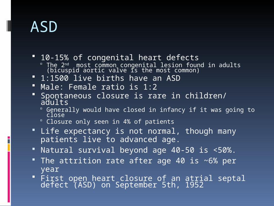
10-15% of congenital heart defects The 2nd most common congenital lesion found in adults
(bicuspid aortic valve is the most common) 1:1500 live births have an ASD Male: Female ratio is 1:2 Spontaneous closure is rare in children/ adults
Generally would have closed in infancy if it was going to close
Closure only seen in 4% of patients Life expectancy is not normal, though many
patients live to advanced age. Natural survival beyond age 40-50 is <50%. The attrition rate after age 40 is ~6% per year First open heart closure of an atrial septal
defect (ASD) on September 5th, 1952
Embryology
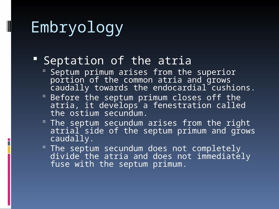
Septation of the atria Septum primum arises from the superior portion of
the common atria and grows caudally towards the endocardial cushions.
Before the septum primum closes off the atria, it develops a fenestration called the ostium secundum.
The septum secundum arises from the right atrial side of the septum primum and grows caudally.
The septum secundum does not completely divide the atria and does not immediately fuse with the septum primum.

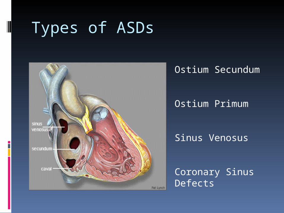
Types of ASDs
Ostium Secundum
Ostium Primum
Sinus Venosus
Coronary Sinus Defects

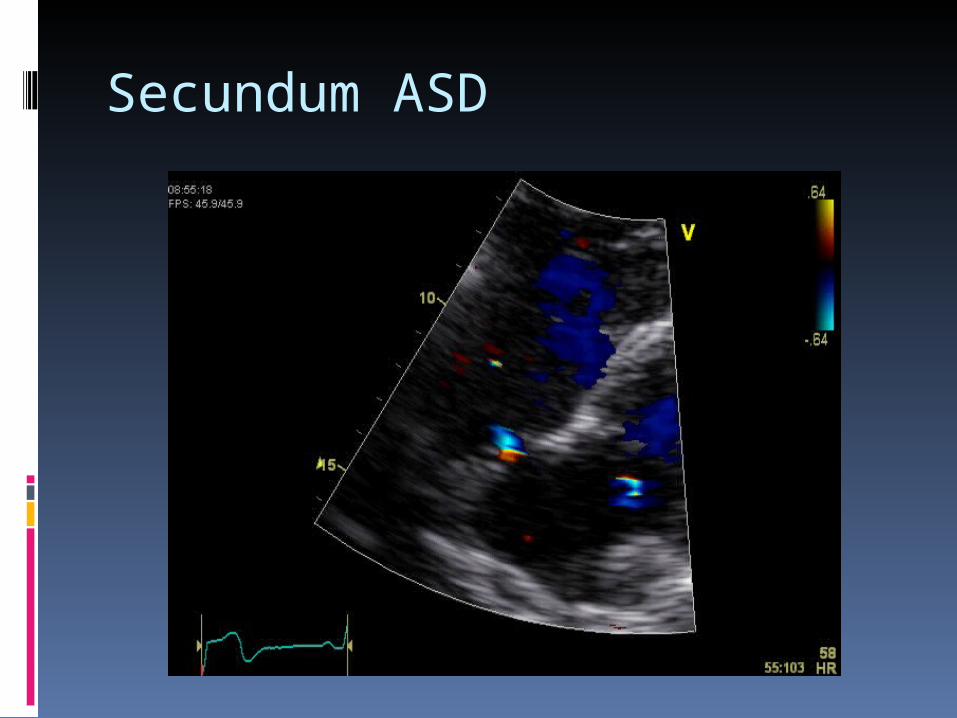
Secundum ASD
Most common type of ASD (70-75%) ~7% of all congenital heart defects Female predominance (2:1) Includes all defects located in the area
of the foramen ovales Mechanisms of formation:
Septum secundum does not grow to cover the ostium secundum.
Ostium secundum is too large for the septum secundum to cover and so is left exposed despite a fully formed septum secundum.
Secundum ASD
Associatd findings: MVP is present in 70% of pts with this type of ASD Partial anomalous pulmonary venous connection
(rare) Specific EKG findings
Right atrial abnormality Prolonged PR Right axis deviation (>100 degrees) rSR’ in v1 (incomplete RBBB) Notching of R wave peak (“crochetage sign”)
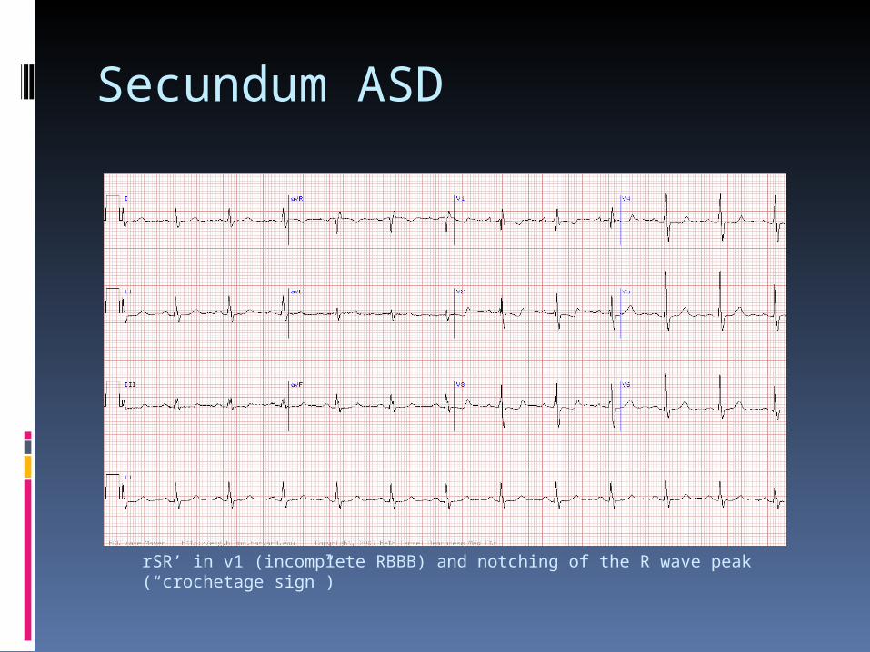
Secundum ASD
rSR’ in v1 (incomplete RBBB) and notching of the R wave peak (“crochetage sign”)
Secundum ASD
Secundum ASD

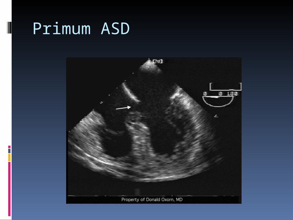
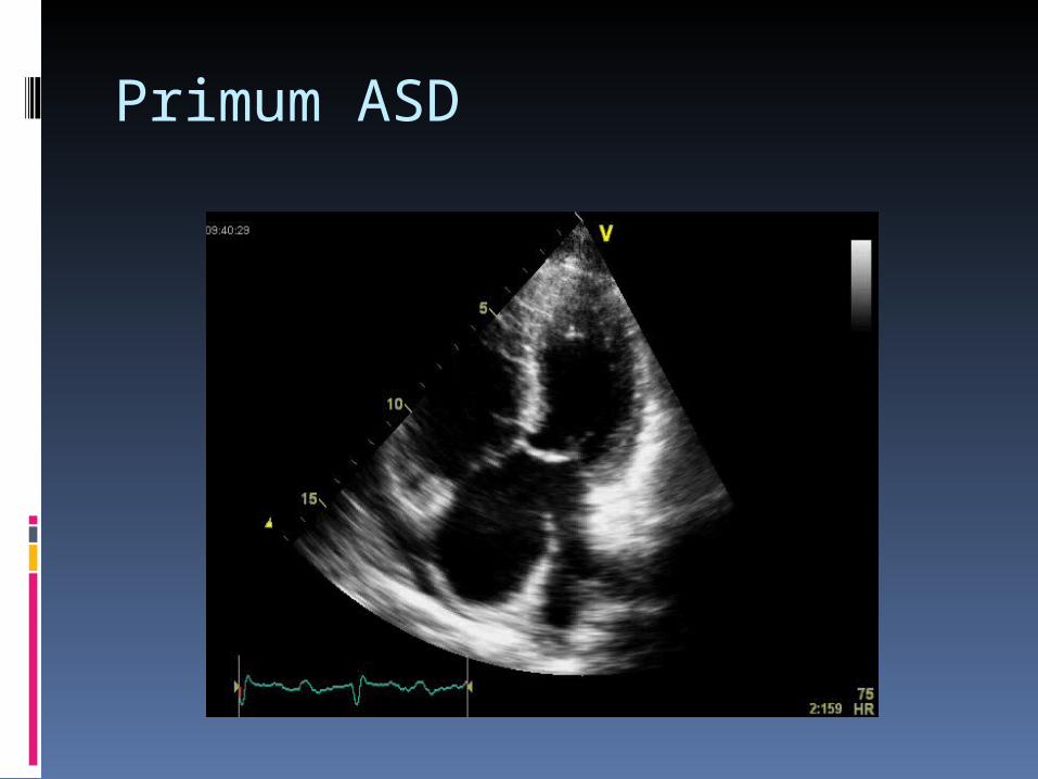
Primum ASD
15-20% of all ASDs Female to Male ratio is 1:1 Simplest form of AV canal defect Generally associated with other anomalies
Commonly have AV valve defects, most notably a cleft in the anterior mitral valve leaflet
Defects of the ventriular septum Common AV canal
Seen commonly in trisomy 21 40-50% of pts w/ Downs syndrome have CHD. Of these
pts 65% are AV canal defects Usually not a subtle finding

Primum ASD
Mechanism of formation: Failure of the septum primum to fuse with the
endocardial cushions (i.e. the ostium primum remains unclosed)
EKG findings: PR prolongation Right atrial enlargement Left axis deviation rSR’ (incomplete RBBB)

Primum ASD
Left axis deviation, rSR’ (incomplete RBBB), and PR prolongation
Primum ASD
Primum ASD
Sinus Venosus Defect
1% of all congenital heat defects in the United States
Account for 10% of all ASDs Not truly considered an ASD:
Abnormality in the insertion of the superior or inferior vena cava (which overrides the interatrial septum)
Two types: Superior sinus venosus defects, located in the atrial
septum immediately below the SVC Inferior sinus venosus defects (less common),
located in the atrial septum immediately above the IVC

Sinus Venosus Defect
Associated Findings Both defects are often associated with a partial
anomalous pulmonary vein connection with abnormal drainage Pulmonary veins may be directed into the right
atrium even if they are in the normal position Pulmonary veins may also be completely displaced
and insert into either vena cava EKG changes
P wave negative in III and aVF and positive in Avl Junctional/ low atrial rhythms

Sinus Venosus Defect

Sinus Venosus Defect
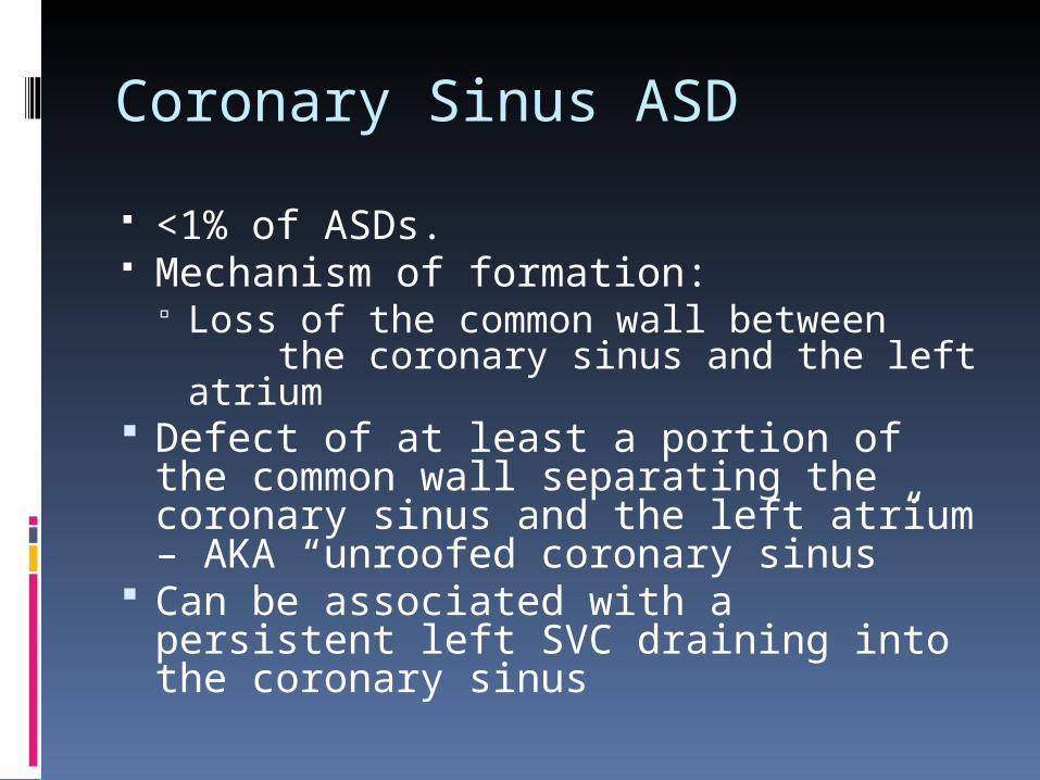
Coronary Sinus ASD
<1% of ASDs. Mechanism of formation:
Loss of the common wall between the coronary sinus and the left atrium
Defect of at least a portion of the common wall separating the coronary sinus and the left atrium – AKA “unroofed coronary sinus”
Can be associated with a persistent left SVC draining into the coronary sinus

Shunting
Degree of shunt has implications as to whether to repair as ASD
Qp/Qs ratio correlates to the size of the ASD. This falls apart when pulmonary hypertension is
present Repair of ASD when Qp/Qs (ratio of
pulmonary flow to system flow) > 2:1 although some papers argue for 1.5:1 AHA recommends >1.5:1, but this excludes
individuals over 21 yrs of age Canadian Cardiac Society recommends Qp/Qs >2:1,
or >1.5:1 in the presence of reversible pulmonary hypertension
Recalculation of Qp/Qs every 2-3 yrs
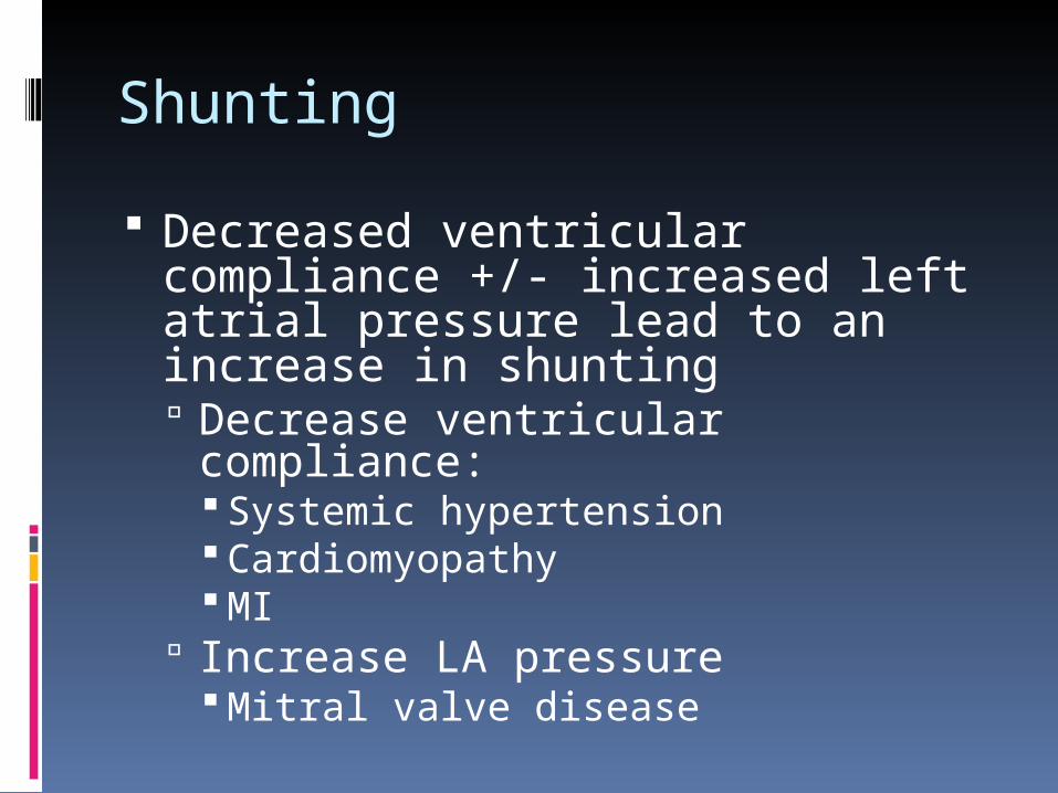
Shunting
Decreased ventricular compliance +/- increased left atrial pressure lead to an increase in shunting Decrease ventricular compliance:
Systemic hypertension Cardiomyopathy MI
Increase LA pressure Mitral valve disease
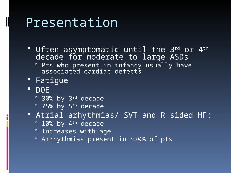
Presentation
Often asymptomatic until the 3rd or 4th decade for moderate to large ASDs Pts who present in infancy usually have associated
cardiac defects Fatigue DOE
30% by 3rd decade 75% by 5th decade
Atrial arhythmias/ SVT and R sided HF: 10% by 4th decade Increases with age Arrhythmias present in ~20% of pts
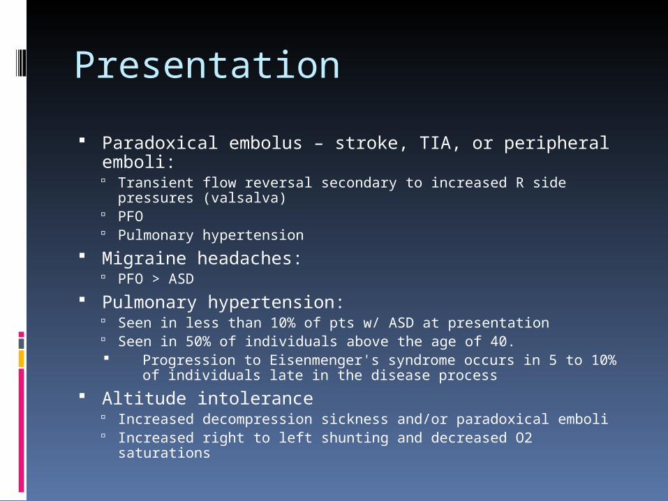
Presentation
Paradoxical embolus – stroke, TIA, or peripheral emboli: Transient flow reversal secondary to increased R side pressures
(valsalva) PFO Pulmonary hypertension
Migraine headaches: PFO > ASD
Pulmonary hypertension: Seen in less than 10% of pts w/ ASD at presentation Seen in 50% of individuals above the age of 40. Progression to Eisenmenger's syndrome occurs in 5 to 10% of
individuals late in the disease process
Altitude intolerance Increased decompression sickness and/or paradoxical emboli Increased right to left shunting and decreased O2 saturations
Physical Findings
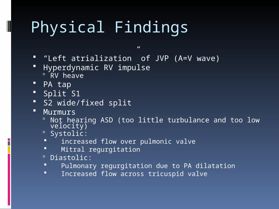
“Left atrialization” of JVP (A=V wave) Hyperdynamic RV impulse
RV heave PA tap Split S1 S2 wide/fixed split Murmurs
Not hearing ASD (too little turbulance and too low velocity) Systolic: increased flow over pulmonic valve Mitral regurgitation Diastolic: Pulmonary regurgitation due to PA dilatation Increased flow across tricuspid valve

Echocardiographic Evaluation Subcostal view most reliable: US
beam perpendicular to plane of IAS Other views may have loss of signal from the atrial
septum from parallel alignment Secundum ASD: central portion of
atrial septum (89% sensitivity) Primum ASD: adjacent to AV valve
annuli (100% sensitivity) Sinus Venosus defects: difficult to
visualize on TTE (44% sensitivity)
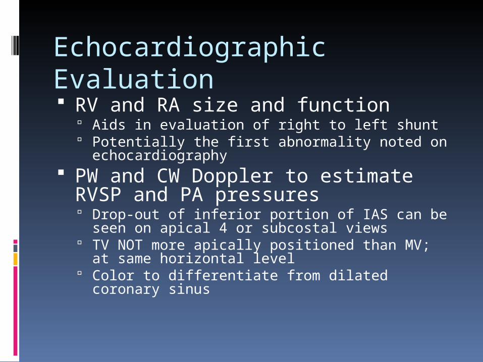
Echocardiographic Evaluation RV and RA size and function
Aids in evaluation of right to left shunt Potentially the first abnormality noted on
echocardiography PW and CW Doppler to estimate RVSP
and PA pressures Drop-out of inferior portion of IAS can be seen on
apical 4 or subcostal views TV NOT more apically positioned than MV; at same
horizontal level Color to differentiate from dilated coronary sinus

Echo:
Identify: Coronary sinus Entrance of pulmonary veins Primum portion of atrial septum Drop-out of inferior portion of IAS can be seen on
apical 4 or subcostal views TV NOT more apically positioned than MV; at same
horizontal level Color to differentiate from dilated coronary sinus

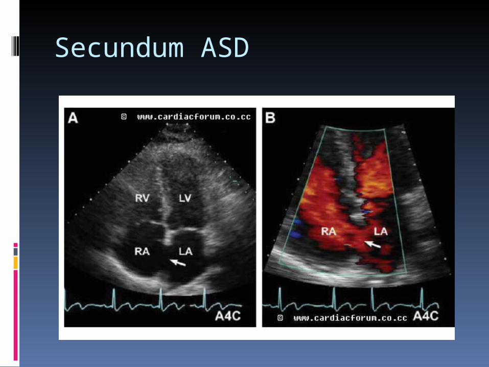
Echo: Secundum ASD
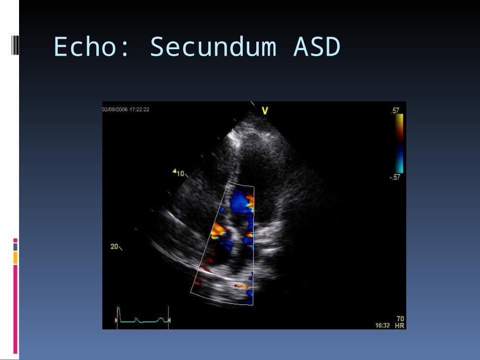
Echo: Secundum ASD
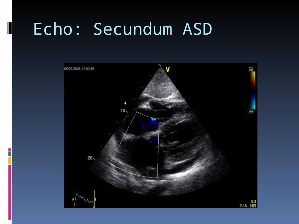
Echo: Secundum ASD

Echo: Primum ASD
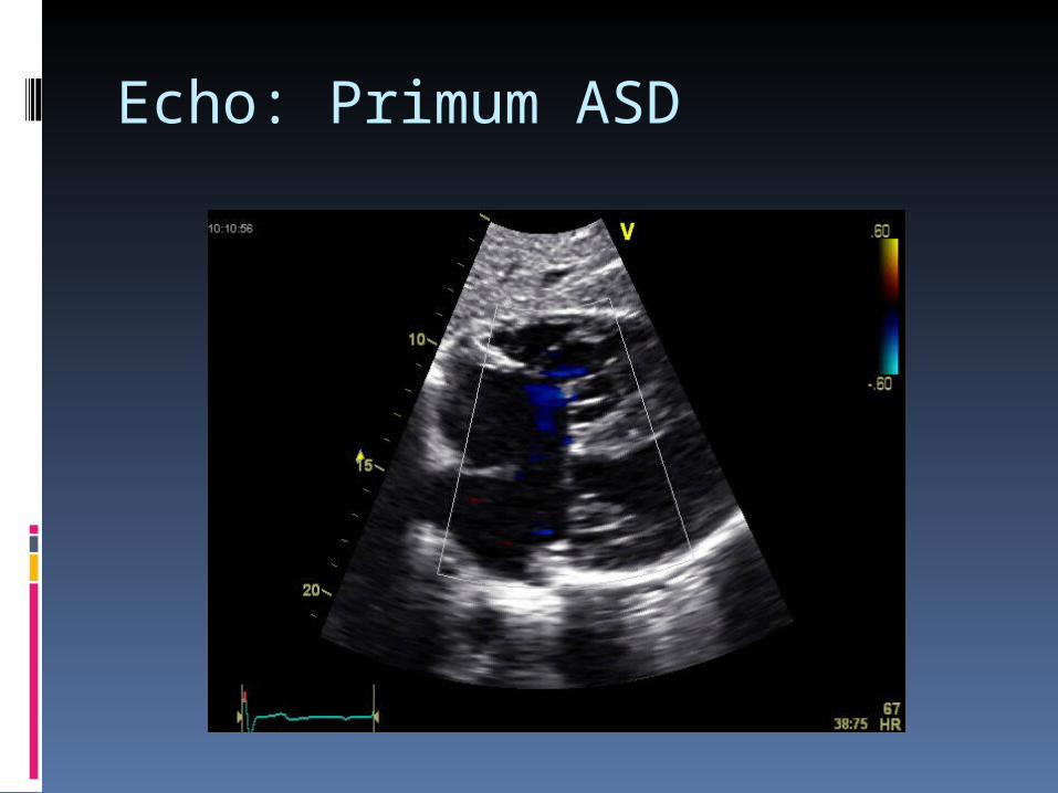
Echo: Primum ASD

Echo: Primum ASD

Echo: Primum ASD

Doppler Echocardiography
Color Doppler can identify left to right flow
Subcostal view is best Multiple views needed:
Low-velocity flow signal between atria
SVC flow along IAS can be mistaken for shunting
TR jet directed toward IAS can also be confused as a shunt

Doppler Echocardiography
Location and timing of flow critical (as oppsed to the velocity) Flow from L -> R atrium in both
systole and diastole More prominent diastolic component Can extend across open TV in diastole
into RV Seen in larger shunts Flow acceleration on side of LA
Absolute velocity of flow less important

Doppler Echocardiography
Shunt calculation: Can be performed utilizing these
equations to relate pulmonic CO and systemic CO
Qp = TVI pulm X PULd Qs = TVI lvot X LVOTd Qp/Qs = shunt fraction Significant usually if > 1.5/1.0 in
ASD

Constast Echocardiography Microbubbles seen across IAS
Even if shunting predominantly L to R
RA pressure transiently > LA pressure
“Negative” contrast jet: Flow from LA to RA appears as area
with no echo contrast Rarely needed for ASD - more
useful for smaller shunts (PFO’s)
TEE
Needed when TTE images are suboptimal Usually necessary to see sinus venosus
defect or partial anomalous pulmonary venous return
To locate small secundum ASDs Device sizing before percutaneous closure Estimation of defect size using the
diameter of the Doppler color flow jet correlates with surgical findings
TEE is often used when contrast echo suggests shunting, but a defect can’t be visualized on TTE. The TEE then helps to differentiate between a PFO and a true ASD
MRI
Phase constrast MRI compares well against the gold standard (invasive measurement) 93% sensitivity and specificity
for Qp/Qs > or = 1.5:1 100% sensitivity and
specificity for Qp/Qs > or = 1.7:1

MRI
Correlation of PC-MRI to TEE and IVBM (invasive balloon measurement) measurements of ASD size
Some studies have noted MR to have better correlation to balloon sizing of ASDs than TEE
MR also able to provide information about shape of ASD and proximity to adjacent structures Possible that TEE will not be able to measure the
largest section of the ASD if it is not round
ASD Closure
Percutaneous ASD closure was first performed 30 years ago First report percutaneous ASD closure of via Amplatzer septal
occluder in 1997 Successful closure in >80% of secundum ASDs Compared to surgical approach
Decreased LOS Decreased complication rate Same success rate
Determining factors: Location Size
<30 – 40 mm by TEE Rim
Initial studies used rim of ~5mm in all directions Some authors have proposed that it is the posterior inferior rim in
particular that must be of adequate size for successful transcatheter closure
ASD Closure
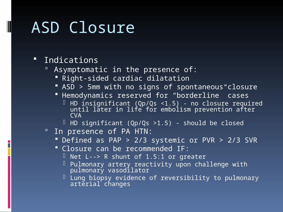
Indications Asymptomatic in the presence of:
Right-sided cardiac dilatation ASD > 5mm with no signs of spontaneous closure Hemodynamics reserved for “borderline” cases
HD insignificant (Qp/Qs <1.5) - no closure required until later in life for embolism prevention after CVA
HD significant (Qp/Qs >1.5) - should be closed In presence of PA HTN:
Defined as PAP > 2/3 systemic or PVR > 2/3 SVR Closure can be recommended IF:
Net L--> R shunt of 1.5:1 or greater Pulmonary artery reactivity upon challenge with pulmonary
vasodilator Lung biopsy evidence of reversibility to pulmonary arterial
changes
ASD Closure
Percutaneous indications: Only for Secundum ASD with
stretch diameter < 41 mm Need adequate rims to enable
secure device deployment Cannot have anomalous pulm
venous connection, be too proximal to AV valves, coronary sinus, or systemic venous drainage
~2/3rds of secundum ASDs meet this criteria

ASD Closure
Introduced in 1996. Approved for
percutaneous ASD closure in 2001 by F.D.A.
Over 90,000 have been manufactured and delivered to date.
Consists of two round disks made of Nitinol (nickel + titanium) wire mesh linked together by a short connecting waist.
ASD Closure
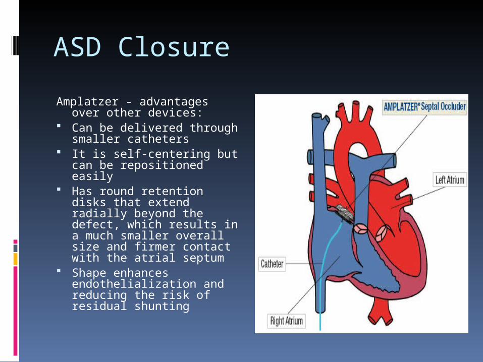
Amplatzer - advantages over other devices:
Can be delivered through smaller catheters
It is self-centering but can be repositioned easily
Has round retention disks that extend radially beyond the defect, which results in a much smaller overall size and firmer contact with the atrial septum
Shape enhances endothelialization and reducing the risk of residual shunting

ASD Closure
Complications of percutaneous closure: Thrombus formation on the device leading to CVA
Decreased in newer devices ASA and plavix after procedure for ~6 months Heart block, effusion, and thrombus formation in LA
(2.4%) Device embolization and/ or malposition (2.4%) Atrial fibrillation (2.4%) Erosion (0.1%):
aortic to right or left atrial fistula Free-wall perforation of the atria resulting in tamponade Factors associated with erosion:
Amplatzer Septal Occluder size greater than 4 mm larger than the unstretched ASD
Device size greater than 1.5 times the size of the unstretched ASD
ASD Closure
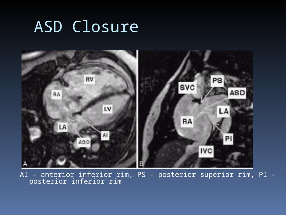
AI – anterior inferior rim, PS – posterior superior rim, PI – posterior inferior rim

ASD Closure
TEE Can be used to evaluate suitability of
transcutaneous approach Monitoring during interventional
procedure Measure stretch diameter of ASD Doppler to look for residual shunting
during occlusion of the ASD with the balloon
Doppler to look for residual shunting after occluder is in place

ASD Closure
Decreased rim A more technically difficult
transcutaneous procedure with higher rates of failure
Long term complications are increased Erosion of device through cardiac
wall and formation of fistulas.
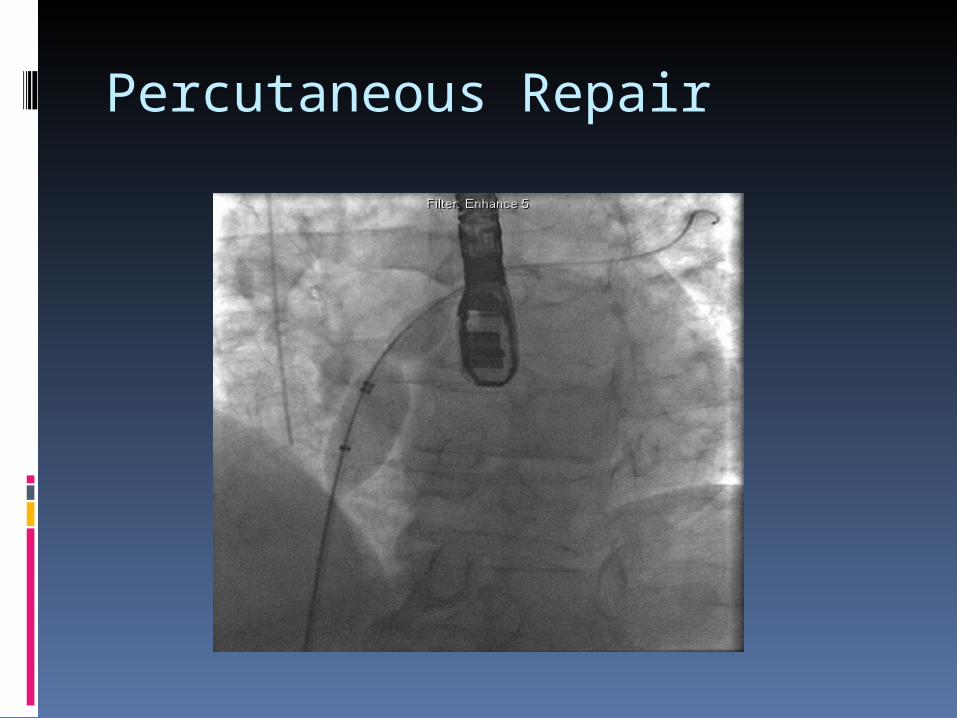
Percutaneous Repair
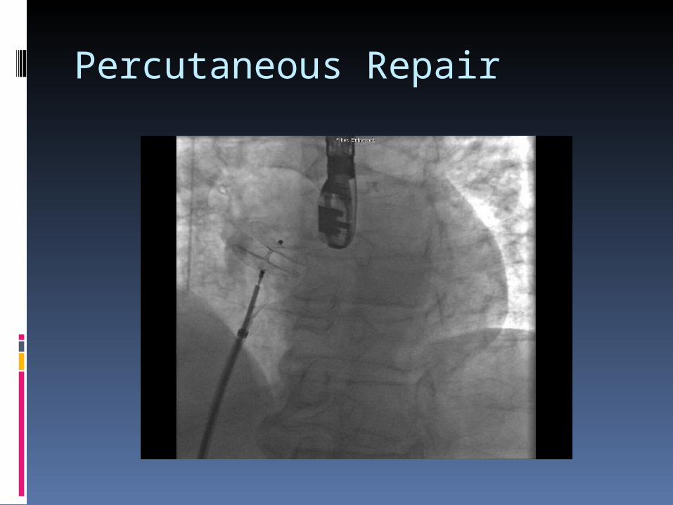
Percutaneous Repair
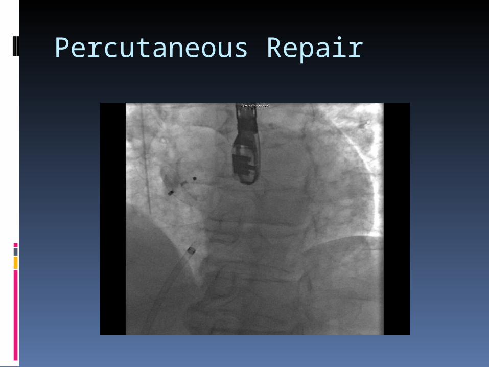
Percutaneous Repair
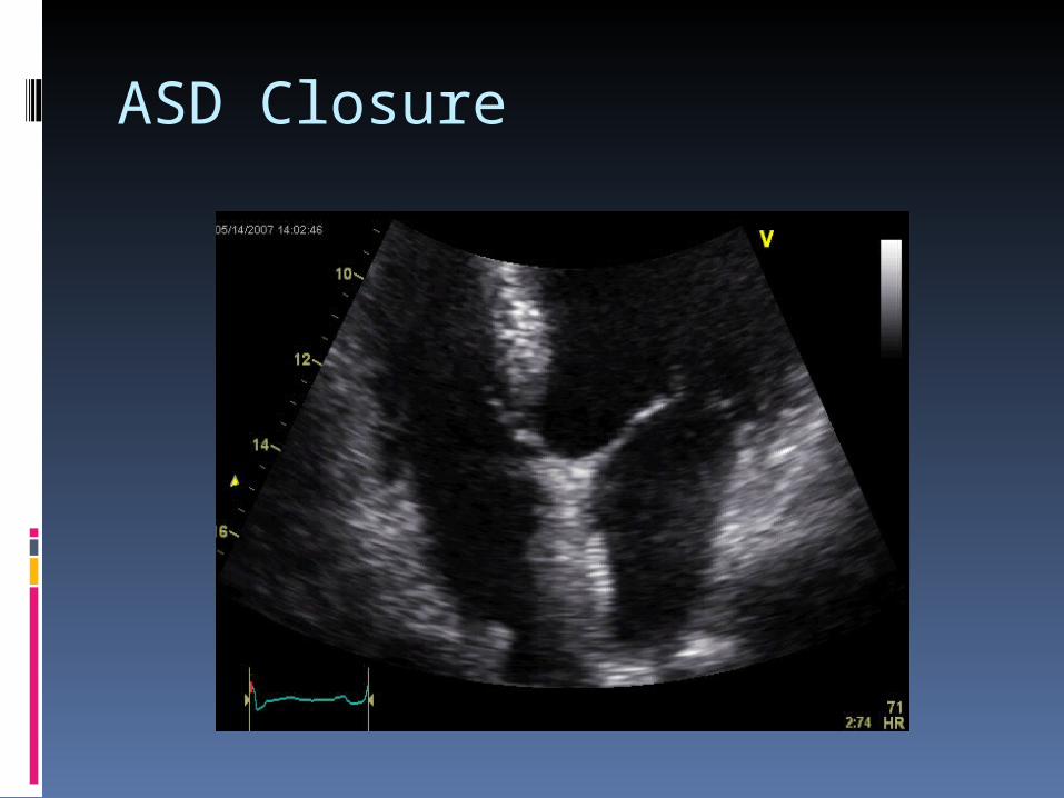
ASD Closure

ASD Closure
Surgical Indications Reserved for cases that are not
candidates for percutaneous closures: Non-secundum ASDs Secundum ASDs with unsuitable anatomy Primary suture vs tissue/synthetic patch Symptomatic improvement seen Does not prevent AF/aflutter in adults
(especially >40 years old) Concomitant MAZE a consideration
ASD Closure
Surgical outcomes: Surgery before the age of 25 yields in 30-year
survival rates comparable to age- and sex-matched controls.
At 25-40 years of age, surgical survival is reduced, though not significantly if PA pressures are normal.
If PASP > 40 mmHg, late survival is 50% less than control rates, though life expectancy in surgically treated older patients is better than that of medically treated patients.
No benefit of surgery in reducing the incidence of AF, though the patient’s age at the time of closure is the most important predictor of the development of atrial arrhythmias.

Stroke Risk
Data are widely conflicting on the relationship between PFO, atrial septal aneurysm, and/or ASD and recurrent cerebral emboli. Increased prevalence of PFO and ASA in cryptogenic
stroke; less clear for ASD. The role of defect closure vs. medical therapy
for prevention of recurrent stroke is not well defined.
Aspirin is often used in setting of PFO or an isolated atrial septal aneurysm, and especially if PFO + ASA. Role of coumadin is not as clear – coumadin recommended if patient has a documented DVT/PE. Less data available for ASDs.
Surgical excision of an atrial septal aneurysm (without PFO or ASD) may be considered if aspirin or coumadin fail to prevent a recurrent embolic event.