atrial septal defects david m. chaky, md. terminology ► asd = defect in the atrial septum of the...
TRANSCRIPT

Atrial septal defectsAtrial septal defects
David M. Chaky, MDDavid M. Chaky, MD

TerminologyTerminology
► ASD = defect in the atrial septum of the ASD = defect in the atrial septum of the heart which can be isolated anomaly or heart which can be isolated anomaly or associated with other congenital heart associated with other congenital heart lesionslesions
► 10% of congenital heart lesions in children 10% of congenital heart lesions in children yet 30% of congenital heart lesions in adultsyet 30% of congenital heart lesions in adults

Types of ASDTypes of ASD► Foramen ovale is nl interatrial communication that in Foramen ovale is nl interatrial communication that in
utero allows flow from IVC to enter the left atriumutero allows flow from IVC to enter the left atrium
► Secundum ASD is oval defect bordered by fossa Secundum ASD is oval defect bordered by fossa ovalisovalis
► Ostium primum defect occurs in anterior and inferior Ostium primum defect occurs in anterior and inferior portion of the septumportion of the septum
► AV canal defect involves atrial and septal portions of AV canal defect involves atrial and septal portions of the septumthe septum
► Sinus benosus defect occurs superiorly in atrial Sinus benosus defect occurs superiorly in atrial septum near SVCseptum near SVC
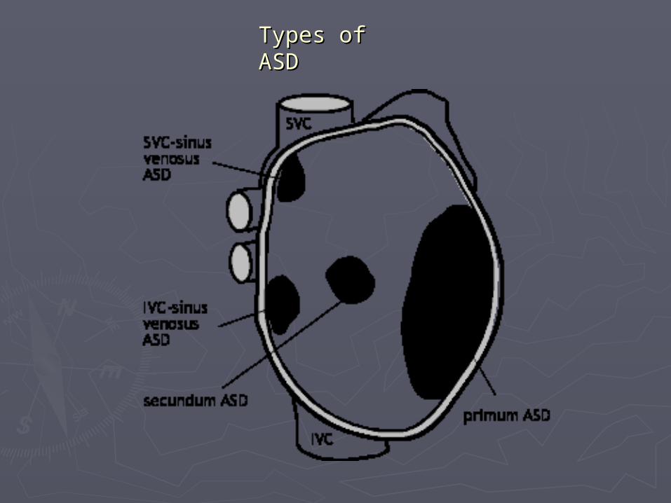
Types of Types of ASDASD

PathologyPathology
► Embryological defect occurs in 5Embryological defect occurs in 5thth week of week of gestationgestation
► ASD: low pressure shuntsASD: low pressure shunts► VSD, AVSD: high pressure shuntsVSD, AVSD: high pressure shunts► Eventually all shunts lead to pulmonary HTN Eventually all shunts lead to pulmonary HTN
if untreatedif untreated►Holt Oram syndrome = ASD with upper Holt Oram syndrome = ASD with upper
extremity anomaliesextremity anomalies►Ostium primum and AVSD associated with Ostium primum and AVSD associated with
trisomy 21 in 65% of childrentrisomy 21 in 65% of children

Imaging ASDImaging ASD
►Primary diagnosis made by Primary diagnosis made by echocardiography in infants and echocardiography in infants and childrenchildren
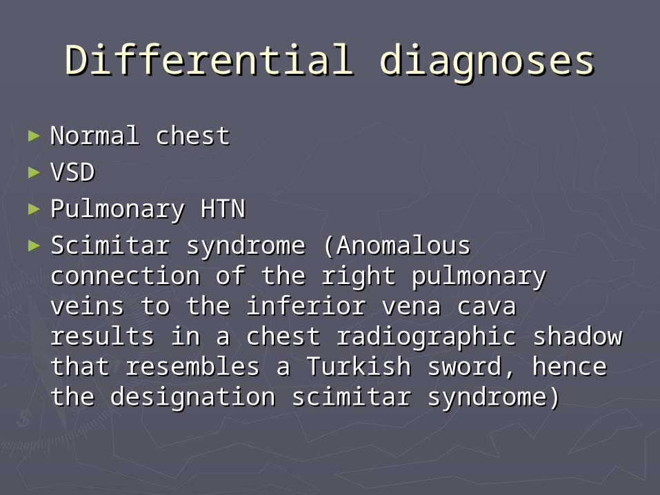
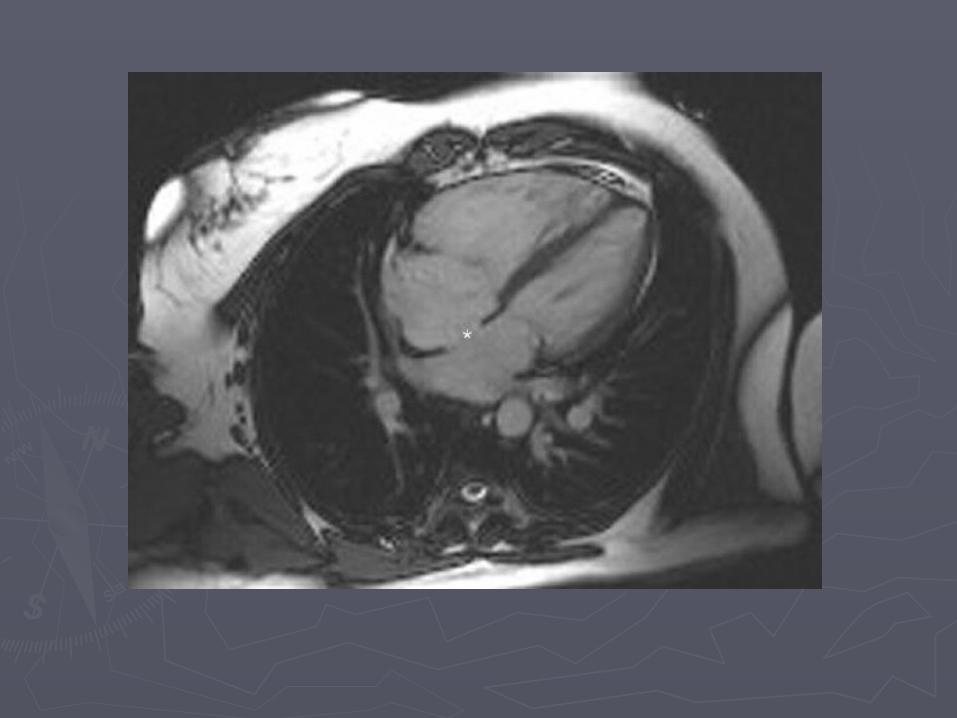
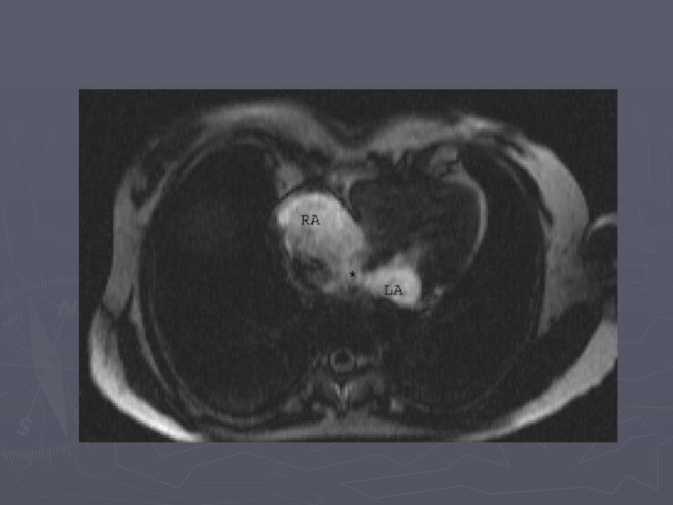
►MRI is emerging as accurate MRI is emerging as accurate alternative for depiction of function, alternative for depiction of function, flow, and anatomy in older patientsflow, and anatomy in older patients

TreatmentTreatment
►Secundum = many spontaneously close, Secundum = many spontaneously close, transcatheter percutaneous closure transcatheter percutaneous closure device if possible, large defects may device if possible, large defects may need Dacron patch or direct closureneed Dacron patch or direct closure
►Primum = surgery at age 3-5 years (too Primum = surgery at age 3-5 years (too close to AV valve for device closure)close to AV valve for device closure)
►AV canal = surgery in 1AV canal = surgery in 1stst year of life year of life►Sinus venosus = surgerySinus venosus = surgery

FindingsFindings
► XR – small to moderate defects have normal XR – small to moderate defects have normal radiographsradiographs
► XR - large defects have cardiomegaly with XR - large defects have cardiomegaly with the main PA normal or enlarged, with shunt the main PA normal or enlarged, with shunt vascularity, later on PA and RV enlargevascularity, later on PA and RV enlarge
► CTA – defect in atrial septum, enlargement CTA – defect in atrial septum, enlargement of RA, RV and PAof RA, RV and PA
►MR – can quantify shunt volume on MR – can quantify shunt volume on volumetric cine MR or velocity encoded cine volumetric cine MR or velocity encoded cine MRMR

FindingsFindings
►Echo – “drop out” in atrial septum best Echo – “drop out” in atrial septum best seen on apical 4 chamber viewseen on apical 4 chamber view
►Angio – can also be used for Angio – can also be used for transcatheter percutaneous treatmenttranscatheter percutaneous treatment
Differential diagnosesDifferential diagnoses
►Normal chestNormal chest►VSDVSD►Pulmonary HTNPulmonary HTN►Scimitar syndrome (Anomalous Scimitar syndrome (Anomalous
connection of the right pulmonary veins connection of the right pulmonary veins to the inferior vena cava results in a to the inferior vena cava results in a chest radiographic shadow that chest radiographic shadow that resembles a Turkish sword, hence the resembles a Turkish sword, hence the designation scimitar syndrome)designation scimitar syndrome)


► Frontal chest radiograph in adult patient with the same entity (image Frontal chest radiograph in adult patient with the same entity (image 3) shows moderate to severe cardiomegaly with enlarged central 3) shows moderate to severe cardiomegaly with enlarged central pulmonary arteries and peripheral tapering of pulmonary vessels. pulmonary arteries and peripheral tapering of pulmonary vessels.

ASDS, particularly the sinus venosus defects, are commonly associated with anomalous connection of the right pulmonary veins, pariticularly the upper lobe veinds to either the right atrium or SVC.

ReferencesReferences
►Kuhn, JP. CAFFEY'S PEDIATRIC Kuhn, JP. CAFFEY'S PEDIATRIC DIAGNOSTIC IMAGING, 10 ed., Mosby, DIAGNOSTIC IMAGING, 10 ed., Mosby, 2004, pg 1381 Diangostic Imaging 2004, pg 1381 Diangostic Imaging Pediatrics, Donnelly, Lane, MD Section Pediatrics, Donnelly, Lane, MD Section 3 pages 104-106, 2005. 3 pages 104-106, 2005.