attenuation of intermittent hypoxia-induced apoptosis and
TRANSCRIPT

RESEARCH ARTICLE Open Access
Attenuation of intermittent hypoxia-induced apoptosis and fibrosis inpulmonary tissues via suppression of ERstress activationZhihui Shi1,2,3†, Linhao Xu3,4†, Hui Xie3, Ruoyun Ouyang1,2, Ya Ke3, Rui Zhou1,2* and Wing-Ho Yung3*
Abstract
Background: Obstructive sleep apnea (OSA) is associated with pulmonary fibrosis and endothelial apoptosis inpulmonary tissues. Chronic intermittent hypoxia (IH) is considered to be the primary player in OSA, but themechanisms underlying its effect on pulmonary tissues are unknown. Endoplasmic reticulum (ER) stress inducedby IH treatment plays an important role in accelerating the process of fibrosis and induction of apoptosis.
Methods: Mice were placed in IH chambers for 4 weeks with an oscillating oxygen (O2) concentration between 5and 21%, cycling every 90s for 8 h daily. Mice were randomly divided into four groups: control group (normaloxygen), tauroursodeoxycholic acid (TUDCA) group (normal oxygen intraperitoneally injected with TUDCA), IHgroup and IH + TUDCA group. After 4 weeks, the proteins in three branch signaling pathways of ER stress, includingprotein kinase RNA (PKR)-like/Pancreatic ER kinase (PERK), activating transcription factor 6 (ATF-6) and inositol-requiring enzyme 1 (IRE-1), were evaluated. The cleaved caspase-3, caspase-12 and TUNNEL staining was assessed.Furthermore, the expression of transforming growth factor-β1 (TGF-β1) and thrombospondin-1(TSP-1), twoextracellular matrix proteins that play critical role in fibrosis, were examined. Finally, Masson’s trichrome stainingwas performed to detect the expression of collagen.
Results: After 4 weeks of IH treatment, the expressions of two ER stress markers, glucose regulated protein-78(Grp78) and transcription factor C/EBP homologous protein (CHOP) were increased which was prevented byadministration of the ER stress attenuator, TUDCA. The expressions of PERK, but not those of ATF-6 and IRE-1,were increased. The effects of IH were accompanied by an increased number of apoptotic cells and increasedexpressions of cleaved caspase-3 and caspase-12 in pulmonary tissues. In addition, histological examinationsuggested the presence of fibrosis after chronic IH treatment, indicated by increased expression of collagen, whichwas associated with the up-regulation of TGF-β1 and TSP-1 that are known to promote fibrosis. Similarly, TUDCAcould reduce the extent of fibrotic area and the expression levels of these proteins.(Continued on next page)
© The Author(s). 2020 Open Access This article is licensed under a Creative Commons Attribution 4.0 International License,which permits use, sharing, adaptation, distribution and reproduction in any medium or format, as long as you giveappropriate credit to the original author(s) and the source, provide a link to the Creative Commons licence, and indicate ifchanges were made. The images or other third party material in this article are included in the article's Creative Commonslicence, unless indicated otherwise in a credit line to the material. If material is not included in the article's Creative Commonslicence and your intended use is not permitted by statutory regulation or exceeds the permitted use, you will need to obtainpermission directly from the copyright holder. To view a copy of this licence, visit http://creativecommons.org/licenses/by/4.0/.The Creative Commons Public Domain Dedication waiver (http://creativecommons.org/publicdomain/zero/1.0/) applies to thedata made available in this article, unless otherwise stated in a credit line to the data.
* Correspondence: [email protected]; [email protected]†Zhihui Shi and Linhao Xu contributed equally to this work.1Department of Respiratory and Critical Care Medicine, The Second XiangyaHospital, Central-South University, Changsha, China3School of Biomedical Sciences, Faculty of Medicine, The Chinese Universityof Hong Kong, Shatin, Hong Kong, SAR, ChinaFull list of author information is available at the end of the article
Shi et al. BMC Pulmonary Medicine (2020) 20:92 https://doi.org/10.1186/s12890-020-1123-0
(Continued from previous page)
Conclusions: It reveals the roles of ER stress, especially the PERK pathway, in IH induced apoptosis and fibrosis inpulmonary tissues that might underlie the pulmonary complications observed in OSA.
Keywords: Obstructive sleep apnea, Endoplasmic reticulum stress, Intermittent hypoxia, Apoptosis, Fibrosis
BackgroundObstructive sleep apnea (OSA) is a common disordercharacterized by repetitive collapse of the pharyngeal air-way during sleep, resulting in intermittent hypoxia (IH)and reoxygenation. Several previous reports have indi-cated that the morphology and function of the lung arealtered in OSA patients and IH animal models, includingreductions in lung volumes and induction of pulmonaryhypertension [1–3]. These symptoms were associatedwith pulmonary fibrosis and endothelial apoptosis [4–6].According to previous studies, endoplasmic reticulum(ER) stress activation had been found in IH model andplays a critical role in apoptosis [7, 8].It is well known that ER stress is caused by conditions
that perturb the processing and folding of proteins,resulting in the accumulation of misfolded proteins [9].Under IH condition, increased reactive oxygen species(ROS) production caused by the inhibition of complex Iactivity may lead to a loss of ER homeostasis and accu-mulation of misfolded proteins, and in turn activates ERstress [10, 11]. At the early stage of ER stress, unfoldedprotein response (UPR) was activated to enhance ERchaperone protein production, such as glucose regulatedprotein-78 (Grp78). This molecular chaperone could re-store ER function via facilitating protein folding [12]. Ifthe activation of ER stress is prolonged, ER stress-mediated apoptosis can be induced via three pathways,including protein kinase RNA (PKR)-like/Pancreatic ERkinase (PERK), activating transcription factor 6 (ATF-6)and inositol-requiring enzyme 1 (IRE-1) pathways [13].These three pathways activate pro-apoptotic via the in-duction of the pro-apoptotic transcription factor C/EBPhomologous protein (CHOP) [14, 15].On the other hand, once ER stress is initiated, it is be-
lieved that fibrosis will be induced due to the accelerationof fibroblast proliferation and the expression of extracellu-lar matrix protein, such as transforming growth factor-β1(TGF-β1) and thrombospondin-1(TSP-1) [16, 17]. In theclinics, pulmonary fibrosis was noted in OSA patients [4,18]. However, the major mechanisms underlying the ef-fects of IH treatment on the pathological changes and dys-function of pulmonary tissue are still largely unknown.We hypothesized that ER stress has a major contributionsince inhibition of ER stress activation could potentiallyattenuate fibrosis and apoptosis [19].At present, the most commonly used methods in the
clinical treatment of OSA are surgery and continuous
positive airway pressure (CPAP); however, each of thesetwo methods has their own shortcomings and it is noteffective for all patients [20, 21]. Therefore, there is anurgency in seeking a new therapeutic approach. Fordrug-based therapy, there have only been a few studiesup to now. In order to identify precise and therefore bet-ter target sites for drugs, it is worth investigating themolecular mechanisms of pulmonary fibrosis and apop-tosis in OSA.Therefore, in order to investigate whether ER stress is
present and its role in fibrosis and apoptosis in lung tis-sue after exposure to IH, tauroursodeoxycholic acid(TUDCA), a chemical chaperone that has been shown toreduce ER stress by facilitating proper protein foldingand trafficking [22], was used to suppress ER stress acti-vation and to observe the effect of reducing the numberof apoptotic neurons and the progression of fibrosis.
Materials and MethodsAnimalsThirty-two C57BL/6 male mice (20–22 g) werepurchased from the Animal Center of the ChineseUniversity of Hong Kong (CUHK). The mice were keptin plastic cages under specific pathogen-free conditionswith controlled lighting (12 h per day) and temperature(21 ± 2 °C). They were allowed free access to standard la-boratory food and water at the animal laboratories of theCUHK. Four mice were placed in one cage. The proce-dures of experimentation were conducted with approvalof the Animal Experimentation and Ethics Committee ofthe CUHK.
ReagentsTUDCA was purchased from Sigma-Aldrich (St. Louis,MO). The BCA protein assay kit was purchased fromPierce Biotechnology (Rockford, USA). Antibodies, in-cluding glucose regulated protein-78 (Grp78, cat:3177),p-PERK (cat: 3179), activating transcription factor4(ATF-4, cat:11815), phosphorylated eukaryotic initi-ation factor 2 alpha (p-elf2α, cat: 3398), caspase-3(cat:9661) were purchased from Cell Signaling Technology(Beverly, MA). Caspase-12 (cat: ab62484) and transcrip-tion factor C/EBP homologous protein (CHOP cat:ab11419) were purchased from Abcam (Cambridge,USA). TUNEL kit was purchased from MilliporeCorporation (Massachusetts, USA).
Shi et al. BMC Pulmonary Medicine (2020) 20:92 Page 2 of 11

Model of chronic intermittent hypoxia and TUDCAtreatmentThe protocol was well-accepted and utilized by other re-searchers to generate a sleep apnea model in rodent [8,23–25]. Ordinary cages were placed in computer-controlled ventilation chambers (Oxycycler modelA48XOV, Redfield, USA) to achieve IH exposures in theanimals. The concentration of oxygen (O2) was main-tained between 5 and 21%, cycling every 90s for 8 h (8:00 A.M. to 4:00 P.M. Fig. 1). Control animals were ex-posed to alternating periods of room air in identicalchambers. The intermittent hypoxia treatment lasted for4 weeks.According to our previous study [8], TUDCA dis-
solved in phosphate buffer saline (PBS) was intra-peritoneally injected daily (100 mg/kg) at 7:30 AM to 8:00 AM before IH treatment. The control group receivedPBS alone. The mice were randomly assigned into fourgroups: PBS-treated group (control group), TUDCA-treated group (TUDCA group), PBS-treated IH group(IH group) and TUDCA-treated IH group (IH + TUDCAgroup).
Tissue processingAfter 4 weeks of IH exposure, the mice were deeplyanesthetized with pentobarbital sodium salt (Sigma-Al-drich, Darmstadt, Germany) at the dosage of 30 mg/kgby intraperitoneal injection. Then, four mice of eachgroup were decapitated to obtain pulmonary tissues(upper lobe of left lung), which were stored at − 70 °Cfor Western blot analysis and quantitative real-time RT-PCR analysis. The thoracic cavities of another four micein each group were opened by surgical scissors and per-fused with 30–40ml fixative containing 4% paraformal-dehyde in 0.1 M phosphate buffer (pH 7.4) through thecardiac aorta. After perfusion, the left lung was removed.
The tissue was post-fixed in the same fixative for 6 hand embedded in paraffin, and cut into 4 μm sections.
Western blot analysisProteins were obtained from pulmonary tissue, whichwas homogenized with ice-cold radioimmunoprecipita-tion assay buffer containing 1 mM of PMSF and phos-phatase inhibitor cocktail (Roche, Germany). BCAProtein Assay Reagent Kit (Pierce Biotechnology,Rockford, USA) was used to measure the protein con-centration. Protein was separated by 12% sodiumdodecyl sulfate-polyacrylamide gel electrophoresis andtransferred to a PVDF membrane (Millipore Corpor-ation; Billerica, MA, USA). Non-fat dry milk (5%) wasused to block the menbranes. The blocked membraneswere then incubated with the primary antibody (Grp-78,p-PERK, ATF-4, p-elf2α, caspase-3, caspase-12 andCHOP) at 4 °C overnight. After stringent washes withTris-buffered saline containing 0.1% Tween-20 (TBST),blots were incubated with a fluorescent secondary anti-body (LI − COR Biotechnology, Lincoln, USA) for 1 h atroom temperature. An Odyssey scanner was used to de-tect the intensities of the specific bands (LI −COR Bio-technology, Lincoln, USA). Equal amount of targetprotein was confirmed by β-actin antibody (Bio-Rad La-boratories, California, USA).
Histological stainingParaffin sections were rinsed in 0.01M PBS andmounted onto 0.02% poly-L-lysine-coated slides. The ex-pression of collagen was detected by Masson’s trichromestaining which stains cell nuclei, cytoplasm, and collagenin dark brown, red and blue color respectively.Section was firstly stained by Weigert’s working
hematoxylin for 10 min. After washing with distilledwater, they were then stained with Biebrich scarlet for 5
Fig. 1 Schematics of the intermittent hypoxia (IH) model used in this study. a Conventional ventilated cages that mimic usual housing conditionsare placed in computerized hypoxic chambers to achieve IH exposures in mice. The system is composed of the Oxycycler, which is responsiblefor gas (N2, O2), air-inlet regulation, gas tank (N2, O2), computer (not shown) and the intermittent hypoxia chamber. b The paradigm ofintermittent hypoxia in OSA model. The concentration of oxygen (O2) was maintained between 5 and 21% cycling in every 90s
Shi et al. BMC Pulmonary Medicine (2020) 20:92 Page 3 of 11
min. Finally, the sections were treated with phosphomo-lybdic acid and differentiated by aniline blue. After stain-ing, the sections were dehydrated with absolute alcoholand xylene, then cover-slipped with Permount, and thesections were observed under a light mi croscope (ZeissMicroscope Axiophot 2) by an investigator blind to thetreatment. Three or four slices from each mouse werequantifed and a total of 15 slices from four mice in eachgroup were analyzed. The area of collagen expressionwas automatically identified by Metamorph 7.5 softwarewith the function of colocalization and deconvolution(Molecular Devices, USA).
Quantitative real-time RT-PCR analysisTotal RNA was isolated from pulmonary tissue homoge-nates using TRIzol reagent (Invitrogen, USA). The RNAconcentration was measured by NanoDrop (Thermo,USA) and complementary DNA (cDNA) was synthesizedusing a Transcriptor First Strand cDNA Synthesis Kit(TaKaRa, Japan). For real-time qPCR, cDNA was firstlydenatured by heating to 94 °C for 3 min and amplified by40 cycles of PCR (denaturation at 94 °C for 15 s, anneal-ing at 60 °C for 1 min, and extension at 72 °C for 30 s). Asingle production was confirmed by the dissociationcurve. PCR primers (Invitrogen, USA) used in this studyare as follows:Grp-78: (forward: 5′-GGTGCAGCAGGACATCAA
GTT-3′;reverse: 5′-CCCACCTCCAATATCAACTTGA-3′);CHOP: (forward: 5′-CTGCCTTTCACCTTGGAGAC-3′;reverse: 5′- CGTTTCCTGGGGATGAGATA - 3′);Frameshift splice X box-binding protein 1 (XBP1-s):
(forward: 5′ -AAGAACACGCTTGGGAATGG-3′;reverse: 5′- ACTCCCCTTGGCCTCCAC -3′);ATF4: (forward: 5′ -GCAGCAGCACCAGGCTCT-3′;reverse: 5′-TTGTCCGTTACAGCAACA CTG-3′);TSP-1: (forward: 5′-CACCTCTCCGGGTTACTGAG-3′;reverse: 5′-GCAACAGGAACAGGACACCTA-3′);TGF-β1: (forward: 5′-CCGCAACAACGCCATCTA
TG-3′;reverse: 5′-CTCTGCACGGGACAGCAAT-3′);β-actin: (forward: 5′-ACCCACACTGTGCCCATCTA-3′;reverse: 5′- CACGCTCGGTCAGGATCTTC-3′).The results were calculated by the 2-△△Ct method
based on previous study [7].
TUNEL stainingApoptosis was detected by in situ terminal transferasemediated dUTP nick end labeling (TUNEL technique),using an ApopTag1 kit (Millipore Corporation, MA) andfollowing the instructions of the manufacturer. First, thesection (5 μm thick) of pulmonary tissue were incubatedwith proteinase-K (20 μg/ml in PBS) for 15 min at roomtemperature. Second, the sections were washed by PBS
and then quenched in 3% H2O2 in PBS for 5 min.. Third,they were then treated with biotin–deoxyuridine triphos-phate in the working solution of deoxynucleotidyl trans-ferase for 1 h in a humidified chamber at 37 °C. Fouth,the reaction was stopped with buffer solution at roomtemperature. The sections were then rinsed with PBS forthree times, and anti-digoxigenin-peroxidase wereapplied and incubated for 30 min at room temperature.Finally, the sections were reacted with 0.05% diamino-benzidine with 0.01% H2O2, and counterstained in 0.5%methyl green. After processing and dehydration, theslides were mounted with Permount and observed undera light microscope (Zeiss Microscope Axiophot 2, USA).Three or four slices from each mouse were quantifiedand a total of 15 slices from four mice in each groupwere analyzed.
Statistical analysisData are presented as means ± standard error of means.The results were analyzed by two-way ANOVA followedby a Newman-Keuls post hoc test for multiple compari-sons. Difference with P-value less that 0.05 was consid-ered significant.
ResultsChronic IH induces ER stress in pulmonary tissueTo test whether ER stress could be triggered in pulmon-ary tissue by chronic IH, mice were subject to 4 weeks ofIH exposure, which consisted of daily 8 h of fluctuatingO2 level between 21 and 5% in every 90s in the ambientenvironment (see Materials and Methods and Fig. 1).Western blot and real-time PCR were used to explorethe protein and gene expression levels respectively ofGrp78 and CHOP. As shown in Fig. 2a, after 4 weeks ofIH treatment, two-way ANOVA disclosed that bothTUDCA administration and IH treatment had signifi-cantly effects on the protein level of Grp78 [TUDCA ad-ministration: F (1,12) = 5.10, P = 0.0433; IH treatment: F(1,12) = 8.91, P = 0.0114]. However, two-way ANOVAconfirmed that only IH treatment had a significant effecton the protein level of CHOP [TUDCA administration:F (1,12) = 0.47, P = 0.5046; IH treatment: F (1,12) = 26.57,P < 0.001; Fig. 2b]. Post hoc Newman-Keuls revealed thatwhile administration of the ER stress inhibitor TUDCAhad no effect on the expression levels of these two mole-cules in naïve animals, co-treatment of TUDCA duringIH exposure could significantly suppress the increase ofthese two molecues when compared with control group(P < 0.01, Fig. 2a and b). At the same time, the mRNAlevels of Grp78 and CHOP were significantly affectedafter 4 weeks of IH treatment by two-way ANOVA ana-lysis [F (1,12) = 10.89, P = 0.0063 for the expression ofGrp78 mRNA; F (1,12) = 11,14, P = 0.0059 for the ex-pression of CHOP, Fig. 2c and d]. However, TUDCA
Shi et al. BMC Pulmonary Medicine (2020) 20:92 Page 4 of 11

administration did not affect these two mRNA levels [F(1,12) = 2.12, P = 0.1713 for Grp78 mRNA; F (1,12) =3.24; P = 0.0971 for CHOP mRNA]. Post hoc Newman-Keuls revealed TUDCA treatment could suppress in-creased Grp78 and CHOP mRNA expression induced byIH (P < 0.05, Fig. 2c and d). These findings confirm thatchronic IH exposure activates ER stress in pulmonarytissue.
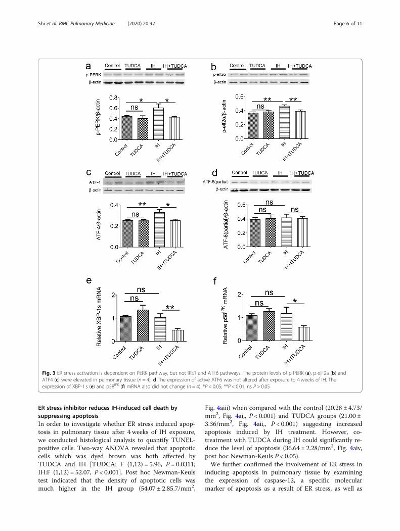
IH-induced ER stress is dependent on the PERK but notthe IRE1 and ATF6 pathwaysTo dissect the exact pathways leading to ER stress inpulmonary tissue, we examined the protein or gene ex-pression levels of specific molecules in these differentpathways. First, a number of proteins involved in thePERK pathways were assessed. As shown in Fig. 3a-c,compared with the control, two-way ANOVA revealedthat TUDCA could affect the expression of p-PERK,ATF-4, but not p-elf2α[F (1, 12) = 5.79, P = 0.0331 for p-PERK; F (1,12) = 7.56, P = 0.0176 for ATF-4; F (1,12) =3.19, P = 0.0995 for p-elf2α], meanwhile, IH treatmenthad a significant effect on the expression of p-elf2α andATF-4 [F (1,12) = 12.9, P = 0.0036 for p-elf2α; F (1,12) =
6.81, P = 0.0228 for ATF-4; F (1,12) = 4.10, P = 0.0659 forp-PERK]. Post hoc Newman-Keuls revealed the expres-sion of p-PERK, ATF-4 and p-elf2α were significantly in-creased after IH exposure, which were rectified byTUDCA treatment (P < 0.01). In contrast, the level ofATF6 was not affected by TUDCA or IH treatment[TUDCA: F (1,12) = 0.006, P = 0.9383; IH: F (1,12) = 0.13,P = 0.7224, Fig. 2d]. For the IRE1 pathway, the mRNAlevels of XBP-1 s were also altered by IH application[TUDCA: F (1,12) = 0.31, P = 0.5897; IH: F (1,12) = 18.91,P < 0.001], However, the mRNA levels of p58IPK was notaffected by TUDCA and IH [TUDCA:F (1,12) = 1.96,P = 0.1871; IH: F (1,12) = 3.94, P = 0.07]. Post hoc New-man–Keuls’s test indicated that these two mRNA levelswere unaffected in IH group when compared with con-trol group (P > 0.05, Fig. 3e and f). The only significanteffect that we observed was a decrease in XBP-1 s andp58IPK level in the IH group in the presence of TUDCAwhen compared with IH group (P < 0.05, Fig. 3e and f).Together, these results indicate that IH-induced ERstress in pulmonary tissue is dependent specifically onthe PERK pathway but not the IRE1 and ATF6pathways.
Fig. 2 ER stress activation in pulmonary tissue after exposure to 4 weeks of intermittent hypoxia (IH). The expression levels of Grp78 (a) andCHOP (b) in whole pulmonary tissue homogenates were detected by Western blot. Grp78 and CHOP were upregulated after 4 weeks of IHtreatment, which were prevented by the injection of TUDCA (n = 4). The expression of Grp78 (c) and CHOP (d) mRNA in pulmonary tissue wasincreased after 4 weeks of IH treatment (n = 4).. *P < 0.05; **P < 0.01; ***P < 0.001; ns P > 0.05
Shi et al. BMC Pulmonary Medicine (2020) 20:92 Page 5 of 11
ER stress inhibitor reduces IH-induced cell death bysuppressing apoptosisIn order to investigate whether ER stress induced apop-tosis in pulmonary tissue after 4 weeks of IH exposure,we conducted histological analysis to quantify TUNEL-positive cells. Two-way ANOVA revealed that apoptoticcells which was dyed brown was both affected byTUDCA and IH [TUDCA: F (1,12) = 5.96, P = 0.0311;IH:F (1,12) = 52.07, P < 0.001]. Post hoc Newman-Keulstest indicated that the density of apoptotic cells wasmuch higher in the IH group (54.07 ± 2.85.7/mm2,
Fig. 4aiii) when compared with the control (20.28 ± 4.73/mm2, Fig. 4ai,, P < 0.001) and TUDCA groups (21.00 ±3.36/mm2, Fig. 4aii,, P < 0.001) suggesting increasedapoptosis induced by IH treatment. However, co-treatment with TUDCA during IH could significantly re-duce the level of apoptosis (36.64 ± 2.28/mm2, Fig. 4aiv,post hoc Newman-Keuls P < 0.05).We further confirmed the involvement of ER stress in
inducing apoptosis in pulmonary tissue by examiningthe expression of caspase-12, a specific molecularmarker of apoptosis as a result of ER stress, as well as
Fig. 3 ER stress activation is dependent on PERK pathway, but not IRE1 and ATF6 pathways. The protein levels of p-PERK (a), p-eIF2a (b) andATF4 (c) were elevated in pulmonary tissue (n = 4). d The expression of active ATF6 was not altered after exposure to 4 weeks of IH. Theexpression of XBP-1 s (e) and p58IPK (f) mRNA also did not change (n = 4). *P < 0.05; **P < 0.01; ns P > 0.05
Shi et al. BMC Pulmonary Medicine (2020) 20:92 Page 6 of 11

cleaved caspase-3, an important protein marker of apop-tosis. As shown in Fig. 5, two-way ANOVA indicatedthat the expression of caspase-12 and cleaved caspase-3were affected by IH treatment [F (1,12) = 5.30, P = 0.04for caspase-12; F (1,12) = 6.96, P = 0.0217 for cleavedcaspsae-3], but not TUDCA [F (1,12) = 0.60, P = 0.4548for caspase-12; F (1,12) = 2.18, P = 0.1653 for cleavedcaspsae-3]. Post hoc Newman-Keuls test indicated that
the protein levels of these two markers were significantlyincreased by chronic IH treatment, which were sup-pressed by TUDCA treatment (P < 0.05, Fig. 5).
ER stress activates TGF-β/TSP-1 pathway-dependentfibrosis in chronic IHTo examine if pulmonary fibrosis is indeed a conse-quence of ER stress induced by chronic IH, we used
Fig. 4 ER stress induced cell death after exposure to chronic IH. a A small number of apoptotic cells were observed around pulmonary alveoli inthe control normoxia group (i) and TUDCA treatment (ii). Significantly increased number of apoptotic alveolar cells were observed in the IHgroup (iii); however, apoptotic cell counts were lower in the IH+ TUDCA group when compared to the IH group. The inset of each picture isenlarged and displayed on the right. Most of the apoptotic alveolar cells (indicated by arrowheads) were type II alveolar epithelial cells. Thepooled data from 4 mice for each group are summarized in b (n = 4). Scale bar: 25 μm. *P < 0.05; **P < 0.01; ns P > 0.05
Fig. 5 Western blot analysis of caspase-12 and cleaved caspase-3 in pulmonary tissue homogenates in cchronic IH. Caspase-12 (a) and cleavedcaspase-3 (b) were both up-regulated after 4 weeks of IH treatment. Western blot bands were normalized to β-actin. These effects wereprevented by administration of TUDCA. (n = 4) . *P < 0.05; **P < 0.01; ns P > 0.05
Shi et al. BMC Pulmonary Medicine (2020) 20:92 Page 7 of 11

Masson’s trichrome staining to detect the expression ofcollagen. As shown in Fig. 6a, a few blue spots of stainedcollagen were seen around the pulmonary alveoli, andon average occupying 7.18 ± 0.47% and 6.61 ± 0.31% ofareas in the control and TUDCA group (Fig. 6ai and ii).However, increased expression of collagen was observedin the pulmonary alveoli and alveolar of IH group, occu-pying 15.01 ± 1.32% of area (Fig. 6aiii) and suggesting fi-brosis 6. Two-way ANOVA analysis of the percentage ofarea of fibrosis revealed significant alternation of colla-gen expression [TUDCA: F (1,12) = 7.24, P = 0.0196; IH:F (1,12) = 45.73, P < 0.01]. Meanwhile, the expression ofcollagen was significantly reduced by TUDCA applica-tion when compared with the IH group occupying10.08 ± 1.05% of area (Fig. 6aiv, post hoc Newman-Keuls, P < 0.05,), indicating that the level of fibrosis wasattenuated.In order to gain insight into the mechanism of
TUDCA on reducing collagen, some extracellular matrixproteins, including transforming growth factor-β1 (TGF-
β1) and thrombospondin-1 (TSP-1), were measured. In-deed, two-way ANOVA revealed that the mRNA levelsof TGF-β1 and TSP-1 were only altered by TUDCA [F(1,12) = 12.42, P = 0.0042 for the mRNA level of TGF-β;F (1,12) = 10.68, P = 0.0067 for the mRNA level of TSP-1]. Furthermore, post hoc Newman–Keuls’s test indi-cated that the mRNA levels of TGF-β and TSP-1 wereboth elevated in the IH group when compared with con-trol group (P < 0.05, Fig. 6c and d), and TUDCA couldreduce the expression of these mRNAs (P < 0.05, Fig. 6cand d). These findings suggest that ER stress-induced fi-brosis is triggered in our model and the ER stress inhibi-tor TUDCA could substantially attenuate the fibrosislevel via the TGF-β/TSP-1 pathway.
DiscussionPrevious studies hinted that ER stress could be activatedin some organs under chronic IH, such as the brain andmyocardium [7, 8]. However, whether ER stress is in-duced in lung tissue under the same condition is
Fig. 6 TUDCA attenuates the progression of fibrosis inpulmonary tissue via suppressing TSP-1/TGF-β1 pathway. a Representative micrographswere obtained from (i) the control group; (ii) TUDCA group; (iii) IH group; and (iv) IH+ TUDCA group. The expression of pulmonary fibrosis wasidentified using Masson’s trichrome staining. The bright blue color represents the distribution of collagen. Scale bars: 50 μm. b The percent areaof collagen was significantly increased after 4 weeks of IH exposure, which was reduced by TUDCA application (n = 4). c-d mRNA expressions ofTGF-β1(c) and TSP-1 (d) in pulmonary tissue. Analysis of the mRNA expressions reveals that TGF-β1and TSP-1mRNA were significantly elevated inthe IH group when compared with the control and TUDCA groups, but prevented by the application of TUDCA (n = 4). *P < 0.05; **P < 0.01;***P < 0.001; ns P > 0.05
Shi et al. BMC Pulmonary Medicine (2020) 20:92 Page 8 of 11

unknown. This study is the first to observe ER stress ac-tivation in pulmonary tissue in an animal model of IH.We found that ER stress-induced apoptosis and fibrosiswere present after 4 week’s IH exposure, which could beabolished by TUDCA, a common and widely used ERstress inhibitor.The endoplasmic reticulum (ER) is an important or-
ganelle which modulates protein biosynthesis, folding,lipid biosynthesis, apoptosis and calcium homeostasis.Once the homeostasis is upset under cellular stress con-ditions such as hypoxia, accumulation of unfolded andmisfolded proteins in the ER could lead to ER stress.When ER stress is triggered, increased Grp78 is dissoci-ated from PERK, IRE1 and ATF6 to bind with the un-folded or misfolded protein. In our study, the expressionof Grp78 was indeed increased after exposure to 4 weeksof IH treatment. This is accompanied by elevated ex-pression of CHOP which is well known to promote celldeath. TUDCA treatment could suppress the expressionof these two proteins, which was consistent with ourprevious study that TUDCA could suppress ER stressactivation via decreasing the expression of Grp78 andCHOP [8]. These pieces of evidence together suggestthat chronic IH treatment could activate ER stress inpulmonary tissue.It is commonly accepted that the PERK, ATF-6 and
IRE1 pathways are all capable of inducing the expressionof CHOP [14, 15]. First, PERK phosphorylates eukaryoticinitiation factor 2 alpha (elf2α), which leads to regulationof the translation of several genes. The most studied ofthese genes is ATF4, which encodes CHOP transcrip-tion. Second, active ATF6 then moves to the nucleusand induces genes with an ER stress response element intheir promoter, including CHOP and X box-bindingprotein 1 (XBP-1). Finally, the endonuclease activity ofIRE1 removes a 26-nucleotide intron from the XBP1mRNA previously induced by ATF6, to generate frame-shift splice variant (XBP-1 s) which has diverse targetssuch as p58IPK, and finally activates the expression ofCHOP [15]. In our results, it was shown that the expres-sion levels of p-PERK, p-elf2 and ATF-4 were all en-hanced after IH-induced treatment; however, theexpressions of XBP-1 s, p58IPK and active ATF6 werenot elevated, suggesting that the IRE1α–XBP1 and ATF6pathways are probably not involved. Therefore, the in-creased CHOP expression was due to the activation ofthe PERK pathway, but not the IRE1 and ATF6 path-ways in our model. One potential explanation is that, ac-cording to some previous studies, the IRE1α–XBP1 andATF6 pathways are turned off in cells undergoing pro-longed ER stress, whereas PERK signaling is sustained inthe pro-apoptotic phase [26–28]. Although studies onthe mechanisms of IRE1–XBP1 and ATF6 pathways de-activation are sparse, IRE1–XBP1 and ATF6 pathways
were reported to promote cell survival instead of indu-cing cell death under the condition of chronic ER stress[14, 27]. In contrast, PERK signaling was found to inhibittranslation and induce pro-apoptotic transcription regu-lator CHOP [27, 28], which is consistent with our re-sults. It is noteworthy that a previous study reportedthat IH exposure decreases rather than increases ERstress markers [29]. This disparity could be due to thatthe duration of IH treatment in this study was only 3days which is quite different from our model. In fact,from our previous study, we did not observe ER stressactivation in the hippocampus after 7 days of IH treat-ment, but was significantly activated after 14 days of IHtreatment [8]. Also, it has been reported that PERK-elf2-ATF-4 arm is activated to induce apoptosis in prolongedIH treatment [30]. Caspase-12 has been proposed as akey mediator under the execution phase of ER stress-induced apoptosis, with its activation by means of exces-sive ER stress and in turn activating pro-caspase-3, lead-ing to apoptosis [31]. We observed that the expressionlevels of caspase-12 and cleaved caspase-3 were signifi-cantly elevated in the IH group, while the number ofTUNEL-positive alveolar epithelial cells increased, whichcould be suppressed by TUDCA treatment. Apoptosishas a critical role in lung function, since it has beenfound that increased apoptotic cell count could lead tolung dysfunction in LPS-induced inflammatory models.Furthermore, reduction of apoptotic cells by saturatedhydrogen saline could improve oxygenation and rescueabnormal pulmonary structure [32]. Although there areno previous studies demonstrating that apoptosis ispresent in pulmonary tissues of OSA patients and IHanimal models, a large amount of free DNA was foundin the serum of OSA patients implicating the presenceof apoptosis [33]. In this work, we demonstrated thatapoptosis was indeed activated in pulmonary tissue ofthe IH model which was induced by ER stress activation.In addition to apoptosis, ER stress activation plays a
critical role in the development of pulmonary fibrosis[34, 35]. In the clinics, it was found that idiopathic pul-monary fibrosis is a characteristic of OSA patients [18].It is well known that increased collagen deposition andsynthesis are critical contributors of fibrosis [36]. In thisstudy, we observed that the expression of collagen as de-tected by Masson trichrome staining was elevated in theIH group. At the same time, TUDCA application couldreduce collagen expression, which was consistent with aprevious study [37]. Although the precise mechanism ofER stress-induced fibrosis is not fully elucidated, someinvestigators suggested that epithelial mesenchymal tran-sition (EMT) induced by ER stress contributes to fibrosis[34]. According to previous study, it has been shownthat pharmacological inhibition of ER stress preventstransforming growth factor β-1 (TGF-β1)-inducted EMT
Shi et al. BMC Pulmonary Medicine (2020) 20:92 Page 9 of 11

features [35]. TGF-β1 is a well-known profibrotic cyto-kine that activates fibroblasts and leads to tissue fibrosis[38]. TSP-1, a multifunctional glycoprotein, is a majoractivator of TGF-β1 [39]. In this study, the results of thereal time PCR revealed that TGF-β1 and TSP-1 mRNAswere increased after exposure to 4 weeks of IH treatmentand reduced by administration of TUDCA. Although theunderlying mechanism was not revealed in this study,we speculate that PERK-elf2-ATF-4-CHOP signalingpathway plays an important role since only PERK signal-ing pathway was activated in our model. A large numberof studies have shown that interference or knockout ofCHOP could significantly decrease the production ofTGF-β1 [40, 41], which could be associated with sup-pressing NF-κB signaling [39] since NF-κB is a majorregulator to modulate the expression of TSP-1 andTGF-β1 [42, 43].It is well known that NF-κB is a common proinflam-
matory transcription factor and was activated in OSApatients [44]. Indeed, OSA appears to have an inflamma-tory component. In addition to NF-κB activation, manyproinflammatory cytokine, such as tumor necrosisfactor-α (TNF-α), interleukin-1 beta (IL-1β) andinterleukin-6 (IL-6), are all found in high concentrationsin OSA patients [45]. However, the participation of thismechanism has not yet been clarified. Some researchershave found that an increase in ROS production underIH cycles could induce inflammatory pathways thatactivate multiple proinflammatory cytokines [46]. Underconditions of ER stress, additional misfolded or unfoldedproteins are synthesized and known to generate add-itional ROS as a byproduct [47]. Also, according to ourprevious study, TUDCA could reduce the production ofROS [8]. Therefore, the effect of TUDCA on attenuatingfibrosis might be associated with suppressing inflamma-tion and ROS production. Finally, as shown in this study,apoptosis and fibrosis in the lung are two consequencesof IH treatment. Whether there is a causal relationshipbetween these two phenomena would need furtherinvestigation.
ConclusionIn summary, the present study demonstrates that ERstress is activated, and could be a major factor, in thepathogenesis of IH-induced apoptosis and fibrosis inpulmonary tissues that might underlie pulmonary com-plications observed in OSA. TUDCA, a well-known ERchemical chaperone, inhibits PERK pathway-dependentER stress activation in our model. Moreover, TUDCAattenuates IH-induced pulmonary fibrosis by suppres-sion of the TSP-1/TGF-β1 pathway. Thus, chemicalchaperones may have potential preventive and thera-peutic uses for protecting against pulmonary apoptosisand fibrosis in IH models.
AbbreviationsATF-4: Activating transcription factor 4; ATF-6: Activating transcription factor6; CHOP: Transcription factor C/EBP homologous protein; EMT: Epithelialmesenchymal transition; CPAP: Continuous positive airway pressure;ER: Endoplasmic reticulum; IH: Intermittent hypoxia; IRE-1: Inositol-requiringenzyme 1; IL-1β: Interleukin-1 beta; IL-6: Interleukin-6; Grp78: Glucoseregulated protein-78; OSA: Obstructive sleep apnea; p-elf2α: Phosphorylatedeukaryotic initiation factor 2 alpha; PERK: Protein kinase RNA (PKR)-like/Pancreatic ER kinase; ROS: Reactive oxygen species; UPR: Unfold proteinresponse; TGF-β1: Transforming growth factor-β1; TNF-α: Tumor necrosisfactor-α; TSP-1: Thrombospondin-1; TUDCA: Tauroursodeoxycholic acid; XBP-1: X box-binding protein 1
AcknowledgementsNot applicable.
Authors’ contributionsWHY and RZ designed the study. LHX collected and analyzed the data. ZHSand LHX participated in performing the main experiments, includinginduction of the IH model, western blot, and qRT-PCR. HX and RYOperformed TUNEL staining and Masson’s staining. ZHS and LHX drafted andwrote the manuscript. YK and WHY revised the manuscript critically forintellectual content. All authors reviewed the manuscript and gave finalapproval prior to publication.
FundingThe present work was supported by a Health and Medical Research Grant ofthe Hong Kong Government (No: 04153446). The funders had no role instudy design, data collection and analysis, analysis, interpretation of data andpreparation of the manuscript.
Availability of data and materialsThe data that support the findings of this study are available from School ofBiomedical Sciences, the Chinese university of Hong Kong. The datasets usedand analyzed during the current study are available from the correspondingauthor (Wing-Ho Yung) on reasonable request.
Ethics approval and consent to participateThe procedures of experimentation were approved by the AnimalExperimentation and Ethics Committee of the Chinese University of HongKong.
Consent for publicationNot applicable.
Competing interestsThe authors declare that they have no competing interests.
Author details1Department of Respiratory and Critical Care Medicine, The Second XiangyaHospital, Central-South University, Changsha, China. 2Research Unit ofRespiratory Disease, Central-South University, Changsha, China. 3School ofBiomedical Sciences, Faculty of Medicine, The Chinese University of HongKong, Shatin, Hong Kong, SAR, China. 4Department of Cardiology, AffiliatedHangzhou First People’s Hospital, Zhejiang University School of Medicine,Hangzhou, China.
Received: 28 August 2019 Accepted: 25 March 2020
References1. Appelberg J, Janson C, Lindberg E, Pavlenko T, Hedenstierna G. Lung
aeration during sleep in patients with obstructive sleep apnoea. Clin PhysiolFunct Imaging. 2010;30(4):301–7.
2. Series F, Cormier Y, La Forge J. Role of lung volumes in sleep apnoea-related oxygen desaturation. Eur Respir J. 1989;2(1):26–30.
3. Zielinski J. Effects of intermittent hypoxia on pulmonary haemodynamics:animal models versus studies in humans. Eur Respir J. 2005;25(1):173–80.
4. Isaiah A, Daher A, Sharma PB, Naqvi K, Mitchell RB. Predictors of sleephypoxemia in children with cystic fibrosis. Pediatr Pulmonol. 2019;54(3):273–9.
Shi et al. BMC Pulmonary Medicine (2020) 20:92 Page 10 of 11
5. Reinke C, Bevans-Fonti S, Grigoryev DN, Drager LF, Myers AC, Wise RA, et al.Chronic intermittent hypoxia induces lung growth in adult mice. Am JPhysiol Lung Cell Mol Physiol. 2011;300(2):L266–73.
6. May AM, Van Wagoner DR, Mehra R. OSA and cardiac Arrhythmogenesis:mechanistic insights. Chest. 2017;151(1):225–41.
7. Ding W, Zhang X, Huang H, Ding N, Zhang S, Hutchinson SZ, et al.Adiponectin protects rat myocardium against chronic intermittent hypoxia-induced injury via inhibition of endoplasmic reticulum stress. PLoS One.2014;9(4):e94545.
8. Xu LH, Xie H, Shi ZH, Du LD, Wing YK, Li AM, et al. Critical role ofendoplasmic reticulum stress in chronic intermittent hypoxia-induceddeficits in synaptic plasticity and long-term memory. Antioxid Redox Signal.2015;23(9):695–710.
9. Chaudhari N, Talwar P, Parimisetty A, Lefebvre d'Hellencourt C, Ravanan P. Amolecular web: endoplasmic reticulum stress, inflammation, and oxidativestress. Front Cell Neurosci. 2014;8:213.
10. Prabhakar NR. Sensory plasticity of the carotid body: role of reactive oxygenspecies and physiological significance. Respir Physiol Neurobiol. 2011;178(3):375–80.
11. Mello T, Zanieri F, Ceni E, Galli A. Oxidative stress in the healthy andwounded hepatocyte: a cellular organelles perspective. Oxidative Med CellLongev. 2016;2016:8327410.
12. Gupta MK, Tahrir FG, Knezevic T, White MK, Gordon J, Cheung JY, et al.GRP78 interacting partner Bag5 responds to er stress and protectscardiomyocytes from er stress-induced apoptosis. J Cell Biochem. 2016;117(8):1813–21.
13. Walter P, Ron D. The unfolded protein response: from stress pathway tohomeostatic regulation. Science. 2011;334(6059):1081–6.
14. Szegezdi E, Logue SE, Gorman AM, Samali A. Mediators of endoplasmicreticulum stress-induced apoptosis. EMBO Rep. 2006;7(9):880–5.
15. Lee AH, Iwakoshi NN, Glimcher LH. XBP-1 regulates a subset of endoplasmicreticulum resident chaperone genes in the unfolded protein response. MolCell Biol. 2003;23(21):7448–59.
16. Luo T, Kim JK, Chen B, Abdel-Latif A, Kitakaze M, Yan L. Attenuation of ERstress prevents post-infarction-induced cardiac rupture and remodeling bymodulating both cardiac apoptosis and fibrosis. Chem Biol Interact. 2015;225:90–8.
17. Borok Z, Horie M, Flodby P, Wang H, Liu Y, Ganesh S, et al. Grp78 loss inepithelial progenitors reveals an age-linked role for endoplasmic reticulumstress in pulmonary fibrosis. Am J Respir Crit Care Med. 2020;201(2):198–211.
18. Cardoso AV, Pereira N, Neves I, Santos V, Jesus JM, Melo N, et al. Obstructivesleep apnoea in patients with fibrotic diffuse parenchymal lung disease-characterization and treatment compliance assessment. Can J Respir Ther.2018;54(2):35–40.
19. Ranga Rao S, Subbarayan R, Ajitkumar S, Murugan Girija D. 4PBA stronglyattenuates endoplasmic reticulum stress, fibrosis, and mitochondrialapoptosis markers in cyclosporine treated human gingival fibroblasts. J CellPhysiol. 2018;233(1):60–6.
20. Carlucci A, Ceriana P, Mancini M, Cirio S, Pierucci P, D'Artavilla Lupo N, et al.Efficacy of Bilevel-auto treatment in patients with obstructive sleep apneanot responsive to or intolerant of continuous positive airway pressureventilation. J Clin Sleep Med. 2015;11(9):981–5.
21. Epstein LJ, Kristo D, Strollo PJ Jr, Friedman N, Malhotra A, Patil SP, et al.Clinical guideline for the evaluation, management and long-term care ofobstructive sleep apnea in adults. J Clin Sleep Med. 2009;5(3):263–76.
22. Ozcan U, Yilmaz E, Ozcan L, Furuhashi M, Vaillancourt E, Smith RO, et al.Chemical chaperones reduce ER stress and restore glucose homeostasis in amouse model of type 2 diabetes. Science. 2006;313(5790):1137–40.
23. Goldbart A, Row BW, Kheirandish L, Schurr A, Gozal E, Guo SZ, et al.Intermittent hypoxic exposure during light phase induces changes in cAMPresponse element binding protein activity in the rat CA1 hippocampal region:water maze performance correlates. Neuroscience. 2003;122(3):585–90.
24. Gozal D, Row BW, Kheirandish L, Liu R, Guo SZ, Qiang F, et al. Increasedsusceptibility to intermittent hypoxia in aging rats: changes in proteasomal activity,neuronal apoptosis and spatial function. J Neurochem. 2003;86(6):1545–52.
25. Xie H, Leung KL, Chen L, Chan YS, Ng PC, Fok TF, et al. Brain-derivedneurotrophic factor rescues and prevents chronic intermittent hypoxia-induced impairment of hippocampal long-term synaptic plasticity.Neurobiol Dis. 2010;40(1):155–62.
26. Hetz C. The unfolded protein response: controlling cell fate decisions underER stress and beyond. Nat Rev Mol Cell Biol. 2012;13(2):89–102.
27. Lin JH, Li H, Yasumura D, Cohen HR, Zhang C, Panning B, et al. IRE1signaling affects cell fate during the unfolded protein response. Science.2007;318(5852):944–9.
28. Lin JH, Li H, Zhang Y, Ron D, Walter P. Divergent effects of PERK and IRE1signaling on cell viability. PLoS One. 2009;4(1):e4170.
29. Chang JC, Hu WF, Lee WS, Lin JH, Ting PC, Chang HR, et al. Intermittenthypoxia induces autophagy to protect Cardiomyocytes from endoplasmicreticulum stress and apoptosis. Front Physiol. 2019;10:995.
30. Song S, Tan J, Miao Y, Sun Z, Zhang Q. Intermittent-hypoxia-inducedautophagy activation through the er-stress-related PERK/elf2α/ATF4pathway is a protective response to pancreatic β-cell apoptosis. Cell PhysiolBiochem. 2018;51(6):2955–71.
31. Shiraishi H, Okamoto H, Yoshimura A, Yoshida H. ER stress-inducedapoptosis and caspase-12 activation occurs downstream of mitochondrialapoptosis involving Apaf-1. J Cell Sci. 2006;119(Pt 19):3958–66.
32. Zhang Y, Liu Y, Zhang J. Saturated hydrogen saline attenuates endotoxin-induced lung dysfunction. J Surg Res. 2015;198(1):41–9.
33. Bauça JM, Yañez A, Fueyo L, de la Peña M, Pierola J, Sánchez-de-la-Torre A,et al. Cell death biomarkers and obstructive sleep apnea: implications in theacute coronary syndrome. Sleep. 2017;40(5).
34. Tanjore H, Blackwell TS, Lawson WE. Emerging evidence for endoplasmicreticulum stress in the pathogenesis of idiopathic pulmonary fibrosis. Am JPhysiol Lung Cell Mol Physiol. 2012;302(8):L721–9.
35. Wang YC, Dong J, Nie J, Zhu JX, Wang H, Chen Q, et al. Amelioration ofbleomycin-induced pulmonary fibrosis by chlorogenic acid throughendoplasmic reticulum stress inhibition. Apoptosis. 2017;22(9):1147–56.
36. Briones AM, Arribas SM, Salaices M. Role of extracellular matrix in vascularremodeling of hypertension. Curr Opin Nephrol Hypertens. 2010;19(2):187–94.
37. Tanaka Y, Ishitsuka Y, Hayasaka M, Yamada Y, Miyata K, Endo M, et al. Theexacerbating roles of CCAAT/enhancer-binding protein homologous protein(CHOP) in the development of bleomycin-induced pulmonary fibrosis andthe preventive effects of tauroursodeoxycholic acid (TUDCA) againstpulmonary fibrosis in mice. Pharmacol Res. 2015;99:52–62.
38. Samarakoon R, Overstreet JM, Higgins PJ. TGF-beta signaling in tissuefibrosis: redox controls, target genes and therapeutic opportunities. CellSignal. 2013;25(1):264–8.
39. Poczatek MH, Hugo C, Darley-Usmar V, Murphy-Ullrich JE. Glucosestimulation of transforming growth factor-beta bioactivity in mesangial cellsis mediated by thrombospondin-1. Am J Pathol. 2000;157(4):1353–63.
40. Zhang M, Guo Y, Fu H, Hu S, Pan J, Wang Y, et al. CHOP deficiency preventsUUO-induced renal fibrosis by attenuating fibrotic signals originated fromHmgb1/TLR4/NFκB/IL-1β signaling. Cell Death Dis. 2015;6:e1847.
41. Kato M, Wang M, Chen Z, Bhatt K, Oh HJ, Lanting L, et al. An endoplasmicreticulum stress-regulated lncRNA hosting a microRNA megacluster inducesearly features of diabetic nephropathy. Nat Commun. 2016;7:12864.
42. Zhang Y, Huang W. Transforming growth factor β1 (tgf-β1)-stimulatedintegrin-linked kinase (ilk) regulates migration and epithelial-mesenchymaltransition (emt) of human lens epithelial cells via nuclear factor κB (NF-κB).Med Sci Monit. 2018;24:7424–30.
43. Xing T, Wang Y, Ding WJ, Li YL, Hu XD, Wang C, et al. Thrombospondin-1production regulates the inflammatory cytokine secretion in THP-1 cellsthrough NF-κB signaling pathway. Inflammation. 2017;40(5):1606–21.
44. Lu D, Abulimiti A, Wu T, Abudureyim A, Li N. Pulmonary surfactant-associated proteins and inflammatory factors in obstructive sleep apnea.Sleep Breath. 2018;22(1):99–107.
45. de Lima FF, Mazzotti DR, Tufik S, Bittencourt L. The role inflammatoryresponse genes in obstructive sleep apnea syndrome: a review. SleepBreath. 2016;20(1):331–8.
46. Lee EJ, Heo W, Kim JY, Kim H, Kang MJ, Kim BR, et al. Alteration of inflammatorymediators in the upper and lower airways under chronic intermittent hypoxia:preliminary animal study. Mediat Inflamm. 2017;2017:4327237.
47. Bhandary B, Marahatta A, Kim HR, Chae HJ. An involvement of oxidativestress in endoplasmic reticulum stress and its associated diseases. Int J MolSci. 2012;14(1):434–56.
Publisher’s NoteSpringer Nature remains neutral with regard to jurisdictional claims inpublished maps and institutional affiliations.
Shi et al. BMC Pulmonary Medicine (2020) 20:92 Page 11 of 11