author manuscript nih public access 1,2,3 ming–hu han
TRANSCRIPT

Neurobiology of Resilience
Scott J. Russo1,*, James W. Murrough1,2, Ming–Hu Han1,3, Dennis S. Charney1,2,3, and EricJ. Nestler1,2,3
1Department of Neuroscience, Friedman Brain Institute, Mount Sinai School of Medicine, NewYork, NY 100292Department of Psychiatry, Friedman Brain Institute, Mount Sinai School of Medicine, New York,NY 100293Department of Pharmacology and Systems Therapeutics, Friedman Brain Institute, Mount SinaiSchool of Medicine, New York, NY 10029
AbstractHumans exhibit a remarkable degree of resilience in the face of extreme stress, with most resistingthe development of neuropsychiatric disorders. Over the past 5 years, there has been increasinginterest in the active, adaptive coping mechanisms of resilience; however, in humans, the majorityof published work focuses on correlative neuroendocrine markers that are associated with aresilient phenotype. In this review, we highlight a growing literature in rodents that is starting tocomplement the human work by identifying the active behavioral, neural, molecular, andhormonal basis of resilience. The therapeutic implications of these findings are important and canpave the way for an innovative new approach to drug development for a range of stress–relatedsyndromes.
Keywordsdepression; anxiety; major depressive disorder (MDD); post–traumatic stress disorder (PTSD);stress disorders; sex differences; stress inoculation; stress mastery; mesolimbic dopamine system;synaptic plasticity; structural plasticity; glutamatergic neurotransmission; hypothalamic–pituitary–adrenal (HPA) axis; hypothalamic–pituitary–gonadal (HPG) axis; cortisol; corticosterone;estrogen; testosterone; progesterone; social defeat stress; chronic unpredictable stress; learnedhelplessness; early intermittent stress; neuroendocrine; glucocorticoid
IntroductionOver the past decade, there has been increasing attention paid to the phenomenon ofresilience, the fact that most people, when exposed even to extraordinary levels of stress andtrauma, manage to maintain normal psychological and physical functioning and avoidserious mental illness. While resilience has been identified across the spectrum ofpsychiatric disorders, we focus here on resilience as it relates to posttraumatic stress disorder(PTSD) and major depressive disorder (MDD). In this context, resilience refers to thecapacity of an individual to avoid negative social, psychological, and biologicalconsequences of extreme stress that would otherwise compromise their psychological orphysical well being. Recent reports indicate that resilience in humans represents an active,adaptive process, and not simply the absence of pathological responses that occur in moresusceptible individuals1, 2 (Fig. 1). The concept of resilience is difficult to operationalize,
*Correspondence should be addressed to SJR ([email protected]).
NIH Public AccessAuthor ManuscriptNat Neurosci. Author manuscript; available in PMC 2013 November 01.
Published in final edited form as:Nat Neurosci. 2012 November ; 15(11): 1475–1484. doi:10.1038/nn.3234.
NIH
-PA Author Manuscript
NIH
-PA Author Manuscript
NIH
-PA Author Manuscript
since it encapsulates many divergent behavioral phenotypes. Indeed, the study of humanresilience is still a mostly phenomenological literature which has only begun to characterizebiological factors in resilient individuals that are associated with more successful copingresponses. As will be seen, most of these studies have focused, by necessity, on peripheralneuroendocrine changes that are predictive of resilience or on genetic variations that arelinked—albeit still preliminarily—with resilient outcomes.
Over the past ~5 years, neural and molecular mechanisms related to stress resilience havebeen investigated in laboratory animals. This work has provided more causal informationabout neuroadaptations in brain and their neuroendocrine output that contribute to resilience(Fig. 1). Work to date has demonstrated that such resilience—the ability to avoid deleteriousbehavioral changes in response to chronic stress—is mediated not only by the absence ofkey molecular abnormalities which occur in susceptible animals to impair their copingability, but also by the presence of novel molecular adaptations which occur uniquely inresilient individuals to help promote normal behavioral function. The former can be seen asmechanisms of passive resilience; the latter as mechanisms of active resilience. Certainactive resilience mechanisms have been shown to counteract maladaptive molecular changesseen in susceptible animals, thus providing mechanistic insight into the biological basis ofactive coping.
Here we review the evolving biological understanding of resilience by integrating findingsfrom humans and animals and identify key areas for future investigation. We emphasizeknown active processes correlated with resilience in humans and provide newer evidence inrodent models for the molecular and cellular mechanisms that underlie active resilience. Thereader is referred elsewhere for the vast literature of stress–induced changes that have notbeen explicitly related to active resilience per se. Finally, we conclude with a discussion ofhow this growing body of knowledge can guide the development of novel treatments for arange of stress–related disorders by enhancing such natural mechanisms of resilience.
Neuroendocrine Findings in Human ResilienceHypothalamic–pituitary–adrenal (HPA) axis
A major mediator of the impact of stress on brain and behavior is activation of the HPAaxis, which results in widespread hormonal, neurochemical, and physiological alterations3.Glucocorticoids, released from the adrenal cortex as a consequence of HPA axis activation,interact with steroid receptors expressed throughout brain that function primarily astranscription factors to regulate cellular function beyond the time scale of acute stresseffects. In particular, glucocorticoid receptors (GRs) and mineralocorticoid receptors (MRs),which also respond to glucocorticoids, are expressed at high levels in hippocampus,amygdala, prefrontal cortex (PFC), and other limbic and midbrain structures where theymodulate the neural circuitry and neuroendocrine systems that underlie behavioral responsesto stress.
The effects of stress on the HPA axis depend upon the developmental timing of the stress, aswell as other critical factors such as stress magnitude, type, and duration (see “stressinoculation” and “stress mastery” below). Most research on the effects of stress on HPA axisfunction in humans has concentrated on MDD and PTSD. Numerous studies have reportedelevated blood glucocorticoid levels in roughly two–thirds of individuals with MDD,although a smaller subset of depressed individuals show reduced glucocorticoid levels andtypically display less severe symptoms4. Hypocortisolemia has also been reported widely inPTSD; however, here too the findings have been mixed5.
Russo et al. Page 2
Nat Neurosci. Author manuscript; available in PMC 2013 November 01.
NIH
-PA Author Manuscript
NIH
-PA Author Manuscript
NIH
-PA Author Manuscript
The variable findings of glucocorticoid levels in MDD and PTSD have posed challenges forunderstanding the role of the HPA axis in risk or resistance to the development of stress–related disorders. Indeed, the distinction between MDD and PTSD is not clear—there are noobjective biological measures that differentiate these syndromes—and stress exposure andadverse life events are important risk factors for both disorders. Increased cerebrospinalfluid levels of corticotropin releasing hormone (CRH) have been documented moregenerally in adults who report a history of childhood abuse6, and thus does not alwayscorrelate with the development of MDD or PTSD7, 8. While the HPA axis is central tonormal stress responses, the relationship between HPA axis function and resilience to stress–related affective illness is still unclear. Recent studies suggest that exogenous glucocorticoidreplacement can protect against PTSD in trauma–exposed humans, however, it is unknownwhether these mechanisms occur naturally to promote stress resilience. Nevertheless, as willbe seen below, human and animal studies have recently identified active biologicalresponses that can blunt stress–induced HPA activation to promote resilience.
Dehydroepiandrosterone (DHEA)DHEA is a precursor for the synthesis of anabolic steroids and is co–released with cortisol(corticosterone in rodents) from the adrenal cortex in response to stress. It may also actdirectly on several steroid hormone receptors. While the physiological consequences ofDHEA are not completely understood, DHEA might counter the actions of cortisol as wellas exert anti–oxidant and anti–inflammatory effects. Reports have shown that blood DHEAlevels increase under acute stress and that a higher level of DHEA, or a higher DHEA tocortisol ratio, is associated with less dissociative symptoms and superior performance inhealthy subjects undergoing military survival training9.
Initial research in PTSD supported the hypothesis that DHEA or the DHEA to cortisol ratiomay represent a resilience factor. It has been reported that DHEA responses toadrenocorticotropic hormone (ACTH) were elevated in PTSD and negatively correlated withthe severity of symptoms, suggesting that DHEA release during stress may buffer theseverity of PTSD 9. Consistent with this interpretation, a separate study found that DHEAlevels were elevated in PTSD but that higher levels were correlated with symptomimprovement and better coping while a lower DHEA to cortisol ratio was positivelycorrelated with the severity of PTSD symptoms10. However, a contrary report linkedelevated DHEA to increased suicidality in male veterans with PTSD11, and an initialrandomized trial of DHEA supplementation in men undergoing military survival trainingwas negative12. Future work is therefore needed to determine whether DHEA is indeed acausative factor in positive coping.
TestosteroneTestosterone has been strongly linked to social rank and aggression. In both men andwoman, testosterone increases in “winners” following an athletic competition and, onaverage, athletes with the highest saliva testosterone levels have higher rankings within theteam13. Testosterone levels are also positively correlated with the degree of socialconnectedness with teammates, greater feelings of personal success and dominance14.However, more causative studies are needed to demonstrate this definitively. Given its rolein social behavior and positive mood, it is not surprising that blood and saliva testosteronelevels decrease following stress15 and that lower circulating levels are often found inindividuals with PTSD or MDD16, 17. Early studies in men suggest that testosterone may beeffective in treatment–resistant depression and as an adjunct to SSRI treatment17. Althoughmuch future work is needed, testosterone may serve as a pro–resilience factor by promotingpositive mood and social connectedness.
Russo et al. Page 3
Nat Neurosci. Author manuscript; available in PMC 2013 November 01.
NIH
-PA Author Manuscript
NIH
-PA Author Manuscript
NIH
-PA Author Manuscript
Neuropeptide YNeuropeptide Y (NPY), a peptide neurotransmitter, modulates stress responses in animals,and studies in humans support the possibility that NPY may represent a protective factor inthe face of stress18, 19. In one study, higher blood NPY levels were shown to predict betterperformance under stress during military survival training and were found in Special Forcessoldiers, compared to their non–Special Forces military counterparts18. A follow–up studyreported that higher levels of NPY in response to acute stress predicted less psychologicaldistress and fewer symptoms of dissociation19. These studies suggest a protective role forNPY under conditions of high stress, which is consistent with tentative human geneticstudies that weakly implicate variations in the NPY gene in emotional behavior and stressresponses (discussed below)20, 21.
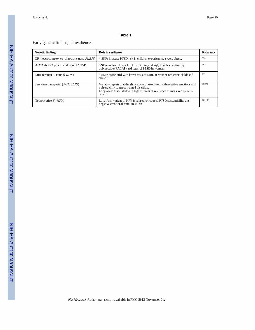
Early Genetic Findings in Human ResilienceGenetic factors are important determinants for the risk or resilience to psychiatric disorders.Most work thus far has focused on candidate genes with relatively weak associationsreported. Some recent examples of genes related to the HPA axis, serotonergic systems, orneuropeptide Y that show weak to moderate associations with resilient phenotypes are listedin Table 1. The field is now pivoting increasingly to genome–wide studies on large numbersof people to parse the complex genetic contributions to mood or anxiety disorders. Weanticipate exciting findings in the coming years as the genetic basis of resilience becomesbetter understood.
Animal Models of ResilienceDefinition of resilience in animals
As with humans, chronic stress leads to the development of depression– or anxiety–likebehaviors in only a subset of laboratory animals22–27. The remaining animals, which havebeen termed resilient in some studies, usually exhibit some deleterious symptoms inresponse to the stress, but do not exhibit deficits in key behavioral domains. For example,following chronic social defeat stress, all genetically inbred C57BL6/J male mice exhibit aconstellation of symptoms including heightened reactivity of the HPA axis, deficits inexploratory–based behavior that are interpreted as increased anxiety, and stress–inducedpolydipsia24. However, ~35% of the stressed mice, considered “resilient,” do not exhibitsocial avoidance, hyperthermia elicited by social interactions, anhedonia–like symptoms(reduced interest in sucrose, high fat food, or sex), or a metabolic syndrome characterized byover–eating, obesity, and central leptin resistance24, 28. Using this classification, resilientanimals are not devoid of symptoms and, in fact, exhibit some behavioral adaptations thatappear maladaptive, but they exhibit clear resistance to many other maladaptive sequelae ofthe chronic social stress.
Other stress paradigms have been used to study resilience in animals. It has long beenknown that inbred rodents subjected to learned helplessness models display a range ofresponses. Depending on the severity and duration of exposure to inescapable foot shock, asubset of animals, ~30% in some studies, develop learned helplessness—they fail to escapewhen escape becomes possible, while another subset (termed resilient) escapes withlatencies seen in unstressed animals29. Maier and colleagues view the risk of learnedhelplessness to be highly dependent upon the animal’s own control over the stress, withresilience promoted by control over cessation of the stress (For review see 30). Cohen et al.23
recently utilized a predator odor to induce a stress response and then classified rats into 3groups; one-third each that was extremely disrupted, partially disrupted, or minimallydisrupted. The classifications were based largely on the number and type of behavioraldeficits and the degree of change in brain NPY levels. The animals classified as extremely
Russo et al. Page 4
Nat Neurosci. Author manuscript; available in PMC 2013 November 01.
NIH
-PA Author Manuscript
NIH
-PA Author Manuscript
NIH
-PA Author Manuscript
disrupted exhibited anxiety–like behaviors and increased acoustic startle responses, as wellas large reductions in NPY across multiple brain regions. Both the partially and minimallydisrupted groups exhibited mixed deficits within these domains. In another recent study,Delgado et al.26 utilized a chronic “mild” stress (CMS) paradigm (where rats were exposedto varying physical and psychosocial stresses) and defined resilient animals as those that didnot exhibit significant anhedonia measured by reduced sucrose consumption. This form ofstress induced anhedonia–like symptoms in 70% of exposed rats. They went on to usestructural magnetic resonance imaging (MRI) and spectroscopy to show that CMS did notreduce hippocampal volume or alter glutamate metabolism in resilient mice as seen insusceptible mice.
An equally important literature has focused on strain differences in relative risk orresilience, studies that have shed light on potential genetically controlled traits that make ananimal more or less susceptible to stress. In the social defeat stress model, the relativedistribution of resilience differs across mouse strains. For example, 10 days of social defeatin C57BL6/J mice results in ~35% resilient mice, while other strains such as CD1 or FVBare closer to 100% resilient 27. These ratios are also a function of the severity and durationof the defeat episodes. In a related study, Vidal et al. (2011) found that better copingstrategies might make Sprague Dawley rats more resilient to social defeat stress than Wistarrats as measured in fear– and anxiety–based domains31. Strain–dependent responses to otherphysical stressors such as restraint or CMS have been shown, underscoring the role ofcomplex genetics in regulating risk and resilience to chronic stress32–34.
The central question concerning all animal studies of resilience is how they relate toresilience as defined in humans. As seen from the above examples, the definition ofresilience, and the percent of animals exhibiting resilience, varies across several stressparadigms. Most studies of resilience to date have focused on the absence of somebehavioral or molecular abnormality in a subset of stressed animals, and only more recentlyhave turned to more active mechanisms of resilience—protective changes that occuruniquely in resilient individuals. The crucial criterion for defining resilience in animals,however, as noted earlier, is the ability to avoid some or all of the deleterious behavioraleffects of chronic stress. The ability to avoid such deleterious consequences of stress in turndepends on both passive and active resilience mechanisms. Experimentally, it is thusessential to evaluate whether the absence (passive mechanism) or presence (activemechanism) of a given stress–induced molecular, cellular, or circuit change exerts a positiveeffect on the animal’s behavior, that is, whether it makes the animal less prone to exhibitmaladaptive behavioral traits. Such a definition would be analogous to the Diagnostic andStatistical Manual of Mental Disorders–IV (DSM–IV), where behavioral symptoms reachthreshold for diagnosis only when they cause significant distress or impairment in theindividual’s ability to function. Even this is complex, since there is an ongoing debate thatquestions whether anxiety and depression symptoms are maladaptive from a ethologicalperspective or whether they serve an evolutionary purpose such as avoidance in the face ofthreat or by reducing expenditure of resources35. Our group has defined resilience to chronicsocial defeat stress as the absence of social avoidance, anhedonia, and metabolic syndrome,which are highly correlated with one another24, 27, 28, 36. The anhedonic and metabolicsymptoms are clearly maladaptive and indeed are reversed by chronic administration ofantidepressants to susceptible mice36, 37. Interpretation of social avoidance is morecomplicated, even though it too is reversed by chronic antidepressant treatment, since onecould argue that avoiding an unfamiliar mouse after 10 days of aggressive encounters isadaptive, not maladaptive. However, we have shown that susceptible mice also avoid non–aggressive C57BL6/J littermates, and that such social avoidance is essentially permanent—itpersists for >6 months—despite housing with non–aggressive littermates, suggesting that itdoes reflect a maladaptive response36, 38. Therefore, when defining resilience in non–human
Russo et al. Page 5
Nat Neurosci. Author manuscript; available in PMC 2013 November 01.
NIH
-PA Author Manuscript
NIH
-PA Author Manuscript
NIH
-PA Author Manuscript

species, it is essential to utilize a more nuanced definition, considering not only the presenceor absence of depression– or anxiety–like behaviors, but also their ethological relevance tothe psychological and physical health of the organism. In the following sections, wehighlight recent work that has begun to identify active, adaptive coping mechanisms—behavioral, neural, molecular, and hormonal, which promote such behavioral resilience.
Stress over the life cycle in the development of resilienceDevelopmental and psychological research in humans over the last two decades hasdemonstrated that, rather than being a rare trait, resilience in both children and adults is acommon outcome following adversity – representing successful, adaptive coping in the faceof stress39, 40. Within the general population, between 50–60% experience a severe trauma,yet the prevalence of illness is estimated to be only 7.8%41. Children in particular displayremarkable resilience across a range of negative environmental stressors39. One importantset of observations concerns the potential pro–resilience effect of encountering andovercoming stress–inducing situations during development. According to this model, copingwith moderate amounts of stress leads to an individual sense of mastery and promotesresilience in the future. It might be that more moderate exposure to stress allows for a senseof stress mastery enhancing one’s perception of control. This inverted U shape relationshipbetween stress and coping can be observed in all organisms throughout their lifetime andsuggests that maintaining optimal stress exposure might prevent the development of majorpsychiatric dysfunction.
A potentially related phenomenon observed in laboratory animals, sometimes referred to as“stress inoculation,” was first described by Levine et al. who showed that infant rats exposedto intermittent foot shocks subsequently respond more effectively when confronted withnovel situations compared to their non–stressed counterparts42. Subsequent work hasexamined in detail the long–term effects of brief intermittent mother–offspring separationsin squirrel monkeys (for review see43). In a common experimental design, socially housedmonkeys are randomized to either brief intermittent separations or a non–separated controlcondition at 17 weeks of age. Over 10 separation sessions, a monkey is removed from thegroup for one hour per week. At nine months of age, behavioral and hormonal parametersare measured in separated and non–separated monkeys in a novel environment stress test44.Compared to non–separated monkeys, previously separated monkeys showed fewer signs ofanxiety and increased exploration of the environment coupled with diminished plasma levelsof cortisol and ACTH44. Separated monkeys also demonstrated enhanced responseinhibition to previously rewarding stimuli, suggesting improved cognitive control ofbehavior45. These and related findings highlight the critical window of stress exposure andsuggest that early intermittent separations enhance stress tolerance, i.e., promotes resilience.
The development of standardized rodent models of early life stress will greatly increase ourability to study the mechanisms of stress inoculation. A recent study showed that maternaldeprivation during early periods combined with chronic unpredictable stress (CUS; exposureto varying physical stresses) during juvenile periods promoted a greater degree of stressresilience than maternal deprivation alone or when combined with adult CUS46. This workpoints to critical developmental mechanisms engaged during early stress exposure, however,much future work is needed to make these paradigms more routine, particularly in mice.
While a relatively large animal and human literature has shown that chronic exposure ofadults to high levels of stress is usually associated with increased susceptibility to mood,anxiety, and addiction disorders6, 24, 36, 47–49, there is some evidence that more gradedexposure of adults to stress might reduce such vulnerabilities, that is, promote resilience.Over several decades, McEwen and colleagues systematically measured the type, length,and quality of stress experience on multiple rat behaviors and found that stress affects most
Russo et al. Page 6
Nat Neurosci. Author manuscript; available in PMC 2013 November 01.
NIH
-PA Author Manuscript
NIH
-PA Author Manuscript
NIH
-PA Author Manuscript
behavioral domains with an inverted U–shape curve, where low and high levels of stressboth impair behavior, while intermediate levels actually promote positive coping responses(for review see50). The shape of the inverted U differs by many factors including sex, strain,and behavioral domain in question (Fig. 2). For example, while female rodents tend to bemore susceptible to stress–induced dysfunction in emotional domains (i.e., sucrosepreference and forced swim test), they tend to show a greater degree of stress resilience incognitive domains (i.e., object placement and recognition) (for review see51). These findingssuggest that some level of stress and the context in which it is experienced may help adultanimals to develop better coping responses to future stress experiences.
Although further research is necessary to confirm some of these hypotheses, it seems clearthat moderate degrees of stress exposures during early life, adolescence, and adulthood canshift an individual’s stress–vulnerability curve to the right or broaden the curve byincreasing the range of tolerable stress for the organism. The ability to harness suchapproaches might have important therapeutic applications.
Neurobiological Findings in Animal Models of ResilienceIn recent years, the aforementioned animal models have just begun to be used to define theneural circuits and molecular adaptations within these circuits that contribute to resilience.In several cases, key findings in animals have been demonstrated in human postmortembrain, which provides an important measure of validation. As mentioned above, resilience isoften defined as the absence of behavioral symptoms in a subset of animals subjected tochronic stress. Indeed, numerous studies have identified pro–susceptibility neural andmolecular factors, which, if removed, promote stress resilience52–54. However, this approachsuggests that stress resilience is solely a passive process, whereby an animal’s lack ofresponse is adaptive. While this is no doubt true for certain conditions and biologicalpathways, there is increasing evidence that stress resilience also arises from active copingstrategies, both behavioral and molecular. For example, many have argued that, duringchronic exposure to stress, behavioral strategies that limit the stress experience couldpromote resilience. During social defeat stress, animals that engage in less submissiveposturing during the attack show less social avoidance, suggesting that this behavioralcoping strategy may affect the aggressive interaction and thereby lessen the effects of thestress55. While other examples of behavioral coping strategies have been described56, onlyrecently have neurobiological mechanisms of active resilience been characterized. In turnsout that resilience to chronic social defeat stress is associated with large numbers of uniquechanges in gene expression and chromatin modifications in specific brain regions that arenot seen in susceptible animals24, 57. In this section, we focus on such active mechanisms ofresilience, first considering the evolving neural circuitry implicated in resilience and thenpresenting several specific active molecular processes that have been shown recently tomediate a resilient phenotype in rodent models.
Glutamatergic signaling and synaptic connectivityDepression and anxiety are heterogeneous disorders marked by deficits in many behavioraldomains and controlled by multiple brain structures (Fig. 3). Numerous studies suggest thatdepression and anxiety in humans result in part from hypo–activation and reduced volume offrontal cortical and hipppocampal regions that control subcortical structures such as nucleusaccumbens (NAc) and amygdala, although hyper–activation of certain PFC regions (e.g.,subgenual area of anterior cingulate cortex) is also involved58–61. In addition, fMRI studieshave shown that both depression and anxiety are associated with hyperactivity ofamygdala 62. While imaging studies in NAc are less clear, most would argue forhypoactivity of NAc in depression, which is supported by deep brain stimulation studies thatshow electrical stimulation of the anterior limb of the internal capsule (which includes NAc)
Russo et al. Page 7
Nat Neurosci. Author manuscript; available in PMC 2013 November 01.
NIH
-PA Author Manuscript
NIH
-PA Author Manuscript
NIH
-PA Author Manuscript
alleviates symptoms of depression and anxiety. fMRI blood oxygen level dependent(BOLD) signals in humans provide an understanding of which structures are more or lessactive based on oxygen utilization, however, it is unclear whether these adaptations occur ininhibitory or excitatory neurons, or for that matter glia, and thus whether they reflect anoverall increase or decrease in net circuit activity. To address this question, investigators arestudying rodent models to understand circuit–level synaptic changes in glutamate systemswith far greater precision. In general, the literature supports the idea that chronic stressreduces dendritic arborization and glutamatergic dendritic spine density of pyramidalneurons in PFC and hippocampus, and reduces hippocampus neurogenesis, while increasingdendritic spine number or branching in amygdala and NAc (for review see63). HypoactivePFC and hippocampal inputs to neurons in these subcortical structures may mediate theiractivation and the subsequent activity–dependent structural plasticity of their dendrites,although further research is needed in this area.
Despite this work focused on stress susceptibility, few reports have to date examinedwhether resilience is associated with any active mechanisms within this defined braincircuitry. Recent studies have shown a greater degree of c–Fos, FosB, or ΔFosB expressionin glutamatergic neurons of mPFC (infralimbic, paralimbic PFC) of resilient mice followingchronic predator or social defeat stress25, 64, 65. Increased expression of these immediateearly genes would suggest increased neuronal activation within this brain region, whichmight represent a pro–resilience adaptation. Consistent with this hypothesis, Covington et al.showed that direct optogenetic stimulation of mPFC neurons with channel rhodopsin (ChR2)promotes resilience to social defeat stress65. While this initial work did not distinguishbetween glutamatergic vs. GABAergic neurons, more recent work using a viral vector thatspecifically targets ChR2 to glutamatergic neurons in mPFC shows that optogeneticstimulation of the glutamatergic microcircuit from PFC to NAc is antidepressant(Christoffel, Soc. Neurosci. Abstr., 2012). Future studies examining the contribution of otherglutamatergic microcircuits to resilient responses, using this powerful methodology, willfurther delineate the circuit basis of resilience.
Early intermittent maternal separations that promote resilience increase cortical volumewithin ventromedial PFC (VMPFC)66, changes opposite in direction to those seen indepressed humans and stressed rodents. Understanding the molecular basis of these activerestructuring processes may shed light on the hypofrontality observed in depression oranxiety disorders67, 68, as well as the circuit mechanisms underlying stress inoculation.Notably, environmental enrichment similarly increases the complexity of the dendritic tree,dendritic spine density, and synaptic protein levels of pyramidal neurons in hippocampusand PFC, suggesting that this may be a shared feature of resilience under these two distinctconditions69. Likewise, in rodents, enrichment increases expression of FosB and ΔFosB inmPFC and is reported to confer resilience to stress–induced increases in depression– andanxiety–like behaviors across multiple domains25 (Fig. 4). These data provide an importantcontrast to earlier studies of the deleterious effects of early life stress on neural structuresinvolved in emotional behavior and stress responses. Rats exposed to severe chronic stressduring gestation, which promotes depression–like behaviors later in life, exhibit decreaseddendritic spine density in the anterior cingulate gyrus and orbitofrontal cortex70. Thestriking contrasts between the neural and behavioral effects observed in the context ofdifferent stress paradigms highlights the markedly divergent consequences resulting fromdifferences in the developmental timing and the type, magnitude, and duration of stressexposure, as stated above.
While susceptibility to chronic social defeat stress is associated with increased glutamatergictone, including greater frequency of excitatory currents and number of glutamatergicsynapses, on medium spiny neurons in NAc, there is recent evidence that resilience is
Russo et al. Page 8
Nat Neurosci. Author manuscript; available in PMC 2013 November 01.
NIH
-PA Author Manuscript
NIH
-PA Author Manuscript
NIH
-PA Author Manuscript
mediated in part via an active adaptation that opposes this susceptibility mechanism52, 71.ΔFosB is induced in medium spiny NAc neurons preferentially in resilient animals, where itpromotes resilience partly by inducing expression of GluA2 (GluR2), an AMPA glutamatereceptor subunit that reduces the Ca2+ permeability and overall conductance of AMPAchannels71. Gene expression arrays have identified many other targets for ΔFosB in NAc ofresilient animals, which now warrant examination for their possible role in mediatingresilience as well. Indeed, reduced levels of ΔFosB and GluA2 have been documented inNAc of depressed humans examined postmortem71 (Fig. 4). The selective induction ofΔFosB in NAc of resilient animals is mediated via the activation of serum response factor(SRF) under these conditions72, although the mechanism responsible for SRF’s selectiveinduction in resilient individuals remains unknown.
K+ channel driven intrinsic excitability of neuronsAnother active neural mechanism of resilience is the normalization of firing rate of ventraltegmental area (VTA) dopamine neurons in the chronic social defeat stress paradigm24 (Fig.4). Our initial analysis showed that the firing rate of VTA dopamine neurons was normal inresilient animals and increased in susceptible animals, which would appear to reflect theabsence of a stress–induced change in resilience. However, upon closer examination, there isan independent, active process occurring in resilient mice that normalizes VTA firing tocontrol levels and thus prevents social avoidance and sucrose preference deficits. Thus, thehyper–excitability of VTA dopamine neurons in susceptible mice is mediated in part byinduction of hyperpolarization–activated cation current (Ih), which increases the intrinsicexcitability of these neurons73, Friedman Soc. Neurosci. Abstr., 907.26, 2011 Surprisingly, thiscurrent is similarly increased in VTA neurons of resilient mice, suggesting the existence ofan additional ionic mechanism, which counteracts the increased Ih function and normalizesfiring rate in resilient mice. Our early microarray analyses identified large increases in acluster of K+ channel subunits, including KCNF1, KCNH3, KCNK4 and KCNQ3, in theresilient VTA24. This finding was functionally confirmed by electrophysiological studies,and such induction of K+ channels functionally occludes the increased Ih current and therebyactively promotes behavioral resilience24, 73.
It is surprising that increased excitability of VTA dopamine neurons mediates susceptibility,with normalization mediating resilience, since such firing is generally seen as promotingreward and motivation, both implicated in resilience2. However, increased firing of VTAdopamine neurons has long been documented in response to rewards as well as to aversivestimuli, and it has been suggested that they increase salience for both types ofstimuli24, 73–76. One possible explanation of this paradox is that different subsets of VTAdopamine neurons may show activation by rewarding vs. aversive stimuli77. This highlightsthe fact that, across numerous laboratories and experimental conditions, there is not a one–to–one correspondence between reward and resilience. Not all molecular changes in theVTA or NAc that increase drug or natural reward promote resilience, and vice versa.Optogenetic studies, which now make it possible to selectively activate subsets of VTAdopamine neurons, or their efferent or afferent connections, should help our understandingof the circuit mechanisms by which VTA dopamine neuron firing regulates responses torewarding and aversive stimuli and determine stress susceptibility vs.resilience78, Chaudhury, Soc. Neurosci. Abstr., 907.27, 2011.
K+ channel–mediated adaptations have also been observed in NAc after prolonged socialisolation of adult mice or rats, which induces depression– and anxiety–like behavioralabnormalities49 and promotes susceptibility to chronic social defeat stress71. Induction ofparticular K+ channel subunits in NAc in response to adult social isolation was shown tocontribute to depression–like symptoms seen in this paradigm, while normalization of K±
channel function contributes to antidepressant responses49.
Russo et al. Page 9
Nat Neurosci. Author manuscript; available in PMC 2013 November 01.
NIH
-PA Author Manuscript
NIH
-PA Author Manuscript
NIH
-PA Author Manuscript

Consistent with this work in VTA and NAc, there is increasing evidence that several typesof K+ channels function as general gatekeepers of neuronal excitability in numerousexperimental systems79, 80. The findings centered around K+ channels underscore the valueof focusing on the molecular mechanisms of resilience and warrant attention as potentialtargets for the development of new treatments of stress–associated mental disorders.
Neuroendocrine mechanismsAs mentioned above, the HPA axis plays a critical role in mediating stress responses, withdisruption of normal HPA function (up or down) associated with both depressive andanxiety syndromes in humans. Rodent models have largely supported this literature. Forexample, prenatal exposure to glucocorticoids increases CRH levels in the central nucleus ofamygdala and reduces volume of PFC structures in adulthood70. Elevated glucocorticoidlevels may mediate the ability of stress to reduce the dendritic spine density of PFCpyramidal neurons in parallel with hypertrophic effects in basolateral amygdala, asdescribed above70. There is also evidence for a possible role for the HPA axis in resilience.For example, work from Meaney and coworkers have characterized the effects of early lifematernal care in rats on glucocorticoid receptor expression in hippocampus and onemotional behavior. They find that high levels of maternal care are associated withdecreased DNA methylation of the GR gene, higher levels of GR expression, greaterfeedback inhibition of the HPA axis, and resilient stress responses in adulthood (for reviewsee 81). The GR gene is likely just one of many epigenetic targets of greater maternal carethat promote resilience later in life.
Less is known about HPA axis adaptations in adult animals that might contribute toresilience82. One recent paper found an epigenetic mechanism, induced by chronic stress inresilient mice only, that controls HPA axis hyperactivity83 (Fig. 4). The authors showed that,after chronic social defeat stress, Crh gene expression is increased in the paraventricularnucleus (PVN) of hypothalamus of susceptible animals and that this adaptation is necessaryfor the development of social avoidance83. Notably, the Crh gene is hypermethylated andsilenced to prevent Crh induction in the subset of animals termed resilient for their lack ofsocial avoidance. Expression of a small interfering RNA (RNAi) to decrease CRHexpression was sufficient to prevent social avoidance in susceptible mice. Consistent withthese observations, environmental enrichment paradigms that promote resilience in rodentsalso reduce ACTH and corticosterone responses to stress, suggesting an interesting linkamong genetic, experience–based, and epigenetic factors84.
As alluded to earlier, male rodents are more resilient than females with respect to the effectsof chronic stress on emotional aspects of depression– and anxiety–like behavioral domains.For example, we have shown that sub–chronic unpredictable stress induces anhedonia(decreased sucrose preference), increased immobility on the forced swim test, and increasedanxiety (greater latency to feed in a novel environment and decreased time grooming in thesplash test). The data clearly show that females are more sensitive than males in thesedomains85, Hodes, Soc. Neurosci. Abstr. 219.01, 2011. However, as mentioned above, stressedfemales tend to perform better on non–aversive cognitive or memory tasks compared tomales. Stress enhances the performance of female rodents on the radial arm maze, Morriswater maze, Y–maze, non–associative learning, and object placement tasks, whereas stressimpairs male performance in these assays86–88. Conversely, in tests of acute stress oraversive conditioning, stress enhances learning in males and impairs it in females89, 90.These data highlight the possibility that males and females may use different copingstrategies in the face of stress, which has led to the hypothesis that gonadal hormones, suchas testosterone in males, might promote resilience to deficits on emotional domains, whileestrogen or progesterone may promote stress resilience in females within cognitive domains.
Russo et al. Page 10
Nat Neurosci. Author manuscript; available in PMC 2013 November 01.
NIH
-PA Author Manuscript
NIH
-PA Author Manuscript
NIH
-PA Author Manuscript
Moreover, the literature suggests that on cognitive domains females cope better with chronicforms of stress, whereas males tend to cope better with acute stress.
While most human work is limited to correlative studies, there is evidence that testosteronein males promotes resilience in MDD and PTSD, potentially consistent with epidemiologicaldata showing that woman are significantly more vulnerable to developing these disordersthan men 16, 17. Based on the animal work stated above, future studies in humans shouldinvestigate sex differences in vulnerability to stress–related deficits in cognitive andemotional domains. Indeed, evidence from women across the reproductive lifespan suggeststhat fluctuating ovarian hormones are likely a biological source of increased prevalence forthese disorders. Work in rodents largely confirms this, showing that removal of ovarianhormones decreases the pro–depressant or anxiogenic effects of stress, while simultaneouslyincreasing the negative effects of stress on spatial and non–spatial memory85, 91, 92. As well,maternal experience in female rodents seems to promote resilience to the effects of stress oncognition, which is likely in part through hormonal mechanisms regulating oxytocin93.While much future work is needed to understand these interesting roles for gonadalhormones in promoting or opposing resilience, studies of the underlying mechanisms of sexdifferences in stress responses can provide us with unique biological information about themechanisms of coping in depression and anxiety disorders.
Therapeutic ImplicationsAn important finding from animal studies of neurobiological and neuroendocrinemechanisms of resilience–like behavioral adaptations is that resilience is likely mediated, inlarge part, via active adaptations that occur selectively in resilient individuals (Fig. 1).Indeed, genome–wide studies in the chronic social defeat paradigm have identified a rangeof gene expression changes and chromatin modifications in VTA and NAc that occuruniquely in resilience24, 57. Examples include ΔFosB and SRF, discussed above, as well asHDAC2 (histone deacetylase–2) and the WNT (wingless)–DVL (disheveled)–GSK3β(glycogen synthase kinase–3β) signaling cascade71, 72, 94 In fact, there is significant overlapbetween genes that are regulated in resilience and those that are regulated by chronicantidepressant treatment of susceptible individuals57, raising the possibility that one way inwhich current antidepressants work is by inducing in depressed individuals some of thesame adaptations that occur naturally in inherently resilient individuals. These insights thussuggest a new path forward for the development of new treatments of stress–relateddisorders: in addition to looking for ways to prevent or reverse the deleterious effects ofstress, it should be possible to induce natural mechanisms of resilience, distinct from currentantidepressant actions, in more vulnerable populations.
There is already considerable behavioral evidence for this approach. Stress resilience isenhanced in specific populations, such as military personnel and rescue workers, throughcontrolled exposure to stress–related stimuli. Similarly, behavior therapy uses controlledstress exposure as one means to treat symptoms of mood and anxiety disorders. Forexample, exposure therapy and cognitive behavioral therapy can aid individuals with PTSDthrough cognitive restructuring and relaxation techniques following a traumatic event topromote recovery. These effects are well documented to reverse hyperactivity of PFC–amygdala microcircuits shown to be overactive in PTSD (for review see62). It is possiblethat similar behavioral approaches might be adopted in at risk populations to enhanceresilience to subsequent stressful life events and prevent the development of these disorders.
Such treatments might be understood as being analogous to stress inoculation in thatexperiencing more moderate levels of stress, combined with techniques that reducephysiological and psychological perception of the trauma, can promote positive coping
Russo et al. Page 11
Nat Neurosci. Author manuscript; available in PMC 2013 November 01.
NIH
-PA Author Manuscript
NIH
-PA Author Manuscript
NIH
-PA Author Manuscript
responses. The observation that such approaches significantly reduce PTSD severitysupports the view that targeting underlying biological mechanisms of stress inoculation mayprovide us with novel protein targets for new medications that further promote resilience inat–risk populations. Likewise, identification of pro–resilience factors should make itpossible to identify predictive biomarkers of resilience, which should greatly aid inrecognizing at–risk populations.
Future DirectionsAs we learn more about the neurobiological mechanisms that confer resilience on anindividual, the goal to develop treatment strategies to restore or enhance coping resourcesshould improve the efficacy of treatment. However, we are just beginning to identify suchresilience factors. A major gap in the field is the lack of coordination between human andanimal studies. Human research has identified several tentative neuroendocrineconcomitants of resilience (e.g., testosterone, NPY), which have not yet been adequatelyinvestigated mechanistically in animal models, while it remains challenging toexperimentally interrogate the vast majority of neurobiological mechanisms discovered inanimals (e.g., ΔFosB, K+ channels) in living humans.
Nevertheless, work to date identifies several important areas for future investigation. First,there is a great need for human brain imaging studies to determine the brain structures andcircuits that mediate stress resilience. Deep brain stimulation in humans60 could potentiallybe used to provide highly valuable causal information about dysfunction of brain structuresand circuits in depression and anxiety. This information could then be used in conjunctionwith optogenetic studies in rodent models, where we can more definitively describe theneural circuitry of resilience. Second, it is crucial to identify the range of heritable factorsthat help determine an individual’s capacity for resilience. Extrapolating from geneticstudies of other complex human traits, it is likely that complex combinations of perhapshundreds of genetic variations, rare and common in the population, comprise this geneticbasis of resilience. Third, we must characterize the epigenetic mechanisms that control thedegree to which this genetic predilection for resilience become manifest. Part of thisepigenetic control of gene expression will occur in response to a host of environmentalstimuli throughout life, but a portion may occur through random events during braindevelopment. Fourth, far more insight is needed into the genetic, epigenetic,neurobiological, and neuroendocrine basis of sex differences in stress susceptibility vs.resilience. Finally, we need to better define how just the right type and level of stressinoculation, through this complex interplay of mechanisms, can promote resilience.
In the end, studies of resilience have unleashed a fundamentally novel way of understandingan individual’s responses to adverse life events, and have ushered in an exciting new era instudies of MDD, PTSD, and other stress–related disorders.
AcknowledgmentsPreparation of this review was supported by grants from the National Institute of Mental Health: R01 MH090264(SJR), K23 MH094707 (JWM), R01 MH092306 (MHH), and R01 MH51399, P50 MH66172, and P50 MH96890(EJN).
References1. Charney DS. Am J Psychiatry. 2004; 161:195–216. [PubMed: 14754765]
2. Feder A, Nestler EJ, Charney DS. Nat Rev Neurosci. 2009; 10:446–457. [PubMed: 19455174]
3. Herman JP, Cullinan WE. Trends Neurosci. 1997; 20:78–84. [PubMed: 9023876]
4. Stetler C, Miller GE. Psychosom Med. 2011; 73:114–126. [PubMed: 21257974]
Russo et al. Page 12
Nat Neurosci. Author manuscript; available in PMC 2013 November 01.
NIH
-PA Author Manuscript
NIH
-PA Author Manuscript
NIH
-PA Author Manuscript
5. Meewisse ML, Reitsma JB, de Vries GJ, Gersons BP, Olff M. Br J Psychiatry. 2007; 191:387–392.[PubMed: 17978317]
6. Heim C, Newport DJ, Mletzko T, Miller AH, Nemeroff CB. Psychoneuroendocrinology. 2008;33:693–710. [PubMed: 18602762]
7. Heim C, Newport DJ, Miller AH, Nemeroff CB. JAMA. 2000; 284:2321. [PubMed: 11066180]
8. Yehuda R, Golier JA, Kaufman S. Am J Psychiatry. 2005; 162:998–1000. [PubMed: 15863805]
9. Rasmusson AM, Vythilingam M, Morgan CA 3rd. CNS Spectr. 2003; 8:651–656. 665–657.[PubMed: 15079139]
10. Yehuda R, Brand SR, Golier JA, Yang RK. Acta Psychiatr Scand. 2006; 114:187–193. [PubMed:16889589]
11. Butterfield MI, et al. Am J Psychiatry. 2005; 162:380–382. [PubMed: 15677605]
12. Taylor MK, et al. Stress. 2011
13. Oliveira T, Gouveia MJ, Oliveira RF. Psychoneuroendocrinology. 2009; 34:1056–1064. [PubMed:19278791]
14. Edwards DA, Wetzel K, Wyner DR. Physiol Behav. 2006; 87:135–143. [PubMed: 16233905]
15. Morgan CA 3rd, et al. Biol Psychiatry. 2000; 47:891–901. [PubMed: 10807962]
16. Mulchahey JJ, et al. Psychoneuroendocrinology. 2001; 26:273–285. [PubMed: 11166490]
17. Pope HG Jr, Cohane GH, Kanayama G, Siegel AJ, Hudson JI. Am J Psychiatry. 2003; 160:105–111. [PubMed: 12505808]
18. Morgan CA 3rd, et al. Biol Psychiatry. 2000; 47:902–909. [PubMed: 10807963]
19. Morgan CA 3rd, et al. Biol Psychiatry. 2002; 52:136–142. [PubMed: 12114005]
20. Zhou Z, et al. Nature. 2008; 452:997–1001. [PubMed: 18385673]
21. Mickey BJ, et al. Arch Gen Psychiatry. 2011; 68:158–166. [PubMed: 21300944]
22. Taliaz D, et al. J Neurosci. 2011; 31:4475–4483. [PubMed: 21430148]
23. Cohen H, et al. Neuropsychopharmacology. 2011
24. Krishnan V, et al. Cell. 2007; 131:391–404. [PubMed: 17956738]
25. Lehmann ML, Herkenham M. J Neurosci. 2011; 31:6159–6173. [PubMed: 21508240]
26. Delgado y Palacios R, et al. Biol Psychiatry. 2011; 70:449–457. [PubMed: 21762877]
27. Golden SA, Covington HE 3rd, Berton O, Russo SJ. Nat Protoc. 2011; 6:1183–1191. [PubMed:21799487]
28. Lutter M, et al. Nat Neurosci. 2008; 11:752–753. [PubMed: 18552842]
29. Berton O, et al. Neuron. 2007; 55:289–300. [PubMed: 17640529]
30. Fleshner M, Maier SF, Lyons DM, Raskind MA. Stress. 2011; 14:498–502. [PubMed: 21790482]
31. Vidal J, Buwalda B, Koolhaas JM. Behav Processes. 2011; 88:76–80. [PubMed: 21854839]
32. Uchida S, et al. Neuron. 2011; 69:359–372. [PubMed: 21262472]
33. Mozhui K, et al. J Neurosci. 2010; 30:5357–5367. [PubMed: 20392957]
34. Andrus BM, et al. Mol Psychiatry. 2012; 17:49–61. [PubMed: 21079605]
35. Nesse RM. Arch Gen Psychiatry. 2000; 57:14–20. [PubMed: 10632228]
36. Berton O, et al. Science. 2006; 311:864–868. [PubMed: 16469931]
37. Covington HE 3rd, et al. J Neurosci. 2009; 29:11451–11460. [PubMed: 19759294]
38. Covington HE 3rd, Vialou VF, LaPlant Q, Ohnishi YN, Nestler EJ. Neurosci Lett. 2011; 493:122–126. [PubMed: 21335060]
39. Masten AS. Am Psychol. 2001; 56:227–238. [PubMed: 11315249]
40. Bonanno GA. Am Psychol. 2004; 59:20–28. [PubMed: 14736317]
41. Kessler RC, Sonnega A, Bromet E, Hughes M, Nelson CB. Arch Gen Psychiatry. 1995; 52:1048–1060. [PubMed: 7492257]
42. Levine S. Science. 1962; 135:795–796. [PubMed: 14464660]
43. Lyons DM, Parker KJ, Schatzberg AF. Dev Psychobiol. 2010; 52:616–624. [PubMed: 20957724]
44. Parker KJ, Buckmaster CL, Schatzberg AF, Lyons DM. Arch Gen Psychiatry. 2004; 61:933–941.[PubMed: 15351772]
Russo et al. Page 13
Nat Neurosci. Author manuscript; available in PMC 2013 November 01.
NIH
-PA Author Manuscript
NIH
-PA Author Manuscript
NIH
-PA Author Manuscript
45. Parker KJ, Buckmaster CL, Justus KR, Schatzberg AF, Lyons DM. Biol Psychiatry. 2005; 57:848–855. [PubMed: 15820705]
46. Ricon T, Toth E, Leshem M, Braun K, Richter–Levin G. Stress. 2011
47. Bradley RG, et al. Arch Gen Psychiatry. 2008; 65:190–200. [PubMed: 18250257]
48. Caspi A, Hariri AR, Holmes A, Uher R, Moffitt TE. Am J Psychiatry. 2010; 167:509–527.[PubMed: 20231323]
49. Wallace DL, et al. Nat Neurosci. 2009; 12:200–209. [PubMed: 19151710]
50. McEwen BS, Gianaros PJ. Annu Rev Med. 2011; 62:431–445. [PubMed: 20707675]
51. Luine V. Stress. 2002; 5:205–216. [PubMed: 12186683]
52. Christoffel DJ, et al. J Neurosci. 2011; 31:314–321. [PubMed: 21209217]
53. Tsankova NM, et al. Nat Neurosci. 2006; 9:519–525. [PubMed: 16501568]
54. Christoffel DJ, et al. Neuropsychopharmacology. 2012
55. Wood SK, Walker HE, Valentino RJ, Bhatnagar S. Endocrinology. 2010; 151:1795–1805.[PubMed: 20160137]
56. Ono Y, et al. Stress. 2011
57. Wilkinson MB, et al. J Neurosci. 2009; 29:7820–7832. [PubMed: 19535594]
58. Price JL, Drevets WC. Neuropsychopharmacology. 2010; 35:192–216. [PubMed: 19693001]
59. Murrough JW, Iacoviello B, Neumeister A, Charney DS, Iosifescu DV. Neurobiol Learn Mem.2012
60. Mayberg HS. J Clin Invest. 2009; 119:717–725. [PubMed: 19339763]
61. van Tol MJ, et al. Biol Psychiatry. 71:593–602. [PubMed: 22206877]
62. Linden DE. Mol Psychiatry. 2006; 11:528–538. [PubMed: 16520823]
63. Christoffel DJ, Golden SA, Russo SJ. Rev Neurosci. 2011; 22:535–549. [PubMed: 21967517]
64. Adamec R, Toth M, Haller J, Halasz J, Blundell J. Physiol Behav. 2012
65. Covington HE 3rd, et al. J Neurosci. 2010; 30:16082–16090. [PubMed: 21123555]
66. Katz M, et al. Dev Neurosci. 2009; 31:293–299. [PubMed: 19546566]
67. Milad MR, Orr SP, Pitman RK, Rauch SL. Psychophysiology. 2005; 42:456–464. [PubMed:16008774]
68. Rauch SL, et al. Neuroreport. 2005; 16:1909–1912. [PubMed: 16272877]
69. Kozorovitskiy Y, et al. Proc Natl Acad Sci U S A. 2005; 102:17478–17482. [PubMed: 16299105]
70. Lupien SJ, McEwen BS, Gunnar MR, Heim C. Nat Rev Neurosci. 2009; 10:434–445. [PubMed:19401723]
71. Vialou V, et al. Nat Neurosci. 2010; 13:745–752. [PubMed: 20473292]
72. Vialou V, et al. J Neurosci. 2011; 30:14585–14592. [PubMed: 20980616]
73. Cao JL, et al. J Neurosci. 2010; 30:16453–16458. [PubMed: 21147984]
74. Goto Y, Otani S, Grace AA. Neuropharmacology. 2007; 53:583–587. [PubMed: 17709119]
75. Grace AA, Floresco SB, Goto Y, Lodge DJ. Trends Neurosci. 2007; 30:220–227. [PubMed:17400299]
76. Brischoux F, Chakraborty S, Brierley DI, Ungless MA. Proc Natl Acad Sci U S A. 2009;106:4894–4899. [PubMed: 19261850]
77. Lammel S, Ion DI, Roeper J, Malenka RC. Neuron. 70:855–862. [PubMed: 21658580]
78. Shumake J, Ilango A, Scheich H, Wetzel W, Ohl FW. J Neurosci. 2010; 30:5876–5883. [PubMed:20427648]
79. Luscher C, Slesinger PA. Nat Rev Neurosci. 2010; 11:301–315. [PubMed: 20389305]
80. Balana B, et al. Proc Natl Acad Sci U S A. 2011; 108:5831–5836. [PubMed: 21422294]
81. Weaver IC, et al. J Neurosci. 2005; 25:11045–11054. [PubMed: 16306417]
82. Meaney MJ, Szyf M. Dialogues Clin Neurosci. 2005; 7:103–123. [PubMed: 16262207]
83. Elliott E, Ezra–Nevo G, Regev L, Neufeld–Cohen A, Chen A. Nat Neurosci. 2011; 13:1351–1353.[PubMed: 20890295]
Russo et al. Page 14
Nat Neurosci. Author manuscript; available in PMC 2013 November 01.
NIH
-PA Author Manuscript
NIH
-PA Author Manuscript
NIH
-PA Author Manuscript
84. Moncek F, Duncko R, Johansson BB, Jezova D. J Neuroendocrinol. 2004; 16:423–431. [PubMed:15117335]
85. LaPlant Q, et al. Biol Psychiatry. 2009; 65:874–880. [PubMed: 19251249]
86. Conrad CD, Grote KA, Hobbs RJ, Ferayorni A. Neurobiol Learn Mem. 2003; 79:32–40. [PubMed:12482677]
87. Galea LA, et al. Neuroscience. 1997; 81:689–697. [PubMed: 9316021]
88. Bowman RE, Beck KD, Luine VN. Horm Behav. 2003; 43:48–59. [PubMed: 12614634]
89. Wood GE, Shors TJ. Proc Natl Acad Sci U S A. 1998; 95:4066–4071. [PubMed: 9520494]
90. Wood GE, Beylin AV, Shors TJ. Behav Neurosci. 2001; 115:175–187. [PubMed: 11256441]
91. Autry AE, Adachi M, Cheng P, Monteggia LM. Biol Psychiatry. 2009; 66:84–90. [PubMed:19358977]
92. Bowman RE, Ferguson D, Luine VN. Neuroscience. 2002; 113:401–410. [PubMed: 12127097]
93. Douglas AJ, Brunton PJ, Bosch OJ, Russell JA, Neumann ID. Endocrinology. 2003; 144:5268–5276. [PubMed: 12960085]
94. Wilkinson MB, et al. J Neurosci. 2011; 31:9084–9092. [PubMed: 21697359]
95. Binder EB, et al. JAMA. 2008; 299:1291–1305. [PubMed: 18349090]
96. Ressler KJ, et al. Nature. 2011; 470:492–497. [PubMed: 21350482]
97. Polanczyk G, et al. Arch Gen Psychiatry. 2009; 66:978–985. [PubMed: 19736354]
98. Stein MB, Campbell–Sills L, Gelernter J. Am J Med Genet B Neuropsychiatr Genet. 2009; 150B:900–906. [PubMed: 19152387]
99. Murrough JW, Charney DS. Biol Psychiatry. 2011; 69:510–512. [PubMed: 21353836]
100. Domschke K, et al. Eur Neuropsychopharmacol. 2010; 20:301–309. [PubMed: 19854625]
Russo et al. Page 15
Nat Neurosci. Author manuscript; available in PMC 2013 November 01.
NIH
-PA Author Manuscript
NIH
-PA Author Manuscript
NIH
-PA Author Manuscript

Figure 1. Schematic of gene x environment interactions that promote resilienceThe scheme describes how behavioral strategies through stress inoculation can interact withan individual’s genetic constitution to control expression of key genes—via epigeneticprocesses—in the brain’s limbic regions to mount active, adaptive molecular and cellularchanges that mediate resilience.
Russo et al. Page 16
Nat Neurosci. Author manuscript; available in PMC 2013 November 01.
NIH
-PA Author Manuscript
NIH
-PA Author Manuscript
NIH
-PA Author Manuscript
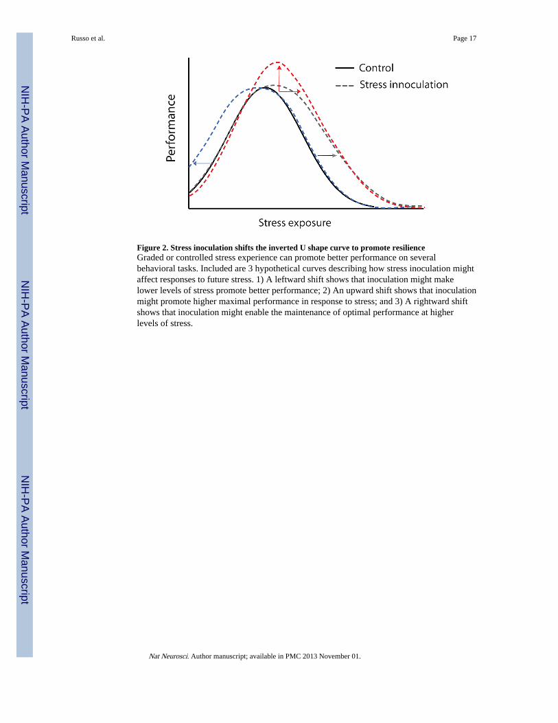
Figure 2. Stress inoculation shifts the inverted U shape curve to promote resilienceGraded or controlled stress experience can promote better performance on severalbehavioral tasks. Included are 3 hypothetical curves describing how stress inoculation mightaffect responses to future stress. 1) A leftward shift shows that inoculation might makelower levels of stress promote better performance; 2) An upward shift shows that inoculationmight promote higher maximal performance in response to stress; and 3) A rightward shiftshows that inoculation might enable the maintenance of optimal performance at higherlevels of stress.
Russo et al. Page 17
Nat Neurosci. Author manuscript; available in PMC 2013 November 01.
NIH
-PA Author Manuscript
NIH
-PA Author Manuscript
NIH
-PA Author Manuscript
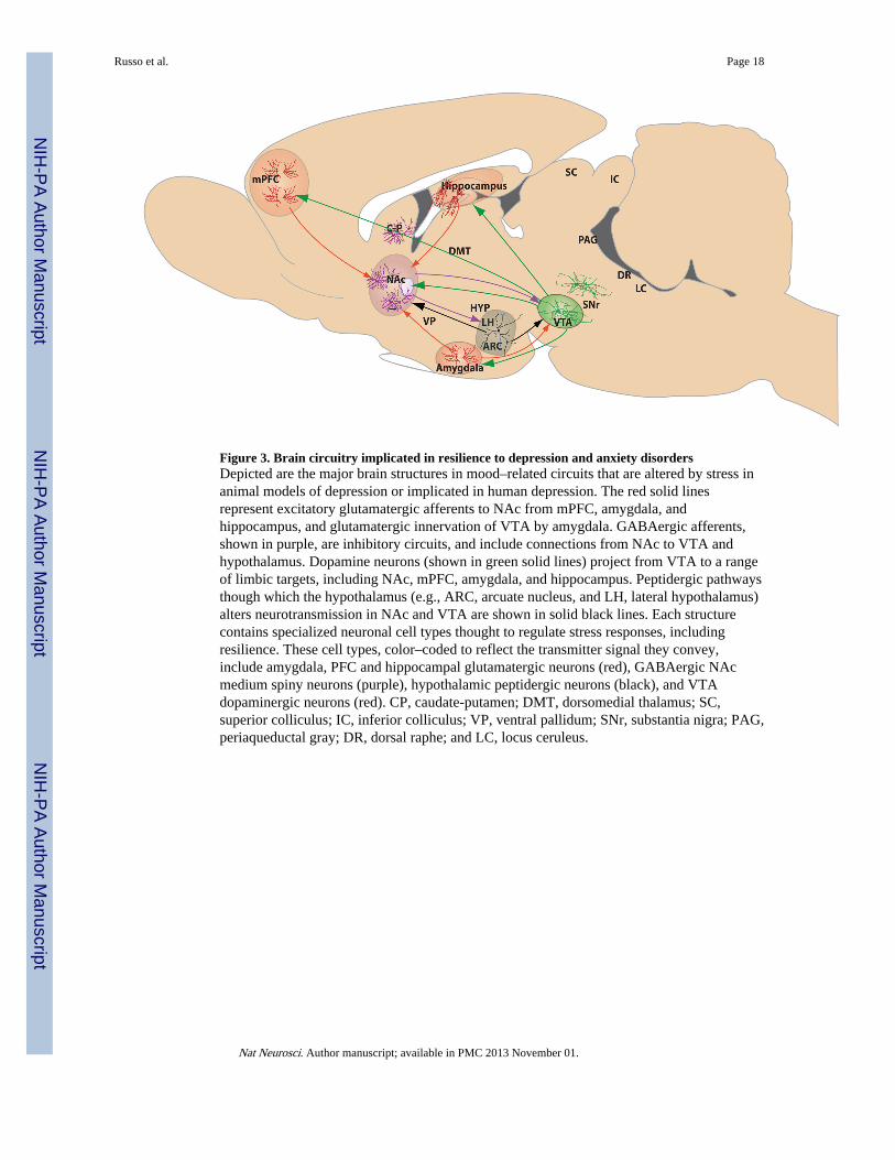
Figure 3. Brain circuitry implicated in resilience to depression and anxiety disordersDepicted are the major brain structures in mood–related circuits that are altered by stress inanimal models of depression or implicated in human depression. The red solid linesrepresent excitatory glutamatergic afferents to NAc from mPFC, amygdala, andhippocampus, and glutamatergic innervation of VTA by amygdala. GABAergic afferents,shown in purple, are inhibitory circuits, and include connections from NAc to VTA andhypothalamus. Dopamine neurons (shown in green solid lines) project from VTA to a rangeof limbic targets, including NAc, mPFC, amygdala, and hippocampus. Peptidergic pathwaysthough which the hypothalamus (e.g., ARC, arcuate nucleus, and LH, lateral hypothalamus)alters neurotransmission in NAc and VTA are shown in solid black lines. Each structurecontains specialized neuronal cell types thought to regulate stress responses, includingresilience. These cell types, color–coded to reflect the transmitter signal they convey,include amygdala, PFC and hippocampal glutamatergic neurons (red), GABAergic NAcmedium spiny neurons (purple), hypothalamic peptidergic neurons (black), and VTAdopaminergic neurons (red). CP, caudate-putamen; DMT, dorsomedial thalamus; SC,superior colliculus; IC, inferior colliculus; VP, ventral pallidum; SNr, substantia nigra; PAG,periaqueductal gray; DR, dorsal raphe; and LC, locus ceruleus.
Russo et al. Page 18
Nat Neurosci. Author manuscript; available in PMC 2013 November 01.
NIH
-PA Author Manuscript
NIH
-PA Author Manuscript
NIH
-PA Author Manuscript

Figure 4. Active molecular mechanisms in limbic brain circuits that promote resilience in animalmodelsFour examples of active adaptive molecular processes that confer resilience to chronic socialdefeat stress are shown. In mPFC (infralimbic and paralimbic), resilient animals displaymolecular evidence of increased neural activity, which has been shown via optogenetictechniques to promote resilience. The cell type exhibiting this hyperactivity is not known(GABAerigc versus glutamatergic). VTA dopamine neurons of resilient animals showincreased transcription of K+ channel subunits, which normalizes the stress–inducedincrease in VTA firing rate that drives deleterious responses to stress. In NAc, resilience isassociated with increased ΔFosB–mediated transcription of GluA2, a Ca2+–impermeableAMPA glutamate receptor subunit that counteracts glutamate hyperactivity found insusceptible mice. In hypothalamus, resilience is associated with hypermethylation of the Crhgene to suppress its transcription and reduce HPA hyperactivity found in susceptible mice.
Russo et al. Page 19
Nat Neurosci. Author manuscript; available in PMC 2013 November 01.
NIH
-PA Author Manuscript
NIH
-PA Author Manuscript
NIH
-PA Author Manuscript
NIH
-PA Author Manuscript
NIH
-PA Author Manuscript
NIH
-PA Author Manuscript
Russo et al. Page 20
Table 1
Early genetic findings in resilience
Genetic findings Role in resilience Reference
GR–heterocomplex co–chaperone gene FKBP5 4 SNPs increase PTSD risk in children experiencing severe abuse. 95
ADCYAP1R1 gene encodes for PACAP. SNP associated lower levels of pituitary adenylyl cyclase–activatingpolypeptide (PACAP) and rates of PTSD in woman.
96
CRH receptor–1 gene (CRHR1) 3 SNPs associated with lower rates of MDD in women reporting childhoodabuse.
97
Serotonin transporter (5–HTTLRP) Variable reports that the short allele is associated with negative emotions andvulnerability to stress–related disorders.Long allele associated with higher levels of resilience as measured by self–report.
98, 99
Neuropeptide Y (NPY) Long form variant of NPY is related to reduced PTSD susceptibility andnegative emotional states in MDD.
20, 100
Nat Neurosci. Author manuscript; available in PMC 2013 November 01.