author’s choice half-life ... · pathway ... approved for the management of hemophilia b. fusion...
TRANSCRIPT

Half-life– extended recombinant coagulation factor IX–albumin fusion protein is recycled via the FcRn-mediatedpathwayReceived for publication, September 12, 2017, and in revised form, February 28, 2018 Published, Papers in Press, March 9, 2018, DOI 10.1074/jbc.M117.817064
Jenny Chia‡, Jade Louber§1, Isabelle Glauser‡1, Shirley Taylor‡1, Greg T. Bass‡¶, Steve K. Dower‡, Paul A. Gleeson§,and Anne M. Verhagen‡2
From the ‡CSL Limited, Research, Bio21 Molecular Science and Biotechnology Institute, Melbourne, Victoria 3010, Australia, the§Department of Biochemistry and Molecular Biology, Bio21 Molecular Science and Biotechnology Institute, University ofMelbourne, Melbourne, Victoria 3010, Australia, and the ¶Department of Biomedical Engineering, University of Melbourne,Melbourne, Victoria 3010, Australia
Edited by Peter Cresswell
The neonatal Fc receptor (FcRn) has a pivotal role in albuminand IgG homeostasis. Internalized IgG captured by FcRn underacidic endosomal conditions is recycled to the cell surface whereexocytosis and a shift to neutral pH promote extracellular IgGrelease. Although a similar mechanism is proposed for FcRn-mediated albumin intracellular trafficking and recycling, thispathway is less well defined but is relevant to the development oftherapeutics exploiting FcRn to extend the half-life of short-lived plasma proteins. Recently, a long-acting recombinantcoagulation factor IX–albumin fusion protein (rIX-FP) has beenapproved for the management of hemophilia B. Fusion to albu-min potentially enables internalized proteins to engage FcRnand escape lysosomal degradation. In this study, we present forthe first time a detailed investigation of the FcRn-mediatedrecycling of albumin and the albumin fusion protein rIX-FP. Wedemonstrate that following internalization via FcRn at low pH,rIX-FP, like albumin, is detectable within the early endosomeand rapidly (within 10 –15 min) traffics into the Rab11� recy-cling endosomes, from where it is exported from the cell. Simi-larly, rIX-FP and albumin taken up by fluid-phase endocytosis atphysiological pH traffics into the Rab11� recycling compart-ment in FcRn-positive cells but into the lysosomal compartmentin FcRn-negative cells. As expected, recombinant factor IX(without albumin fusion) and an FcRn interaction– defectivealbumin variant localized to the lysosomal compartments ofboth FcRn-expressing and nonexpressing cells. These resultsindicate that FcRn-mediated recycling via the albumin moiety isa mechanism for the half-life extension of rIX-FP observed inclinical studies.
The neonatal Fc receptor (FcRn)3 is a major histocompatibil-ity class I–like molecule that consists of a transmembraneheavy chain that is noncovalently associated with the common�2-microglobulin chain. It is expressed widely and has multipleand diverse functions. Initially identified for its role in maternaltransfer of IgG, from circulation to the fetus and from ingestedcolostrum across the neonatal gut epithelium, it is now knownto mediate IgG transcytosis across multiple membrane barriersand throughout the lifetime of a mammal (1, 2). Even morecritical is the role of FcRn in the rescue of albumin and IgG fromlysosomal degradation (3–6), resulting in a prolonged half-lifeof 19 –21 days in humans and high plasma concentrations of 10and 30 mg/ml, respectively (7, 8). In recent years, there has beensignificant interest in exploiting FcRn to extend the half-life oftherapeutic proteins, with fusion to the Fc region of IgG oralbumin providing an opportunity to engage with FcRn andescape lysosomal degradation (9 –11).
Several studies have used fluorescence imaging to investigateFcRn-mediated recycling of IgG following either fluid-phaseendocytosis or uptake via FcRn at acidic pH (12–16). The FcRn-IgG complex, supported by the acidic conditions of the early endo-some, is sorted into common recycling endosomes that divertcargo away from lysosomes. Subsequent exocytosis and exposureto the neutral pH of the extracellular milieu allows IgG to dissoci-ate from FcRn. Albumin and IgG interact with different residueswithin FcRn and can bind to FcRn concurrently (17). It is widelyassumed that an equivalent pathway exists for FcRn-mediated sal-vage of albumin, although to date, there has been a lack of imagingstudies that have directly demonstrated the intracellular transportand recycling of internalized albumin.
Importantly, there appear to be differences in the relativecontribution of different cell types to FcRn-mediated IgG andalbumin homeostasis. Whereas hematopoietic cells and endo-thelial cells account for the majority of FcRn-mediated IgGhomeostasis and are likely to play some role in FcRn-mediatedalbumin homeostasis (18, 19), the kidney has also been identi-fied as an important site for FcRn-mediated albumin homeo-
This work was supported in part by Linkage Grant LP130100531 from theAustralian Research Council and by Internship Scholarship AMSIIntern INT-0265 (to G. T. B.). J. C., I. G., S. T., S. K. D., and A. M. V. are employees of CSLLimited and are able to partake in employee share option schemes. J. L.and P. A. G. are supported by research funding from CSL Limited throughan Australian Research Council linkage grant collaboration.Author’s Choice—Final version free via Creative Commons CC-BY license.
This article contains Figs. S1 and S2.1 These authors contributed equally to this work.2 To whom correspondence should be addressed: CSL Limited, Research,
Bio21 Institute, 30 Flemington Rd., Parkville, VIC 3010, Australia. Tel.: 61-3-9389-1784; Fax: 61-3-9389-1610; E-mail: [email protected].
3 The abbreviations used are: FcRn, neonatal Fc receptor; rIX-FP, recombinantcoagulation factor IX–albumin fusion protein; FIX, coagulation factor IX;rFIX, recombinant FIX; LRP1, low-density lipoprotein–related protein-1;HSA, human serum albumin; AF, Alexa Fluor.
croARTICLEAuthor’s Choice
J. Biol. Chem. (2018) 293(17) 6363–6373 6363© 2018 by The American Society for Biochemistry and Molecular Biology, Inc. Published in the U.S.A.
by guest on April 3, 2019
http://ww
w.jbc.org/
Dow
nloaded from
stasis (20). Unlike IgG, significant amounts of the smaller pro-tein albumin can pass through the podocyte slit diaphragm ofthe kidney into the glomerular filtrate. There, it is activelyretrieved and internalized by the megalin– cubulin complexexpressed on proximal tubular capillary cells and subsequentlytranscytosed by FcRn back into the circulation (21).
Hemophilia B is a congenital bleeding disorder caused bygene mutations within the X-linked gene encoding coagulationfactor (F)IX and affecting �1:30,000 males. Until recently, thestandard therapy for patients involves replacement therapyusing recombinant FIX (rFIX) or plasma-derived FIX, withpatients suffering from more severe symptoms requiring pro-phylactic administration of FIX concentrates to prevent spon-taneous bleeding. However, as a result of the short serum half-life of traditional FIX treatments (�18 h in humans) (22, 23),prophylactic regimes with these products required intravenousadministration every 2–3 days (24).
To reduce the frequency of dosing, a recombinant fusionprotein linking coagulation factor IX with albumin, rIX-FP, wasdeveloped and is now licensed for use in the United States,Canada, Europe, Australia, and Japan. rIX-FP is produced as asingle protein with a cleavable linker between FIX and albumin.The short linker peptide, derived from an endogenous activa-tion peptide in native FIX, enables in vivo cleavage of activatedFIX from the albumin moiety by FXIa when required for coag-ulation (25, 26). rIX-FP has demonstrated prolonged pharma-cokinetics and pharmacodynamics, when compared with rFIXin preclinical studies (25, 27, 28) and in clinical trials (29, 30).Most recently, a 4 –5-fold half-life extension was demonstratedin phase III studies in patients with severe hemophilia B, trans-lating to a once every 14 days dosing regime (31).
Previous biosensor analysis has shown that the albumin moi-ety of rIX-FP supports interaction with FcRn under acidic con-ditions.4 Furthermore, the half-life extension of rIX-FP in vivorecently observed in clinical trials is consistent with FcRn-me-diated recycling. However, the proposed cellular mechanism ofhalf-life extension has not been directly demonstrated. In thisstudy, we have established in vitro cellular systems to investi-gate the interaction of rIX-FP (and other albumin- or Fc-fusionproteins) with FcRn and the recycling through the FcRn-medi-ated salvage system. Our results demonstrate that FcRnengages with rIX-FP at acidic pH, diverting it from the lyso-somal degradation pathway into the recycling endosomes fortransport out of the cell. These data provide strong support forthe contribution of the FcRn salvage pathway to the prolongedhalf-life of the FIX–albumin fusion in vivo and provide a cellsystem to rapidly analyze a range of albumin fusion proteins fortheir recycling efficiency.
Results
rIX-FP binds to cell-surface– expressed FcRn in a pH-dependentmanner, like IgG and albumin
To investigate the interactions of albumin- and Fc-fusionproteins with FcRn, we generated a stable cell line expressing
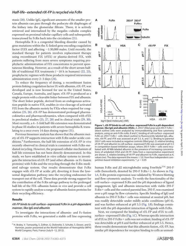
human FcRn and �2 microglobulin using FreeStyleTM 293-Fcells (henceforth, denoted by 293-F FcRn�). As shown in Fig.1A, FcRn protein expression was validated by Western blottingand flow cytometric analysis. To verify the functionality of thecell-surface– expressed FcRn and the pH dependence of ligandengagement, IgG and albumin interaction with viable 293-FFcRn� cells and the control parental line, 293-F, was examinedover a pH range by flow cytometry. Specific binding of IgG andalbumin to 293-F FcRn� cells was minimal at neutral pH, butwas readily detectable under mildly acidic conditions (pH 6),and was further enhanced at pH 5.5 (Fig. 1B); findings consis-tent with the pH-dependent interaction of ligands with FcRn.
Next, we compared the binding of rIX-FP and rFIX to cell-surface– expressed FcRn (Fig. 1C). Whereas specific interactionof rFIX to 293-F FcRn� cells was not evident, binding of rIX-FPwas detectable at pH 6 and further enhanced at pH 5.5. Hence,these results demonstrate that this albumin fusion, rIX-FP, hassimilar pH dependence for receptor binding to cells as unmod-
4 H. Braley, V. Tomasetig, S. Zollner, G. Dickneite, S. Schulte, S. Dower, and D.Hartman, poster presented at the World Federation of Hemophilia (WFH)World Congress, Paris, France (July 8 –12, 2012).
IgG HSA rIX-FP FIX
pH
7p
H 6
pH
5.5
pH
7p
H 6
pH
5.5
0 1 2 3 4 5log10 [test molecule] (nM)
HS
A-A
F488
HSArIX-FP
HSA rIX-FP0
200400600800
100012001400
Ki (
nM)
*879 +/- 136
193 +/- 36
rela
tive
ce
ll n
um
ber
293-F 293-F FcRn+
FcRn
tubulin
FcRn A
B C
ED
Figure 1. rIX-FP binds to cell-surface– expressed FcRn in a pH-dependentmanner, like IgG and albumin (HSA). A, 293-F (filled gray) and 293-F FcRn�(black outline) cells were analyzed by immunoblotting and flow cytometryanalysis, using an anti-FcRn mAb. B and C, binding of cell-surface– expressedFcRn on 293-F FcRn� cells (black outline) to fluorescently labeled IgG, albu-min, rIX-FP, and rFIX was assessed at pH 5.5, 6, and 7.2. Binding to parental293-F cells were included as negative controls (filled gray). D, relative bindingof rIX-FP and albumin to cell-surface– expressed FcRn was assessed at pH 5.5in competition-based inhibition assays, where 293-F FcRn� cells were incu-bated with AF488-labeled albumin in the presence of increasing concentra-tions of unlabeled rHSA and rIX-FP. Data from a representative experimentare shown. E, relative binding of rIX-FP and albumin to FcRn, expressed as Kivalues (nM). The data represent the means � S.E. from four independent com-petition-based inhibition experiments. *, p � 0.05
Half-life– extended rIX-FP is recycled via FcRn
6364 J. Biol. Chem. (2018) 293(17) 6363–6373
by guest on April 3, 2019
http://ww
w.jbc.org/
Dow
nloaded from

ified albumin. Based on these results, pH 5.5 was selected forcell-based FcRn-binding studies.
To further examine the interaction of albumin and rIX-FPwith cellular FcRn, we adapted a competition based inhibitionassay from Mathur et al. (33), originally developed to evaluatethe binding of IgG-based therapeutics for FcRn. In our assay,test molecules containing albumin compete with fluorescentlylabeled albumin (albumin-AF488) for binding to cell-surface–expressed FcRn at pH 5.5 (Fig. 1D), and the relative bindingaffinity to cell-surface– expressed FcRn is inferred by the indi-vidual Ki values of the molecules. As shown in Fig. 1E, rIX-FP(Ki of 193 � 36 nM) binds to cell-surface– expressed FcRn witha stronger apparent affinity than albumin (Ki of 879 � 136 nM).Previous biosensor analyses using soluble FcRn have alsoderived a higher affinity for rIX-FP,4 although the differencebetween rIX-FP and albumin was only 2-fold (�5 and 10 �M forrIX-FP and albumin, respectively, at pH 6). When examiningligand interaction with cell surface FcRn, however, it is possiblethat additional electrostatic or Gla domain–phospholipidinteractions may occur, mediated by the FIX component ofrIX-FP, thereby creating some binding avidity in the bifunc-tional fusion protein that may lower the Kd (34). Nevertheless,these interactions are presumably too weak to be detectable fornative FIX alone.
Endogenous Rab11 is a marker for recycling endosomes andthe FcRn-mediated recycling pathway in 293-F FcRn� cells
Having demonstrated the interaction between FcRn and thealbumin/Fc-containing cargo on 293-F FcRn� cells, we soughtto determine whether receptor-bound cargo could then beinternalized and recycled via the FcRn-mediated recyclingpathway. To track the movement of internalized proteinsthrough the intracellular recycling and/or degradation path-ways in 293-F FcRn� cells, we assessed a number of differentantibodies raised against specific endosomal compartmentsthat were suitable for paraformaldehyde-fixed samples. Weselected a panel of three antibodies for co-localization experi-ments: anti-EEA1, anti-Rab11, and anti-CD63 as markers forthe early endosomes, recycling endosomes, and late endo-somes/lysosomes, respectively (Fig. 2, A and B). Importantly,co-staining cells with EEA1 (Fig. 2A) and CD63 (Fig. 2B)revealed minimal co-localization with Rab11.
Although EEA1 and CD63 antibodies have been used previ-ously for identifying endosomal compartments in human cells,this is the first time, to the best of our knowledge, that a Rab11antibody has been used to detect endogenous Rab11 in recy-cling endosomes as a marker for the FcRn-mediated recyclingof cargo. Other publications have, however, used fluorescentlytagged Rab11 constructs transfected into their cell line of inter-est (35). To confirm the specificity of the Rab11 antibody andvalidate its use in 293-F FcRn� cells, we performed a knock-down experiment using siRNA against Rab11. Cells that weretransfected with Rab11-specific siRNA resulted in almost com-plete loss of antigen detection by immunofluorescence (Fig.2C), confirming antibody specificity.
To confirm whether 293-F FcRn� cells are functionallycapable of mediating intracellular trafficking and recycling, weexamined IgG and transferrin, because the movement of these
proteins via the Rab11 compartment through engagement withFcRn and transferrin receptors respectively is well docu-mented. As shown in Fig. 2D, following internalization (10 –15min at pH 5.5 for IgG or pH 7 for transferrin) and a 10-minchase, both IgG and transferrin were shown to co-localize withRab11� recycling endosomes in 293-F FcRn� cells, kineticsconsistent with the trafficking of cargo from the cell surface tothe recycling endosomes via the early endosomes.
Like IgG, albumin is internalized and traffics via the early andrecycling endosomes of the FcRn-mediated recycling processin 293-F FcRn� cells
To evaluate the intracellular trafficking of albumin, 293-FFcRn� cells were pulsed with AF488-labeled albumin at pH
C
D
E
A
B
EEA1 Rab11 overlay
CD63 Rab11 overlay
untransfected control siRNA Rab11siRNA
Transferrin Rab11 overlay
IgG Rab11 overlay
Figure 2. Endogenous Rab11 is a marker for recycling endosomes andthe FcRn-mediated recycling pathway in 293-F FcRn� cells. A and B, con-focal images of 293-F FcRn� cells stained with anti-EEA1 (green) and anti-Rab11 (red) antibodies (A) or anti-CD63 (green) and anti-Rab11 (red) antibod-ies (B). No co-localization was evident between the organelle-specificmarkers. C, scramble negative control and Rab11 siRNA transfected 293-FFcRn� cells were stained with anti-Rab11 antibody. D, 293-F FcRn� cellswere pulsed with AF594-labeled transferrin (green) at neutral pH for 15 min at37 °C, the cargo was removed, and the cells were incubated in completemedia, pH 7, for a further 10 min, before fixing and staining with anti-Rab11antibody (red). E, 293-F FcRn� cells were pulsed with AF488-labeled IgG(green) at pH 5.5 for 10 min (37 °C), the cargo was removed, and the cells wereincubated in complete media, pH 7, for a further 10 min, before fixing andstaining with anti-Rab11 antibody (red). The cell nuclei were labeled withHoechst 33342 (blue). All images were acquired on a Leica TCS SP5 confocalmicroscope with 63� magnification and a 1.4 NA oil-immersion objective asdescribed in detail under “Experimental procedures.” Representative confo-cal middle sections are shown. Scale bar, 10 �m.
Half-life– extended rIX-FP is recycled via FcRn
J. Biol. Chem. (2018) 293(17) 6363–6373 6365
by guest on April 3, 2019
http://ww
w.jbc.org/
Dow
nloaded from

5.5, for 10 min to allow the internalization of cargo via thesurface FcRn receptor (minimal internalization by parental293-F cells was observed under these conditions). Excess cargowas then removed and replaced with complete growth media(pH 7) for various chase periods. The cells were then fixedand stained for the organelle markers EEA1, Rab11, andCD63 (Fig. 3).
At the earliest time point (10-min pulse, 0-min chase), albu-min was readily detectable within early endosomes (Fig. 3, A,top row, and D), decreasing thereafter. After a 5-min chase, thepresence of cargo in the recycling endosome was clearly evident(Fig. 3, B, second row, and D). This was also observed at the20-min chase period (Fig. 3, B, third row, and D), although theoverall signal for albumin was markedly reduced, presumablybecause much had already been exported from the cells by thistime (Fig. 3E). Co-localization at this later time point wasobserved as orange (rather than yellow/pale green) because ofthe dominance of the Rab11 signal (in red) over the faint albu-min signal (in green) and is highlighted with arrows for individ-ual and combined channels. By the 45-min chase, only traces ofcargo were evident, consistent with observations for IgG (datanot shown). Throughout the time course, minimal albumin was
detected within the CD63� lysosomal compartment (Fig. 3, Cand D). Taken together, these data demonstrate that albumin isefficiently internalized and directed via the Rab11� recyclingendosomes in human cells expressing FcRn, similar to previousreports for IgG (12–16).
rIX-FP also traffics via early and recycling endosomes in 293-FFcRn� cells, with similar kinetics to albumin
Next, 293-F FcRn� cells were used to determine whetherrIX-FP has the potential to be recycled via the FcRn pathway. Asabove for albumin, 293-F FcRn� cells were pulsed with AF488-labeled rIX-FP at pH 5.5 for 10 min, and the transport of rIX-FPthrough intracellular organelles were examined after a series ofchase intervals (Fig. 4). After the pulse (10-min pulse, 0-minchase), and as observed for albumin, rIX-FP was readilydetected within the early endosome (Fig. 4, A, top row, and D).After a 5-min chase and from then on, rIX-FP was detected inthe recycling endosome (Fig. 4, B, second and third rows, and D).As for albumin, after a 20-min chase, the amount of AF488-labeled rIX-FP cargo detected was markedly reduced (Fig. 4Eand Fig. S1; arrows highlight regions of co-localization withRab11� endosome) and by 45 min was virtually undetectable
A B
C D
E
Ch
ase
tim
e fo
llow
ing
init
ial 1
0’ p
uls
e5’
20’
0’
HSA EEA1 overlay
Ch
ase
tim
e fo
llow
ing
init
ial 1
0’ p
uls
e5’
20’
0’
HSA Rab11 overlay
Ch
ase
tim
e fo
llow
ing
init
ial 1
0’ p
uls
e5’
20’
0’
HSA CD63 overlay
0 5 10 15 20Pulse
020406080
100
% c
o-l
oca
lisat
ion
Chase (minutes)
EEA1
CD63 Rab11
0 5 10 15 20Pulse
020406080
100
tota
l sig
nal
(%
)
Chase (minutes)
Figure 3. Like IgG, albumin traffics via the early and recycling endosomes in 293-F FcRn� cells. A–C, 293-F FcRn� cells were pulsed with AF488-labeledalbumin at pH 5.5 (green) and chased for various times at pH 7. The cells were then fixed and stained with the following organelle specific markers (red): EEA1for detection of early endosomes (A), Rab11 for recycling endosomes (B), or CD63 for late endosomes/lysosomes (C). The cell nuclei were labeled with Hoechst33342 (blue). Representative confocal middle sections for each time point are shown. Scale bar, 10 �m. Arrowheads indicate co-localization. Insets showmagnified images of selected areas denoted by the white boxes, where the signal for albumin has also been enhanced to highlight co-localization with Rab11.D, the proportion of albumin in each organelle after the 0-, 5-, and 20-min chase time points, as quantified according to “Experimental procedures” is expressedas % co-localization values. E, the amount of albumin remaining in 293-F FcRn� cells at each time point is expressed as the relative number of objects/cell, asquantified according to “Experimental procedures.” For D and E, the data represent the means � S.E. from three independent experiments, where for eachexperiment, an average value was determined from two to five images (each containing 13 or more cells) for each time point.
Half-life– extended rIX-FP is recycled via FcRn
6366 J. Biol. Chem. (2018) 293(17) 6363–6373
by guest on April 3, 2019
http://ww
w.jbc.org/
Dow
nloaded from

(data not shown). Throughout the time course, rIX-FP waslargely excluded from CD63� lysosomal compartments (Fig. 4,C and D), suggesting minimal degradation.
We also examined the co-trafficking of albumin and rIX-FP,where cells were simultaneously pulsed with differentiallylabeled cargoes at acidic pH (Fig. 5). Following FcRn-mediateduptake, the itinerary and kinetics of intracellular transport ofAF594-labeled albumin and AF488-labeled rIX-FP were verysimilar, and a high degree of co-localization of the two mole-cules was observed.
Albumin is rescued via FcRn interaction from lysosomaldegradation following fluid-phase endocytosis, in 293-FFcRn� cells
Although the internalization of albumin and IgG via FcRn atacidic pH may occur in some physiological settings, fluid-phaseendocytosis, such as pinocytosis or macropinocytosis, is likelyto have a greater role in internalization of these proteins in vivofrom which subsequent rescue by FcRn may occur (36 –38). Toexamine the cellular transport of albumin ligands under physi-ological conditions, cell monolayers were incubated with albu-min at neutral pH for prolonged periods. Internalization ofcargo was observed in both parental 293-F and 293-F FcRn�
cells, although the amount of cargo detected in the parentalcells was notably higher (Fig. 6A; �3-fold increased levels ofalbumin in 293-F cells than 293-F FcRn� cells). This is consis-tent with an absence of recycling in cells not expressing FcRn,where accumulation within the CD63� compartments (wheredegradation occurs more slowly) was clearly evident (Fig. 6A,top row). In contrast, in 293-F FcRn� cells, albumin wasdetected mainly in Rab11� recycling endosomes with a weakersignal, presumably resulting from constant export from the celland an absence of accumulation within the lysosomal compart-ment (Fig. 6A, bottom row). Co-localization with Rab11 wasobserved as pale red/orange given the dominance of the Rab11signal (in red) over that of the ligand (faint green) (see arrows inFig. 6A).
Under conditions of fluid-phase endocytosis, we were alsoable to investigate the intracellular fate of an albumin variantH464Q that cannot interact with FcRn (39). This mutant accu-mulated within the lysosomal compartment in both parentaland 293-F FcRn� cells and was not detected within the Rab11�recycling endosome (Fig. 6B). Minimal difference was observedin the total signal detected between the two cells lines. Thesefindings clearly demonstrate that the intracellular interaction
A B
C D
E
Ch
ase
tim
e fo
llow
ing
init
ial 1
0’ p
uls
e5’
20’
0’
rIX-FP EEA1 overlay
Ch
ase
tim
e fo
llow
ing
init
ial 1
0’ p
uls
e5’
20’
0’
rIX-FP Rab11 overlay
Ch
ase
tim
e fo
llow
ing
init
ial 1
0’ p
uls
e5’
20’
0’
rIX-FP CD63 overlay
0 5 10 15 20Pulse
020406080
100
% c
o-l
oca
lisat
ion
Chase (minutes)
EEA1
CD63 Rab11
0 5 10 15 20Pulse
020406080
100
tota
l sig
nal
(%
)
Chase (minutes)
Figure 4. rIX-FP traffics through 293-F FcRn� cells via a similar route to albumin. A–C, 293-F FcRn� cells were pulsed with AF488-labeled rIX-FP at pH 5.5(green) and chased for various times at pH 7. The cells were then fixed and stained with the following organelle specific markers (red): EEA1 for detection of earlyendosomes (A), Rab11 for recycling endosomes (B), or CD63 for late endosomes/lysosomes (C). The cell nuclei were labeled with Hoechst 33342 (blue).Representative confocal middle sections for each time point are shown. Scale bar, 10 �m. Arrowheads indicate co-localization. Insets show magnified images ofselected areas denoted by the white boxes, where the signal for rIX-FP has also been enhanced to highlight co-localization with Rab11. D, the proportion ofrIX-FP in each organelle after the 0-, 5-, and 20-min chase time points, as quantified according to “Experimental procedures,” is expressed as % co-localizationvalues. E, the amount of rIX-FP remaining in 293-F FcRn� cells at each time point is expressed as the relative number of objects/cell, as quantified according to“Experimental procedures.” For D and E, the data represent the means � S.E. from three independent experiments, where for each experiment, an averagevalue was determined from two to five images (each containing 13 or more cells) for each time point.
Half-life– extended rIX-FP is recycled via FcRn
J. Biol. Chem. (2018) 293(17) 6363–6373 6367
by guest on April 3, 2019
http://ww
w.jbc.org/
Dow
nloaded from

of albumin with FcRn is essential for diversion of the endocy-tosed albumin from the endolysosomal pathway to the recy-cling endosomes.
Following fluid-phase endocytosis, albumin fusion rescuesrIX-FP from cellular degradation, in cells expressing FcRn
Finally, we used our cellular system to determine the intra-cellular fate of rIX-FP following fluid-phase endocytosis. LikeWT unmodified albumin, rIX-FP accumulated within the lyso-some in the absence of FcRn expression and was detectedwithin the Rab11� compartment in 293-F FcRn� cells (Fig. 7Aand Fig. S2). The amount of cargo detected in the absence ofexogenous FcRn expression was notably higher than in 293-FFcRn� cells, again consistent with a lack of recycling in 293-Fcells (�3-fold more rIX-FP in 293-F cells). We also investigatedthe cellular fate of the unconjugated form of rFIX followingfluid-phase endocytosis. Compellingly, rFIX accumulatedwithin the lysosome in both parental and 293-F FcRn� cells(Fig. 7B), and minimal difference was observed in the total sig-nal detected between the two cell lines. Together, these resultsstrongly support FcRn-mediated salvage via the albumin moi-ety as a mechanism for the half-life extension of rIX-FPobserved in clinical studies.
Discussion
The direct genetic fusion of biopharmaceuticals to albuminor the Fc portion of IgG has been increasingly explored inrecent years as a strategy to improve plasma half-life. Severalfusion proteins have been approved for use in various indica-tions (9 –11), including two albumin fusion proteins: Albiglu-tide, a GLP-1–albumin fusion protein for the treatment of type2 diabetes (40), and rIX-FP for the treatment of hemophilia B.
All have shown improved pharmacokinetics compared withnonfused counterparts in clinical studies, and although it isgenerally accepted that their half-life extension is due at least inpart to FcRn-mediated salvage, FcRn recycling of albumin oralbumin-fusion proteins at a cellular level has not been directlyexamined. In the current study, we have developed an in vitrocellular system that can be used to directly investigate the itin-erary and kinetics of FcRn-mediated intracellular trafficking ofalbumin, IgG, and genetically engineered fusion proteins withalbumin or Fc moieties.
Studies into the intracellular trafficking of cargo have com-monly investigated recycling by co-localization with pathway-specific signature cargoes, such as transferrin and its receptoror, alternatively, the lack of co-localization with markers of thelysosomal degradation pathway (12, 13, 41– 43). Recently,Schmidt et al. (43) have used this approach for investigationsof albumin, demonstrating in cells overexpressing FcRndecreased co-localization with LAMP1� lysosomes for ahigh binding albumin variant. In our study, we have investi-gated the recycling of albumin ligands and demonstrated itstransit through the distinguishing organelle of this route, therecycling endosome (35, 44 – 46). The specificity of theRab11 antibody used to directly detect endogenous Rab11, aRas-like GTPase, as a marker for recycling endosomes wasconfirmed by the loss of signal in cells depleted of Rab11using Rab11 siRNA, but not a control siRNA. In addition,following their uptake, transferrin and IgG were as previouslyreported found to move into the Rab11� recycling compart-ment with rapid kinetics (Fig. 2).
Although multiple studies have demonstrated the itineraryfor IgG recycling (12–14), this is the first time imaging studieshave been thoroughly described for the recycling of albuminand fusion proteins thereof. Following internalization, bothalbumin and rIX-FP were shown to traffic via the early endo-somes into the recycling endosomes before their disappearancefrom the cell, presumably as a consequence of cell-surfacedelivery and exocytosis. After a 20-min chase, signals for bothalbumin and rIX-FP were markedly reduced, and by 45 min,only traces remained. In addition, when cells were pulsedsimultaneously with albumin and IgG, the two proteins fol-lowed the same intracellular trafficking route and with similarkinetics. Although the timing of endocytic routes has not beenextensively described for FcRn recycling, the kinetics weobserved are similar to those generally described for other recy-cling pathways (47).
Despite a 4 –5-fold half-life extension, rIX-FP is clearedmuch more quickly than albumin (half-life of �102 h inhumans (31) compared with 19 days for albumin (8)), suggest-ing a very active clearance mechanism for this coagulation fac-tor. Although clearance pathways may be amenable to half-lifeextension via FcRn, recycling is a competitive process and lessthan 100% efficient. Therefore, a protein that undergoes rapidand continuous internalization will ultimately be more quicklydegraded. The mechanism by which FIX is normally removedfrom the circulation is poorly understood. Although in vitrostudies have suggested a role for the asialoglycoprotein recep-tor in binding and clearing FIX (48, 49), in vivo studies havedemonstrated no difference in plasma levels of FIX in asialogly-
rIX-FP HSA overlay
5’20
’45
’0’
Ch
ase
tim
e fo
llow
ing
init
ial 1
0 m
inu
te p
uls
e
Figure 5. Co-trafficking of HSA and rIX-FP in 293-F FcRn� cells. 293-FFcRn� cells were pulsed simultaneously with AF488-labeled rIX-FP (green)and AF594-labeled albumin (red) at pH 5.5 for 10 min and chased in freshcomplete media for various times at pH 7. The cells were then fixed, and thenuclei were stained with Hoechst 33342 (blue) and visualized by confocalmicroscopy. Representative confocal middle sections for each time point areshown, where results are representative of two independent experiments.Scale bar, 10 �m. Arrowheads indicate co-localization.
Half-life– extended rIX-FP is recycled via FcRn
6368 J. Biol. Chem. (2018) 293(17) 6363–6373
by guest on April 3, 2019
http://ww
w.jbc.org/
Dow
nloaded from

coprotein receptor– deficient mice (49, 50). Activated FIX hasbeen shown to interact with low-density lipoprotein–relatedprotein-1 (LRP1) through an epitope exposed on the active pro-tease, but an association has not been demonstrated for FIXzymogen (51), and no changes in FIX levels have been observedin mice deficient for LRP1 (52). The binding of FIX to vascular
endothelial cells via collagen IV interaction has also been exam-ined but does not appear to be the primary clearance pathwayfor FIX (53). A better understanding of the normal routesof clearance of FIX is clearly required to further elucidatethe mechanisms and limitations of half-life extension of FIXthrough albumin fusion.
A
B
HSA Rab11 CD63 HSA / Rab11 HSA / CD63
HSA Rab11 CD63 HSA / Rab11 HSA / CD6329
3-F
293
-F F
cRn+
CD63
H464Q Rab11 CD63 H464Q / Rab11 H464Q / CD63
H464Q Rab11 H464Q / Rab11 H464Q / CD63
293-
F 2
93-F
FcR
n+
Rab11 CD63 0
50
100
% c
o-lo
calis
atio
n
Rab11 CD63 0
50
100
% c
o-lo
calis
atio
n
Rab11 CD63 0
50
100
% c
o-lo
calis
atio
n
Rab11 CD63 0
50
100
% c
o-lo
calis
atio
n
Figure 6. Albumin is rescued from lysosomal degradation, following fluid-phase endocytosis, in cells expressing FcRn. Fluid-phase uptake ofalbumin variants by 293-F and 293-F FcRn� cells was examined. The cells were incubated with AF488-labeled albumin (HSA, A)) or FcRn interactiondefective albumin variant H464Q (B) at neutral pH for 5– 6 h, prior to fixation and staining with organelle markers (red). The cell nuclei were labeled withHoechst 33342 (blue). Representative confocal middle sections are shown. Scale bar, 10 �m. Arrowheads indicate co-localization. Insets show magnifiedimages of selected areas denoted by the white boxes, where the signal for albumin has also been enhanced to highlight co-localization with Rab11. Theproportion of cargo in each organelle, as quantified according to “Experimental procedures,” is expressed as % co-localization values. The datarepresent the means � S.E. from three independent experiments, where for each experiment, an average value was determined from two to five images(each containing 13 or more cells).
Rab11 CD63 0
50
100
% c
o-l
oca
lisat
ion
Rab11 CD63 0
50
100
% c
o-l
oca
lisat
ion
Rab11 CD63 0
50
100
% c
o-l
oca
lisat
ion
Rab11 CD63 0
50
100
% c
o-l
oca
lisat
ion
A
B
rIX-FP Rab11 CD63 rIX-FP / Rab11 rIX-FP / CD63
rIX-FP Rab11 CD63 rIX-FP / Rab11 rIX-FP / CD63
293-
F 2
93-F
FcR
n+
rFIX Rab11 CD63 rFIX / Rab11 rFIX / CD63
rFIX Rab11 CD63 rFIX / Rab11 rFIX / CD63
293-
F 2
93-F
FcR
n+
Figure 7. FcRn dependent salvage of rIX-FP following internalization via fluid-phase endocytosis. Fluid-phase uptake of FIX variants by 293-F and 293-FFcRn� cells was examined. The cells were incubated with AF488-labeled rIX-FP (A) and rFIX (B) at neutral pH for 5– 6 h, prior to fixation and staining withorganelle markers (red). The cell nuclei were labeled with Hoechst 33342 (blue). Representative confocal middle sections are shown. Scale bar, 10 �m.Arrowheads indicate co-localization. Insets show magnified images of selected areas denoted by the white boxes, where the signal for rIX-FP has also beenenhanced to highlight co-localization with Rab11. The proportion of cargo in each organelle as quantified according to “Experimental procedures” is expressedas % co-localization values. The data represent the means � S.E. from three independent experiments, where for each experiment, an average value wasdetermined from two to five images (each containing 13 or more cells).
Half-life– extended rIX-FP is recycled via FcRn
J. Biol. Chem. (2018) 293(17) 6363–6373 6369
by guest on April 3, 2019
http://ww
w.jbc.org/
Dow
nloaded from
Although it remains a topic of debate (37, 38), it is generallyaccepted that fluid-phase endocytosis is the primary mediatorof IgG and albumin entry into cells from plasma and would alsobe expected to have an important role in internalization of FIXas for any plasma protein. An essential role for endothelial andhematopoietic cells has been demonstrated in maintainingboth IgG and albumin homeostasis using mice with tissue-spe-cific Tie2 Cre-mediated deletion of FcRn (19). Because thesecells are generally found in environments at near neutral pHwhen negligible binding of IgG and albumin to FcRn isobserved, and given the very high levels of these proteins inplasma, the consensus view is that pinocytic uptake is the pri-mary mediator of ligand entry into these cells. Albumin has alsobeen shown to be able to be internalized by other cells, and themegalin– cubulin scavenger receptor, in particular, has beenshown to have a critical role in salvage of albumin from glomer-ular filtrate by kidney tubular epithelial cells, followed by sub-sequent FcRn-mediated transcytosis back into the circulation(20, 54, 55). However, for cells bathed in acidic pH, such asneonatal gut epithelial cells, FcRn-mediated internalization ofIgG and albumin may occur and is likely to mediate the tran-scytosis of maternal IgG to the neonatal circulation. Impor-tantly, regardless of the uptake mechanism, an engagement ofthe ligand with FcRn within the acidic environment of the earlyendosomes, is essential for diverting the ligand away from thelysosomal degradation pathway and back to the cell surface,where exocytosis and a shift to neutral pH facilitates ligandrelease (13). Exocytosis of IgG has been shown to occur viadifferent mechanisms, ranging from a complete fusion of exo-cytic vesicles with the plasma membrane (resulting in release allIgG at once), to a slower-release mode where secretory vesiclesfollow a complex form of kiss-and-run fusion (with only partialrelease of cargo at each event) (56). Consistently, we have dem-onstrated that albumin and rIX-FP, either internalized via FcRnat acidic pH or internalized by fluid-phase endocytosis, aredirected through the intracellular recycling pathway in anFcRn-dependent manner (Figs. 4 and 7).
Importantly, albumin and Fc fusion technology can result inserum persistence in ways other than engaging the FcRn recy-cling machinery. An increase in the hydrodynamic volume ofthe therapeutic protein to prevent clearance through the kidneycan play an important role, especially for small proteins. With amolecular mass of 55 kDa, it is possible that some FIX is nor-mally cleared via kidney filtration, for which the cut-off is �70kDa, but can also vary depending on the charge of the molecule.There is evidence for some clearance of coagulation factor VII,which is of a similar size and composition to FIX, through thekidney (57). It is therefore possible that prevention of clearancevia the kidney may also contribute to the increased half-life ofrIX-FP.
In conclusion, we present evidence to support the hypothesisthat rIX-FP can exploit the FcRn-mediated recycling pathwaynormally reserved for IgG and albumin, providing a mechanismfor the 4 –5-fold half-life extension recently demonstrated inclinical trials (31). In addition, the cell-based assays we havedescribed provide a valuable platform for assessing the recy-cling capacity of novel half-life– extended therapeutics.
Experimental procedures
Materials
Recombinant albumin (human) and monoclonal IgG (humanIgG1) were expressed in FreeStyleTM 293-F Cells (Life Technol-ogies) and purified as previously described (58). rIX-FP (Idel-vion) was obtained from CSL Behring (GmbH, Germany)and rFIX (BeneFIX�) from Pfizer Pharma (GmbH, Berlin,Germany). Human transferrin conjugated to Alexa Fluor�594 (AF594) was purchased from Thermo Fisher (MolecularProbes, T13343).
Recombinant albumin, albumin variant H464Q, IgG, rIX-FP,and rFIX were labeled with Alexa Fluor� 488 (AF488) NHSester (succinimidyl ester) (Life Technologies, A-20000) orAlexa Fluor� 594 (AF594) NHS ester (succinimidyl ester)(Life Technologies, A-37572), according to the manufactu-rer’s protocol. For flow cytometry and Western blotting anal-ysis, the following antibodies were used: mouse anti-FcRn anti-body (Acris Antibodies, AM26754PU-N), anti-tubulin-HRPantibody (Abcam, ab185067), anti-mouse IgG-HRP (JacksonImmunoResearch, 715-035-150), and anti-mouse IgG-AF488(Molecular Probes, A-11029).
Generation of stable FcRn-expressing cells
FreeStyleTM 293-F cells were grown under adherent condi-tions in growth medium containing RPMI supplemented withGlutaMAXTM (Gibco) and 10% fetal bovine serum (Sigma–Aldrich, 12003C) in a humidified 5% CO2 incubator at 37 °C.The cells were transfected with linearized plasmids containinghuman FcRn and �2 microglobulin sequences using Lipo-fectamine 2000 (Thermo Fisher, 11668019) and maintained ingrowth medium containing 0.5 mg/ml G418 (Thermo Fisher,10131027). Single-cell clones were individually picked fromtransfection cultures and expanded under constant antibioticselection. The clones with high expression levels were identi-fied by on a FACS by binding to AF488-labeled human IgG atacidic pH. One of the high expressing clones was selected foruse in binding, trafficking, and recycling assays (henceforthdenoted by 293-F FcRn�).
To confirm FcRn expression, 293-F and 293-F FcRn� cellswere stained for 30 min on ice with 10 �g/ml mouse anti-FcRnantibody (Acris Antibodies, AM26754PU-N), followed by a30-min incubation with secondary antibody, anti-mouse IgG-AF488 (4 �g/ml, Molecular Probes, A-11029). After the incu-bation, the cell-bound fluorescence was analyzed on a LSRFortessaTM cell analyzer (BD Biosciences). Expression was alsoconfirmed by Western blotting analysis.
Rab11 siRNA transient transfections
To knock down Rab11 in 293-F FcRn� cells, siRNA duplexestargeting Rab11A (5�-AAUGUCAGACAGACGCGAAAA-[dT][dT]-3�) and Rab11B (5�-AAGCACCUGACCUAUGAG-AAC[dT][dT]-3�) (Sigma–Aldrich) were delivered to the cellsat a final concentration of 50 nM, using DharmaFECT 1 trans-fection reagent (GE Dharmacon, T-2001-2). At 48 h post-trans-fection, the cells were analyzed for Rab11 expression by immu-nofluorescence microscopy, using a rabbit anti-Rab11 antibody(Abcam, ab3612). A nontargeting siRNA duplex (5�-AGGUC-
Half-life– extended rIX-FP is recycled via FcRn
6370 J. Biol. Chem. (2018) 293(17) 6363–6373
by guest on April 3, 2019
http://ww
w.jbc.org/
Dow
nloaded from

GGUGUGCUCUUGUUGG[dT][dT]-3�) (Sigma–Aldrich) wasincluded as a negative control.
In vitro cell-based FcRn-binding assays
For binding assays, 293-F FcRn� cells were cultured in suspen-sion in FreeStyleTM 293 expression medium (Thermo Fisher,12338018) supplemented with 0.1% Pluronic (ThermoFisher, 24040032), antibiotic–antimycotic solution (ThermoFisher, 15240), and 0.5 mg/ml G418 (Thermo Fisher, 10131027),in a humidified 8% CO2 orbital shaker–incubator (150 rpm), forat least 3–5 days prior to the experiment.
To assess the pH specificity of cargo binding to cell-surface–expressed FcRn, 293-F FcRn� cells were resuspended in Dul-becco’s PBS (Sigma–Aldrich, D8537) at a pH of 5.5, 6.0, or 7(2 � 105 cells/100 �l/reaction) and incubated with 50 nM ofAF488-labeled material. After an hour of incubation on ice, thecells were washed in the same buffer of corresponding pH,before their cell-bound fluorescence was analyzed on a LSR-FortessaTM cell analyzer (BD Biosciences).
To perform the competition-based inhibition assay, 293-FFcRn� cells were resuspended at a density of 8 � 106 cells/ml inserum-free assay medium (Dulbecco’s PBS at pH 5.5). The cellswere then plated in U-bottom wells of a 96-well plate contain-ing mixtures of labeled competitor molecule (albumin-AF488)and unlabeled test molecules (albumin or rIX-FP). In a typicalassay, 25 �l of cell suspension (2 � 105 cells/well), 25 �l ofalbumin-AF488 diluted from stock in assay media to give a finalconcentration of 1 �g/ml (15.15 nM), and 50 �l of varying con-centrations of albumin-containing test molecule were added towells in a total volume of 100 �l/well. The assay mixture wasincubated for 2 h at 4 °C with constant shaking. After the incu-bation, the cell-bound fluorescence in each well was read on theLSR FortessaTM cell analyzer. The mean fluorescence intensitywas obtained from each experiment and analyzed usingGraphPad Prism. The equilibrium dissociation constant, Ki, foreach test molecule was determined according to the one-site fitKi model for competitive binding experiments.
Intracellular trafficking assays following FcRn-mediateduptake at acidic pH
For intracellular trafficking assays, 293-F FcRn� cells wereplated in 8-well chamber NuncTM Lab-TekTMII CC2TM cham-ber slide system (Thermo Fisher, 154941) and grown to �80%confluency. Protein cargo was diluted in assay medium at thefollowing concentrations: 20 �g/ml AF594-labeled transferrin,0.1 �M AF488-labeled IgG, 2– 4 �M AF488/AF594-labeledalbumin, or 0.5–1 �M AF488-labeled rIX-FP and incubatedwith cells in a humidified 5% CO2 incubator at 37 °C. After a10 –15-min pulse at the specified pH (pH 5.5 for IgG, albumin,and rIX-FP; pH 7 for transferrin), the supernatant containingexcess cargo (that had not been internalized) was removed andreplaced with prewarmed complete growth medium. Internal-ized cargo was allowed to traffic for various time periods (chase)at 37 °C in a humidified 5% CO2 incubator, before incubationwas stopped. The cells were fixed with 4% paraformaldehydefor 15 min and either visualized by confocal microscopy or fur-ther processed for intracellular staining of organelles.
For intracellular staining of organelles, the cells were per-meabilized in 0.5% Triton X-100/PBS for 5 min and blocked in1% BSA/PBS at 4 °C overnight. Monolayers were incubatedwith 0.5 �g/ml mouse anti-EEA1 (BD Biosciences, 610457), 0.2�g/ml mouse anti-CD63 (Santa Cruz, sc-5275), or 2.5 �g/mlrabbit anti-Rab11 (Abcam, ab3612) for 1.5 h at room tempera-ture or at 4 °C overnight, followed by a 45-min incubation withsecondary antibodies, anti-mouse IgG-AF647 (4 �g/ml, Molec-ular Probes, A-21236), anti-rabbit IgG-AF647 (4 �g/ml, Molec-ular Probes, A-21245), and anti-rabbit IgG-AF546 (4 �g/ml,Molecular Probes, A-11035). After the intracellular staining(EEA1, Rab11, or CD63), the cells were incubated for 15 minwith Hoechst 33342 diluted in PBS (10 �g/ml, MolecularProbes, H3570). Following the intracellular staining, the cellswere mounted with ProLong Gold antifade mountant (Molec-ular Probes, P36930), and a coverslip was applied. The cellswere examined using a Leica TCS SP5 confocal microscope(Leica Microsystems) equipped with DIC and fluorescenceoptics, diode 405-nm, argon 488-nm, diode pumped solid state561-nm, and HeNe 633-nm lasers. The fluorescence imageswere collected with a 63� magnification and 1.4 NA oil-im-mersion objective at 37 °C using sPMT (spectral detectors) andT-PMT (transmitted light) and acquisition software LAS AF(Leica Application Suite Advanced Fluorescence) version2.6.0.7266. Time-course images for each cargo were taken usingthe same laser intensity, exposure, and gain settings to allow fordirect comparison.
Quantitation of the co-localization between internalizedcargo and fluorescent organelle markers was performed usingthe plugin organelle-based co-localization as described byWoodcroft et al. (32), on the FIJI program (National Institutesof Health public domain software). Two to five images (eachcontaining �13 cells) from each experiment were analyzed atevery time point, because preliminary experiments revealed nosignificant difference in quantitation when cells were analyzedindividually or together as an image. Images from each time-course experiment were analyzed under identical conditions,with constant threshold values used to identify the cargo andorganelles. Co-localization values were expressed as a percent-age of total cargo, determined by taking the sum of overlappingpixels between the cargo and respective markers, divided by thetotal number of cargo pixels. The average number of objects percells was also calculated for each experiment by dividing thenumber of objects (defined as a minimum of five pixels) by thetotal number of cell nuclei per image and expressed relative tothe first time point taken (T0). All data are expressed asmeans � S.E. of three independent experiments.
Fluid-phase endocytosis assays in 293-F and 293-F FcRn� cells
293-F FcRn� cells and 293-F cells were plated in 8-wellchamber NuncTM Lab-TekTM II CC2TM chamber slide systemand grown to �80% confluency. Protein cargo was diluted inprewarmed complete growth medium at the following concen-trations: 2 �M AF488-labeled albumin, 2 �M AF488-labeledalbumin variant H464Q (39), 1 �M AF488-labeled rIX-FP, or 1�M AF488-labeled rFIX and incubated with cells in a humidi-fied 5% CO2 incubator at 37 °C. After a continuous pulse of 5– 6h, the supernatant containing excess cargo was removed, and
Half-life– extended rIX-FP is recycled via FcRn
J. Biol. Chem. (2018) 293(17) 6363–6373 6371
by guest on April 3, 2019
http://ww
w.jbc.org/
Dow
nloaded from
cells were fixed and further processed for intracellular stainingas described above. Imaging and quantitation of the co-local-ization between internalized cargo and fluorescent organellemarkers was also performed as described above. Importantly,the laser intensity, exposure, gain, and threshold settings werekept constant for each cargo analyzed, to allow for direct com-parison between cell lines.
Author contributions—J. C. designed and performed research, ana-lyzed and interpreted data, and wrote the manuscript. J. L. and I. G.designed and/or performed experiments and analyzed and inter-preted data. S. T. contributed vital reagents. G. T. B. helped analyzequantitative data. S. K. D. and P. A. G. helped design research andedited the manuscript. A. M. V. designed research, analyzed andinterpreted data, and wrote the manuscript.
Acknowledgments—We acknowledge the Protein Expression and Pro-tein Technologies groups at CSL research for the expression and puri-fication of recombinant proteins; Tony Rowe for a role in project man-agement; Paul McMillan and the Biological Optical MicroscopyPlatform at the University of Melbourne, where imaging experimentswere conducted; and Justine Mintern for helpful discussions.
References1. Challa, D. K., Velmurugan, R., Ober, R. J., and Sally Ward, E. (2014) FcRn:
from molecular interactions to regulation of IgG pharmacokinetics andfunctions. Curr. Top. Microbiol. Immunol. 382, 249 –272 Medline
2. Ward, E. S., and Ober, R. J. (2009) Chapter 4: Multitasking by exploitationof intracellular transport functions the many faces of FcRn. Adv. Immunol.103, 77–115 CrossRef Medline
3. Gastinel, L. N., Simister, N. E., and Bjorkman, P. J. (1992) Expression andcrystallization of a soluble and functional form of an Fc receptor related toclass I histocompatibility molecules. Proc. Natl. Acad. Sci. U.S.A. 89,638 – 642 CrossRef Medline
4. Raghavan, M., Bonagura, V. R., Morrison, S. L., and Bjorkman, P. J. (1995)Analysis of the pH dependence of the neonatal Fc receptor/immunoglob-ulin G interaction using antibody and receptor variants. Biochemistry 34,14649 –14657 CrossRef Medline
5. Chaudhury, C., Mehnaz, S., Robinson, J. M., Hayton, W. L., Pearl, D. K.,Roopenian, D. C., and Anderson, C. L. (2003) The major histocompatibil-ity complex-related Fc receptor for IgG (FcRn) binds albumin and pro-longs its lifespan. J. Exp. Med. 197, 315–322 CrossRef Medline
6. Junghans, R. P., and Anderson, C. L. (1996) The protection receptor forIgG catabolism is the �2-microglobulin-containing neonatal intestinaltransport receptor. Proc. Natl. Acad. Sci. U.S.A. 93, 5512–5516 CrossRefMedline
7. Watson, D. (1965) Albumin and “total globulin” fractions of blood. Adv.Clin. Chem. 8, 237–303 Medline
8. Peters, T., Jr. (1985) Serum albumin. Adv. Protein Chem. 37, 161–245CrossRef Medline
9. Sand, K. M., Bern, M., Nilsen, J., Noordzij, H. T., Sandlie, I., and Andersen,J. T. (2014) Unraveling the interaction between FcRn and albumin: oppor-tunities for design of albumin-based therapeutics. Front. Immunol. 5, 682Medline
10. Rath, T., Baker, K., Dumont, J. A., Peters, R. T., Jiang, H., Qiao, S. W.,Lencer, W. I., Pierce, G. F., and Blumberg, R. S. (2015) Fc-fusion proteinsand FcRn: structural insights for longer-lasting and more effective thera-peutics. Crit. Rev. Biotechnol. 35, 235–254 CrossRef Medline
11. Sleep, D. (2015) Albumin and its application in drug delivery. Expert Opin.Drug Deliv. 12, 793– 812 CrossRef Medline
12. Ward, E. S., Zhou, J., Ghetie, V., and Ober, R. J. (2003) Evidence to supportthe cellular mechanism involved in serum IgG homeostasis in humans.Int. Immunol. 15, 187–195 CrossRef Medline
13. Ober, R. J., Martinez, C., Vaccaro, C., Zhou, J., and Ward, E. S. (2004)Visualizing the site and dynamics of IgG salvage by the MHC class I-re-lated receptor, FcRn. J. Immunol. 172, 2021–2029 CrossRef Medline
14. Tzaban, S., Massol, R. H., Yen, E., Hamman, W., Frank, S. R., Lapierre,L. A., Hansen, S. H., Goldenring, J. R., Blumberg, R. S., and Lencer, W. I.(2009) The recycling and transcytotic pathways for IgG transport by FcRnare distinct and display an inherent polarity. J. Cell Biol. 185, 673– 684CrossRef Medline
15. Claypool, S. M., Dickinson, B. L., Wagner, J. S., Johansen, F. E., Venu, N.,Borawski, J. A., Lencer, W. I., and Blumberg, R. S. (2004) Bidirectionaltransepithelial IgG transport by a strongly polarized basolateral mem-brane Fcgamma-receptor. Mol. Biol. Cell 15, 1746 –1759 CrossRefMedline
16. Jerdeva, G. V., Tesar, D. B., Huey-Tubman, K. E., Ladinsky, M. S., Fraser,S. E., and Bjorkman, P. J. (2010) Comparison of FcRn- and pIgR-mediatedtransport in MDCK cells by fluorescence confocal microscopy. Traffic 11,1205–1220 CrossRef Medline
17. Oganesyan, V., Damschroder, M. M., Cook, K. E., Li, Q., Gao, C., Wu, H.,and Dall’Acqua, W. F. (2014) Structural insights into neonatal Fc receptor-based recycling mechanisms. J. Biol. Chem. 289, 7812–7824 CrossRefMedline
18. Akilesh, S., Christianson, G. J., Roopenian, D. C., and Shaw, A. S. (2007)Neonatal FcR expression in bone marrow-derived cells functions to pro-tect serum IgG from catabolism. J. Immunol. 179, 4580 – 4588 CrossRefMedline
19. Montoyo, H. P., Vaccaro, C., Hafner, M., Ober, R. J., Mueller, W., andWard, E. S. (2009) Conditional deletion of the MHC class I-related recep-tor FcRn reveals the sites of IgG homeostasis in mice. Proc. Natl. Acad. Sci.U.S.A. 106, 2788 –2793 CrossRef Medline
20. Sarav, M., Wang, Y., Hack, B. K., Chang, A., Jensen, M., Bao, L., and Quigg,R. J. (2009) Renal FcRn reclaims albumin but facilitates elimination of IgG.J. Am. Soc. Nephrol. 20, 1941–1952 CrossRef Medline
21. Amsellem, S., Gburek, J., Hamard, G., Nielsen, R., Willnow, T. E., Devuyst,O., Nexo, E., Verroust, P. J., Christensen, E. I., and Kozyraki, R. (2010)Cubilin is essential for albumin reabsorption in the renal proximal tubule.J. Am. Soc. Nephrol. 21, 1859 –1867 CrossRef Medline
22. White, G. C., 2nd, Beebe, A., and Nielsen, B. (1997) Recombinant factorIX. Thromb. Haemost. 78, 261–265 Medline
23. Ewenstein, B. M., Joist, J. H., Shapiro, A. D., Hofstra, T. C., Leissinger,C. A., Seremetis, S. V., Broder, M., Mueller-Velten, G., Schwartz, B. A., andMononine Comparison Study Group (2002) Pharmacokinetic analysis ofplasma-derived and recombinant F IX concentrates in previously treatedpatients with moderate or severe hemophilia B. Transfusion 42, 190 –197CrossRef Medline
24. Björkman, S., Shapiro, A. D., and Berntorp, E. (2001) Pharmacokinetics ofrecombinant factor IX in relation to age of the patient: implications fordosing in prophylaxis. Haemophilia 7, 133–139 CrossRef Medline
25. Metzner, H. J., Weimer, T., Kronthaler, U., Lang, W., and Schulte, S.(2009) Genetic fusion to albumin improves the pharmacokinetic proper-ties of factor IX. Thromb. Haemost. 102, 634 – 644 Medline
26. Schulte, S. (2009) Half-life extension through albumin fusion technolo-gies. Thromb. Res. 124, S6 –S8 CrossRef Medline
27. Nolte, M. W., Nichols, T. C., Mueller-Cohrs, J., Merricks, E. P., Pragst, I.,Zollner, S., and Dickneite, G. (2012) Improved kinetics of rIX-FP, a recom-binant fusion protein linking factor IX with albumin, in cynomolgus mon-keys and hemophilia B dogs. J. Thromb. Haemost. 10, 1591–1599 CrossRefMedline
28. Herzog, E., Harris, S., Henson, C., McEwen, A., Schenk, S., Nolte, M. W.,Pragst, I., Dickneite, G., Schulte, S., and Zollner, S. (2014) Biodistributionof the recombinant fusion protein linking coagulation factor IX with al-bumin (rIX-FP) in rats. Thromb. Res. 133, 900 –907 CrossRef Medline
29. Santagostino, E., Negrier, C., Klamroth, R., Tiede, A., Pabinger-Fasching,I., Voigt, C., Jacobs, I., and Morfini, M. (2012) Safety and pharmacokineticsof a novel recombinant fusion protein linking coagulation factor IX withalbumin (rIX-FP) in hemophilia B patients. Blood 120, 2405–2411CrossRef Medline
30. Martinowitz, U., Lissitchkov, T., Lubetsky, A., Jotov, G., Barazani-Brut-man, T., Voigt, C., Jacobs, I., Wuerfel, T., and Santagostino, E. (2015)
Half-life– extended rIX-FP is recycled via FcRn
6372 J. Biol. Chem. (2018) 293(17) 6363–6373
by guest on April 3, 2019
http://ww
w.jbc.org/
Dow
nloaded from

Results of a phase I/II open-label, safety and efficacy trial of coagulationfactor IX (recombinant), albumin fusion protein in haemophilia B pa-tients. Haemophilia 21, 784 –790 CrossRef Medline
31. Santagostino, E., Martinowitz, U., Lissitchkov, T., Pan-Petesch, B.,Hanabusa, H., Oldenburg, J., Boggio, L., Negrier, C., Pabinger, I., vonDepka Prondzinski, M., Altisent, C., Castaman, G., Yamamoto, K., Álva-rez-Roman, M. T., Voigt, C., et al. (2016) Long acting recombinant coag-ulation factor IX albumin fusion protein (rIX-FP) in hemophilia B: resultsof a phase 3 trial. Blood 127, 1761–1769 CrossRef Medline
32. Woodcroft, B. J., Hammond, L., Stow, J. L., and Hamilton, N. A. (2009)Automated organelle-based colocalization in whole-cell imaging. Cytom-etry 75, 941–950 Medline
33. Mathur, A., Arora, T., Liu, L., Crouse-Zeineddini, J., and Mukku, V. (2013)Qualification of a homogeneous cell-based neonatal Fc receptor (FcRn)binding assay and its application to studies on Fc functionality of IgG-based therapeutics. J. Immunol. Methods 390, 81–91 CrossRef Medline
34. Zwaal, R. F., Comfurius, P., and Bevers, E. M. (1998) Lipid-protein inter-actions in blood coagulation. Biochim. Biophys. Acta 1376, 433– 453CrossRef Medline
35. Ward, E. S., Martinez, C., Vaccaro, C., Zhou, J., Tang, Q., and Ober, R. J.(2005) From sorting endosomes to exocytosis: association of Rab4 andRab11 GTPases with the Fc receptor, FcRn, during recycling. Mol. Biol.Cell 16, 2028 –2038 CrossRef Medline
36. Ward, E. S., Devanaboyina, S. C., and Ober, R. J. (2015) Targeting FcRn forthe modulation of antibody dynamics. Mol. Immunol. 67, 131–141CrossRef Medline
37. Anderson, C. L. (2014) There’s been a flaw in our thinking. Front. Immu-nol. 5
38. Ward, E. S., and Ober, R. J. (2015) Commentary: “There’s been a flaw inour thinking.” Front. Immunol. 6
39. Andersen, J. T., Dalhus, B., Cameron, J., Daba, M. B., Plumridge, A., Evans,L., Brennan, S. O., Gunnarsen, K. S., Bjørås, M., Sleep, D., and Sandlie, I.(2012) Structure-based mutagenesis reveals the albumin-binding site ofthe neonatal Fc receptor. Nat. Commun. 3, 610 CrossRef Medline
40. Trujillo, J. M., and Nuffer, W. (2014) Albiglutide: a new GLP-1 receptoragonist for the treatment of type 2 diabetes. Ann. Pharmacother. 48,1494 –1501 CrossRef Medline
41. Weflen, A. W., Baier, N., Tang, Q. J., Van den Hof, M., Blumberg, R. S.,Lencer, W. I., and Massol, R. H. (2013) Multivalent immune complexesdivert FcRn to lysosomes by exclusion from recycling sorting tubules. Mol.Biol. Cell 24, 2398 –2405 CrossRef Medline
42. Fujimoto, K., Ida, H., Hirota, Y., Ishigai, M., Amano, J., and Tanaka, Y.(2015) Intracellular dynamics and fate of a humanized anti-interleukin-6receptor monoclonal antibody, tocilizumab. Mol. Pharmacol. 88,660 – 675 CrossRef Medline
43. Schmidt, E. G. W., Hvam, M. L., Antunes, F., Cameron, J., Viuff, D., An-dersen, B., Kristensen, N. N., and Howard, K. A. (2017) Direct demonstra-tion of a neonatal Fc receptor (FcRn)-driven endosomal sorting pathwayfor cellular recycling of albumin. J. Biol. Chem. 292, 13312–13322CrossRef Medline
44. Sönnichsen, B., De Renzis, S., Nielsen, E., Rietdorf, J., and Zerial, M. (2000)Distinct membrane domains on endosomes in the recycling pathway vi-
sualized by multicolor imaging of Rab4, Rab5, and Rab11. J. Cell Biol. 149,901–914 CrossRef Medline
45. Stenmark, H. (2009) Rab GTPases as coordinators of vesicle traffic. Nat.Rev. Mol. Cell Biol. 10, 513–525 CrossRef Medline
46. Gan, Z., Ram, S., Ober, R. J., and Ward, E. S. (2013) Using multifocal planemicroscopy to reveal novel trafficking processes in the recycling pathway.J. Cell Sci. 126, 1176 –1188 CrossRef Medline
47. Maxfield, F. R., and McGraw, T. E. (2004) Endocytic recycling. Nat. Rev.Mol. Cell Biol. 5, 121–132 CrossRef Medline
48. Blasko, E., Brooks, A. R., Ho, E., Wu, J. M., Zhao, X. Y., and Subramanyam,B. (2013) Hepatocyte clearance and pharmacokinetics of recombinant fac-tor IX glycosylation variants. Biochem. Biophys. Res. Commun. 440,485– 489 CrossRef Medline
49. Grewal, P. K. (2010) The Ashwell-Morell receptor. Methods Enzymol.479, 223–241 CrossRef Medline
50. Grewal, P. K., Uchiyama, S., Ditto, D., Varki, N., Le, D. T., Nizet, V., andMarth, J. D. (2008) The Ashwell receptor mitigates the lethal coagulopathyof sepsis. Nat. Med. 14, 648 – 655 CrossRef Medline
51. Neels, J. G., van Den Berg, B. M., Mertens, K., ter Maat, H., Pannekoek, H.,van Zonneveld, A. J., and Lenting, P. J. (2000) Activation of factor IXzymogen results in exposure of a binding site for low-density lipoproteinreceptor-related protein. Blood 96, 3459 –3465 Medline
52. Bovenschen, N., Herz, J., Grimbergen, J. M., Lenting, P. J., Havekes, L. M.,Mertens, K., and van Vlijmen, B. J. (2003) Elevated plasma factor VIII in amouse model of low-density lipoprotein receptor-related protein defi-ciency. Blood 101, 3933–3939 CrossRef Medline
53. Gailani, D. (2009) Factor IX binding to collagen. J. Thromb. Haemost. 7,1840 –1842 CrossRef Medline
54. Tenten, V., Menzel, S., Kunter, U., Sicking, E. M., van Roeyen, C. R.,Sanden, S. K., Kaldenbach, M., Boor, P., Fuss, A., Uhlig, S., Lanzmich, R.,Willemsen, B., Dijkman, H., Grepl, M., Wild, K., Kriz, W., Smeets, B.,Floege, J., and Moeller, M. J. (2013) Albumin is recycled from the primaryurine by tubular transcytosis. J. Am. Soc. Nephrol. 24, 1966 –1980 CrossRefMedline
55. Pyzik, M., Rath, T., Kuo, T. T., Win, S., Baker, K., Hubbard, J. J., Grenha, R.,Gandhi, A., Krämer, T. D., Mezo, A. R., Taylor, Z. S., McDonnell, K.,Nienaber, V., Andersen, J. T., Mizoguchi, A., et al. (2017) Hepatic FcRnregulates albumin homeostasis and susceptibility to liver injury. Proc.Natl. Acad. Sci. U.S.A. 114, E2862–E2871 CrossRef Medline
56. Ober, R. J., Martinez, C., Lai, X., Zhou, J., and Ward, E. S. (2004) Exocytosisof IgG as mediated by the receptor, FcRn: an analysis at the single-mole-cule level. Proc. Natl. Acad. Sci. U.S.A. 101, 11076 –11081 CrossRefMedline
57. Seested, T., Appa, R. S., Jacobsen, C., and Christensen, E. I. (2011) Recom-binant activated factor VII is reabsorbed in renal proximal tubules and is aligand to megalin and cubilin. Nephron Exp. Nephrol. 117, e82–92CrossRef Medline
58. Panousis, C., Dhagat, U., Edwards, K. M., Rayzman, V., Hardy, M. P.,Braley, H., Gauvreau, G. M., Hercus, T. R., Smith, S., Sehmi, R., McMillan,L., Dottore, M., McClure, B. J., Fabri, L. J., Vairo, G., et al. (2016) CSL311,a novel, potent, therapeutic monoclonal antibody for the treatment ofdiseases mediated by the common � chain of the IL-3, GM-CSF and IL-5receptors. mAbs 8, 436 – 453 CrossRef Medline
Half-life– extended rIX-FP is recycled via FcRn
J. Biol. Chem. (2018) 293(17) 6363–6373 6373
by guest on April 3, 2019
http://ww
w.jbc.org/
Dow
nloaded from

Dower, Paul A. Gleeson and Anne M. VerhagenJenny Chia, Jade Louber, Isabelle Glauser, Shirley Taylor, Greg T. Bass, Steve K.
recycled via the FcRn-mediated pathwayalbumin fusion protein is−extended recombinant coagulation factor IX−Half-life
doi: 10.1074/jbc.M117.817064 originally published online March 9, 20182018, 293:6363-6373.J. Biol. Chem.
10.1074/jbc.M117.817064Access the most updated version of this article at doi:
Alerts:
When a correction for this article is posted•
When this article is cited•
to choose from all of JBC's e-mail alertsClick here
http://www.jbc.org/content/293/17/6363.full.html#ref-list-1
This article cites 56 references, 24 of which can be accessed free at
by guest on April 3, 2019
http://ww
w.jbc.org/
Dow
nloaded from