author's personal copy - Главная | ИСТИНА · author's personal copy 262 e.a....
TRANSCRIPT
This article appeared in a journal published by Elsevier. The attachedcopy is furnished to the author for internal non-commercial researchand education use, including for instruction at the authors institution
and sharing with colleagues.
Other uses, including reproduction and distribution, or selling orlicensing copies, or posting to personal, institutional or third party
websites are prohibited.
In most cases authors are permitted to post their version of thearticle (e.g. in Word or Tex form) to their personal website orinstitutional repository. Authors requiring further information
regarding Elsevier’s archiving and manuscript policies areencouraged to visit:
http://www.elsevier.com/copyright
Author's personal copy
Sensors and Actuators B 159 (2011) 261– 270
Contents lists available at ScienceDirect
Sensors and Actuators B: Chemical
journa l h o mepage: www.elsev ier .com/ locate /snb
Screen-printed carbon electrode for choline based on MnO2 nanoparticles andcholine oxidase/polyelectrolyte layers
E.A. Dontsovaa, Y.S. Zeifmana, I.A. Budashovb, A.V. Eremenkoc, S.L. Kalnovb, I.N. Kurochkina,∗
a Department of Chemistry, M.V. Lomonosov Moscow State University, Leninskie gory 1/3, 119991 Moscow, Russiab Emanuel Institute of Biochemical Physics of Russian Academy of Sciences, Kosygina 4, 119991 Moscow, Russiac A.N. Belozersky Institute of Physico-Chemical Biology, Moscow State University, Leninskie gory 1/40, 119991 Moscow, Russia
a r t i c l e i n f o
Article history:Received 17 November 2010Received in revised form 28 June 2011Accepted 1 July 2011Available online 7 July 2011
Keywords:Choline biosensorLayer-by-layer assemblyManganese dioxide nanoparticlesmediating layerScreen-printed carbon electrode
a b s t r a c t
This paper presents the amperometric biosensor that determines choline and cholinesterase activityusing a screen printed graphite electrode. In order to detect H2O2 we have blanket modified the electrodematerial with manganese dioxide nanoparticles layer. Using layer-by-layer technique on the developedhydrogen peroxide sensitive electrode surface choline oxidase was incorporated into the interpolyelec-trolyte nanofilm. Its ability to serve as a detector of choline in bulk analysis and cholinesterase assay wasinvestigated. We examined the interferences from red-ox species and heavy metals in the blood and inthe environmental sample matrixes. The sensor exhibited a linear increase of the amperometric signalat the concentration of choline ranging from 1.3 × 10⁻⁷ to 1.0 × 10⁻⁴ M, with a detection limit (evaluatedas 3�) of 130 nM and a sensitivity of 103 mA M−1 cm−2 under optimized potential applied (480 mV vs.Ag/AgCl). The biosensor retained its activity for more than 10 consecutive measurements and kept 75% ofinitial activity for three weeks of storage at 4 ◦C. The R.S.D. was determined as 1.9% for a choline concen-tration of 10−4 M (n = 10) with a typical response time of about 10 s. The developed choline biosensor wasapplied for butyrylcholinesterase assay showing a detection limit of 5 pM (3�). We used the biosensor todevelop the cholinesterase inhibitor assay. Detection limit for chlorpyrifos was estimated as 50 pM.
© 2011 Elsevier B.V. All rights reserved.
1. Introduction
Cholinesterases have important physiological functions: acetyl-cholinesterase (AChE) is involved in neural signal transductioncatalyzing hydrolysis of neurotransmitter acetylcholine [1,2],butyrylcholinesterase (BChE) act as stoichiometric scavenger oforganophosphates [3,4]. These enzymes can be inhibited by pes-ticides. Currently, activities of blood esterases and concentrationsof their inhibitors are determined by using techniques for registra-tion of ‘exogenous’ choline generated by biocatalytic hydrolysis ofcholine esters. The application of modern electrochemical biosen-sors is a good alternative to routinely used methods due to highsensitivity achieved in recent years.
Thus significance of ‘exogenous’ choline has attracted muchinterest in developing simple and fast analytical methods for selec-tive monitoring of the cholinesterase inhibitors, as well as theprompt analysis of blood esterase activities in surveillance of indi-viduals, exposed to cholinergic and neuropathic organophosphoruscompounds [5].
∗ Corresponding author. Tel.: +7 939 43 91; fax: +7 939 40 42.E-mail address: [email protected] (I.N. Kurochkin).
Enzyme based amperometric biosensors are traditionally usedfor a reliable and rapid detection of choline in aqueous medium.The biosensor detection of choline is based on its biocatalytic oxi-dation by choline oxidase, immobilized on the electrode surfacegenerating hydrogen peroxide. The following registration of hydro-gen peroxide can be achieved by direct, mediated and horseradishperoxidase containing electrodes [6]. The recent application ofmediating layers in hydrogen peroxide sensing electrodes fabri-cation resulted in the lowest hydrogen peroxide detection limitsranging from 4 to 60 nM [7]. Previous work on the mediatorbased biosensors containing hydrogen peroxide sensitive layersfocused on the metal–organic complexes of Ni2+ [8], Cu2+ [9], Fe3+
(ferrocene) [10] and Co2+ (phthalocyanine) [11], complexes of tran-sition metals and polymers (Os3+) [12], Prussian blue [13–16],oxides of cobalt [17,18], manganese [7,19–22] iridium [23] andvanadium [24]. Most of the mentioned above devices used elec-trochemical/chemical deposition for mediator layer fabrication[20,25]. Several methods for choline oxidase immobilization ona surface of electrochemical sensors included covalent immobi-lization [26–28], incorporation into membrane pores [29] andelectropolymerized films [30,31], cross-linking immobilization inthick films [15] and, a more recent, layer-by-layer (LBL) method[25,32,33]. In general, the modified electrodes showed improvedelectrocatalytic responses with reduced overpotentials and
0925-4005/$ – see front matter © 2011 Elsevier B.V. All rights reserved.doi:10.1016/j.snb.2011.07.001
Author's personal copy
262 E.A. Dontsova et al. / Sensors and Actuators B 159 (2011) 261– 270
increased voltammetric signals allowing low detection limits andhigh sensitivities.
The problem is that the proposed immobilization techniques forthe deposition of hydrogen peroxide oxidation mediating layersand choline oxidase are hard and time-consuming to reproduce,have poor enzyme activity, and insufficient spatially controlleddeposition. These disadvantages led to the development of thenew technological approach to the electrochemical biosensors. Thisapproach focuses on immobilization technique of the main sen-sor components: enzymes and mediators. More specifically, ourautomatic methods are based on mechanical plotting of smallliquid volumes. One of them, the ink-jet printing, is accurate, sim-ple for control and applicable for wide range of sensor surfaces[34]. The ink-jet printing requires the development of a specialcomposite water-based material, containing the panel of desirednanosized components for immobilization. Previous research [21]showed that MnO2 nanoparticles have special physical and chem-ical properties, that are different from common MnO2 powders.On the one hand, they provide significant improvement of mediat-ing properties, on the other hand, they simplify the stable waterand organic mixtures. For example, MnO2 nanoparticles can behomogeneously dispersed in water colloidal Na-montmorillonitesolution [22] or reversed micellar solution of MnO2 nanoparticlescan be prepared in hexane [7]. In both cases their properties assistat the preparation of the stable and uniform hydrogen peroxideoxidation mediating layers. These layers are more permeable andconductive than Nafion films, which are commonly used to mod-ify electrodes. The process of preparation of hydrogen peroxidemediating layers based on MnO2 nanoparticles can be simplifiedand improved by depletion of additional stabilizers from colloidalmixtures: Na-montmorillonite or organic solvents.
Researchers have investigated mediator properties of amor-phous, �-phase and �-phase modifications of MnO2 nanoparticlesdeveloped for H2O2 and choline [31,35,36]. The results showed thatMnO2 can be applied as a mediator for the oxidative [31] and reduc-ing [31,35,36] H2O2 decomposition. They reached low detectionslimits for choline (1.0 �M and 0.3 �M, respectively) having reducedthe decomposition of H2O2 on glassy carbon electrode, modifiedwith �-phase and �-phase MnO2. Further, the room temperaturesynthesis is known to result in the production of a highly catalyti-cally active gamma-phase MnO2 [37]. However, there was no dataon the appropriate mediator properties of �-phase MnO2 for H2O2and choline detection.
The present research investigates red-ox mediator properties ofgamma-phase MnO2 produced at the room temperature in watersolution for the biosensor application. This paper introduces thescreen-printed amperometric sensor based on aqueous �-phase-MnO2 as hydrogen peroxide oxidation mediating reagent. Further,we focus on the electrostatic assembly of a choline oxidase onthe interpolyelectrolyte nanofilm. The proposed approach for thebiosensor manufacturing is technological and highly productive. Atthe same time, it achieves an excellent sensitive measurement ofcholine, butyrylcholinesterase and its inhibitors.
2. Experimental sections
2.1. Materials
We have used potassium permanganate (Chimmed, Rus-sia) and manganese acetate tetra hydrate (Acros, Belgium) formanganese dioxide sol solutions preparation. Choline oxidase(ChOx) (from Alcaligenes sp., E.C. 1.1.3.17, 11.6 U mg−1) wasobtained from Fluka (Germany). Butyrylcholinesterase (BChE)(from equine serum, E.C. 3.1.1.8, 264 U mg−1), (4-(2-hydroxyethyl)-1-piperazineethanesulfonic acid), (Hepes), choline was obtained
from Sigma (USA). Hydrogen peroxide (30%, w/w water solution)was purchased from Merck (Germany), bovine serum albumin(BSA) was obtained from Serva (USA). Polydimethyldiallyl ammo-nium chloride (PDDA) (20%, w/w water solution) (Sigma–Aldrich,Germany) and sodium polyanethol sulfonate (PAS) (Serva, USA)were used for sensor preparation. Butyrylcholine chloride wasobtained from the organic synthesis group of Chemical Enzymol-ogy Division of the MSU (Russia). All other reagents (ascorbic anduric acids and inorganic salts) were of analytical grade. All solutionswere prepared with doubly distilled water.
2.2. Apparatus
Transmission electron microscopy (TEM, LEO912 AB OMEGA),spectrophotometry (spectrophotometer Carry-100) and quasi-elastic dynamic scattering (ALV DLS/SLS-SP 5022F, He–Ne laser ofgoniometer ALV-SP 125 with wave-length 632.8 nm, correlator ALV5000/E) were used to characterize MnO2 sol solutions. In orderto characterize the surface of screen-printed electrodes we haveutilized the scanning electron microscopy (SEM, Carl Zeiss Supra40). A potentiostate IPC-2000 (Kronas Ltd., Russia) was used forelectrochemical measurements.
2.3. Preparation and characterization of MnO2 sol solutions
We have obtained manganese dioxide sol solutions by mixingdiluted water solutions of manganese (II) acetate and potassiumpermanganate. The choice of manganese acetate and potassiumpermanganate concentrations in the initial solutions was based onthe stoichiometric ratio of the reagents in the reaction:
3(CH3COO)2Mn·4H2O + 2KMnO4 → 5MnO2 + 2CH3COOK
+ 4CH3COOH + 2H2O
The concentration range for KMnO4 was 0.01–0.5 mM andfor Mn(Ac)2 0.015–0.75 mM subsequently. The optimal concen-trations obtained for KMnO4 and Mn(Ac)2 were 0.25 mM and0.375 mM, consequently. Reagent solutions were mixed (0.5 mlof each reagent) and shaken for 5 min. A freshly prepared MnO2solution was used for the sensor preparation. The optical density(OD) (at � = 366 nm) characterizes the corresponding MnO2 con-centration in obtained sols. OD was measured with microplatespectrophotometer xMark (Bio-rad, USA).
To obtain TEM images of MnO2, 5 �l drop of MnO2 sol solu-tion with OD = 1.6 was dried on the surface of the cupper grid(200 mesh, Pelco, USA) with the support film of formvar stabilizedwith the carbon at a room temperature, then it was rinsed withthe bidistilled water and dried. For spectrophotometric studies wehave utilized MnO2 sol solution with estimated OD = 0.8. For quasi-elastic dynamic scattering we have filtered the initial KMnO4 andMn(Ac)2 solutions through a 0.2 �m porous membrane three times.To obtain scanning electron microscopy (SEM) surface images oflayers formed with developed manganese dioxide nanoparticles,a drop of MnO2 sol solution (OD = 1.6) was dried on the surfaceof freshly split high oriented pirrolytic graphite (HOPG) (NT-MDT,Russia) or screen-printed carbon electrode, rinsed with bidistilledwater and dried.
The measurements of �-potential of manganese dioxidenanoparticles were carried out on a Malvern Zetasizer Nano-ZS(Malvern Instruments, UK). Before measurement the MnO2 solsolutions (OD = 1.6) were diluted in 20 times with bidistilled water.
2.4. Fabrication of biosensors
Screen-printed carbon electrodes (SPE) were made using semi-automated machine Winon (model WSC-160B, China) with 200
Author's personal copy
E.A. Dontsova et al. / Sensors and Actuators B 159 (2011) 261– 270 263
mesh screen stencil. Polyvinyl chloride substrate of 0.2 mm thick-ness and conductive graphite paste (Coates Screen, Germany) wasused. Each SPE consisted of a round-shaped working area (3 mmdiameter), a conductive track (30 mm × 1.5 mm), and a squareextremity (3 mm × 7 mm) for the electrical contact.
Peroxide-sensitive layer was formed by dropping 5 �l of MnO2sol solution on the working area of the electrode followed by dryingat a room temperature for 40 min. Then the electrode was rinsedwith bidistilled water and dried at a temperature of 60 ◦C for 1 h.Linear voltammograms of electrodes were measured from 250 mVto +750 mV with scan rate 20 mV s−1.
We have used MnO2-modified SPE in order to make choline oxi-dase sensors according to the following procedure. Choline oxidasewas dissolved in 50 mM Hepes buffer, containing 30 mM KCl (pH7.5). Polyelectrolytes (PDDA and PAS) were dissolved in bidistilledwater at concentration 5 mg ml−1. For preparation of PDDA/ChOxnanofilms, a 5 �l drop of PDDA solution was put on the surface ofMnO2-modified electrodes and in 10 min (before the drop dried)the electrodes were rinsed with bidistilled water for 1–2 min.The electrodes were then dried and a 5 �l drop of ChOx solutionwas put on the electrodes’ surface (the optimal concentration ofChOx in the solution was estimated as 0.5 mg/ml). After 10 minof adsorption, the electrodes were rinsed with water and dried.The same procedure was used to prepare complex nanofilms con-taining several interpolyelectrolyte layers (PDDA/PAS)2 and severalenzyme/polyelectrolyte layers (PDDA/ChOx)n on the electrode sur-faces.
2.5. Electrochemical measurements
Amperometric measurements were performed with IPC-2000at room temperature in 1 ml electrochemical cell with magneticstirrer in 50 mM Hepes buffer (30 mM KCl, pH 7.0–8.2). We haveemployed a two-electrode configuration that consisted of a screen-printed carbon modified (3 mm diameter) electrode serving as aworking electrode and Ag/AgCl reference electrode. A workingpotential in the range of 150–550 mV (vs. Ag/AgCl reference elec-trode) was applied to the electrode. Analytical response constituteda difference between the current in the presence and the absenceof hydrogen peroxide or choline.
2.6. Butyrylcholinesterase assay
We have utilized the following two approaches to conductbutyrylcholinesterase (BChE) assay. To determine the BChE activ-ity in spike-solutions of mice blood haemolysates (kindly providedby Institute of Physiologically Active Compounds RAS), by ‘endpoint’ variant, the BChE solutions (0.5–2.5 nM) were prepared in50 mM Hepes buffer (30 mM KCl, 1 mg ml−1 BSA, pH 7.5). We haveinitiated the reaction by addition of butyrylcholine (final concen-tration 10 mM) to mixture, containing 20 �l of haemolysate and20 �l of BChE solution. After 30 min incubation at room temper-ature choline concentration was measured using amperometricbiosensor.
The estimation of BChE activity by kinetic method was con-ducted in the mentioned above Hepes buffer, using amperometricbiosensor at butyrylcholine final concentration 1.8 mM. In this casethe analytical response was proportional to the rate of choline gen-eration. The rate of the non-enzymatic hydrolysis was validated bythe analytical response in the absence of the enzyme.
2.7. Choline esterases inhibitor assay
Chlorpyrifos after 10 min oxidation by ‘bromide water’ (0.1% Br2in 0.5 M KBr solution) at room temperature was used as an inhibitorin choline esterases inhibitor assay. We have used the following
SPE in the inhibitor analysis: MnO2/(PDDA/PAS)2/(PDDA/ChOx)3.We performed the analysis by measuring the degree of inhibition(i.e. ratio of BChE activity after 10 min incubation with chlorpyrifosto enzyme activity without inhibitor).
3. Results and discussion
In the process of assembling screen-printed choline oxidase– based electrodes we have performed several optimizations toobtain a reliable composite sensor surface containing: (a) hydro-gen peroxide sensitive layer based on MnO2 nanoparticles, and (b)layer-by-layer assembled enzyme and polyelectrolyte.
3.1. Synthesis, stabilization and characterization of MnO2nanoparticles
For synthesis of MnO2 nanoparticles a comproportionation reac-tion (using KMnO4 as the oxidant and Mn2+ salts as reducing agent)in aqueous solutions at ambient temperature was applied. The reac-tion leads to formation of manganese dioxide nanoparticles reddishbrown dispersion in dilute solutions. These nanoparticles in watersolution present typical colloidal system that is are called hydrosol.The stability of the MnO2 hydrosol is crucial for assembling ahydrogen peroxide sensitive layer onto the electrode surface. Theaddition of various ions (capable to complete a crystal lattice or atleast to be adsorbed on a surface of a crystal phase) to the colloidalsystem allows the stabilization of hydrosols and the preventionof the particles’ aggregation. One of the commonly used methodsis the stabilization of hydrosols by controlling the surface chargeproperties of the nanoparticles using different anions.
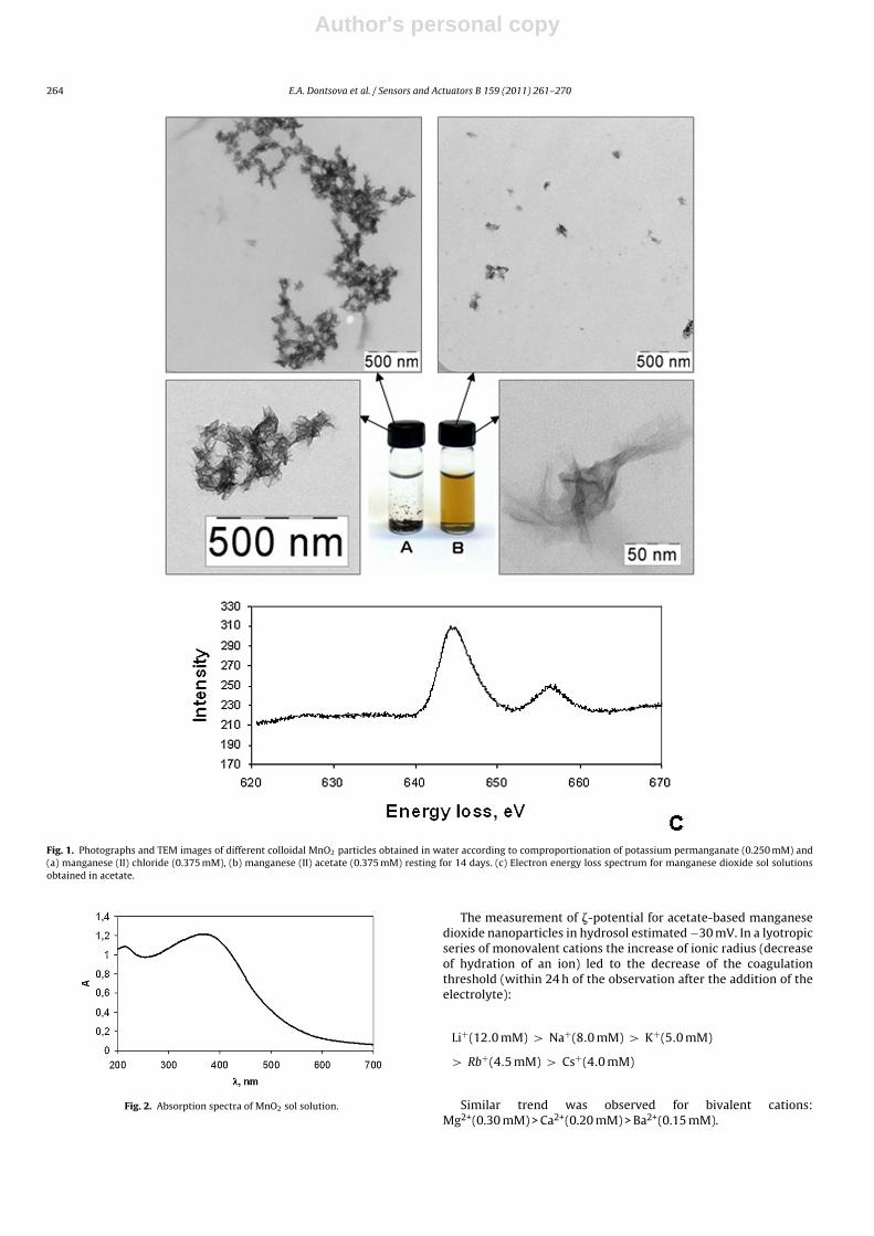
The interaction of KMnO4 with manganese (II) chloride resultedin rapid coagulation of sol forming a pellet. We have observedthe same effect in the presence of sulfate, dodecylsulphate, fluo-ride, and carbonate ions. However, acetate, hydrocarbonate, hydro-and dihydrophosphate ions are promoted stabilization and formedmanganese dioxide hydrosol. Moreover, stable (for several weeks)manganese dioxide hydrosol was obtained during the introductioninto the reaction of manganese (II) acetate instead of manganesechloride. The images of manganese dioxide hydrosol prepara-tions (Fig. 1) obtained using manganese chloride (Fig. 1(a)) andmanganese acetate (Fig. 1(b)) reflect sedimentation and stable col-loidal solution subsequently. We propose that the stabilization ofnanoparticles using manganese acetate is provided by a high degreeof acetate-ion hydration in accordance with Hofmeister series [38].Thus, in the presence of acetate charge screen on the particles sur-face is lowered, protecting them from aggregation.
According to TEM data, most of chloride-based MnO2 nanoparti-cles represent aggregates with dimensions from deciles of micronsto several microns. Acetate-based MnO2 nanoparticles representwrinkled lamellar structures with estimated thickness 0.3–0.6 nmand characteristic dimensions 50–120 nm. The particles have poly-crystalline nature; the crystalline sizes are about 2 nm. We haveobtained electron energy loss spectrum with TEM (Fig. 1(c)). Thepeaks observed at 645 eV and 655 eV corresponded to manganese(according to TEM database).
The crystal phase of the MnO2 was analyzed by powder X-raydiffraction. The diffraction peaks can be indexed to orthorhombicsymmetry and corresponding to �-MnO2 (ICDD –JCPDS Card No.72-1983).
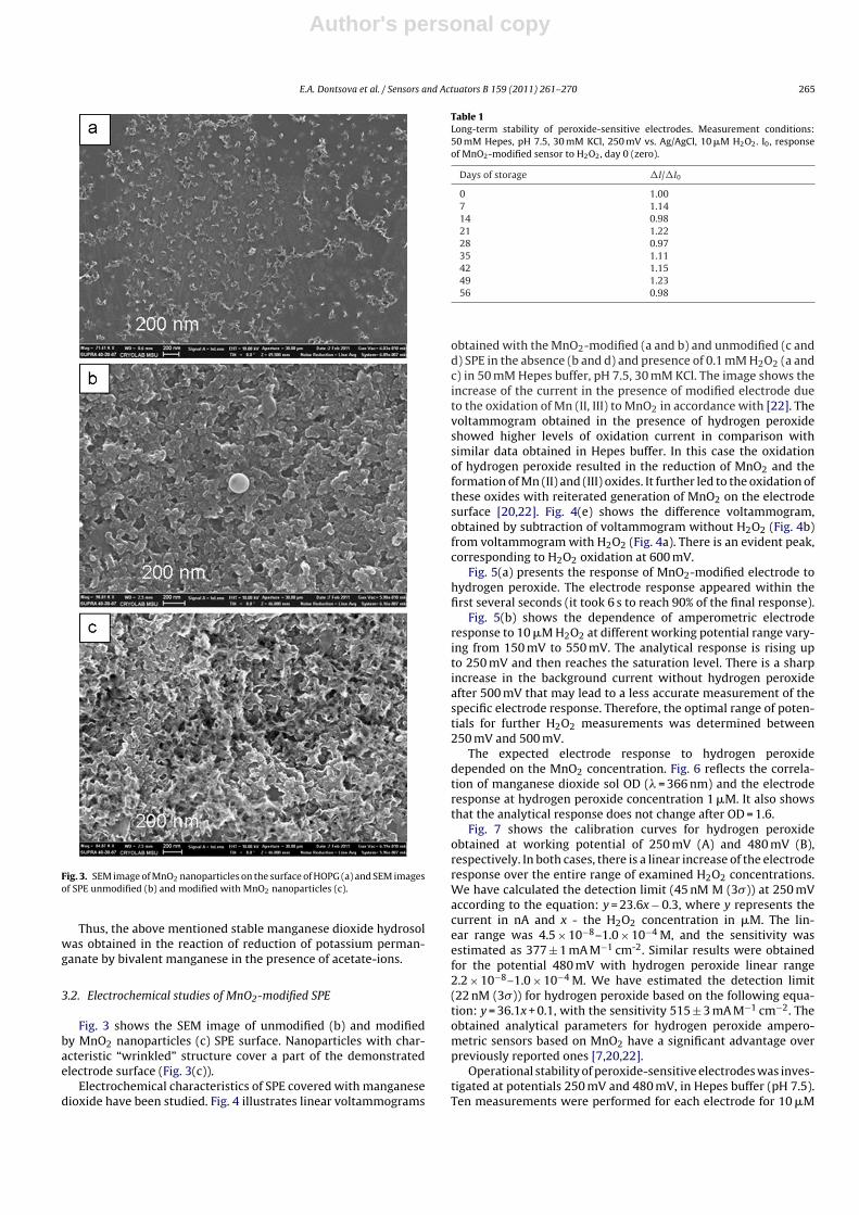
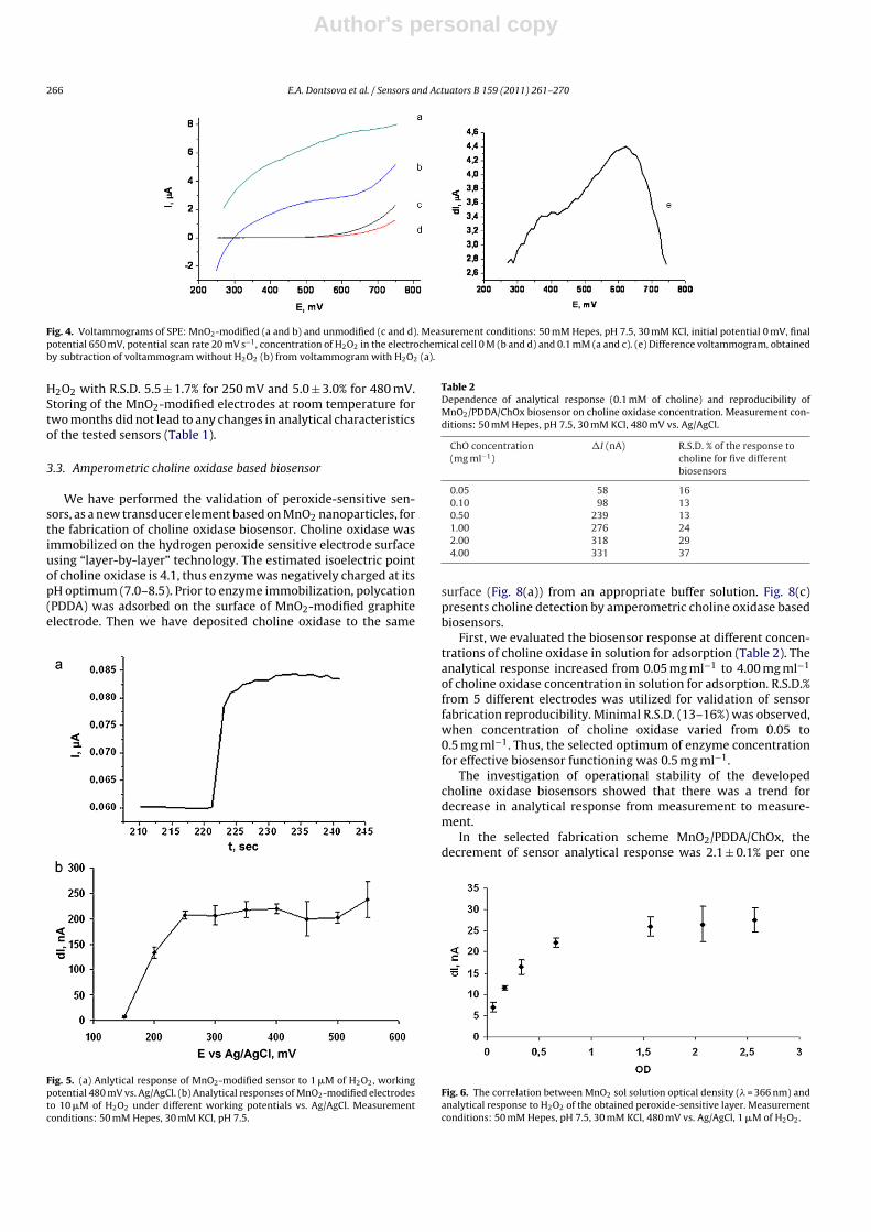
Fig. 2 presents spectrophotometrical data for hydrosol (Fig. 2).The absorption peak at 366 nm confirmed, that hydrosol con-tained MnO2 [39]. The hydrodynamic radius of manganese dioxidenanoparticles, estimated via light scattering, was 64 ± 1 nm.Fig. 3(a) shows the SEM image of hydrosol dried on HOPG surface.The dimensions of separate particles are about 60–100 nm.
Author's personal copy
264 E.A. Dontsova et al. / Sensors and Actuators B 159 (2011) 261– 270
Fig. 1. Photographs and TEM images of different colloidal MnO2 particles obtained in water according to comproportionation of potassium permanganate (0.250 mM) and(a) manganese (II) chloride (0.375 mM), (b) manganese (II) acetate (0.375 mM) resting for 14 days. (c) Electron energy loss spectrum for manganese dioxide sol solutionsobtained in acetate.
Fig. 2. Absorption spectra of MnO2 sol solution.
The measurement of �-potential for acetate-based manganesedioxide nanoparticles in hydrosol estimated −30 mV. In a lyotropicseries of monovalent cations the increase of ionic radius (decreaseof hydration of an ion) led to the decrease of the coagulationthreshold (within 24 h of the observation after the addition of theelectrolyte):
Li+(12.0 mM) > Na+(8.0 mM) > K+(5.0 mM)
> Rb+(4.5 mM) > Cs+(4.0 mM)
Similar trend was observed for bivalent cations:Mg2+(0.30 mM) > Ca2+(0.20 mM) > Ba2+(0.15 mM).
Author's personal copy
E.A. Dontsova et al. / Sensors and Actuators B 159 (2011) 261– 270 265
Fig. 3. SEM image of MnO2 nanoparticles on the surface of HOPG (a) and SEM imagesof SPE unmodified (b) and modified with MnO2 nanoparticles (c).
Thus, the above mentioned stable manganese dioxide hydrosolwas obtained in the reaction of reduction of potassium perman-ganate by bivalent manganese in the presence of acetate-ions.
3.2. Electrochemical studies of MnO2-modified SPE
Fig. 3 shows the SEM image of unmodified (b) and modifiedby MnO2 nanoparticles (c) SPE surface. Nanoparticles with char-acteristic “wrinkled” structure cover a part of the demonstratedelectrode surface (Fig. 3(c)).
Electrochemical characteristics of SPE covered with manganesedioxide have been studied. Fig. 4 illustrates linear voltammograms
Table 1Long-term stability of peroxide-sensitive electrodes. Measurement conditions:50 mM Hepes, pH 7.5, 30 mM KCl, 250 mV vs. Ag/AgCl, 10 �M H2O2. I0, responseof MnO2-modified sensor to H2O2, day 0 (zero).
Days of storage �I/�I0
0 1.007 1.1414 0.9821 1.2228 0.9735 1.1142 1.1549 1.2356 0.98
obtained with the MnO2-modified (a and b) and unmodified (c andd) SPE in the absence (b and d) and presence of 0.1 mM H2O2 (a andc) in 50 mM Hepes buffer, pH 7.5, 30 mM KCl. The image shows theincrease of the current in the presence of modified electrode dueto the oxidation of Mn (II, III) to MnO2 in accordance with [22]. Thevoltammogram obtained in the presence of hydrogen peroxideshowed higher levels of oxidation current in comparison withsimilar data obtained in Hepes buffer. In this case the oxidationof hydrogen peroxide resulted in the reduction of MnO2 and theformation of Mn (II) and (III) oxides. It further led to the oxidation ofthese oxides with reiterated generation of MnO2 on the electrodesurface [20,22]. Fig. 4(e) shows the difference voltammogram,obtained by subtraction of voltammogram without H2O2 (Fig. 4b)from voltammogram with H2O2 (Fig. 4a). There is an evident peak,corresponding to H2O2 oxidation at 600 mV.
Fig. 5(a) presents the response of MnO2-modified electrode tohydrogen peroxide. The electrode response appeared within thefirst several seconds (it took 6 s to reach 90% of the final response).
Fig. 5(b) shows the dependence of amperometric electroderesponse to 10 �M H2O2 at different working potential range vary-ing from 150 mV to 550 mV. The analytical response is rising upto 250 mV and then reaches the saturation level. There is a sharpincrease in the background current without hydrogen peroxideafter 500 mV that may lead to a less accurate measurement of thespecific electrode response. Therefore, the optimal range of poten-tials for further H2O2 measurements was determined between250 mV and 500 mV.
The expected electrode response to hydrogen peroxidedepended on the MnO2 concentration. Fig. 6 reflects the correla-tion of manganese dioxide sol OD (� = 366 nm) and the electroderesponse at hydrogen peroxide concentration 1 �M. It also showsthat the analytical response does not change after OD = 1.6.
Fig. 7 shows the calibration curves for hydrogen peroxideobtained at working potential of 250 mV (A) and 480 mV (B),respectively. In both cases, there is a linear increase of the electroderesponse over the entire range of examined H2O2 concentrations.We have calculated the detection limit (45 nM M (3�)) at 250 mVaccording to the equation: y = 23.6x − 0.3, where y represents thecurrent in nA and x - the H2O2 concentration in �M. The lin-ear range was 4.5 × 10⁻⁸–1.0 × 10⁻⁴ M, and the sensitivity wasestimated as 377 ± 1 mA M−1 cm-2. Similar results were obtainedfor the potential 480 mV with hydrogen peroxide linear range2.2 × 10−8–1.0 × 10⁻⁴ M. We have estimated the detection limit(22 nM (3�)) for hydrogen peroxide based on the following equa-tion: y = 36.1x + 0.1, with the sensitivity 515 ± 3 mA M−1 cm−2. Theobtained analytical parameters for hydrogen peroxide ampero-metric sensors based on MnO2 have a significant advantage overpreviously reported ones [7,20,22].
Operational stability of peroxide-sensitive electrodes was inves-tigated at potentials 250 mV and 480 mV, in Hepes buffer (pH 7.5).Ten measurements were performed for each electrode for 10 �M
Author's personal copy
266 E.A. Dontsova et al. / Sensors and Actuators B 159 (2011) 261– 270
Fig. 4. Voltammograms of SPE: MnO2-modified (a and b) and unmodified (c and d). Measurement conditions: 50 mM Hepes, pH 7.5, 30 mM KCl, initial potential 0 mV, finalpotential 650 mV, potential scan rate 20 mV s−1, concentration of H2O2 in the electrochemical cell 0 M (b and d) and 0.1 mM (a and c). (e) Difference voltammogram, obtainedby subtraction of voltammogram without H2O2 (b) from voltammogram with H2O2 (a).
H2O2 with R.S.D. 5.5 ± 1.7% for 250 mV and 5.0 ± 3.0% for 480 mV.Storing of the MnO2-modified electrodes at room temperature fortwo months did not lead to any changes in analytical characteristicsof the tested sensors (Table 1).
3.3. Amperometric choline oxidase based biosensor
We have performed the validation of peroxide-sensitive sen-sors, as a new transducer element based on MnO2 nanoparticles, forthe fabrication of choline oxidase biosensor. Choline oxidase wasimmobilized on the hydrogen peroxide sensitive electrode surfaceusing “layer-by-layer” technology. The estimated isoelectric pointof choline oxidase is 4.1, thus enzyme was negatively charged at itspH optimum (7.0–8.5). Prior to enzyme immobilization, polycation(PDDA) was adsorbed on the surface of MnO2-modified graphiteelectrode. Then we have deposited choline oxidase to the same
Fig. 5. (a) Anlytical response of MnO2-modified sensor to 1 �M of H2O2, workingpotential 480 mV vs. Ag/AgCl. (b) Analytical responses of MnO2-modified electrodesto 10 �M of H2O2 under different working potentials vs. Ag/AgCl. Measurementconditions: 50 mM Hepes, 30 mM KCl, pH 7.5.
Table 2Dependence of analytical response (0.1 mM of choline) and reproducibility ofMnO2/PDDA/ChOx biosensor on choline oxidase concentration. Measurement con-ditions: 50 mM Hepes, pH 7.5, 30 mM KCl, 480 mV vs. Ag/AgCl.
ChO concentration(mg ml−1)
�I (nA) R.S.D. % of the response tocholine for five differentbiosensors
0.05 58 160.10 98 130.50 239 131.00 276 242.00 318 294.00 331 37
surface (Fig. 8(a)) from an appropriate buffer solution. Fig. 8(c)presents choline detection by amperometric choline oxidase basedbiosensors.
First, we evaluated the biosensor response at different concen-trations of choline oxidase in solution for adsorption (Table 2). Theanalytical response increased from 0.05 mg ml−1 to 4.00 mg ml−1
of choline oxidase concentration in solution for adsorption. R.S.D.%from 5 different electrodes was utilized for validation of sensorfabrication reproducibility. Minimal R.S.D. (13–16%) was observed,when concentration of choline oxidase varied from 0.05 to0.5 mg ml−1. Thus, the selected optimum of enzyme concentrationfor effective biosensor functioning was 0.5 mg ml−1.
The investigation of operational stability of the developedcholine oxidase biosensors showed that there was a trend fordecrease in analytical response from measurement to measure-ment.
In the selected fabrication scheme MnO2/PDDA/ChOx, thedecrement of sensor analytical response was 2.1 ± 0.1% per one
Fig. 6. The correlation between MnO2 sol solution optical density (� = 366 nm) andanalytical response to H2O2 of the obtained peroxide-sensitive layer. Measurementconditions: 50 mM Hepes, pH 7.5, 30 mM KCl, 480 mV vs. Ag/AgCl, 1 �M of H2O2.
Author's personal copy
E.A. Dontsova et al. / Sensors and Actuators B 159 (2011) 261– 270 267
Fig. 7. Calibration plots of MnO2-modified electrodes for H2O2 at 250 mV (a) and480 mV (b) vs. Ag/AgCl. Measurement conditions: 50 mM Hepes, 30 mM KCl, pH 7.5.
measurement (R.S.D. was 7.0 ± 0.3% per ten measurements) atworking potential 480 mV. We have investigated the operationstability of MnO2 layer at different hydrosol concentrations.Concentrations of MnO2 in hydrosol were chosen for linear(OD = 0.2) and saturated (OD = 1.6) response–concentration curveareas (Fig. 6) Sensor operation stability was determined in the sameranges of concentrations, i.e. the decrement of analytical responsewas estimated as 0.2 ± 0.4% (R.S.D. = 2.0 ± 0.1%). All this proves, that
Fig. 8. Architectures of enzyme/polyelectrolyte layers of MnO2-based choline oxi-dase biosensor (a) PDDA/ChOx, (b) (PDDA/PAS)2/PDDA/ChOx and (c) scheme ofcholine detection.
hydrogen peroxide-sensitive layer remained intact. It follows, thatthe observed decay in the sensor response is connected with theenzyme layer. The improved stabilization of choline oxidase lay-ers was carried out with two additional PDDA/PAS layer’s variants(Fig. 8(b)). The decrement of analytical response in this case was0.6 ± 0.2% (R.S.D. 1.9 ± 0.4%) for MnO2/(PDDA/PAS)2/PDDA/ChOxelectrodes, at working potential 480 mV. At working poten-tial 250 mV the decrement of analytical response for electrodesof the same construction (MnO2/(PDDA/PAS)2/PDDA/ChOx) wasestimated 2.1 ± 0.4% (R.S.D. 9.6 ± 3.0%). These values were 3–5fold higher than corresponding data obtained for 480 mV.We have selected 480 mV as an optimal for further cholinedetection.
The increase in the pH dependence values (in the range from7.0 to 8.2.) led to the increase of the electrochemical responsefor MnO2/(PDDA/PAS)2/PDDA/ChOx electrodes. It is known, thatpH-optimum of choline oxidase is about 8.0. However, theobserved operation stability of choline oxidase biosensors wasbetter at pH 7.5 (decrement of analytical response 0.6 ± 0.2% andR.S.D. = 1.9 ± 0.4%), than at pH 8.2 (decrement of analytical response3.2 ± 0.0% and R.S.D. = 11.1 ± 0.4%).
We have evaluated storage stability of dried biosensors at 4 ◦C.Weekly tests showed that the response to 0.1 mM choline of thesebiosensors kept 75% of initial activity for 3 weeks of storage.
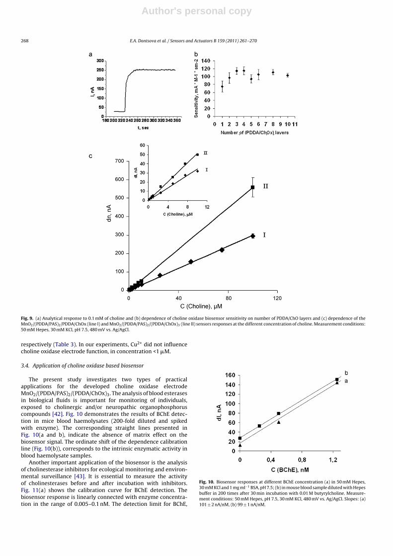
Fig. 9(a) shows a typical steady-state response of choline oxidasebiosensor to 0.1 mM of choline. It took about 10 s to reach 90% ofthe final response.
Fig. 9(b) presents the dependence of choline oxidase biosensorsensitivity on the number of PDDA/ChO layers. The electrode sen-sitivity increased at magnification of PDDA/ChO layers from 1 to 3,and reached the upper limit.
Fig. 9((c), line I) illustrates the dependence of theMnO2/(PDDA/PAS)2/PDDA/ChOx sensors responses at the dif-ferent concentration of choline. Obtained choline calibrationcurve showed a good linearity in a range between 3.0 × 10⁻⁷
and 1.0 × 10⁻⁴ M. The corresponding regression equation was:y = 2.9x + 3.5, where y represents the current in nA and x the cholineconcentration in �M. The sensitivity 59 ± 3 mA M−1 cm−2 and thedetection limit 300 nM (3�) were calculated.
It should be noted, the highest sensitivity of the devel-oped biosensors was obtained during the deposition ofthree enzyme-containing layers on the electrode surface:MnO2/(PDDA/PAS)2/(PDDA/ChOx)3 (Fig. 9(c), line II). In thiscase the detection limit was 130 nM (3�) and the sensitivity was103 ± 3 mA M−1 cm−2. These analytical parameters are the best forthe amperometric choline oxidase – based biosensors.
The interference of sampling “contaminating” compounds,easily oxidized at positive potentials, is an important factor for eval-uating the analytical performance of the electrochemical cholinebiosensor for medical and environmental applications. We haveinvestigated these potential aberrations, introduced by interferingcompounds, using the substances, commonly found in biolog-ical fluids (ascorbic and uric acids) and environmental objects(heavy metals Cd2+, Co2+, Cu2+). Pure, non-contaminated controlsamples were compared to the samples spiked with interfer-ing compounds (see Table 3). The interference for the normalphysiological level of the ascorbic acid (50 �M), increased theMnO2/(PDDA/PAS)2/PDDA/ChOx biosensor response to 394% anddecreased to 0.5% at concentration 0.5�M in the sample spiked withcholine. Similar effect was found for the uric acid (see Table 3). Thus,200-fold dilution of real blood samples is necessary to eliminatethe interference of analogous concomitant compounds on studiedelectrode response.
Heavy metals (Cd2+ and Co2+) are reversible inhibitors of cholineoxidase [40]. The inhibition effect of Cd2+ and Co2+ ions is negligibleat the concentrations, lower than 10 �M Cd2+ and 0.1 mM Co2+,
Author's personal copy
268 E.A. Dontsova et al. / Sensors and Actuators B 159 (2011) 261– 270
Fig. 9. (a) Analytical response to 0.1 mM of choline and (b) dependence of choline oxidase biosensor sensitivity on number of PDDA/ChO layers and (c) dependence of theMnO2/(PDDA/PAS)2/PDDA/ChOx (line I) and MnO2/(PDDA/PAS)2/(PDDA/ChOx)3 (line II) sensors responses at the different concentration of choline. Measurement conditions:50 mM Hepes, 30 mM KCl, pH 7.5, 480 mV vs. Ag/AgCl.
respectively (Table 3). In our experiments, Cu2+ did not influencecholine oxidase electrode function, in concentration <1 �M.
3.4. Application of choline oxidase based biosensor
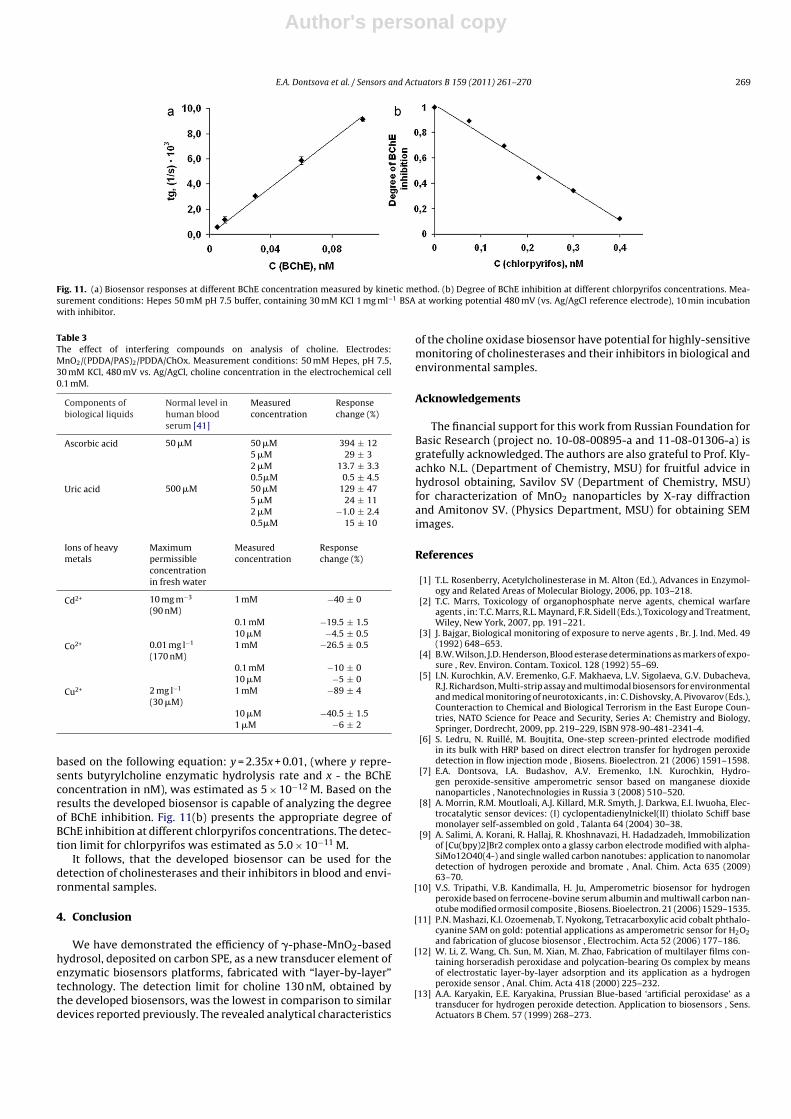
The present study investigates two types of practicalapplications for the developed choline oxidase electrodeMnO2/(PDDA/PAS)2/(PDDA/ChOx)3. The analysis of blood esterasesin biological fluids is important for monitoring of individuals,exposed to cholinergic and/or neuropathic organophosphoruscompounds [42]. Fig. 10 demonstrates the results of BChE detec-tion in mice blood haemolysates (200-fold diluted and spikedwith enzyme). The corresponding straight lines presented inFig. 10(a and b), indicate the absence of matrix effect on thebiosensor signal. The ordinate shift of the dependence calibrationline (Fig. 10(b)), corresponds to the intrinsic enzymatic activity inblood haemolysate samples.
Another important application of the biosensor is the analysisof cholinesterase inhibitors for ecological monitoring and environ-mental surveillance [43]. It is essential to measure the activityof cholinesterases before and after incubation with inhibitors.Fig. 11(a) shows the calibration curve for BChE detection. Thebiosensor response is linearly connected with enzyme concentra-tion in the range of 0.005–0.1 nM. The detection limit for BChE,
Fig. 10. Biosensor responses at different BChE concentration (a) in 50 mM Hepes,30 mM KCl and 1 mg ml−1 BSA, pH 7.5; (b) in mouse blood sample diluted with Hepesbuffer in 200 times after 30 min incubation with 0.01 M butyrylcholine. Measure-ment conditions: 50 mM Hepes, pH 7.5, 30 mM KCl, 480 mV vs. Ag/AgCl. Slopes: (a)101 ± 2 nA/nM, (b) 99 ± 1 nA/nM.
Author's personal copy
E.A. Dontsova et al. / Sensors and Actuators B 159 (2011) 261– 270 269
Fig. 11. (a) Biosensor responses at different BChE concentration measured by kinetic method. (b) Degree of BChE inhibition at different chlorpyrifos concentrations. Mea-surement conditions: Hepes 50 mM pH 7.5 buffer, containing 30 mM KCl 1 mg ml−1 BSA at working potential 480 mV (vs. Ag/AgCl reference electrode), 10 min incubationwith inhibitor.
Table 3The effect of interfering compounds on analysis of choline. Electrodes:MnO2/(PDDA/PAS)2/PDDA/ChOx. Measurement conditions: 50 mM Hepes, pH 7.5,30 mM KCl, 480 mV vs. Ag/AgCl, choline concentration in the electrochemical cell0.1 mM.
Components ofbiological liquids
Normal level inhuman bloodserum [41]
Measuredconcentration
Responsechange (%)
Ascorbic acid 50 �M 50 �M 394 ± 125 �M 29 ± 32 �M 13.7 ± 3.30.5�M 0.5 ± 4.5
Uric acid 500 �M 50 �M 129 ± 475 �M 24 ± 112 �M −1.0 ± 2.40.5�M 15 ± 10
Ions of heavymetals
Maximumpermissibleconcentrationin fresh water
Measuredconcentration
Responsechange (%)
Cd2+ 10 mg m−3
(90 nM)1 mM −40 ± 0
0.1 mM −19.5 ± 1.510 �M −4.5 ± 0.5
Co2+ 0.01 mg l−1
(170 nM)1 mM −26.5 ± 0.5
0.1 mM −10 ± 010 �M −5 ± 0
Cu2+ 2 mg l−1
(30 �M)1 mM −89 ± 4
10 �M −40.5 ± 1.51 �M −6 ± 2
based on the following equation: y = 2.35x + 0.01, (where y repre-sents butyrylcholine enzymatic hydrolysis rate and x - the BChEconcentration in nM), was estimated as 5 × 10⁻¹² M. Based on theresults the developed biosensor is capable of analyzing the degreeof BChE inhibition. Fig. 11(b) presents the appropriate degree ofBChE inhibition at different chlorpyrifos concentrations. The detec-tion limit for chlorpyrifos was estimated as 5.0 × 10⁻¹¹ M.
It follows, that the developed biosensor can be used for thedetection of cholinesterases and their inhibitors in blood and envi-ronmental samples.
4. Conclusion
We have demonstrated the efficiency of �-phase-MnO2-basedhydrosol, deposited on carbon SPE, as a new transducer element ofenzymatic biosensors platforms, fabricated with “layer-by-layer”technology. The detection limit for choline 130 nM, obtained bythe developed biosensors, was the lowest in comparison to similardevices reported previously. The revealed analytical characteristics
of the choline oxidase biosensor have potential for highly-sensitivemonitoring of cholinesterases and their inhibitors in biological andenvironmental samples.
Acknowledgements
The financial support for this work from Russian Foundation forBasic Research (project no. 10-08-00895-a and 11-08-01306-a) isgratefully acknowledged. The authors are also grateful to Prof. Kly-achko N.L. (Department of Chemistry, MSU) for fruitful advice inhydrosol obtaining, Savilov SV (Department of Chemistry, MSU)for characterization of MnO2 nanoparticles by X-ray diffractionand Amitonov SV. (Physics Department, MSU) for obtaining SEMimages.
References
[1] T.L. Rosenberry, Acetylcholinesterase in M. Alton (Ed.), Advances in Enzymol-ogy and Related Areas of Molecular Biology, 2006, pp. 103–218.
[2] T.C. Marrs, Toxicology of organophosphate nerve agents, chemical warfareagents , in: T.C. Marrs, R.L. Maynard, F.R. Sidell (Eds.), Toxicology and Treatment,Wiley, New York, 2007, pp. 191–221.
[3] J. Bajgar, Biological monitoring of exposure to nerve agents , Br. J. Ind. Med. 49(1992) 648–653.
[4] B.W. Wilson, J.D. Henderson, Blood esterase determinations as markers of expo-sure , Rev. Environ. Contam. Toxicol. 128 (1992) 55–69.
[5] I.N. Kurochkin, A.V. Eremenko, G.F. Makhaeva, L.V. Sigolaeva, G.V. Dubacheva,R.J. Richardson, Multi-strip assay and multimodal biosensors for environmentaland medical monitoring of neurotoxicants , in: C. Dishovsky, A. Pivovarov (Eds.),Counteraction to Chemical and Biological Terrorism in the East Europe Coun-tries, NATO Science for Peace and Security, Series A: Chemistry and Biology,Springer, Dordrecht, 2009, pp. 219–229, ISBN 978-90-481-2341-4.
[6] S. Ledru, N. Ruillé, M. Boujtita, One-step screen-printed electrode modifiedin its bulk with HRP based on direct electron transfer for hydrogen peroxidedetection in flow injection mode , Biosens. Bioelectron. 21 (2006) 1591–1598.
[7] E.A. Dontsova, I.A. Budashov, A.V. Eremenko, I.N. Kurochkin, Hydro-gen peroxide-sensitive amperometric sensor based on manganese dioxidenanoparticles , Nanotechnologies in Russia 3 (2008) 510–520.
[8] A. Morrin, R.M. Moutloali, A.J. Killard, M.R. Smyth, J. Darkwa, E.I. Iwuoha, Elec-trocatalytic sensor devices: (I) cyclopentadienylnickel(II) thiolato Schiff basemonolayer self-assembled on gold , Talanta 64 (2004) 30–38.
[9] A. Salimi, A. Korani, R. Hallaj, R. Khoshnavazi, H. Hadadzadeh, Immobilizationof [Cu(bpy)2]Br2 complex onto a glassy carbon electrode modified with alpha-SiMo12O40(4-) and single walled carbon nanotubes: application to nanomolardetection of hydrogen peroxide and bromate , Anal. Chim. Acta 635 (2009)63–70.
[10] V.S. Tripathi, V.B. Kandimalla, H. Ju, Amperometric biosensor for hydrogenperoxide based on ferrocene-bovine serum albumin and multiwall carbon nan-otube modified ormosil composite , Biosens. Bioelectron. 21 (2006) 1529–1535.
[11] P.N. Mashazi, K.I. Ozoemenab, T. Nyokong, Tetracarboxylic acid cobalt phthalo-cyanine SAM on gold: potential applications as amperometric sensor for H2O2
and fabrication of glucose biosensor , Electrochim. Acta 52 (2006) 177–186.[12] W. Li, Z. Wang, Ch. Sun, M. Xian, M. Zhao, Fabrication of multilayer films con-
taining horseradish peroxidase and polycation-bearing Os complex by meansof electrostatic layer-by-layer adsorption and its application as a hydrogenperoxide sensor , Anal. Chim. Acta 418 (2000) 225–232.
[13] A.A. Karyakin, E.E. Karyakina, Prussian Blue-based ‘artificial peroxidase’ as atransducer for hydrogen peroxide detection. Application to biosensors , Sens.Actuators B Chem. 57 (1999) 268–273.
Author's personal copy
270 E.A. Dontsova et al. / Sensors and Actuators B 159 (2011) 261– 270
[14] A.A. Karyakin, E.A. Puganova, I.A. Budashov, I.N. Kurochkin, E.E. Karyakina, V.A.Levchenko, V.N. Matveyenko, S.D. Varfolomeyev, Prussian blue based nano-electrode arrays for H2O2 detection , Anal. Chem. 76 (2004) 474–478.
[15] F. Ricci, A. Amine, G. Palleschi, D. Moscone, Prussian blue based screen printedbiosensors with improved characteristics of long-term lifetime and pH stability, Biosens. Bioelectron. 18 (2003) 165–174.
[16] F. Ricci, G. Palleschi, Sensor and biosensor preparation, optimisation and appli-cations of Prussian Blue modified electrodes , Biosens. Bioelectron. 21 (2005)389–407.
[17] S. Mannino, M.S. Cosio, S. Ratti, Cobalt(II, III)-oxide chemically modified elec-trode as amperometric detector in flow injection systems , Electroanalysis 5(1993) 145–148.
[18] A. Salimi, R. Hallaj, S. Soltanian, H. Mamkhezri, Nanomolar detection of hydro-gen peroxide on glassy carbon electrode modified with electrodeposited cobaltoxide nanoparticles , Anal. Chim. Acta 594 (2007) 24–31.
[19] K. Schachl, H. Alemu, K. Kalcher, J. Jeozkova, I. Svancara, K. Vytoras, Amperomet-ric determination of hydrogen peroxide with a manganese dioxide-modifiedcarbon paste electrode using flow injection analysis , Analyst 122 (1997)985–989.
[20] K. Schachl, H. Alemu, K. Kalcher, H. Moderegger, I. Svancara, K. Vytras,Amperometric determination of hydrogen peroxide with a manganese dioxidefilm-modified screen printed electrode , Fresenius J. Anal. Chem. 362 (1998)194–200.
[21] Sh. Yao, Ju. Xua, X. Yi Wang, Ya. Chena, Sh. Xua, Hua, A highly sensitive hydrogenperoxide amperometric sensor based on MnO2 nanoparticles and dihexadecylhydrogen phosphate composite film , Anal. Chim. Acta 557 (2006) 78–84.
[22] Sh. Yao, Sh. Yuan, Yu. Xu, Yi. Wang, J. Luo, Sh. Hu, A hydrogen peroxide sensorbased on colloidal MnO2/Na-montmorillonite , Appl. Clay Sci. 33 (2006) 35–42.
[23] J.A. Cox, R.K. Jaworski, Voltammetric reduction and determination of hydrogenperoxide at an electrode modified with a film containing palladium and iridium, Anal. Chem. 61 (1989) 2176–2178.
[24] A. Domenech, J. Alarcon, Determination of hydrogen peroxide using glassycarbon and graphite/polyester composite electrodes modified by vanadium-doped zirconias , Anal. Chim. Acta 452 (2002) 11–22.
[25] H. Shi, Yu. Yang, J. Huang, Z. Zhao, X. Xu, J. Anzai, T. Osa, Q. Chen, Amperometriccholine biosensors prepared by layer-by-layer deposition of choline oxidase onthe Prussian blue-modified platinum electrode , Talanta 70 (2006) 852–858.
[26] L. Doretti, D. Ferrara, P. Gattolin, S. Lora, Covalently immobilized enzymes onbiocompatible polymers for amperometric sensor applications , Biosens. Bio-electron. 11 (1996) 365–373.
[27] K.M. Mitchell, Acetylcholine and choline amperometric enzyme sensors char-acterized in vitro and in vivo , Anal. Chem. 76 (2004) 1098–1106.
[28] B.-C. Hsieh, K. Matsumoto, T.-J. Cheng, G. Yuu, R.-L. Chen, Choline biosensorconstructed with chitinous membrane from soldier crab and its applicationin measuring cholinesterase inhibitory activities , J. Pharm. Biomed. Anal. 45(2007) 673–678.
[29] T. Shimomura, T. Itoh, T. Sumiya, F. Mizukami, M. Ono, Amperometric determi-nation of choline with enzyme immobilized in a hybrid mesoporous membrane, Talanta 78 (2009) 217–220.
[30] S. Sen, A. Gulce, H. Gulce, Polyvinylferrocenium modified Pt electrode for thedesign of amperometric choline and acetylcholine enzyme electrodes , Biosens.Bioelectron. 19 (2004) 1261–1268.
[31] Y.-H. Bai, D. Ying, J.-J. Xu, H.-Y. Chen, Choline biosensors based on a bi-electrocatalytic property of MnO2 nanoparticles modified electrodes to H2O2 ,Electrochem. Commun. 9 (2007) 2611–2616.
[32] X. Qina, H. Wang, X. Wang, Sh. Li, Zh. Miao, N. Huang, Q. Chen, Amperometriccholine biosensors based on multi-wall carbon nanotubes and layer-by-layerassembly of multilayer films composed of poly(diallyldimethylammoniumchloride) and choline oxidase , Mater. Sci. Eng. C 29 (2009) 1453–1457.
[33] M.V. Porus, G.V. Dubacheva, L.V. Sigolaeva, A.V. Eremenko, I.N. Kurochkin,Determination of cholinesterase activities by using bielectrode sensor system, Sensornye Sistemy 22 (2008) 88–95.
[34] P. Calvert, Inkjet printing for materials and devices , Chem. Mater. 13 (2001)3299–3305.
[35] Y. Lin, X. Cui, L. Li, Low-potential amperometric determination of hydro-gen peroxide with a carbon paste electrode modified with nanostructuredcryptomelane-type manganese oxides , Electrochem. Commun. 7 (2005)166–172.
[36] Y.-H. Bai, H. Zhang, J.-J. Xu, H.-Y. Chen, Relationship between nanostruc-ture and electrochemical/biosensing properties of MnO2 nanomaterials forH2O2/choline , J. Phys. Chem. 112 (2008) 18984–18990.
[37] X. Fu, J. Feng, H. Wang, K.M. Ng, Room temperature synthesis of a novel �-MnO2
hollow structure for aerobic oxidation of benzyl alcohol , Nanotechnology 20(2009) 375601, doi:10.1088/0957-4484/20/37/375601.
[38] M. Salomäki, P. Tervasmäki, S. Areva, J. Kankare, The Hofmeister anion effectand the growth of polyelectrolyte multilayers , Langmuir 20 (2004) 3679–3683.
[39] T. Fujimoto, Yo. Mizukoshi, Yo. Nagata, Ya. Maeda, R. Oshima, Sonolytical prepa-ration of various types of metal nanoparticles in aqueous solution , ScriptaMater. 44 (2001) 2183–2186.
[40] H. Yamada, N. Mori, Y. Tani, Properties of choline oxidase of cylindrocarpondidymum M-1 , Agric. Biol. Chem. 43 (1979) 2173–2177.
[41] R.K. Murray, D.K. Granner, P.A. Mayes, V.W. Rodwell, Appendix in Harper’sBiochemistry , 21st ed., Mir, Moscow, 1993, pp. 367–384.
[42] G.F. Makhaeva, E. Rudakova, N. Boltneva, L.V. Sigolaeva, A.V. Eremenko, I.N.Kurochkin, R.J. Richardson, Blood Esterases as a complex biomarker for expo-sure to organophosphorus compounds , in: C. Dishovsky, A. Pivovarov (Eds.),Counteraction to Chemical and Biological Terrorism in the East Europe Coun-tries, NATO Science for Peace and Security, Series A: Chemistry and Biology,Springer, Dordrecht, 2009, pp. 177–194, ISBN 978-90-481-2341-4.
[43] S. Andreescu a, J.-L. Marty, Twenty years research in cholinesterase biosen-sors: from basic research to practical applications , Biomol. Eng. 23 (2006)1–15.
Biographies
E.A. Dontsova received her MS degree in Chemistry from Lomonosov MoscowState University, Russia, in 2005. Since September, 2005, she has been workingtowards her Ph.D. degree at the Department of Chemistry in Lomonosov MoscowState University. Her current research interests include electrochemical sensors andbiosensors.
Y.S. Zeifman received her MS degree in Chemistry from Lomonosov Moscow StateUniversity, Russia in 2010. Her research interests include bioanalytical assays andbiosensors.
I.A. Budashov received his MS degree in Biology from Lomonosov Moscow StateUniversity, Russia in 1986. In 1998 he obtained his Ph.D. in Biochemistry from theResearch Center of Molecular Diagnostics and Therapy, Russia. At present he is theleading specialist in the Institute of Biochemical Physics (Russian Academy of Sci-ences). His research interests are biochemistry, physical and chemical methods ofbiology investigations, biosensor systems, and scanning probe microscopy.
A.V. Eremenko received his MS degree in Pharmaceutical Chemistry from the FirstMoscow Medical Institute, Russia in 1982. In 1986 he obtained his Ph.D. in Bio-chemistry and Pharmacology from the Institute of Pharmacology, Russian Academyof Medical Sciences, Moscow. At present he is a Senior Researcher at the Departmentof Biokinetics, A.N. Belozersky Institute of Physico-Chemical Biology, Moscow StateUniversity. His main research interests are biosensors for the detection of pharma-cological and toxicological compounds, electrochemical analyzers for clinical andenvironmental applications.
S.L. Kalnov received his MS degree in Biochemistry from Lomonosov MoscowState University, Russia in 1975. In 1980 he obtained his Ph.D. in Biochem-istry from Lomonosov Moscow State University, Russia. At present he is Head ofthe Laboratory of Diagnostics and Prevention Research Institute; Leading Scien-tist of Emanuel Institute of Biochemical Physics of Russian Academy of Sciences,Moscow.
I.N. Kurochkin received his MS degree in Chemical Enzymology from LomonosovMoscow State University, Russia in 1979. In 1985 he obtained his Ph.D. in Chem-ical Kinetics and Catalysis from Lomonosov Moscow State University, Russia. In2003 he obtained Doctor of Science degree in Chemical Kinetic and Biotech-nology at Lomonosov Moscow State University, Russia. At present he is Headof the laboratory of Postgenomic Chemistry, Professor at Chemical Departmentof Moscow State University, Head of the Department of Physical and ChemicalBiology.