autism associated with conditions characterized by ...sswang/asd/miller_gillberg05... · autism...
TRANSCRIPT

www.elsevier.com/locate/ijdevneu
Int. J. Devl Neuroscience 23 (2005) 201–219
Autism associated with conditions characterized by developmental
errors in early embryogenesis: a mini review
Marilyn T. Millera,*, Kerstin Stromlandb, Liana Venturac, Maria Johanssond,Jose M. Bandime, Christopher Gillbergf,g
aDepartment of Ophthalmology and Visual Sciences, University of Illinois, 1855 West Taylor Street, Rm. 327, Chicago, IL 60612, USAbDepartment of Ophthalmology, Sahlgrenska University Hospital, Goteborg University, Goteborg, Sweden
cDepartment of Pediatric Ophthalmology, Altino Ventura Foundation and Hospital de Olhos de Pernambuco, Pernambuco, BrazildDepartment of Child and Adolescent Psychiatry, Goteborg University, Goteborg, Sweden
eDepartment of Child And Adolescent Neuropsychiatry, Maternal Infantile Institute of Pernambuco,
Federal University of Pernambuco, Pernambuco, BrazilfDepartment of Child and Adolescent Psychiatry, Goteborg University, Goteborg, Sweden
gSt George’s Hospital Medical School, University of London, London, UK
Received 2 March 2004; received in revised form 17 June 2004; accepted 21 June 2004
Abstract
Autism is a complex developmental disorder without an established single etiology but with significant contributions from genetic studies,
functional research, and neuropsychiatric and neuroradiologic investigations. The purpose of this paper is to review the findings in five studies
involving individuals manifesting the characteristic findings of autism spectrum disorder associated with malformations and dysfunctions
known to result from early embryogenic defects. These investigations include two associated with teratogens (thalidomide embryopathy,
Mobius sequence with misoprostol) and three (most Mobius sequence cases, CHARGE association, Goldenhar syndrome) with no known
etiology.
These studies suggest that early embryonic development errors often involving cranial nerve palsies, internal and external ear
malformations, ophthalmologic anomalies, and a variety of systemic malformations may be associated with autism spectrum disorders
statistically more frequently than expected in a normal population. Although the exact time of developmental insult for each condition cannot
be identified, the evidence is that it may occur as early as week 4 to 6+ of embryogenesis.
# 2004 ISDN. Published by Elsevier Ltd. All rights reserved.
Keywords: Autism; Autism spectrum disorders (ASD); Thalidomide embryopathy; Mobius sequence; Misoprostol
1. Introduction
Autism spectrum disorders (ASD) are neurodevelopmen-
tal disorders characterized by impairment in social interac-
tion and communication, and associated with repetitive
Abbreviations: ASD, autism spectrum disorder; CARS, childhood aut-
ism rating scale; CHARGE, colobomas, heart defects, choanal atresia,
retarded growth or development, genital anomalies, and ear abnormalities
and/or hearing loss; DSM, diagnostic and statistical manual of mental
disorders.
* Corresponding author. Tel.: +1 312 996 7445; fax: +1 312 413 4916.
E-mail address: [email protected] (M.T. Miller).
0736-5748/$30.00 # 2004 ISDN. Published by Elsevier Ltd. All rights reserved
doi:10.1016/j.ijdevneu.2004.06.007
behaviors and interests. There are several clinical ASD
phenotypes, including autistic disorder/childhood autism,
Asperger syndrome, and atypical autism (also referred to
as autistic-like condition and pervasive developmental dis-
orders not otherwise specified, or PDD NOS). The patho-
physiology of ASD remains elusive, with clues from genetic
studies, neurochemistry, autopsy reports, functional research,
radiological imaging, research on environmental influences,
and many other approaches. The purpose of this paper is to
summarize studies in which ASD was present in individuals
with conditions and malformations involving brainstem and
systemic structures known to result from early embryonic
.

M.T. Miller et al. / Int. J. Devl Neuroscience 23 (2005) 201–219202
damage. It is hoped that this information might add another
piece to the puzzle of autism by describing associated
developmental errors in some individuals with characteris-
tics of ASD.
The tragic thalidomide epidemic of the 1960s resulted in
an estimated 10,000-affected fetuses and about 6000
reported live births (Lenz and Knapp, 1962; Lenz, 1986).
The drug was distributed worldwide and, because of many
informative cases in which the time of drug intake was
known, it was determined that the teratogenic sensitive
period extended from day 20 to day 36 after fertilization
(34–60 days post last menstrual period) (Lenz and Knapp,
1962; Lenz, 1986). Since the drug is rapidly hydrolyzed the
teratogen effect is short unless there is continual intake.
From the data in the literature, it was known that early
exposure with the drug (days 20–25) resulted in involvement
of the cranial nerves (especially 6 and 7), external ear,
abnormal ocular movement, aberrant lacrimation, and
thumb anomalies (Fig. 1). Later exposure caused upper limb
and eye malformations, systemic anomalies, and finally
lower limb malformations and triphalangeal thumbs (Papst,
1964; Papst and Esslen, 1964; Nowack, 1965; Kida, 1987;
Arimoto, 1987).
Some systemic malformations were responsible for spon-
taneous abortions and early neonatal death, but the critical
period for development of systemic anomalies was more
difficult to determine, although many appeared to be in the
middle of the sensitive period.
During a court trial in the 1960s approximately 100
Swedish children were identified as showing malformations
associated with exposure to thalidomide at an early time in
their mothers’ pregnancies (Stromland and Miller, 1993).
Multiple studies ensued in Sweden involving different med-
ical subspecialties (d’Avignon and Barr, 1964; Winberg,
1964; Zetterstrom, 1966). Between 1989 and 1991, Strom-
land and Miller (1993), pediatric ophthalmologists, con-
ducted an evaluation of 86 individuals of this original cohort
Fig. 1. Thalidomide embryopathy: the
with thalidomide embryopathy, all of whom were then 27–
29 years of age. The aim of the study was to describe the
ocular motility dysfunctions (strabismus) and other eye
anomalies or visual disturbances. From their observations
and the known timetable in the literature, the authors con-
cluded that the ophthalmologic and cranial nerve dysfunc-
tion involving ocular structures occurred from thalidomide
intake in the early sensitive period (Miller, 1991; Miller and
Stromland, 1991; Stromland and Miller, 1993). Four indi-
viduals were noted to have autism associated with ocular
motility and facial nerve involvement typical of the early
sensitive period (Stromland et al., 1994).
Intrigued by the association of autism with an uncommon
type of strabismus and facial nerve palsy, the literature was
reviewed for other conditions with similar findings, and a
few articles were identified that described a connection
between Mobius syndrome and autism (Ornitz et al.,
1977; Gillberg and Winnergard, 1984; Gillberg and Steffen-
burg, 1989). To further study this association, a multidisci-
plinary team initiated a prospective study from 1995 to 1998
of 25 Swedish individuals with Mobius sequence.
Mobius ‘‘syndrome’’ has more recently been designated
‘‘Mobius sequence,’’ since the term ‘‘sequence’’ defines a
cascade of secondary events that occur after a single
embryonic insult from heterogeneous causes. Mobius
sequence may be seen with a variety of systemic and
functional anomalies, but the most accepted clinical criter-
ion for Mobius sequence is evidence of congenital sixth and
seventh cranial nerve involvement. Commonly associated
anomalies include other cranial nerve involvement, limb
defects, usually the amputation or hypoplastic type; cranio-
facial anomalies involving the tongue and lip, and pectoralis
muscle defect (Poland anomaly). Several possible etiologies
have been suggested for some cases of Mobius sequence, but
most appear to be sporadic (Ziter et al., 1977). The systemic
and ocular findings of the 25 Swedish study patients were
fairly consistent with those in the literature, and the presence
historical timetable (literature).

M.T. Miller et al. / Int. J. Devl Neuroscience 23 (2005) 201–219 203
of characteristics of ASD in a significant percentage of
patients reaffirmed the previously reported association
(Johansson et al., 2001; Stromland et al., 2002).
After the Swedish Mobius study, the multidisciplinary
team decided to pursue more clinical research on conditions
that had a reported association of ASD with craniofacial or
other systemic anomalies. They selected Goldenhar syn-
drome (oculo-auriculo-vertebral dysplasia syndrome, OAV,
hemifacial microsomia, HFM) and the CHARGE associa-
tion because of case reports of autism in these conditions by
Landgren et al. (1992) and Fernell et al. (1999). The study
was initiated in 1995.
An ‘‘association’’ is defined as a non-random occurrence
of congenital malformations that are collectively seen
together more often than would be statistically expected.
The CHARGE association is one example, and the acronym,
suggested by Pagon et al. (1981), signified the frequent
presence of colobomas, heart defects, choanal atresia,
retarded growth or development, genital anomalies, and
ear abnormalities and/or hearing loss. More studies have
expanded the observed malformations and possible diag-
nostic criteria (Davenport et al., 1986; Oley et al., 1988;
Blake et al., 1998; Tellier et al., 1998). A number of cases
have been found to have chromosomal anomalies, which
confuses the basic definition of what is an association and
what is a chromosomal or other single etiology syndrome
(Kallen et al., 1999). Graham (2001) suggests there may be a
true syndrome within the CHARGE association.
Hemifacial microsomia (HFM) is a descriptive term used
by Gorlin et al. (1952, 2000) to characterize a group of
patients who manifest a spectrum of malformations invol-
ving the ear, mandible, mouth, eye, and often, cervical spine.
It occurs unilaterally in most, but not all, patients. It is
usually sporadic but family occurrences, especially with
only a few anomalies, are reported (Rollnick and Kaye,
1983). Goldenhar syndrome has been felt by many to
represent a subset of HFM (Gorlin et al., 2000). Goldenhar
(1952) had described a number of patients with a combina-
tion of epibulbar dermoids, lipodermoids, and preauricular
skin tags and fistula. Later, upper lid coloboma, facial, and
vertebral anomalies became appreciated as part of the
syndrome. Duane syndrome has been reported in a number
of patients with Goldenhar syndrome, but is not a common
characteristic (Miller, 1985). Poswillo (1973) has suggested
from animal experiments that a hematoma at the time and
site of fetal artery development may be one etiologic factor
for HFM. A report of autistic behavior in two girls with
Goldenhar syndrome, plus the fact that the clinical char-
acteristics of Goldenhar had some similarity to those in
Mobius sequence and thalidomide embryopathy, prompted
the inclusion of this syndrome in the new study (Landgren
et al., 1992).
In the early 1990s there appeared in the Brazilian litera-
ture case reports of infants born with malformations invol-
ving limbs, cranial nerves, and other anomalies following
self-induced but failed abortions (Fonseca et al., 1991, 1993;
Costa and Vessey, 1993; Genest et al., 1999; Coelho et al.,
1991, 1993, 1994, 2000). The abortifacient drug utilized was
misoprostol (Cytotec1), a prostaglandin type E analogue. In
some of these reports the children exhibited the typical
findings of Mobius syndrome with and without limb anoma-
lies (Gonzalez et al., 1993, 1998; Blanchard et al., 1998;
Marques-Dias, 1999). The lessons learned from the previous
Swedish study on Mobius and the Brazilian reports
prompted the design of a prospective multidiscipline study
in Brazil by Ventura (2001).
The purpose of the Brazilian Mobius sequence study was
to be both descriptive of malformations and functional
disorders in these patients, and to address the hypothesis
that there was a different gestalt and prevalence of mal-
formations in patients with no known etiology compared to
those patients with a history of misoprostol intake during the
mother’s pregnancy. There was also a particular interest in
whether ASD occurred in any patients, and if so, whether it
was present in both groups.
2. Methods
2.1. Swedish thalidomide study (1987–1989)
From 1987 to 1989 a study was conducted to document
the ocular findings of 86 thalidomide affected Swedish
individuals who were then 27–29 years of age. The study
targeted the ophthalmologic findings, but the history and
medical records summarizing the systemic findings were
also included in the data collection. There were about 100
patients originally affected, but a few had left the country,
died of other causes, or refused examination. However, the
medical records of the 14 unexamined patients were
available and there did not seem to be any evidence that
they represented a group with significantly different char-
acteristics. Information from previous studies on this
group of patients done by other disciplines was reviewed.
The ophthalmologic exam consisted of the usual compre-
hensive clinical evaluation that involved visual acuity,
ocular motility, refraction, and examination of the anterior
and posterior segments of the eye. The mothers were not
available, so there was little direct information on when
and how much thalidomide was taken. There was parti-
cular interest in the finding of aberrant lacrimation, which
included either tearing when eating and/or lack of emo-
tional tearing. This unusual finding was particularly pre-
valent in this group of patients. All of the systemic and
ocular exams were done on a database determined before
the onset of the study.
At the end of the study three mentally retarded and deaf
patients who showed the classic symptoms of autism were
examined. Since the ophthalmologists were not trained to do
a formal psychiatric evaluation, a team of psychiatrists
subsequently evaluated these three individuals, and two
more with reported similar behavior. They confirmed that

M.T. Miller et al. / Int. J. Devl Neuroscience 23 (2005) 201–219204
four of these these individuals met the criteria for autism
(Stromland et al., 1994).
2.2. Swedish Mobius study (1995–1998)
The Swedish Mobius study was designed as a prospective
multidiscipline study. The team included ophthalmologists,
neuropsychiatrists, neurologists, pediatricians, dentists, oto-
laryngologists, speech and language therapists, and in some
patients, orthopedists.
The team identified 25 patients who met the study criteria
of Mobius sequence manifesting (1) congenital facial nerve
paresis (7th cranial nerve), either unilateral or bilateral, and
(2) congenital limitation of abduction (6th cranial nerve),
unilateral or bilateral. Tongue and limb anomalies, pector-
alis muscle defects, cleft palate, and other problems were
common, but were not included in the entry criteria. All of
the medical records of these children were available, so other
health problems were known.
A detailed history seeking information of adverse preg-
nancy occurrences was taken and the medical records were
reviewed by an obstetrician for additional information. The
patients were only available for one day by the whole team,
but some of the psychiatric and other evaluations were done
at different times. A common protocol was used for the
history and clinical evaluation, and photographic and video-
tape documentation was done when possible. Radiological
evaluations such as MRI and CT were available only on ten
patients.
Psychiatric examinations were performed on children old
enough to be formally evaluated. It included a structured and
semi-structured interview utilizing the diagnostic and sta-
tistical manual of mental disorders, third edition, revised
(DSM-III-R) (American Psychiatric Association, 1987),
childhood autism rating scale (CARS) (Schopler et al.,
1980, 1988), Autistic Behavior Checklist (ABC) (Krug
et al., 1980), and the Autism Diagnostic Interview-Revised
(ADI-R) (Lord et al., 1993). The criteria for childhood
autism listed in the World Health Organization’s (1992)
International Classification of Disease and disorder (ICD-
10) were checked as operationalized in the ADI-R. Assess-
ment of mental development was based on clinical observa-
tion of patients and test results (including appropriate
developmental/intelligence tests).
2.3. Swedish charge study (1998–2002)
A multidiscipline study in Sweden evaluated patients
referred by the medical profession with a diagnosis of
CHARGE association or with a registered diagnosis of
CHARGE. Although the literature is not consistent as to
what clinical characteristics are necessary to make the
diagnosis, the original description of CHARGE association
(coloboma, heart, choanal atresia, retarded growth and/or
development, genital hypoplasia, and ear anomalies and/or
deafness) was used to establish the key characteristics. Most
patients had four characteristics, but patients with three, and
other commonly associated systemic anomalies were also
included. Thirty-one of 33 referred/registered patients were
considered to meet the minimal requirements for CHARGE
association. The examinations were done on one day by the
multidisciplinary team, except for the psychiatric examina-
tion, which required further contact with the family and was
completed at a later time. The same methodology as in the
Swedish Mobius study was utilized, plus completion of the
DMV-IV criteria (American Psychiatric Association, 1994).
The ophthalmic and systemic evaluations were performed
with predetermined protocols similar to the Swedish Mobius
study, and by essentially the same team.
2.4. Swedish hemifacial microsomia/‘ (oculo-auriculo-
vertebral, OAV, HFM) syndrome study (1998–2002)
The Goldenhar study was done concurrently with the
CHARGE study, and by the same multidisciplinary team.
While the database to be collected was essentially the same,
there were certain modifications because of the differences
in clinical characteristics of these two entities. Vertebral
anomalies, ocular dermoids, and mandible hypoplasia are
characteristic of the Goldenhar syndrome, but not as fre-
quent in the CHARGE association. In contrast, ocular
coloboma, genitourinary, and cardiovascular malformations
are frequently noted in the CHARGE syndrome. However, a
comprehensive exam was done on all patients; the summary
tables represent the frequently-associated anomalies. In the
neuropsychiatric evaluation, the same methodology as in the
CHARGE study was utilized.
In the neuropsychiatric evaluations of all individuals
reported in this paper, care was taken to remove items
and criteria in diagnostic instruments when scoring that
might be affected by cranial nerve palsy and severe visual
and hearing impairment (e.g., items and criteria concerned
with facial mimicry, eye-contact, and intonation of speech).
All Swedish studies were approved by relevant ethics
committees.
2.5. Brazilian Mobius study 2000–2001
A prospective multidiscipline study was performed in
Pernambuco, Brazil and northeastern area of Brazil.
Detailed pregnancy and social history was done, with parti-
cular attention to the timing, dosage, and method of taking
misoprostol or other abortifacient drugs. The inclusion
criterion of Mobius sequence was evidence of involvement
of the 6th and 7th cranial nerves.
The study was initiated in August 2000, and most of the
patients were recruited in the first 6 months. Of the 31
patients presented with a possible diagnosis of Mobius, 28
met the criteria of the study. The patients were divided into
two groups. Group 1 was composed of children without
known exposure to misoprostol, and group 2 were children
with a history of misoprostol exposure by their mothers in

M.T. Miller et al. / Int. J. Devl Neuroscience 23 (2005) 201–219 205
Table 2
Swedish thalidomide study ocular anomalies in Swedish thalidomide study
Anomaly No. (%)
affected
Strabismus (n = 84)
Incomitant strabismus—Duane syndrome and variants 37 (44%)
Horizontal comitant strabismus (all esotropia) 6 (7%)
Aberrant lacrimation 17 (20%)
Coloboma (uveal or optic disc) 4
Microphthalmos 3
Myelinated nerve fiber or ptosis 2
Glaucoma, lipodermoid or hypertelorism 1
early pregnancy. Detailed social, demographic, and psycho-
logical data were obtained on 26 patients and ophthalmo-
logic data on 28 patients. Initially, the mother was
interviewed and a database was gathered about the preg-
nancy, including medical history, genetic background, and
drug exposure. The importance of truth and confidentiality,
and the purpose and methods of the study were explained to
the mother and an informed consent was signed. The
investigators had some concern that there might be reluc-
tance on the part of the mother to admit to taking the drugs.
However, the observation of the team was that most mothers
were very desirous of discussing their problems with a non-
judgmental medical staff. On separate days the children
were examined by a multidiscipline team representing
pediatric, ophthalmology, neurology, cardiology, otolaryn-
gology, dentistry, genetics, psychiatry, speech and language,
and radiology specialties. All examinations were performed
using a preset, constant database similar to that of the
Swedish Mobius study.
Psychiatric and intellectual evaluations were performed
on children old enough to be formally evaluated. It included
an interview utilizing the Diagnostic and Statistical Manual
(DSM-IV) (American Psychiatric Association, 1994) and
childhood autism rating scale (CARS) (Schopler et al., 1988).
The study met the requirements established by the Bra-
zilian National Health Council for Research in Humans.
3. Results
3.1. Thalidomide
Table 1 summarizes the most common systemic anoma-
lies observed in the Swedish thalidomide study. Table 2
Table 1
Swedish thalidomide study ‘‘cranial nerve, systemic malformations and
functional problems’’ (n = 86)
Cranial nerve involvement No. (%) affected
Abducens (6th) 37 (44%)
Facial (7th) 17 (20%)
Hypoglossal (12th) 16 (19%)
Systemic malformations
Thumbs 70 (89%)
Upper limb (excluding thumb) 59 (69%)
Lower limb 21 (24%)
Ears/hearing 33 (38%)
Kidneya 12 (14%)
Cardiovasculara 7 (8%)
Chest/lunga 4 (5%)
Genitaliaa 3 (3%)
Anal atresiaa 4 (5%)
Choanal atresiaa 2 (2%)
Dental anomaliesa 4 (5%)
Functional problems
Mental retardation (moderate to severe) 5 (6%)
Autism 4 (5%)
a By history or medical record.
shows the ocular findings and aberrant innervation. The most
frequently observed ocular motility abnormality was Duane
syndrome, a type of ocular motility disturbance that is felt to
be caused by paradoxical innervation of the lateral rectus
muscle by a branch of the third nerve, and usually combined
with a sixth nerve palsy of nuclear origin that would affect
abduction of the eye (Duane, 1905; Hoyt and Nachtigaller,
1965; Huber, 1974; Hotchkiss et al., 1980; Miller et al.,
1982). The typical clinical findings are limitation of abduc-
tion associated with narrowing of the palpebral fissure on
adduction caused by co-contraction of the medial and lateral
rectus muscles from the aberrant innervation. There are
often associated varying degrees of decrease in adduction
of the eye. The striking finding in the thalidomide study was
that 44% of the patients showed this incomitant type of
strabismus typical of Duane syndrome or an accepted var-
iant. Facial nerve palsy, external ear anomalies, and aberrant
lacrimation were frequent associated findings in these
patients with Duane syndrome. Limb malformations were
the most prevalent finding in the study, with the most
common being hypoplastic deformities of the thumb, fol-
lowed by upper, than lower limb anomalies and an unusual
finding of triphalangeal thumbs.
Table 3 summarizes the findings in the four patients who
had autistic disorder. The same constellation of early tha-
lidomide effects, i.e. external ear, incomitant strabismus,
facial nerve palsy, and aberrant lacrimation were present in
most of these patients. An upper limb anomaly (unilateral)
was noted in only one patient (Fig. 3).
Using the thalidomide timetable from the literature
(Fig. 1), each patient with Duane syndrome was matched
with the malformations on the timetable, and the observation
was that they all fell in the category of early thalidomide
effect. The same was done for facial nerve palsy, lacrima-
tion, and autistic disorder, and they also represented early
thalidomide effects. A summary of the timetable of these
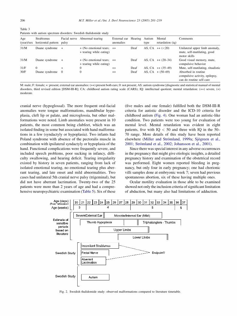
anomalies and functional disorders is shown in Fig. 2.
3.2. Swedish Mobius study
The age of the 25 patients (18 male, 7 female) ranged
from 1 month to 55 years. Table 4 summarizes the prominent
findings. Sixteen patients showed involvement of the 12th
M.T. Miller et al. / Int. J. Devl Neuroscience 23 (2005) 201–219206
Table 3
Patients with autism spectrum disorders: Swedish thalidomide study
Age
(year)/sex
Strabismus
horizontal pattern
Facial nerve
palsy
Abnormal tearing External ear
anomalies
Hearing Autism
type
Mental
retardation (iq)
Comments
31/M Duane syndrome + + (No emotional tears;
+ tearing while eating)
++ Deaf AS, CA ++ (<20) Unilateral upper limb anomaly,
mute, self-mutilating, good
motor skills
31/M Duane syndrome + + (No emotional tears;
+ tearing while eating)
++ Deaf AS, CA ++ (20–34) Good visual memory, mute,
compulsive behavior
31/F 0 + 0 ++ Deaf AS, CA ++ (35–49) Mute, self-mutilating, ritualistic
30/F Duane syndrome 0 0 ++ Deaf AS, CA + (50–69) Absorbed in routine
compulsive activity, epilepsy,
can do routine self-care
M: male; F: female; +: present; external ear anomalies: (++) present both ears; 0: not present; AS: autism syndrome [diagnostic and statistical manual of mental
disorders, third revised edition [DSM-III-R]; CA: childhood autism rating scale (CARS); IQ: intellectual quotient; mental retardation: (++) severe, (+)
moderate.
cranial nerve (hypoglossal). The more frequent oral-facial
anomalies were tongue malformations, mandibular hypo-
plasia, cleft lip or palate, and microglossia, but other mal-
formations were noted. Limb anomalies were present in 10
patients, the most common being clubfeet, which was an
isolated finding in some but associated with hand malforma-
tions in a few (syndactyly or hypoplasia). Two infants had
Poland syndrome with absence of the pectoralis muscle in
combination with ipsilateral syndactyly or hypoplasia of the
hand. Functional complications were frequently severe, and
included speech problems, poor sucking in infancy, diffi-
culty swallowing, and hearing deficit. Tearing irregularity
existed by history in seven patients, ranging from lack of
isolated emotional tearing, no emotional tearing plus aber-
rant tearing, and late onset and mild abnormalities. Two
cases had unilateral 5th cranial nerve palsy (trigeminal), but
did not have aberrant lacrimation. Twenty-two of the 25
patients were more than 2 years of age and had a compre-
hensive neuropsychiatric examination (Table 5). Six of these
Fig. 2. Swedish thalidomide study: observed malf
(five males and one female) fulfilled both the DSM-III-R
criteria for autistic disorder and the ICD-10 criteria for
childhood autism (Fig. 4). One woman had an autistic-like
condition. Two patients were too young for evaluation of
mental level. Mental retardation was evident in eight
patients, five with IQ < 50 and three with IQ in the 50–
70 range. More details of this study have been reported
elsewhere (Miller and Stromland, 1999a; Sjogreen et al.,
2001; Stromland et al., 2002; Johansson et al., 2001).
Since there was special interest in any adverse occurrences
in the pregnancy that might give etiologic insights, a detailed
pregnancy history and examination of the obstetrical record
was performed. Eight women reported bleeding in preg-
nancy, but only four in early pregnancy; one had chorionic
villi samples done at embryonic week 7; seven had previous
spontaneous abortion, six of these having multiple ones.
Ocular motility evaluation in those able to be examined
showed not only the inclusion criteria of significant limitation
of abduction, but many also had limitations of adduction.
ormations compared to literature timetable.
M.T. Miller et al. / Int. J. Devl Neuroscience 23 (2005) 201–219 207
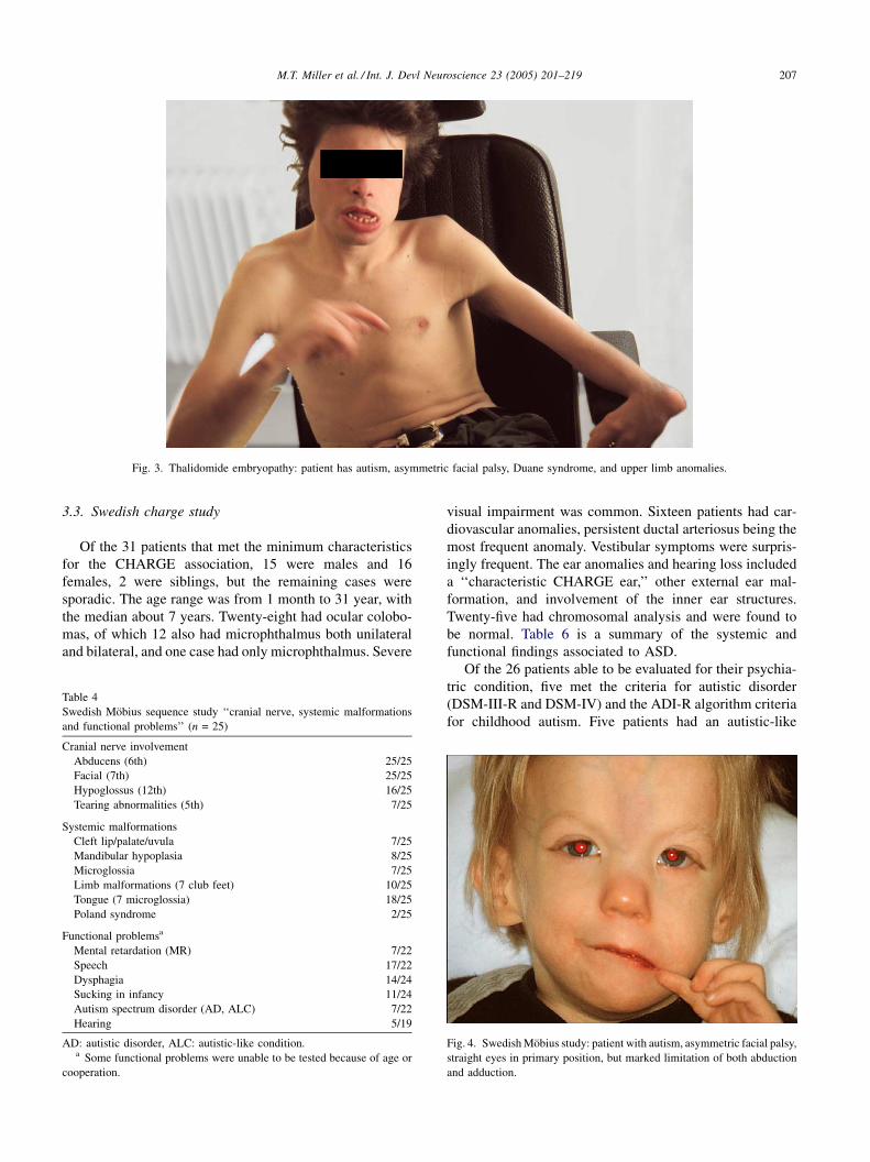
Fig. 3. Thalidomide embryopathy: patient has autism, asymmetric facial palsy, Duane syndrome, and upper limb anomalies.
3.3. Swedish charge study
Of the 31 patients that met the minimum characteristics
for the CHARGE association, 15 were males and 16
females, 2 were siblings, but the remaining cases were
sporadic. The age range was from 1 month to 31 year, with
the median about 7 years. Twenty-eight had ocular colobo-
mas, of which 12 also had microphthalmus both unilateral
and bilateral, and one case had only microphthalmus. Severe
Table 4
Swedish Mobius sequence study ‘‘cranial nerve, systemic malformations
and functional problems’’ (n = 25)
Cranial nerve involvement
Abducens (6th) 25/25
Facial (7th) 25/25
Hypoglossus (12th) 16/25
Tearing abnormalities (5th) 7/25
Systemic malformations
Cleft lip/palate/uvula 7/25
Mandibular hypoplasia 8/25
Microglossia 7/25
Limb malformations (7 club feet) 10/25
Tongue (7 microglossia) 18/25
Poland syndrome 2/25
Functional problemsa
Mental retardation (MR) 7/22
Speech 17/22
Dysphagia 14/24
Sucking in infancy 11/24
Autism spectrum disorder (AD, ALC) 7/22
Hearing 5/19
AD: autistic disorder, ALC: autistic-like condition.a Some functional problems were unable to be tested because of age or
cooperation.
visual impairment was common. Sixteen patients had car-
diovascular anomalies, persistent ductal arteriosus being the
most frequent anomaly. Vestibular symptoms were surpris-
ingly frequent. The ear anomalies and hearing loss included
a ‘‘characteristic CHARGE ear,’’ other external ear mal-
formation, and involvement of the inner ear structures.
Twenty-five had chromosomal analysis and were found to
be normal. Table 6 is a summary of the systemic and
functional findings associated to ASD.
Of the 26 patients able to be evaluated for their psychia-
tric condition, five met the criteria for autistic disorder
(DSM-III-R and DSM-IV) and the ADI-R algorithm criteria
for childhood autism. Five patients had an autistic-like
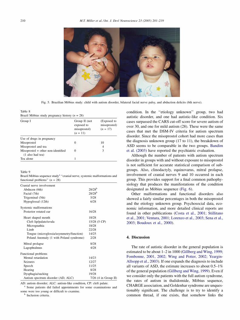
Fig. 4. Swedish Mobius study: patient with autism, asymmetric facial palsy,
straight eyes in primary position, but marked limitation of both abduction
and adduction.

M.T. Miller et al. / Int. J. Devl Neuroscience 23 (2005) 201–219208
Table 5
Patients with autism spectrum disorders Swedish Mobius study
Case Age
(year)/sex
Strabismus Facial
nerve
palsy
Other cranial
nerves
Abnormal
tearing
Tongue
anomaly
Autism
type
Mental
retardation
Comments
Primary Adduct
defect
Abduct
defect
1 4/F Esotropia 0 ++ ++ 10 0 + AD, CA ++ Club feet, seizures,
sucking problems
2 8/M Esotropia 0 R > L ++ 5 (R) 0 0 AD, CA ++ Corneal scar, poor
balance
3 14/M Straight ++ ++ L > R 12 + + AD, CA ++ Mute, ear anomaly,
epilepsy, decreased
hearing, normal MRI,
dental anomalies
4 15/M Small Esotropia ++ ++ ++ 12 + (No emotional;
+ when eating)
+ AD, CA + Mute, uses sign
language, micrognathia,
hypoplasia of the
pectoralis muscle
5 17/M Small esotropia ++ ++ ++ 12 + (No emotional) + AD, CA ++ Mute, mild decreased
hearing, sucking
problems in infancy,
mild ptosis, cerebral
palsy, micrognathia
6 22/M Small esotropia ++ + ++ 0 + (No emotional) 0 AD, CA +
7 1.7/M Esotropia + ++ ++ 12 0 + AD, CA ++ Club feet, sucking
problems in infancy,
eating difficulties
8 55/F Large angel
esotropia
0 ++ ++ 0 0 + ALC + Decreased hearing
M: male; F: female, strabismus: (++) marked decrease abduction or adduction, (+) minimal defect; tongue anomaly: (+) present; 0 = not present; autism type:
AD: autism disorder, CA: childhood autism, ALC: autism-like condition; mental retardation: (++) severe, (+) moderate.
condition. All but one of the patients with ASD had mental
retardation. A comprehensive report of the clinical and
autism data will be described elsewhere (Stromland et al.,
2004).
3.4. Goldenhar (oculo-auriculo-vertebral, OAV) syndrome
study
In the 18 patients in this study, ear anomalies and func-
tional deficits were frequent but varied (microtia, tags and
fistula, abnormal shape, hearing defect). These anomalies
plus a number of cases with microstomia, facial asymmetry,
and vertebral anomalies made the presumptive diagnosis of
hemifacial microsomia. The presence of epibulbar and
ocular dermoids put most into the Goldenhar subgroup of
hemifacial microsomia. Facial nerve palsy, cardiovascular
anomalies, gastrointestinal and genitourinary problems
showed the wide spectrum of less common associated
conditions.
Two children met the criteria for autistic disorder (DSM-
III-R, DSM-IV) and the ADI-R algorithm criteria for child-
hood autism. Both these children were mentally retarded,
one very severely. One further child was diagnosed as
manifesting characteristics of an autistic-like condition with
average IQ. Table 7 summarizes the associated findings in
these cases. A comprehensive report will be described in the
future.
3.5. Brazilian Mobius/misoprostol study
Table 8 shows a summary of the pregnancy findings in the
28 patients in the Brazilian Mobius study group. The most
used drug to induce abortion was misoprostol reported by 17
patients (group II). In 10 cases they used misoprostol only
and in 4 cases they used it plus tea, which was a culturally
popular drug felt to induce abortion (Pinto et al., 2003).
Three patients took misoprostol plus injection of an uni-
dentified medication. One patient on the unexposed group
had also taken tea. Misoprostol was taken both orally and
vaginally in 14 patients (82%); vaginally alone in 2 patients
(12%); and orally alone in 1 patient (6%). The average
number of pills taken was 4.8 (each pill was 200 mg). In the
misoprostol exposure group, 15 cases had a history of
bleeding early in pregnancy, compared to four in the non-
exposed group. The average duration of bleeding was
approximately 9–10 days in both groups. Not surprisingly,
bleeding was more frequent in the attempted abortion (group
2) than the etiology-unknown group. Cramping occurred in
eight of the misoprostol exposed and none in the non-
exposed. Table 9 summarizes the frequent systemic and
functional problems noted.
Common associated anomalies were micrognathia and
posterior rotated ear, with no difference in prevalence
between the two groups. Limb anomalies were present in
22 of the 28 patients in the study, with clubfoot and

M.T. Miller et al. / Int. J. Devl Neuroscience 23 (2005) 201–219 209
Table 6
Swedish CHARGE study autism spectrum disorder
Case Age
(year)/sex
Autism Ocular findings Heart Choanal
atresia
Development
delay (mr)
Genital Ear Other anomalies/
functional problems
Coloboma Microphalmus Ext Hearing
1 5/M AD ++ + � ++ + Cryptorchism
micropenis
+ + Short stature,
spine, hand,
dysphagia
2 6/F AD ++ + + PDA, Fallot + ++ + Labia hypoplasia + + Short stature, spine,
dysphagia
3 7/F AD ++ + + ASD, VSD, PDA 0 ++ 0 + 0 Cleft palate, anal
atresia, renal, spine,
dental, dysphagia
4 13/M AD ++ 0 + PDA + ++ Micropenis,
cryptochisam,
delayed puberty
+ + Cleft palate, trachea
esophageal fistula,
dental, short stature,
dysphagia
5 16/F AD ++ + + PDA, ASD 0 + 0 + 0 Cleft lip/palate,
short stature, dental,
facial nerve,
dysphagia
6 4/M ALC + 0 0 0 + 0 + + Short stature,
dysphagia
7 5/F ALC ++ 0 + PDA, VSD, PS + ++ 0 + + Facial nerve, TE
fistula, anal atresia,
limb, dysphagia
8 14/M ALC 0 0 0 0 0 + Cryptochrism,
micropenis
+ + Facial nerve palsy,
delayed puberty,
balance, short
stature, spine
9 17/F ALC ++ + 0 0 ++ 0 + + Craniosynostosis,
balance
10 18/F ALC ++ 0 0 + ++ + Delayed puberty + + Delayed puberty,
dysphagia
M: male; F: female, autism: AD: Autistic disorder, ALC: autistic-like conditions; coloboma: (++) bilateral, (+) unilateral; +: present, 0: not present; heart: ASD:
atrial septal defect, PDA: patient ductus artenosis, PS: pulmonary stenosis, VSD: ventricular septal defect; development delay: MR: mental retardation: (++)
severe, (+) mild, (0) not present; ear: (+) malformation or functional deficit present, (0) no malformation or functional deficit present.
clinodactyly the most frequent. Abnormal tearing was pre-
sent in both groups. Many patients had oral or dental
malformations including cleft palate, abnormal tongue anat-
omy, altered tongue tone, and poor sucking. The only
statistical difference between the two groups was the low
birth weight, and heart-shaped mouth, which seemed to be
more characteristic of the misoprostol group than the non-
misoprostol group. A detailed analysis of this study is
reported elsewhere by Ventura (2001).
Radiologic imaging was done on 25 of the 28 patients.
The main findings were brain stem calcification present in
Table 7
Swedish Goldenhar study patients with autism spectrum disorder
Case Age
(year)/sex
Ear Decreased
hearing
Dermoid IQ
1 4/M 0 + + Epibulbar
lipodermoid
Severe MR
2 16/F + + + Epibulbar
lipodermoid
Mild MR
3 6/M + (Microtia, ear tags,
fistula, dimple)
+ + Epibulbar
lipodermoid
Average
M: male; F: female; +: anomaly or functional deficit present; 0: not present; AD: a
mental retardation.
six, Dandy–Walker or variant in two, arachnoid cysts in two,
hydrocephalus in three, cerebral atrophy in four, and a
variety of other single anomalies. There did not seem to
be a significant difference between the misoprostol exposed
and the non-exposed groups.
Of the 28 patients, 26 had an evaluation for ASD (Table
10). A few patients were too young or did not attend
examination. In these 26 patients, 5 met the diagnostic
criteria for autism and 2 had an autistic-like condition
(Fig. 5). There was a positive history of misoprostol in three
of the five with autism, and one of the two with autistic-like
Autism type Comments
AD Cardiovascular and gastrointestinal anomaly, mandibular
hypoplasia, microphthalmos
AD Limb anomaly, gastrointestinal and genitourinary
problems, vertebral anomaly
ALC Vertebral anomaly, facial palsy, gastrointestinal problem
utistic disorder; ALC: autism like condition; IQ: intellectual quotient; MR:
M.T. Miller et al. / Int. J. Devl Neuroscience 23 (2005) 201–219210
Table 8
Brazil Mobius study pregnancy history (n = 28)
Group I Group II (not
exposed to
misoprostol)
(n = 11)
(Exposed to
misoprostol)
(n = 17)
Use of drugs in pregnancy
Misoprostol 0 10
Misoprostol and tea 4
Misoprostol + other non-identified
(1 also had tea)
0 3
Tea alone 1
Table 9
Brazil Mobius sequence studya ‘‘cranial nerve, systemic malformations and
functional problems’’ (n = 28)
Cranial nerve involvement
Abducen (6th) 28/28b
Facial (7th) 28/28b
Trigeminal (5th) 5/28
Hypoglossal (12th) 6/28
Systemic malformations
Posterior rotated ear 16/28
Heart shaped mouth 18/28
Cleft lip/palate/uvula 15/28 (5 CP)
Micrognathia 18/28
Limb 22/28
Tongue (microglossia/asymmetry/function) 14/25
Poland Anomaly (1 with Poland syndrome) 2/28
Mitral prolapse 8/28
Lagophtalmus 4/28
Functional problems
Mental retardation 14/23
Seizures 12/27
Speech 11/25
Hearing 8/28
Dysphagia/sucking 19/28
Autism spectrum disorder (AD, ALC) 7/26 (4 in Group II)
AD: autism disorder; ALC: autism-like condition, CP: cleft palate.a Some patients did failed appointments for some examinations and
some were too young or difficult to examine.b Inclusion criteria.
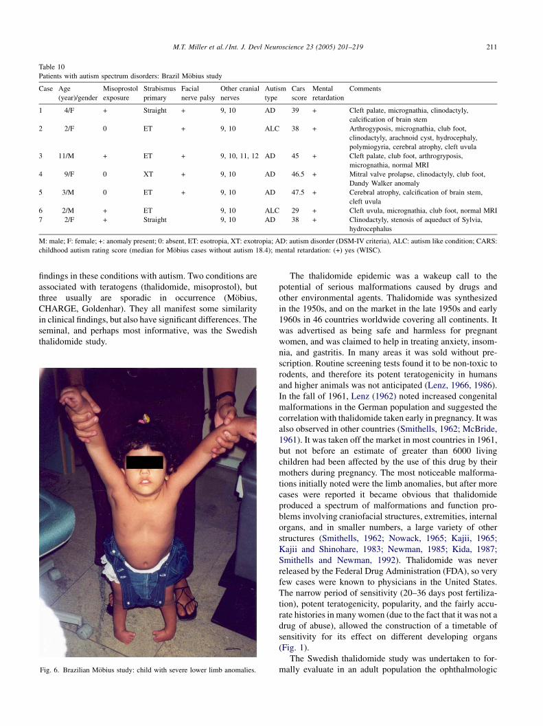
Fig. 5. Brazilian Mobius study: child with autism disorder, bilateral facial nerve palsy, and abduction deficits (6th nerve).
condition. In the ‘‘etiology unknown’’ group, two had
autistic disorder, and one had autistic-like condition. Six
cases surpassed the CARS cut-off score for severe autism of
over 30, and one for mild autism (28). These were the same
cases that met the DSM-IV criteria for autism spectrum
disorder. Since the misoprostol cohort had more cases than
the diagnosis unknown group (17 to 11), the breakdown of
ASD seems to be comparable in the two groups. Bandim
et al. (2003) have reported the psychiatric evaluation.
Although the number of patients with autism spectrum
disorder in groups with and without exposure to misoprostol
is not sufficient for accurate statistical comparison of sub-
groups. Also, clinodactyly, equinovarus, mitral prolapse,
involvement of cranial nerves 9 and 10 occurred in each
group. This provides support for a final common pathophy-
siology that produces the manifestations of the condition
designated as Mobius sequence (Fig. 6).
Other malformations and functional disorders also
showed a fairly similar percentages in both the misoprostol
and the etiology unknown group. Psychosocial data, eco-
nomic information, and more detailed clinical reports are
found in other publications (Costa et al., 2001; Stillitano
et al., 2001; Ventura, 2001; Lorenzo et al., 2003; Sena et al.,
2003; Boudoux et al., 2000).
4. Discussion
The rate of autistic disorder in the general population is
estimated to be about 1–2 in 1000 (Gillberg and Wing, 1999;
Fombonne, 2001, 2002; Wing and Potter, 2002; Yeargin-
Allsopp et al., 2003). If one expands the diagnosis to include
all variants of ASD, the estimate increases to about 0.5–1%
of the general population (Gillberg and Wing, 1999). Even if
we consider only the patients with the full autism syndrome,
the rates of autism in thalidomide, Mobius sequence,
CHARGE association, and Goldenhar syndrome are unques-
tionably significant. The challenge is to try to identify a
common thread, if one exists, that somehow links the
M.T. Miller et al. / Int. J. Devl Neuroscience 23 (2005) 201–219 211
Table 10
Patients with autism spectrum disorders: Brazil Mobius study
Case Age
(year)/gender
Misoprostol
exposure
Strabismus
primary
Facial
nerve palsy
Other cranial
nerves
Autism
type
Cars
score
Mental
retardation
Comments
1 4/F + Straight + 9, 10 AD 39 + Cleft palate, micrognathia, clinodactyly,
calcification of brain stem
2 2/F 0 ET + 9, 10 ALC 38 + Arthrogyposis, micrognathia, club foot,
clinodactyly, arachnoid cyst, hydrocephaly,
polymiogyria, cerebral atrophy, cleft uvula
3 11/M + ET + 9, 10, 11, 12 AD 45 + Cleft palate, club foot, arthrogryposis,
micrognathia, normal MRI
4 9/F 0 XT + 9, 10 AD 46.5 + Mitral valve prolapse, clinodactyly, club foot,
Dandy Walker anomaly
5 3/M 0 ET + 9, 10 AD 47.5 + Cerebral atrophy, calcification of brain stem,
cleft uvula
6 2/M + ET 9, 10 ALC 29 + Cleft uvula, micrognathia, club foot, normal MRI
7 2/F + Straight 9, 10 AD 38 + Clinodactyly, stenosis of aqueduct of Sylvia,
hydrocephalus
M: male; F: female; +: anomaly present; 0: absent, ET: esotropia, XT: exotropia; AD: autism disorder (DSM-IV criteria), ALC: autism like condition; CARS:
childhood autism rating score (median for Mobius cases without autism 18.4); mental retardation: (+) yes (WISC).
findings in these conditions with autism. Two conditions are
associated with teratogens (thalidomide, misoprostol), but
three usually are sporadic in occurrence (Mobius,
CHARGE, Goldenhar). They all manifest some similarity
in clinical findings, but also have significant differences. The
seminal, and perhaps most informative, was the Swedish
thalidomide study.
Fig. 6. Brazilian Mobius study: child with severe lower limb anomalies.
The thalidomide epidemic was a wakeup call to the
potential of serious malformations caused by drugs and
other environmental agents. Thalidomide was synthesized
in the 1950s, and on the market in the late 1950s and early
1960s in 46 countries worldwide covering all continents. It
was advertised as being safe and harmless for pregnant
women, and was claimed to help in treating anxiety, insom-
nia, and gastritis. In many areas it was sold without pre-
scription. Routine screening tests found it to be non-toxic to
rodents, and therefore its potent teratogenicity in humans
and higher animals was not anticipated (Lenz, 1966, 1986).
In the fall of 1961, Lenz (1962) noted increased congenital
malformations in the German population and suggested the
correlation with thalidomide taken early in pregnancy. It was
also observed in other countries (Smithells, 1962; McBride,
1961). It was taken off the market in most countries in 1961,
but not before an estimate of greater than 6000 living
children had been affected by the use of this drug by their
mothers during pregnancy. The most noticeable malforma-
tions initially noted were the limb anomalies, but after more
cases were reported it became obvious that thalidomide
produced a spectrum of malformations and function pro-
blems involving craniofacial structures, extremities, internal
organs, and in smaller numbers, a large variety of other
structures (Smithells, 1962; Nowack, 1965; Kajii, 1965;
Kajii and Shinohare, 1983; Newman, 1985; Kida, 1987;
Smithells and Newman, 1992). Thalidomide was never
released by the Federal Drug Administration (FDA), so very
few cases were known to physicians in the United States.
The narrow period of sensitivity (20–36 days post fertiliza-
tion), potent teratogenicity, popularity, and the fairly accu-
rate histories in many women (due to the fact that it was not a
drug of abuse), allowed the construction of a timetable of
sensitivity for its effect on different developing organs
(Fig. 1).
The Swedish thalidomide study was undertaken to for-
mally evaluate in an adult population the ophthalmologic
M.T. Miller et al. / Int. J. Devl Neuroscience 23 (2005) 201–219212
findings, especially those related to ocular motility distur-
bances (strabismus). Sweden also offered an ideal place for
this study because of the excellent medical records, a stable
population, and the availability of an accurate list of affected
individuals. The finding of three individuals with autism at
the very end of the study was completely unexpected, but
was felt to be an important observation; therefore, psychia-
trists were asked to evaluate these patients and two others
who had been seen previously. They confirmed the diagnosis
of ASD in four cases (Stromland et al., 1994). There was no
attempt to look at the rest of the population in a prospective
manner at this time, so the prevalence in this of ASD could
easily be underestimated.
There were two findings that caught the pediatric ophthal-
mologists’ attention early. They were the presence of an
unusual form of ocular motor imbalance (strabismus) and a
rare finding of aberrant tearing. Incomitant strabismus is a
type of ocular motility disturbance in which the amount of
deviation changes as the individual looks in different fields
of gaze. Compared to comitant deviation, in which there is
no limitation of movement but just the misalignment, con-
genital incomitant deviation makes up less than 10% of the
total types of strabismus seen in children and Duane syn-
drome is a frequent cause of this congenital type of stra-
bismus. Duane syndrome is estimated to occur in 1% of all
strabismus cases, which gives a prevalence of Duane syn-
drome of about 2–3/10,000, since strabismus occurs in 2–4%
of the population (Kirkham, 1970).
Although usually an isolated finding, there is a wide
spectrum of associated anomalies reported in the literature
with Duane syndrome, but the most frequent are those seen
in Goldenhar syndrome (upper lid colobomas, conjunctival
limbal dermoids, neurosensory deafness, and spinal cord
anomalies) (Pfaffenbach et al., 1972; Alexander, 1973;
Aleksic et al., 1976). The thalidomide study showed an
extremely high association of individuals with incomitant
strabismus (44%), usually of the Duane type. The usual
observed associated anomalies were malformations of the
thumb and external ear, facial nerve palsy, and hearing loss.
It was uncommon to see Duane syndrome with other limb
anomalies, unless there was evidence that thalidomide had
been taken through a somewhat longer period of pregnancy.
It was clearly an early teratogenic effect of thalidomide in
days 20–24 or 25. This observation had also been made
previously in a large Japanese study (Arimoto, 1987). These
findings gave considerable insight into Duane syndrome.
Congenital paradoxical lacrimation, a very rare anomaly,
was present in 20% of the thalidomide patients. It was also
noted in the Japanese thalidomide series (Arimoto, 1987).
The clinical symptoms range from isolated lack of emotional
tearing to inappropriate tearing when eating, or both. This
condition also goes under the names of paradoxical lacrima-
tion and crocodile tears. The literature reports an almost
universal combination of this anomaly with Duane syn-
drome, itself a condition caused by misdirection of nerves
involving ocular muscles (Biedner et al., 1979; Brik and
Athayde, 1973; Lutman, 1947; Ramsay and Taylor, 1980;
Tachibana et al., 1984; Zhang, 2002). Some suggest that
paradoxical lacrimation represents an example of aberrant
innervation of the lacrimal system from a branch of the
salivary nucleus (Jampel and Titone, 1962), secondary to
failure of differentiation of the salivary and lacrimal nucleus.
It is interesting that early in embryogenesis the sixth and
seventh cranial and lacrimal nuclei are in close proximity.
Destruction or failure of development of these structures
might result in aberrant repair processes with inappropriate
innervation. In the thalidomide study, most were associated
with early effects such as horizontal incomitant strabismus
(100%), hearing deficit or ear malformation (94%), and
facial nerve palsy (71%).
Aberrant innervation does not occur frequently, and yet
examples of this appear throughout these studies. The most
striking is in thalidomide embryopathy, with both aberrant
tearing (20%) and a high prevalence of Duane syndrome
(44%). Although none of our Goldenhar study patients
demonstrated this finding, the literature reports Duane syn-
drome frequently in association with this syndrome, or
malformation common to this syndrome. There is also a
literature report of another type of paradoxical innervation,
Marcus–Gunn jaw winking, with CHARGE association.
Local vulnerability and critical time may be the necessary
factors for these neurologic mismatches to occur.
The similarity of early thalidomide effects to the classic
Mobius sequence patients, i.e. limitation of abduction and
facial nerve palsy, suggests detailed attention to tearing
abnormalities in the Swedish and Brazilian Mobius studies
might prove fruitful. Amaya et al. (1990) noted excessive
lacrimation in 11 of 18 in a series of Mobius cases, with 3
also manifesting tearing when eating. Careful questioning
did reveal abnormal tearing in about a third of the patients in
the Swedish study, ranging from tearing when eating, lack of
emotional tearing, to late onset of tearing. Of interest is that
four of the six Mobius patients with a diagnosis of autism
had no emotional tearing, and two of the four in thalidomide
embryopathy with autism also lacked emotional tearing.
Aberrant lacrimation was a prominent finding in our thali-
domide embryopathy series (20%). Abnormal tearing symp-
toms were also present in many in the Brazilian study, in
both the misoprostol-related and etiology-unknown groups.
Mobius (1888, 1892), a German neurologist in the late
1800s, described a number of cases with congenital involve-
ment of the sixth and seventh nerve, and his name is
associated with this condition. Most cases have severe
limitation of abduction, and often adduction, but near-nor-
mal vertical movements (Amaya et al., 1990). Many syn-
dromologists believe that Mobius sequence is not a distinct
entity but belongs to a group of conditions characterized by
involvement of craniofacial and oral/dental anomalies (espe-
cially affecting the tongue), Poland syndrome, and hypo-
plastic limb anomalies. Mobius sequence is usually a
sporadic event, but there is literature of some cases with
familial occurrence and also chromosomal anomalies

M.T. Miller et al. / Int. J. Devl Neuroscience 23 (2005) 201–219 213
(Becker-Christensen and Lund, 1974; Baraitser, 1977; Ziter
et al., 1977; McDermot et al., 1991; Slee et al., 1991;
Donahue et al., 1993; Kremer et al., 1996; Nishikawa
et al., 1997; Wilmore et al., 2000; Borck et al., 2001).
The ophthalmologic findings in both of these reported
Mobius studies were similar to those in the literature, with
either esotropia (inward turning of deviated eye) or no
misalignment in the straight-ahead position, with only
occasionally an exotropia (outward turning). The systemic
and functional problems in both studies were again as
expected, with difficulty sucking from the seventh nerve
palsy, an important issue in infancy because of feeding
problems. The frequency of findings might show some
variations because of the recognition of subtle findings noted
by the team of specialists. Abnormalities of the tongue, limb,
and oral structures were often present. The patients in both
the Swedish thalidomide and Mobius cohorts were evaluated
comprehensively by psychiatrists, and the results reported in
the literature (Stromland et al., 1994; Johansson et al., 2001).
Early effects in thalidomide embryopathy and Mobius
sequence imply brainstem dysfunction because of the invol-
vement of a number of cranial nerves.
Although the group of ocular and systemic malforma-
tions seen with CHARGE association had been previously
described by other authors (Hall, 1979; Hittner et al., 1979;
Warburg, 1983), the acronym CHARGE was proposed by
Pagon et al. (1981). The criteria used in this paper for the
diagnosis were the presence of four of the six characteristics
(coloboma, heart malformation, choanal atresia, develop-
mental retardation, genital and ear anomalies), or three
characteristics and other anomalies. Other reports in the
literature have described other malformations, and sug-
gested modifications of the diagnostic criteria (Kallen
et al., 1999; Brock et al., 2003). Byerly and Pauli (1993)
propose that involvement of multiple cranial nerves occurs
frequently. Since the findings cover multiple disciplines of
medicine, there is often a bias of ascertainment in any series
reflecting population evaluated, the interest of investigators
and the sophistication of the examination of any given organ
or structure (Russell-Eggitt et al., 1990). The estimated
prevalence of CHARGE association is about 1:10,000
(Blake et al., 1998).
While the association of ASD with these reported con-
ditions is quite convincing, what connects these conditions
to each other that might give insight into common pathways?
The initial thalidomide literature had many suggested
actions of thalidomide responsible for thalidomide embryo-
pathy. Comprehensive reviews by Zwingenberger and
Wendt (1996), Tseng et al. (1996), Argiles et al. (1998),
Miller and Stromland (1999b), Stephens (1988), Stephens
et al. (2000), and others have summarized more recent
theories. Stephens (2000) noted that although there were
2000 papers published in the last 40 years concerning
thalidomide teratogenicity, the mechanism of action still
remains elusive. He reviewed the present hypotheses, and
summarized their strengths and weaknesses.
However, no matter what the mechanism of teratogenic
action it is clear from the large clinical data in the literature
that thalidomide has a selective action on developing fetal
structures in very specific time frames, and the four patients
with autism seem to occur in the period days 20–25 after
fertilization and associated with cranial nerve involvement,
incomitant strabismus, and ear and thumb malformations.
Mobius sequence with no established single etiology, as
in the Swedish study and group 1 of the Brazilian series, has
received considerable attention and some agreement in the
literature as to probable pathophysiology. A popular expla-
nation is that it belongs in a group of disruption syndromes,
but there is a disagreement as to the causes of the embryonic
disruption. Bamforth (1993) describes a process termed
‘‘organizational disruption’’ (blastogenic disruption) as an
explanation for some of the observed malformations sug-
gesting that it is a better explanation for some phenotypes.
He proposes that there are a group of organizational mole-
cules, highly conserved and determined by chromosomes in
a sequential manner, that are important in the early stage of
organization. This organization is imposed on embryonic
cells by these molecules in a sequence of activation deter-
mined by homebox genes. These molecules or morpho-
genes, as they are sometimes called, at different
concentrations can activate different genes. If something
interferes with setting up the organization of these morpho-
genes, higher or lower concentrations may result, activating
genes at inappropriate times. The changes would not be
visible until differentiation commences and could result in
malformation of organs or histological development. This
theory is perhaps compatible with the observations that some
of the Hox gene defects in the animal models result in brain
stem malformations that are sometimes associated with
autism in humans (Rodier et al., 1996, 1997; Rodier, 2000).
A more commonly suggested mechanism is that of a
vascular disruption in the early embryonic period. Some
investigators refer to it as the ‘‘subclavion disruption syn-
drome’’ (Bavinck and Weaver, 1986; Issaivanan et al.,
2002). In addition to the primary vascular disruption causing
hypoxia from ischemia, edema and hemorrhage, many
secondary events can occur affecting other organs (Shepard,
1991; St. Charles et al., 1993; Matsui et al., 1997). Animal
models have been developed to show how different mal-
formations can occur (Lipson et al., 1989). The timing extent
of this hypoxic event will determine the ultimate malforma-
tions based on the sensitive tissues at the time of the hypoxia
(Leong and Ashwell, 1997).
There are many clinical examples that will support this
vascular disruption concept, but they represent individual
case reports. For instance, malformations suggestive of
Mobius sequence occasionally occur in fetuses exposed to
cocaine, which causes vasoconstriction of the uterine vessels
(Hoyme et al., 1990; Kankirawatana et al., 1993). Also,
chorionic villi sampling has been suggested as an infrequent
cause of limb anomalies and occasionally Mobius sequence,
although there are reports both supporting and refuting the

M.T. Miller et al. / Int. J. Devl Neuroscience 23 (2005) 201–219214
association (Firth et al., 1991; Quintero et al., 1992; Burton
et al., 1993; Froster and Jackson, 1996; Hall, 1996; Holmes,
2002). Mobius cases have been associated with hypovolemia
in a splenic bleed during pregnancy (Lipson et al., 1996),
and to inadvertent exposure to ergotamine, a uterine con-
strictor (Hughes and Goldstein, 1988; Verlos et al., 1990).
Another example was a child with Mobius sequence follow-
ing a history of polyhydramnios in pregnancy (St. Charles
et al., 1993). In the Swedish Mobius study there was an
apparent increase of bleeding in early pregnancy reported
without known precipitating causes, and also one case with a
history of chorionic villi sampling procedure. Courtens et al.
(1992) report a case associated with a history of exposure to
benzodiazepines. The common characteristic of all these
cases in the literature is an early pregnancy event producing
a possible short period of hypoxia brought upon by dis-
turbance in the blood supply from uterine constriction. This
line of reasoning received support with the association of
misoprostol taken early in pregnancy resulting in patients
with characteristic Mobius sequence.
Goldenhar syndrome also has some similarity in clinical
manifestations to Mobius sequence and thalidomide
embryopathy and vascular disruption has been suggested
as a mechanism (Gorlin et al., 2000; Poswillo, 1973).
Though the time of embryonic insult is not as clear in
Mobius sequence or Goldenhar syndrome as it is in thali-
domide embryopathy, it is early in pregnancy, probably
around 4 to 6+ weeks of development. Lam (2000) proposed
a theory that ectodermal non-disjunction involving the otic
placode could produce the same malformations. If correct,
this would explain the multisystem findings and also place
the time early in the 4th week. Another suggestion is that it
can be a result of ‘‘reproductive wastage’’ in high risk
conceptions, based on one case of possible monozygotic
twins conceived by vitro fertilization and embryo transfer
(Jongbloet, 1987). There were a few invitro fertilization
cases in our series.
The pathophysiology of CHARGE association has not
been identified. While most cases are sporadic without a
known etiology, there are some familial cases and a number
with known but different chromosomal anomalies (Mitchell
et al., 1985; Clementi et al., 1991; Slee et al., 1991;
Wieczorek et al., 1997; Sanlaville et al., 2002; Devriendt
et al., 1998). The observation that many of the features
suggest defects in neural crest cell development or migration
might suggest it should be considered in the group of
neurocristopathies (Siebert et al., 1985). Why autism exists
in a significant number of cases is still a mystery. However,
the time of initial embryonic insult is necessarily early,
especially because ocular colobomas are such a significant
finding. Ocular colobomas are caused by failure of closure of
the embryonic fetal fissure sometime before the 6-week-old
embryo, but from the thalidomide study, an earlier insult (25
to 27+ days) can result in an ocular coloboma. The few
ocular colobomas noted in thalidomide cases seem to result
from exposure close to, but a few days later than, the early-
effect malformations of cranial nerves and aberrant tearing.
Although not part of the diagnostic acronym, facial nerve
palsy is a common characteristic of CHARGE (Lacombe,
1994; Byerly and Pauli, 1993). Blake et al. (1998) propose
that cranial nerve dysfunction (anosmia, facial nerve palsy,
sensorineural deafness and vestibular problems, swallowing
difficulties) be considered a major criterion. This involve-
ment of cranial nerves may be a thread that exists with
CHARGE association, thalidomide embryopathy, and
Mobius sequence. A common pathway for CHARGE and
OAV spectrum has been suggested (Van Meter and Weaver,
1996).
Misoprostol as a drug for self-induced abortions has
gained much popularity in South America, especially Brazil,
where abortions are not legal except in a few situations.
Misoprostol was cheap and readily available because of its
accepted use in medical conditions such as gastric ulcers and
arthritis (Grazioli et al., 1993; Raskin et al., 1996; Morgan,
1999). Medically-induced abortions have advantages over
clandestine abortion from unlicensed ‘‘professionals.’’ They
avoid risk of anesthesia and surgical complications in
unclean environments, and perhaps most important, can
be done in privacy. For a number of years it was estimated
to be used in more than 50% of attempted abortions (Coelho
et al., 1993, 1994). It has also been utilized for planned
abortions, conducted by medical professionals in many
countries, but almost always combined with another drug
such as mifrostone (Norman et al., 1991; Spitz et al., 1998;
Wing, 1999). However, misoprostol alone is a poor aborti-
facient drug, and many pregnancies continue to term. Con-
sidering its frequent usage, misoprostol does not appear to
cause many malformations, but because of the tremendous
popularity as an abortifacient drug for self-induced abor-
tions, even low incidence complications such as Mobius
sequence occurred in sizable numbers (Schuler et al., 1997).
The presence of ASD in both the misoprostol-exposed
and non-exposed cases of Mobius sequence in the Brazilian
study gives further evidence of the association of autism
with early embryonic insults, and also the non-specificity of
etiology of Mobius sequence.
While autism is usually associated with cortical dysfunc-
tion, brain stem abnormalities have been suggested as a
possible mechanism in some patients (Rosenhall et al.,
2003). The frontal and temporal lobes, the limbic system
and cerebellum are believed to be crucially dysfunctional in
autism (Gillberg and Coleman, 2000). Theoretically, such
dysfunctions could be related to disrupted brain stem con-
nections projecting to limbic, cortical, and cerebellar struc-
tures.
In the group of studies in this paper, early injuries to the
embryo are associated with disturbances in the brain stem.
Rodier, in a number of papers, summarized the associations
of autism with other early embryologic events (Rodier et al.,
1997, Rodier, 2000, 2004), The brain stem anomalies do not
rule out dysfunction in the forebrain. It is more likely that
abnormal input from the brain stem contributes to forebrain

M.T. Miller et al. / Int. J. Devl Neuroscience 23 (2005) 201–219 215
dysfunction in addition to the more direct effects on cranial
nerve function.
Valproic acid (VPA) is teratogenic to rodents and causes
malformations similar to thalidomide. Exposure to VPA
during pregnancy has been reported to be associated with
autism in children (Moore et al., 2000). Rodier et al. (1996)
and Rodier (2004) used the rat fetus as an animal model and
noted that when the pregnant rat was exposed to VPA at the
time of neural tube closure, there was as an affect on the
cranial nerve motor nuclei.
Rodier et al. (1996) noted almost complete absence of
facial nuclei and shortening of brainstem in a patient with
autism. Radiologic abnormalities, albeit not consistent or
conclusive, have also been reported in Mobius syndrome.
They include abnormalities of the brainstem (Thakkar et al.,
1977; Nardelli et al., 1983; Harbord et al., 1989; Kuhn et al.,
1990; Lengyel et al., 2000; Pedraza et al., 2000; Yoon et al.,
1997). There are a number of literature reports of central
hypoventilation, brainstem changes, and in some, Mobius
sequence (Cohen and Thompson, 1987; Govaert et al.,
1989; Konkol et al., 1991; Cortez and Kinney, 1996;
Igarashi et al., 1997). In the Swedish Mobius study 2 of
the 10 evaluated radiologically revealed brain abnormalities
(agenesis of corpus callosum, hydrocephalus). In the Bra-
zilian Mobius study there were a considerable number of
MRI abnormalities with the most common being brain stem
calcification.
Magnetic resonance imaging (MRI) study by Hashimoto
et al. (1995) and Cody et al. (2002) summarize the MRI
findings in individuals with autism. Some have suggested a
smaller brainstem and cerebellum in patients with autism,
compared to control participants.
The most common thread of the observed anomalies
associated with autism is that they result from an early
adverse embryonic event. The literature can be confusing,
and must be read carefully when it relates to embryonic
timing issues. ‘‘Gestational age,’’ used by obstetricians and
many others, is calculated from the last menstrual period. A
gestational age of 1 month of an embryo is actually 2 weeks
post conception/fertilization. These 2 weeks are certainly
not trivial when looking at early embryonic events. There-
fore, if a woman attempts abortion at 4–6 weeks gestational
age the embryo is actually at only 2–4 weeks of develop-
ment. The studies reported here relate most events to the
actual age of development of the fetus. The most reliable
timing is from the thalidomide data, in which the 4 indivi-
duals with autism condition were 20–25 days post fertiliza-
tion. The next evidence of embryonic timing is from the
misoprostol group. Although less precise, it appears to be 4–
6 weeks (6–8 weeks from the last menstrual cycle). In
Mobius from etiology unknown there is no definite informa-
tion except that it seems consistent with the misoprostol
group as early, e.g. 4–6 weeks. The least established timing
is in the CHARGE group, although we know the ocular
embryonic fissure is closed in 5–6 weeks of embryogenesis,
so the insult must be some time before. From the thalido-
mide timetable it could be as early as the late 4th week. The
Goldenhar group seems to be at 4–6 weeks of embryogen-
esis, based on associated anomalies.
How do we get from early-onset insult that seems to
affect multiple brainstem structures to autism disorders that
involve higher centers not yet formed? Are there a group of
unidentified cells that are even at this time programmed for
higher brain centers, which are damaged, or is there an
interruption in a series of connections ultimately crucial for
higher centers to develop correctly? These are key questions,
but we may only be able to speculate about answers at this
time on some evidence from other studies.
Although autism may result from a variety of mechan-
isms and causes, the combined evidence from the thalido-
mide, Mobius, CHARGE, and Goldenhar studies seems to
establish quite firmly that early insults in embryogenesis,
often involving brainstem structures, are sometimes asso-
ciated with ASD.
Acknowledgements
Marilyn T. Miller, MD, supported in part by grant
U19HD/DC35466, a Collaborative Program of Excellence
in Autism. Also supported in part by core grant EY 1792
from the National Eye Institute, Bethesda, Maryland, by an
unrestricted research grant from Research to Prevent Blind-
ness Inc., New York, and by the Lions of Illinois Foundation,
Maywood, Illinois. Kerstin Stromland, MD, supported in
part by The Goteborg Medical Society. Liana Ventura, MD,
supported in part by the Altino Ventura Foundation, Recife,
Brazil.
References
Aleksic, S., Budzilovich, G., Choy, A., Reuben, R., Randt, C., Finegold, M.,
McCarthy, J., Converse, J., Feigin, I., 1976. Congenital ophthalmoplegia
in oculoauriculovertebral dysplasia-hemifacial microsomia (Golden-
har–Gorlin syndrome): a clinicopathologic study and review of the
literature. Neurology 26, 638–644.
Alexander, J.C., 1973. Ocular abnormalities among congenitally deaf
children. Can. J. Ophthalmol. 8 (3), 428–433.
Amaya, L.G., Walker, J., Taylor, D., 1990. Mobius syndrome. A study and
report of 18 cases. Bin. Vis. Q. 5 (3), 119–132.
American Psychiatric Association, 1987. DSM-III-R. Diagnostic and Sta-
tistical Manual of Mental Disorders,revised third ed. APA, Washington,
DC.
American Psychiatric Association, 1994. DSM-IV-R. Diagnostic and Sta-
tistical Manual of Mental Disorders,revised fourth ed. APA, Washing-
ton, DC.
Argiles, J.M., Carbo, N., Lopez-Soriano, F.J., 1998. Was tumor necrosis
factor-alpha responsible for the fetal malformations associated with
thalidomide in the early 1960s? (Review). Med. Hypotheses 50, 313–
318.
Arimoto, Y., 1987. Ophthalmology in thalidomide embryopathy. In: Tha-
lidomide Embryopathy in Japan, Kadansha, Tokyo, pp. 143–153.
Bamforth, J.S., 1993. Disruption sequences: embryonic vascular accident or
blastogenic disruption sequence? Am. J. Med. Genet. 47, 284–288.
M.T. Miller et al. / Int. J. Devl Neuroscience 23 (2005) 201–219216
Bandim, J.M., Ventura, L.O., Miller, M.T., Almeida, H.C., Costa, A.E.,
2003. Autism and Mobius sequence: an exploratory study of children in
northeastern Brazil. Arquivos de Neuro-Psiquiatria 61 (2A), 181–185.
Baraitser, M., 1977. Genetics of Mobius syndrome. Am. J. Med. Genet. 13,
415–417.
Bavinck, J.N., Weaver, D.D., 1986. Subclavian artery supply disruption
sequence: hypothesis of a vascular etiology for Poland, Klippel–Feil,
and Mobius anomalies. Am. J. Med. Genet. 23, 903–918.
Becker-Christensen, F., Lund, H.T., 1974. A family with Mobius syndrome.
J. Pediatr. 84 (1), 115–117.
Biedner, B., Geltman, C., Rothkoff, L., 1979. Bilateral Duane’s syndrome
associated with crocodile tears. J. Pediatr. Ophthalmol. Strab. 16, 113–
114.
Blake, K.D., Davenport, S.L., Hall, B.D., et al., 1998. CHARGE associa-
tion: an update and review for the primary pediatrician (Review). Clin.
Pediatr. (Phila). 37, 159–173.
Blanchard, K., Winikoff, B., Ellertson, C., 1998. Use of misoprostol during
pregnancy and Mobius’ syndrome in infants (comment: Letter). N. Engl.
J. Med. 339 (21), 1553–1554.
Borck, G., Wirth, J., Hardt, T., Tonnies, H., Brondum-Nielsen, K., Bugge,
M., Tommerup, N., Nothwang, H.G., Ropers, H.H., Haaf, T., 2001.
Molecular cytogenetic characterisation of a complex 46,XY,t(7;8;11;13)
chromosome rearrangement in a patient with Moebius syndrome (Let-
ter). J. Med. Genet. 38 (2), 117–121.
Boudoux, D.D., Matos, M.A.G., Goncalves, E.D., Rocha, M., Ventura, L.O.,
Hinrichsen, S.L., 2000. Sındrome de Moebius relacionada a amcaca de
abortamento. Rev. Bras. Oftalmol. 59 (3), 173–177.
Brik, M., Athayde, A., 1973. Bilateral Duane’s syndrome, paroxysmal
lacrimation and Klippel–Feil anomaly. Ophthalmologica 167, 1–8.
Brock, K.E., Mathiason, M.A., Rooney, B.L., Williams, M.S., 2003.
Quantitative analysis of limb anomalies in CHARGE syndrome: corre-
lation with diagnosis and characteristic CHARGE anomalies. Am. J.
Med. Genet. 123A, 111–121.
Burton, B.K., Schulz, C.J., Burd, L.I., 1993. Spectrum of limb disruption
defects associated with chorionic villus sampling. Pediatrics 91, 989–
993.
Byerly, K.A., Pauli, R.M., 1993. Cranial nerve abnormalities in CHARGE
association. Am. J. Med. Genet. 45, 751–757.
Clementi, M., Tenconi, R., Turolla, L., et al., 1991. Apparent CHARGE
association and chromosome anomaly: change or contiguous gene
syndrome. Am. J. Med. Genet. 41, 246–250.
Cody, H., Pelphrey, K., Piven, J., 2002. Structural and functional magnetic
resonance imaging of autism. Int. J. Dev. Neurosci. 20, 421–438.
Coelho, H.L.L., Misago, C., Fronseca, W.V.C., Souza, D.S.C., Araujo,
J.M.L., 1991. Selling abortifacients over the counter in pharmacies
in Fortaleza, Brazil. Lancet 338, 247.
Coelho, H.L.L., Teixeira, A.C., Cruz, M.F., Gonzaga, S.L., Arrais, O.S.,
Luchini, L., Vecchia, C.L., Tognoni, G., 1994. Misoprostol: the experi-
ence of women in Forteleza, Brazil. Contraception 49 (2), 101–110.
Coelho, H.L.L., Teixeira, A.C., Santos, A.P., Forte, E.B., Morais, S.M.,
LaVecchia, C., Tognoni, G., Herxheimer, A., 1993. Misoprostol and
illegal abortion in Fortazelza, Brazil. Lancet 341, 1261–1263.
Coelho, K.E.F.A., Sarmento, M.F., Veiga, C.M., Speck-Martins, C.E.,
Safatle, H.P.N., Castro, C.V., Niikawa, N., 2000. Misoprostol embry-
otoxicity: clinical evaluations of fifteen patients with arthrogryposis.
Am. J. Med. Genet. 95, 297–301.
Cohen, S.R., Thompson, J.W., 1987. Variants of Mobius’ syndrome and
central neurologic impairment. Lindeman procedure in children. Ann.
Otol. Rhinol. Laryngol. 96, 93–100.
Cortez, S.C., Kinney, H.C., 1996. Brainstem tegmental necrosis and olivary
hypoplasia: a lethal entity associated with congenital apnea. J. Neuro-
pathol. Exp. Neurol. 55, 841–849.
Costa, A.E.S., Ventura, L., Almeda, H.C., Miller, M., Cassia-Lima, R.,
Bandim, J.M., 2001. Sequencia de Mobius: aspectos socialis, psicolo-
gicos E cognitivos (sequence of Mobius: social, psychological and
cognitive aspects). An. Fac. de Med. Univ. Fed. Pernambuco, Recife,
Brazil 46 (2), 137–143.
Costa, S.H., Vessey, M.P., 1993. Misoprostol and illegal abortion in Rio de
Janeiro, Brazil. Lancet 341, 258–1261.
Courtens, W., Vamos, E., Hainaut, M., Vergauwen, P., 1992. Moebius
syndrome in an infant exposed in utero to benzodiazepines. J. Pediatr.
121 (5), 833–834.
Davenport, S.L.H., et al., 1986. The spectrum of clinical features in
CHARGE syndrome. Clin. Genet. 29, 298–310.
d’Avignon, M., Barr, B., 1964. Ear abnormalities and cranial nerve palsies
in thalidomide children. Arch. Otolaryngol. 80, 136–140.
Devriendt, K., Swillen, A., Fryns, J.P., 1998. Deletion in chromosome
region 22q11 in a child with CHARGE association. Clin. Genet. 53,
408–410.
Donahue, S.P., Wenger, S.L., Steele, M.W., Gorin, M.B., 1993. Broad-
spectrum Mobius syndrome associated with a 1;11 chromosome trans-
location. Ophthal. Paediatr. Genet. 14, 17–21.
Duane, A., 1905. Congenital deficiency of abduction associated with
impairment of adduction, retraction movements, contraction of the
palpebral fissure and oblique movements of the eye. Arch. Ophthalmol.
34, 133–159.
Fernell, E., Olsson, V.A., Karlgren-Leitner, C., et al., 1999. Autistic
disorders in children with CHARGE association. Dev. Med. Child.
Neurol. 4, 270–272.
Firth, H.V., Boyd, P.A., Chamberlain, P., MacKenzie, I.K., Lindenbaum,
R.H., Huson, S.M., 1991. Severe limb abnormalities after chorionic
villus sampling at 56–66 days’ gestation. Lancet 337, 762–763.
Fombonne, E., 2002. Prevalence of childhood disintegrative disorder.
Autism 6, 149–157.
Fombonne, E., 2001. Is there an epidemic of autism? Pediatrics 107 (2),
411–413.
Fonseca, W.V.C., Alencar, A.J.C., Mota, F.S.B., Coelho, H.L.L., 1991.
Misoprostol and congenital malformations.. Lancet 338 (8758), 56.
Fonseca, W., Alencar, A.J.C., Pereira, R.M.M., Misago, C., 1993. Con-
genital malformation of the scalp and cranium after failed first trimester
abortion attempt with misoprostol. Clin. Dysmorphol. 2 (1), 76–80.
Froster, U.G., Jackson, L., 1996. Limb defects and chorionic villus sam-
pling: results from an international registry, 1992–1994. Lancet 347,
489–494.
Genest, D.R., Di Salvo, D., Rosenblatt, M.J., Holmes, L.B., 1999. Terminal
transverse limb defects with tethering and omphalocele in a 17 week
fetus following first trimester misoprostol exposure. Clin. Dysmorphol.
8 (1), 53–58.
Gillberg, C., Coleman, M., 2000. The neurology of autism, The Biology of
the Autistic Syndrome, third ed. MacKeith Press, London (Clinics in
Developmental Medicine No. 153/4).
Gillberg, C., Steffenburg, S., 1989. Autistic behaviour in Moebius syn-
drome. Acta Paediatr. 79, 314–326.
Gillberg, C., Wing, L., 1999. Autism: not an extremely rare disorder. Acta
Psychiatr. Scand. 99, 399–406.
Gillberg, C., Winnergard, I., 1984. Childhood psychosis in a case of
Moebius syndrome. Neuropaediatrics 15, 147–149.
Goldenhar, M., 1952. Associations de malformaties l’oeil et de l’oreille en
particulier le syndrome: dermoide epibulbaire, appendices auriculaires,
fistula auri congenita et ses relations avec la dysostose mandibule-
faciale. J. Genet. Hum. 1, 243–282.
Gonzalez, C.H., Marques-Dias, M.J., Kim, C.A., Sugayama, S.M., DaPaz,
J.A., Huson, S.M., Holmes, L.B., 1998. Congenital abnormalities in
Brazilian children associated with misoprostol misuse in first trimester
of pregnancy. Lancet 351 (9116), 1624–1627.
Gonzalez, C.H., Vargas, F.R., Perez, A.B., Kim, C.A., Brunoni, D., Mar-
ques-Dias, M.J., Leone, C.R., Correa Neto, J., Llerena Junior, J.C., de
Almeda, J.C., 1993. Limb deficiency with or without Mobius sequence
in seven Brazilian children associated with Misoprostol use in the first
trimester of pregnancy. Am. J. Med. Genet. 47 (1), 59–64.
Gorlin, R.J., Jue, K.I., Jacobson, U., et al., 1952. Oculoauriculo-vertebral
syndrome. J. Pediatr. 63, 991–999.
Gorlin, R.J., Cohen, M.M., Levin, L.S., 2000. Syndromes of the Head and
Neck, 3rd ed. Oxford University Press, New York, pp. 641–649, 666–672.
M.T. Miller et al. / Int. J. Devl Neuroscience 23 (2005) 201–219 217
Govaert, P., Vanhaesebrouch, P., De Praeter, C., et al., 1989. Moebius
sequence and prenatal brainstem ischemia. Pediatrics 84, 570–573.
Graham Jr., J.M., 2001. A recognizable syndrome within CHARGE asso-
ciation: Hall–Hittner syndrome. Am. J. Med Genet. 99, 120–123.
Grazioli, I., Avossa, M., Bogliolo, A., Broggini, M., Carcassi, A., Carcassi,
U., Cecconami, L., Ligniere, G.C., Colombo, B., Consoli, G., et al.,
1993. Multicenter study of the safety/efficacy of misoprostol in the
prevention and treatment of NSAID-induced gastroduodenal lesions.
Clin. Exp. Rheumatol. 11 (3), 289–294.
Hall, B.D., 1979. Choanal atresia and associated multiple anomalies. J.
Pediatr. 95, 395–398.
Hall, J.G., 1996. Arthrogryposis associated with unsuccessful attempts at
termination of pregnancy. Am. J. Med. Genet. 63, 293–300.
Harbord, M.J., Finn, J.P., Hall-Craggs, M.A., Brett, E.M., Baraitser, M.,
1989. Moebius’ syndrome with unilateral cerebellar hypoplasia. J. Med.
Genet. 26 (9), 579–582.
Hashimoto, T., Tayama, M., Murakawka, K., et al., 1995. Development of
the brainstem and cerebellum in autistic patients. J. Autism Dev. Mental
Disord. 25, 1–18.
Hittner, H.M., et al., 1979. Colobomatous microphthalmus, heart disease,
hearing loss and mental retardation. J. Pediatr. Ophthalmol. Strab. 122–
128.
Holmes, L.B., 2002. Teratogen-induced limb defects. Am. J. Med. Genet.
112 (3), 297–303.
Hotchkiss, M.G., Miller, N.R., Clark, A.W., et al., 1980. Bilateral Duane’s
retraction syndrome: a clinical pathologic case report. Arch. Ophthal-
mol. 98, 870–874.
Hoyme, H.E., Jones, K.L., Dixon, S.D., Jewett, T., Hanson, J.W., Robinson,
L.K., Msall, M.E., Allanson, J.E., 1990. Prenatal cocaine exposure and
fetal vascular disruption. Pediatrics 85, 743–747.
Hoyt, W.F., Nachtigaller, I., 1965. Anomalies of ocular motor nerves:
neuroanatomic correlations of paradoxical innervation in Duane syn-
drome and related congenital ocular motor diseases. Am. J. Ophthalmol.
60, 443–448.
Huber, A., 1974. Electrophysiology of the retraction syndrome. Br. J.
Ophthalmol. 58, 293–300.
Hughes, H.E., Goldstein, D.A., 1988. Birth defects following maternal
exposure of ergotamine, beta blockers and caffeine. J. Med. Genet. 25,
396–399.
Igarashi, M., Rose, D.F., Storgion, S.A., 1997. Moebius syndrome and
central respiratory dysfunction. Pediatr. Neurol. 16 (3), 237–240.
Issaivanan, M., Virdi, V.S., Parmar, V.R., 2002. Subclavian artery supply
disruption sequence—Klippel–Feil and Mobius anomalies. Indian J.
Pediatr. 69 (5), 441–442.
Jampel, R.S., Titone, C., 1962. Congenital paradoxical gustatory-lacrimal
reflex and lateral rectus paralysis. Acta Ophthalmol. 67, 123–126.
Johansson, M., Wentz, E., Fernell, E., Stromland, K., Miller, M.T., Gillberg,
C., 2001. Autistic spectrum disorders in Mobius sequence: a compre-
hensive study of 25 individuals. Dev. Med. Child. Neurol. 43, 338–345.
Jongbloet, P.H., 1987. Goldenhar syndrome and overlapping dysplasias, in
vitro fertilisation and ovopathy. J. Med. Genet. 24, 616–620.
Kajii, T., Shinohare, M., 1983. Thalidomide in Japan. Lancet 1, 501–502.
Kajii, T., 1965. Thalidomide experience in Japan. Ann. Pediatr. 205, 341–
354.
Kallen, K., Robert, E., Mastroiacovo, P., et al., 1999. CHARGE association
in newborns: a registry-based study. Teratology 60, 334–343.
Kankirawatana, P., Tennison, M.B., D’Cruz, O., Greenwood, R.S., 1993.
Mobius syndrome in infant exposed to Cocaine in utero. Pediatr. Neurol.
9 (1), 71–72.
Kida, M., 1987. Thalidomide Embryopathy in Japan, Kodansha Ltd., Tokyo.
Kirkham, T.H., 1970. Inheritance of Duane’s syndrome. Br. J. Ophthalmol.
54, 323–329.
Konkol, R.J., D’Cruz, O., Splaingard, M., 1991. Central respiratory dys-
function in Mobius syndrome. J. Child. Neurol. 6 (1), 73–77.
Kremer, H., Kuyt, L.P., van den Helm, B., van Reen, M., Leunissen, J.A.,
Hamel, B.C., Jansen, C., Mariman, E.C., Frants, R.R., Padberg, G.W.,
1996. Localization of a gene for Mobius syndrome to chromosome 3q
by linkage analysis in a Dutch family. Hum. Mol. Genet. 5, 1367–
1371.
Krug, D.A., Arick, J., Almond, P., 1980. Behaviour checklist for identifying
severely handicapped individuals with high levels of autistic behaviour.
J. Child. Psychol. Psychia. 21, 221–229.
Kuhn, M.J., Clark, H.B., Morales, A., Shekar, C., 1990. Group III Mobius
syndrome: CT and MR findings. AJNR 11, 903–904.
Lacombe, D., 1994. Facial palsy and cranial nerve abnormalities in CHARGE
association (comment: Letter). Am. J. Med. Genet. 49, 351–353.
Lam, C., 2000. A theory on the embryogenesis of oculo-auricular-vertebral
(Goldenhar) syndrome. J. Craniofacial Surg. 11, 547–552.
Landgren, M., Gillberg, C., Stromland, K., 1992. Goldenhar syndrome and
autistic behaviour. Dev. Med. Child. Neurol. 34, 999–1005.
Lengyel, D., Zaunbauer, W., Keller, E., Gottlob, I., 2000. Mobius syndrome:
MRI findings in three cases. J. Pediatr. Ophthalmol. Strab. 37 (5), 305–
308.
Lenz, W., 1986. A short history of thalidomide embryopathy. Teratology 38
(3), 203.
Lenz, W., 1962. Thalidomide and congenital abnormalities. Lancet 1, 271–
272.
Lenz, W., Knapp, K., 1962. Die thalidomide-embryopathie. Deutsch Med.
Wochenschr. 87, 1232–1242.
Lenz, W., 1966. Malformation caused by drugs in pregnancy. Am. J. Dis.
Child. 112, 99–106.
Leong, S., Ashwell, K.W., 1997. Is there a zone of vascular vulnerability in
the fetal brain stem? Neurotoxicol. Teratol. 19 (4), 265–275.
Lipson, A.H., Gillerot, Y., Tannenberg, A.E., Giurgea, S., 1996. Two cases
of maternal antenatal splenic rupture and hypotension associated with
Moebius syndrome and cerebral palsy in offspring: further evidence for
in utero placental vascular aetiology for the Moebius syndrome and
some cases of cerebral palsy. Eur. J. Pediatr. 55, 800–804.
Lipson, A.H., Webster, W.S., Brown-Woodman, P.D.C., Osborn, R.A.,
1989. Moebius syndrome: animal model—human correlations and
evidence for the brainstem vascular etiology. Teratology 40, 339–350.
Lord, C., Ruter, M., LeCouteur, 1993. Autism diagnostic interview-revised:
a revised version of a diagnostic interview for caregivers of individuals
with possible pervasive developmental disorders. J. Autism Dev. Disord.
24 (5), 659–685.
Lorenzo, M., Arruda, P., Almeida, H.C., Ventura, L., Miller, M.T., 2003.
Secuencia de Mobius: hallazgos genetico-clınicos. Rev. Bras. Oftal. 62
(12), 860–970.
Lutman, F.C., 1947. Paroxysmal lacrimation when eating. Am. J. Ophthal-
mol. 30, 1583–1585.
Marques-Dias, M.J., 1999. Fisipathogenia da syndrome de Mobius e da
artrogripose decorrentes da exposicao in utero ao misoprostol (Tese).
Faculdade de Medicina da Universidade de Sao Paulo, Sao Paulo.
Matsui, A., Nakagawa, M., Okuno, M., 1997. Association of atrial septal
defect with Poland–Moebius syndrome: vascular disruption can be a
common etiologic factor. Angiology 48 (3), 269–271.
McBride, W.G., 1961. Thalidomide and congenital abnormalities. Lancet 2,
1358.
McDermot, K.D., Winter, R.M., Taylor, D., Baraitser, M., 1991. Oculofacial
bulbar palsy in a mother and son: literature review of 26 reports of
familial transmission within the Mobius spectrum of defects. J. Med.
Genet. 27, 18–26.
Miller, M.T., Stromland, K., 1991. Ocular motility in thalidomide embryo-
pathy. J. Pediatr. Ophthalmol. Strab. 28 (1), 47–54.
Miller, M.T., Stromland, K., 1999a. The Mobius sequence: a relook. J.
AAPOS 3, 199–208.
Miller, M.T., Stromland, K., 1999b. Teratogen update: thalidomide: a
review, with a focus on ocular findings and new potential uses. Ter-
atology 60 (5), 306–321.
Miller, M.T., 1985. Association of Duane retraction syndrome with cra-
niofacial malformations. J. Craniofacial Genet. 1, 273–283.
Miller, M.T., 1991. Thalidomide embryopathy: a model for the study of
congenital incomitant horizontal strabismus. Trans. Am. Ophthalmol.
Soc. 89, 623–674.

M.T. Miller et al. / Int. J. Devl Neuroscience 23 (2005) 201–219218
Miller, N.R., Kiel, S.M., Green, W.R., et al., 1982. Unilateral Duane’s
retraction syndrome (type 1). Arch. Ophthalmol. 100, 1468–1472.
Mitchell, J.A., Giangiacomo, J., Hefner, M.A., Thelin, J.W., Pickens, J.M.,
1985. Dominant CHARGE association. Ophthal. Paediatr. Genet. 6 (1–
2), 271–276.
Mobius, P.J., 1888. Uber angeborene doppel seitige Abducens-Facialis-
Lahmung. Munch Med. Wochschr. 35, 91–94, 108–111.
Mobius, P.J., 1892. Uber infantile Kernschwund. Munch Med. Wochschr.
39, 17–58.
Moore, S.J., Turnpenny, P., Quinn, A., et al., 2000. A clinical study of 57
children with fetal anticonvulsant syndrome. J. Med. Genet. 27, 489–497.
Morgan, D., 1999. Arthrotec: the evidence speaks for itself. Scand. J.
Rheumatol. Suppl. 109, 13–18.
Nardelli, E., Vio, M., Ghersini, L., Rizzuto, N., 1983. Mobius-like syndrome
due to multiple cerebral abnormalities including hypoplasia of the
descending tracts. J. Neurol. 227, 11–19.
Newman, C.G.H., 1985. Teratogen update: clinical aspects of thalidomide
embryopathy—a continuing preoccupation. Teratology 32, 133–144.
Nishikawa, M., Ichiyama, T., Hayashi, T., Furukawa, S., 1997. Mobius-like
syndrome associated with a 1;2 chromosome translocation. Clin. Genet.
51, 122–123.
Norman, J.E., Thong, K.J., Baird, D.T., 1991. Uterine contractility and
induction of abortion in early pregnancy by misoprostol and mifepris-
tone. Lancet 338, 1233–1236.
Nowack, E., 1965. Die sensible Phase bei der Thalidomid-embryopathie.
Humangenetik 1, 516–536.
Oley, C.A., et al., 1988. A reappraisal of the CHARGE association. J. Med.
Genet. 25, 147–156.
Ornitz, E.M., Guthrie, D., Farley, A.H., 1977. The early development of
autistic children. J. Autism Child. Schizophrenia 7, 207–229.
Pagon, R.A., et al., 1981. Coloboma, congenital heart disease and choanal
atresia with multiple anomalies: CHARGE association. J. Pediatr. 99,
223–227.
Papst, W., 1964. Thalidomid und kongenitale Anomalien der Augen.
Bericht Deutsch Ophthal. Ges. 65, 209–215.
Papst, W.E., Esslen, E., 1964. Symptomatology and therapy in ocular motor
disturbance. Am. J. Ophthalmol. 58, 275–291.
Pedraza, S., Gamez, J., Rovira, A., Zamora, A., Grive, E., Raguer, N.,
Ruscalleda, J., 2000. MRI findings in Mobius syndrome: correlation
with clinical features. Neurology 55 (7), 1058–1060.
Pfaffenbach, D.D., Cross, H.E., Kearns, T.P., 1972. Congenital anomalies in
Duane’s retraction syndrome. Arch. Ophthalmol. 88, 635–639.
Pinto, J.E.P, Cardoso, M.G., Gavilanes, M.L., Silva, R.A., Bertolucci,
S.K.V., Campos, G.A., Shan, A.Y.K.V., Santos, C.D., 2003. Plantas
Medicinais. Available from: http://www.editora.ufla.br/boletim/pdfex-
tensao/bol_70.pdf.
Poswillo, D., 1973. The pathogenesis of the first and second bronchial arch
syndrome. Oral Surg. 35, 202–328.
Quintero, R.A., Romero, R., Mahoney, M.J., Vecchio, M., Holden, J.,
Hobbins, J.C., 1992. Fetal hemorrhagic lesions after chorionic villous
sampling. Lancet 339, 193.
Ramsay, J., Taylor, D., 1980. Congenital crocodile tears: a key to the
etiology of Duane’s syndrome. Br. J. Ophthalmol. 64, 518–522.
Raskin, J.B., White, R.H., Jaszewski, R., Korsten, M.A., Schubert, T.T.,
Fort, J.G., 1996. Misoprostol and ranitidine in the prevention of NSAID-
induced ulcers: a prospective, double-blind, multicenter study (Clinical
Trial. Journal Article. Multicenter Study. Randomized Controlled Trial).
Am. J. Gastroenterol. 91 (2), 223–227.
Rodier, P.M., Ingram, J.L., Tisdale, B., Nelson, S., Romano, J., 1996.
Embryological origin for autism: developmental anomalies of the
cranial nerve motor nuclei. J. Comp. Neurol. 370, 247–261.
Rodier, P.M., Ingram, J.L., Tisdale, B., Croog, V.J., 1997. Linking etiologies
in humans and animal models: studies of autism. Reprod. Toxicol. 11
(2–3), 417–422.
Rodier, P.M., 2000. The early origins of Autism. Sci. Am. 282 (2), 56–63.
Rodier, P.M., 2004. 2003 Warkany lecture: autism as a birth defect. Birth
Defects Res. 70 (1), 1–6.
Rollnick, B.R., Kaye, C.I., 1983. Hemifacial microsomia and variants:
pedigree data. Am. J. Med. Genet. 15, 233–253.
Rosenhall, U., Nordin, V., Brantberg, K., Gillberg, C., 2003. Autism and
auditory brain stem responses. Ear Hear 24, 206–214.
Russell-Eggitt, H.I., Blake, K., Taylor, D., Wyse, R., 1990. The eye in the
CHARGE association. Br. J. Ophthalmol. 74, 421–426.
Sanlaville, D., Romana, S.P., Lapierre, J.M., Amiel, J., Genevieve, D.,
Ozilou, C., Le Lorch, M., Brisset, S., Gosset, P., Baumann, C., Turleau,
C., Lyonnet, S., Vekemans, M., 2002. A CGH study of 27 patients with
CHARGE association. Clin. Genet. 61 (2), 135–138.
Schopler, E., Reichler, R.J., DeVellis, R.F., Daly, K., 1980. Toward objective
classification of childhood autism. childhood autism rating scale
(CARS). J. Autism Dev. Disord. 10, 91–103.
Schopler, E., Reichler, R.J., Renner, B.R., 1988. The childhood autism
rating scale (CARS). Revised. Western Psychol Services.
Schuler, L., Pastuszak, A., Sanseverino, M.T., Orioli, I.M., Brunoni, D.,
Koren, G., 1997. Pregnancy outcome after abortion attempt with mis-
oprostol. Teratology 55, 36 (Abstract).
Sena, M., Ventura, L., Miller, M., Almeida, Henderson, C., Leal, D.B.,
Brandt, C.T., 2003. Perfil socio-demorgrafico e gestacional de pacientes
com a sequencia de Mobius. An. Fac. Med. Univ. Fed. Pernambuco,
Recife 48, 36–41.
Shepard, T.H., 1991. Editorial reply to ‘‘comments on Moebius syndrome
Animal model-human correlations and evidence for a brainstem vas-
cular etiology.’’ Case observation vs. epidemiology studies. Teratology
43, 559–560.
Siebert, J.R., et al., 1985. Pathologic features of the CHARGE asso-
ciation: support for involvement of the neural crest. Teratology 31, 33–
336.
Sjogreen, L., Andersson-Norinder, J., Jacobsson, C., 2001. Development of
speech, feeding, eating, and facial expression in Mobius sequence. Int. J.
Pediatr. Otorhinolaryngol. 28, 197–204.
Slee, J., Smart, R., Viljeon, D., 1991. Deletion of chromosome 13 in Mobius
syndrome. J. Med. Genet. 28, 413–414.
Smithells, R.W., Newman, C.G.H., 1992. Recognition of thalidomide
defects. J. Med. Genet. 29, 716–723.
Smithells, R.W., 1962. Thalidomide and malformations in Liverpool.
Lancet 1, 1270–1273.
Spitz, I.M., Bardin, C.W., Benton, L., Robbins, A., 1998. Early pregnancy
termination with mifepristone and misoprostol in the United States. N.
Eng. J. Med. 338 (18), 1241–1247.
St. Charles, S., DiMario Jr., F.J., Grunnet, M.L., 1993. Mobius sequence:
further in vivo support for the subclavian artery supply disruption
sequence. Am. J. Med. Genet. 47 (2), 289–293.
Stephens, T.D., Bunde, C.J., Fillmore, B.J., 2000. Mechanism of action in
thalidomide teratogenesis. Biochem. Pharmacol. 59 (12), 1489–1499.
Stephens, T.D., 1988. Proposed mechanisms of action in thalidomide
embryopathy. Teratology 38, 229–239.
Stillitano, I.G., Ventura, L.O., Miller, M., Almeida, H., Tavares, S., 2001.
Sequencia de Mobius associada ao uso de misoprostol na gestacao:
deteccao na maternidade. Rev. Bras. Oftal. 60 (11), 812–816.
Stromland, K., Miller, M., 1993. Thalidomide embryopathy: revisited 27
years later. Acta Ophthalmol. (Copenh.) 71 (2), 238–245.
Stromland, K., Sjogreen, L., Miller, M., Gillberg, C., Wentz, E., Johansson,
M., Nylen, O., Danielsson, A., Jacobsson, C., Andersson, J., Fernell, E.,
2002. Mobius sequence—a Swedish multidiscipline study. Eur. J.
Paediatr. Neurol. 6 (1), 35–45.
Stromland, K., Nordin, V., Miller, M., Akerstrom, B., Gillberg, C., 1994.
Autism in thalidomide embryopathy—a population study. Dev. Med.
Child. Neurol. 36, 351–356.
Stromland, K., Joelsson, B.M.E., Sjogreen, L., Miller, M., Johansson, M.,
Danielsson, S., Billstedt, E., Gillberg, C., Jacobsson, C., Andersson, J.,
Granstrom, G., 2004. Malformations and functional deficits in
CHARGE Association. Am. J. Genetics, in press.
Tachibana, M., Hoshino, A., Oshima, W., Nishimura, Mizukoshi, O., 1984.
Duane syndrome associated with crocodile tears and ear malformations.
Arch. Otolaryngol. 110, 761–762.
M.T. Miller et al. / Int. J. Devl Neuroscience 23 (2005) 201–219 219
Tellier, A.L., Cormier-Daire, V., Abadie, V., et al., 1998. CHARGE
syndrome: report of 47 case and review. Am. J. Med. Genet. 76, 402–409.
Thakkar, N., O’Neill, W., Duvally, J., Liu, C., Ambler, M., 1977. Mobius
syndrome due to brain stem tegmental necrosis. Arch. Neurol. 34, 124–
126.
Tseng, S., Pak, G., Washenik, K., Pomeranz, M.K., Shupack, J.L., 1996.
Rediscovering thalidomide: a review of its mechanism of action, side
effects, and potential uses. J. Am. Acad. Dermatol. 35 (6), 969–979.
Van Meter, T.D., Weaver, D.D., 1996. Oculo-auticulo-vertebral spectrum
and the CHARGE association: clinical evidence for a common patho-
genetic mechanism. Clin. Dysmorphol. 5, 187–196.
Ventura, L.M.V.O., 2001. Sequencia de Mobius: estudo comparative das
anomalias e disturbios funcianais em criancas com ou sem uso de
misoprostol, durante e gestacao. [Tese]. Universidade Federal de Minas
Gerais, Belo Horizonte.
Verlos, A., Emonts, P., Dubois, M., Rigo, J., Senterre, J., 1990. Paraplegia
and arthrogryposis multiplex of the lower extremities after intrauterine
exposure to ergotamine. J. Med. Genet. 27, 213–214.
Warburg, M., 1983. Ocular coloboma and multiple congenital anomalies:
the CHARGE association. Ophthal. Paediatr. Genet. 2, 189–899.
Wieczorek, D., Bolt, J., Schwechheimer, K., Gillessen-Kaesbach, G., 1997.
A patient with interstitial deletion of the short arm of chromosome 3
(pter!p21.2:p12!qter) and a CHARGE-like phenotype. Am. J. Med.
Genet. 69, 413–417.
Wilmore, H.P., Smith, M.J., Wilcox, S.A., Bell, K.M., Sinclair, A.H., 2000.
SOX14 is a candidate gene for limb defects associated with BPES and
Mobius syndrome. Hum. Genet. 106 (3), 269–276.
Winberg, A.J., 1964. Utredning rorande det eventuella sambandet mellan
fosterskador och lakemedel. Sartryck ur Svenska Lakartidningen 61,
718–763.
Wing, D.A., 1999. Labor induction with misoprostol. Am. J. Obstet.
Gynecol. 181 (2), 339–345.
Wing, L., Potter, D., 2002. The epidemiology of autistic spectrum disorders:
Is the prevalence rising? Mental Retard. Dev. Disabilities Res. Rev. 8,
151–161.
World Health Organization, 1992. International Classification of Diseases
and Disorders (ICD-10). WHO, Geneva.
Yeargin-Allsopp, M., Rice, C., Karapurkar, T., Doernberg, N., Boyle, C.,
Murphy, C., 2003. Prevalence of autism in a US metropolitan area.
JAMA 289, 49–55.
Yoon, K., Yoo, S.J., Suh, D.C., Lee, Y.A., Kim, K.S., Choe, G., 1997.
Mobius syndrome with brain stem calcification: prenatal and neonatal
sonographic findings. Pediatr. Radiol. 27 (2), 150–152.
Zetterstrom, B., 1966. Ocular malformation caused by thalidomide. Acta
Ophthalmol. 44, 391–395.
Zhang, F., 2002. A clinical analysis of 25 cases with Duane’s retraction
syndrome combined with congenital crocodile tears. Chin. J. Ophthal-
mol. 38 (4), 217–219.
Ziter, F.A., Wiser, W.C., Robinson, A., 1977. Three-generation pedigree of a
Mobius syndrome variant with chromosome translocation. Arch. Neu-
rol. 34, 432–442.
Zwingenberger, K., Wendt, S., 1996. Immunomodulation by thalidomide:
systematic review of the literature and of unpublished observations. J.
Inflamm. 46 (4), 177–211.