autoantibodies: methods and meaning
TRANSCRIPT
Autoantibodies: Methods and Meaning
Gary L. Horowitz, MD Beth Israel Deaconess Medical Center
Boston, MA

Objectives
• Contrast the techniques of immunofluorescence and ELISA in detecting autoantibodies
• List the most common autoantibodies and their associated diseases
• Explain the importance of diluting samples when screening for autoantibodies
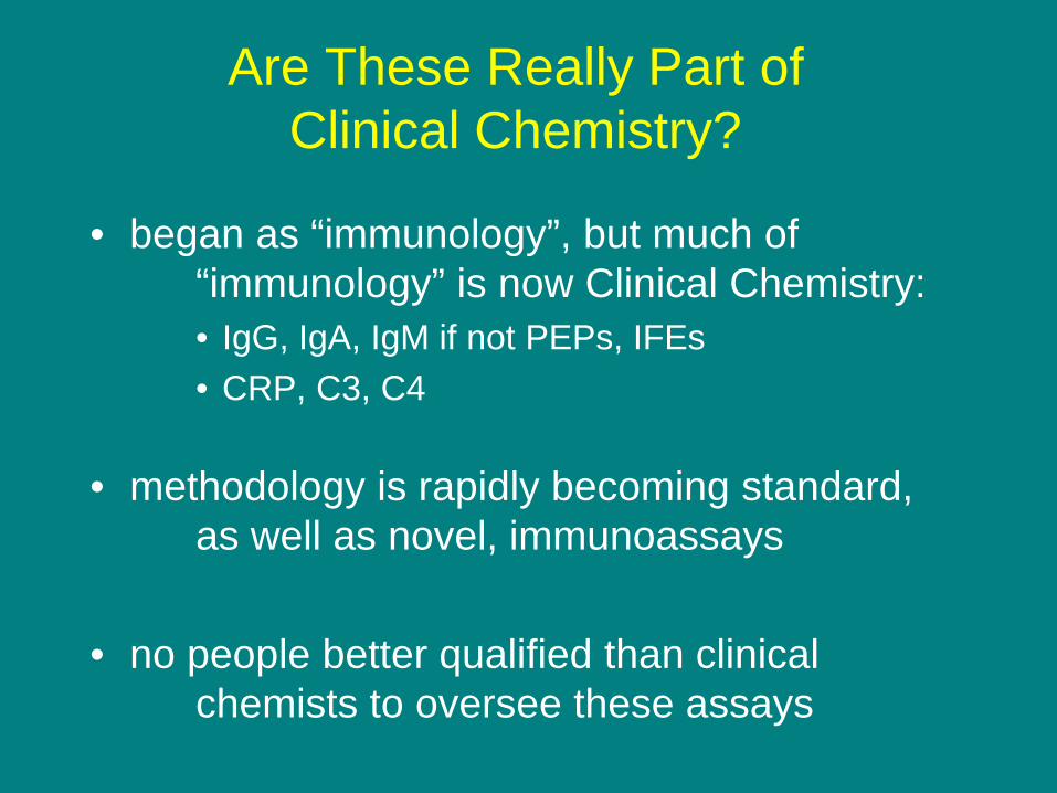
Are These Really Part of Clinical Chemistry?
• began as “immunology”, but much of “immunology” is now Clinical Chemistry:
• IgG, IgA, IgM if not PEPs, IFEs • CRP, C3, C4
• methodology is rapidly becoming standard,
as well as novel, immunoassays
• no people better qualified than clinical
chemists to oversee these assays

Autoantibodies
• antibodies directed against “self”
• give rise to a number of different diseases – Some relatively common (rheumatoid arthritis) – Some relatively serious (systemic lupus erythematosis)
• help to understand, diagnose, and monitor disease
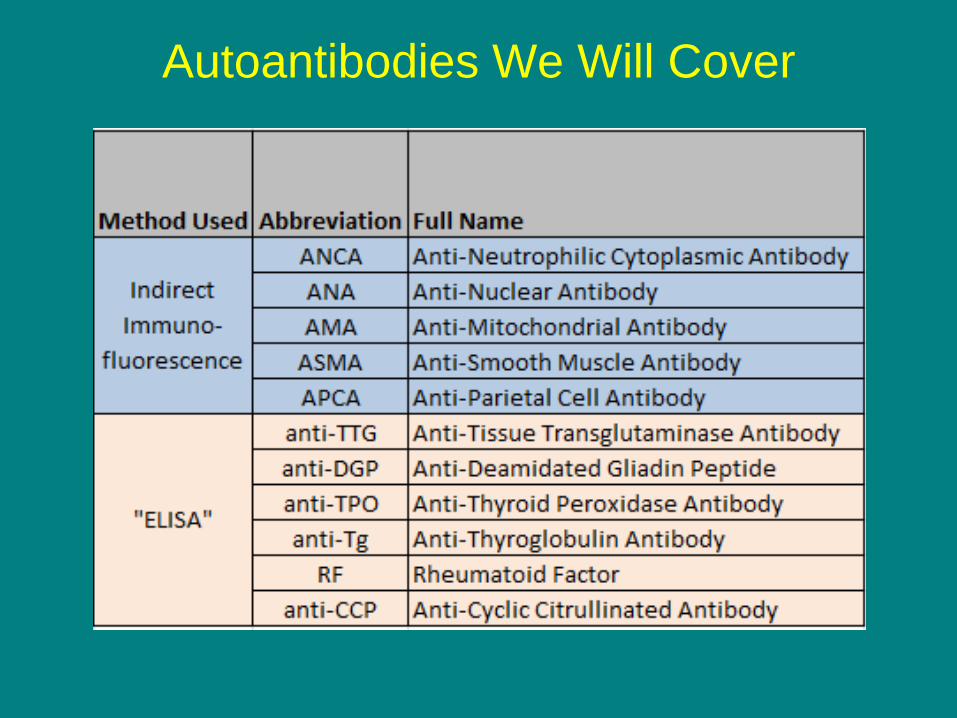
Autoantibodies We Will Cover
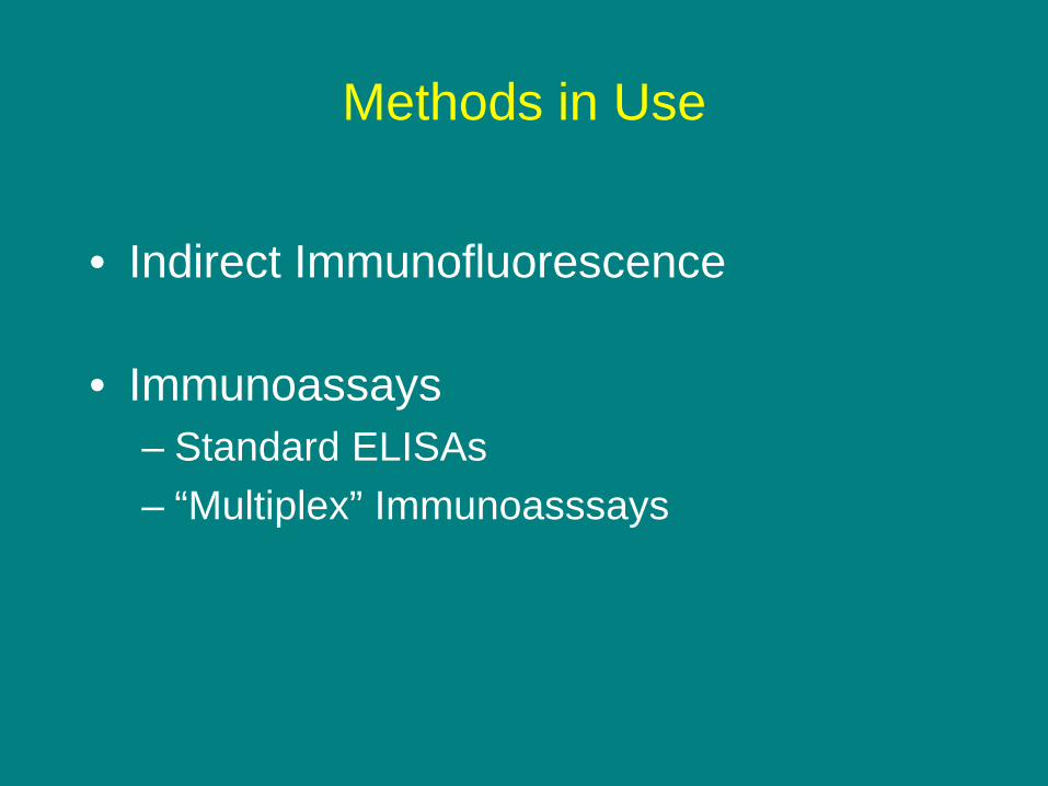
Methods in Use
• Indirect Immunofluorescence
• Immunoassays – Standard ELISAs – “Multiplex” Immunoasssays
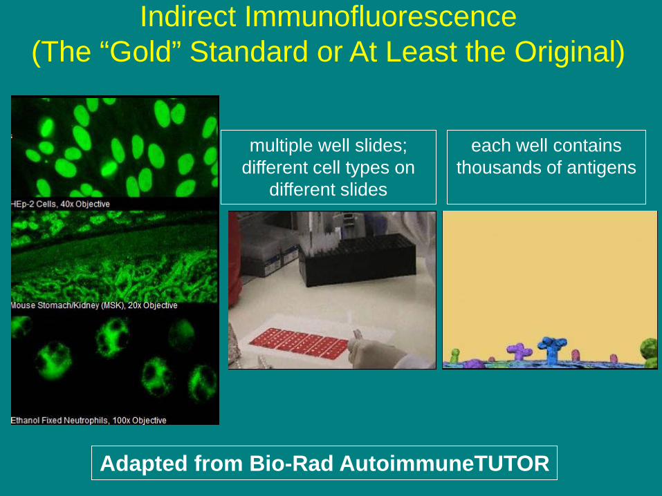
Adapted from Bio-Rad AutoimmuneTUTOR
each well contains thousands of antigens
multiple well slides; different cell types on
different slides
Indirect Immunofluorescence (The “Gold” Standard or At Least the Original)
Adapted from Bio-Rad AutoimmuneTUTOR
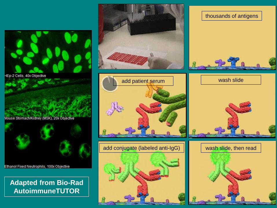
thousands of antigens
add patient serum wash slide
add conjugate (labeled anti-IgG) wash slide, then read
Immunofluorescence Technique Notes
• Using different substrates (cell types), one “captures” antibodies with different specificities

HEp2 Cells: Anti-Nuclear Antibodies (ANA) Mouse Stomach/Kidney Cells: Anti-Mitochondrial Antibodies (AMA) Anti-Smooth Muscle Antibodies (ASMA) Anti-Parietal Cell Antibodies (APCA) Neutrophils Anti-Neutrophil Cytoplasmic Antibodies (ANCA)
Substrate Specificity
Adapted from Bio-Rad AutoimmuneTUTOR

anti-mitochondrial antibody
anti-smooth muscle antibody
smooth muscle
parietal cells
kidney
Substrate Specificity
Adapted from Bio-Rad AutoimmuneTUTOR

Immunofluorescence Technique Notes
• Using different substrates (cell types), one “captures” antibodies with different specificities
• In some cases, the “pattern” of staining may be helpful, suggesting one disease over another

Patterns Can Sometimes Be Helpful (ANCA)
Perinuclear ANCA (P-ANCA) Pattern suggests vasculitis other than Wegener’s Granulomatosis
Cytoplasmic ANCA (C-ANCA) Pattern strongly suggests Wegener’s Granulomatosis
Adapted from Bio-Rad AutoimmuneTUTOR

Nucleolar
Speckled
Homogeneous
Patterns Can Sometimes Be Helpful (ANA)
Other Notes on “Patterns”
• Although patterns are frequently definitive, sometimes they can be ambiguous, so the interpretations become subjective
• In some cases, more than one autoantibody is present, which can definitely obscure the patterns
• If more specificity is needed, it is probably better to rely on immunoassays directed at specific antigens (later)

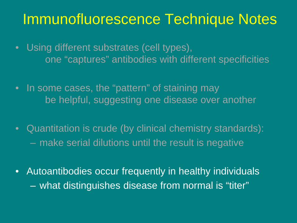
Immunofluorescence Technique Notes
• Using different substrates (cell types), one “captures” antibodies with different specificities
• In some cases, the “pattern” of staining may be helpful, suggesting one disease over another
• Quantitation is crude (by clinical chemistry standards): – make serial dilutions until the result is negative – i.e., 1:40, 1:80, 1:160, 1:320, 1:640, 1:1280, etc.
Immunofluorescence Technique Notes
• Using different substrates (cell types), one “captures” antibodies with different specificities
• In some cases, the “pattern” of staining may be helpful, suggesting one disease over another
• Quantitation is crude (by clinical chemistry standards): – make serial dilutions until the result is negative
• Autoantibodies occur frequently in healthy individuals
– what distinguishes disease from normal is “titer”

Distribution of Values from Patients with Disease
cut-off that detects 99% of patients

Add Distribution of Values from Healthy People
1% false negative 20% false positive
7.5% false negative 2.5% false positive

One Specific Example: ANA
• At 1:40 dilution, 20% of normals are POSITIVE • At 1:160 dilution, 5% of normals are POSITIVE
• In the absence of strong clinical suspicion of an
autoantibody-mediated disease (i.e., for “screening”), it’s probably best to use 1:160 as your lowest titer
• Most labs, including mine, start at 1:40, because the physicians “don’t want to miss any cases”
• My compromise: all “positive” ANAs are reported with the disclaimer that roughly 20% of healthy people will have positive ANAs with titers of 1:40 or 1:80

Methods in Use
• Indirect Immunofluorescence
• Immunoassays – Standard ELISAs – “Multiplex” Immunoasssays
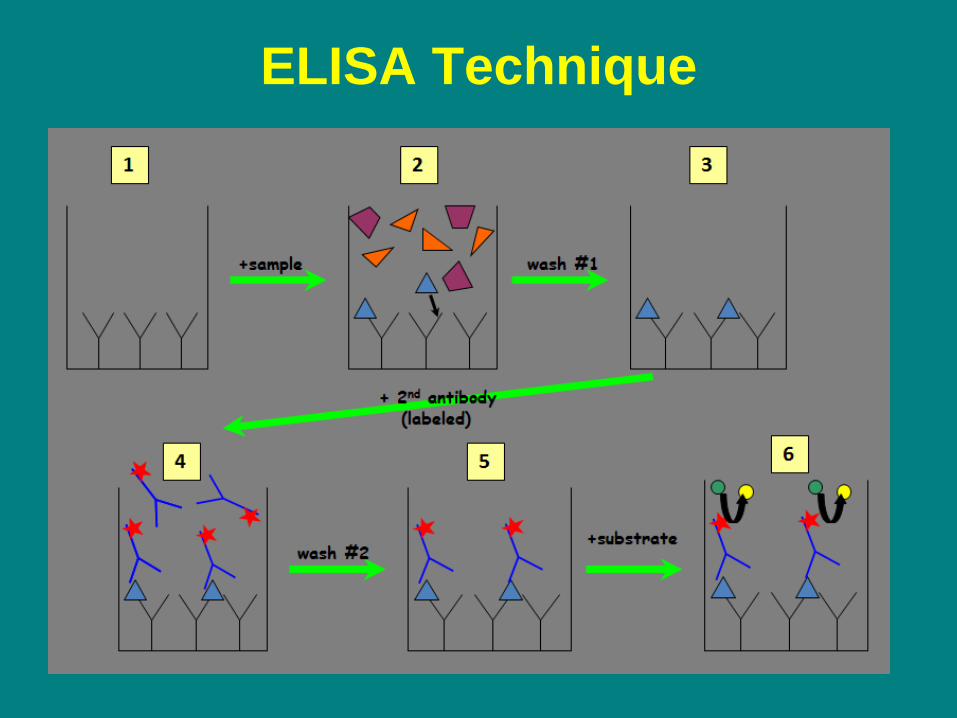
ELISA Technique

ELISAs For Distinguishing P-ANCA and C-ANCA
• we saw the bottom halves of these images earlier • using ELISAs for MPO and PR3 accomplish the same thing
P-ANCA pattern
MPO ELISA
PR3 ELISA
C-ANCA pattern
Adapted from Bio-Rad AutoimmuneTUTOR

So Why Not Just Do the ELISAs?(1)
• Some samples have positive indirect immunofluorescence but are negative for the specific antigens – e.g., positive ANCA but negative for MPO and PR3 – defined as “atypical ANCA” – clearly, biologic substrate has many more antigens – the clinical significance of such antibodies is unclear – but these “atypical ANCAs” are associated with vasculitis
• This phenomenon occurs with many, if not all, autoantibodies; it is not limited to ANCA
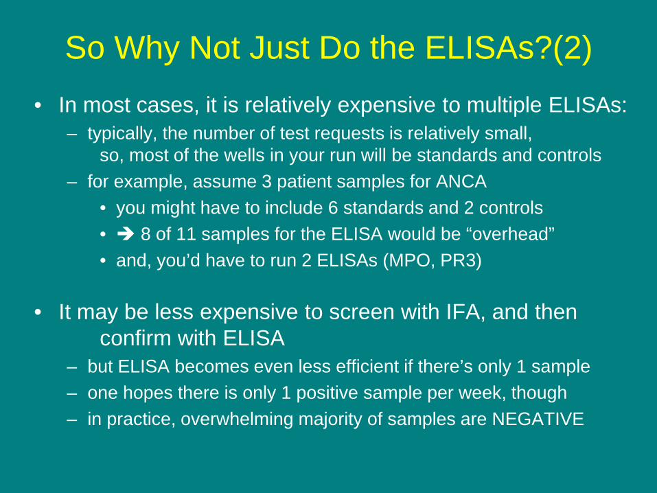
So Why Not Just Do the ELISAs?(2) • In most cases, it is relatively expensive to multiple ELISAs:
– typically, the number of test requests is relatively small, so, most of the wells in your run will be standards and controls
– for example, assume 3 patient samples for ANCA • you might have to include 6 standards and 2 controls • 8 of 11 samples for the ELISA would be “overhead” • and, you’d have to run 2 ELISAs (MPO, PR3)
• It may be less expensive to screen with IFA, and then confirm with ELISA – but ELISA becomes even less efficient if there’s only 1 sample – one hopes there is only 1 positive sample per week, though – in practice, overwhelming majority of samples are NEGATIVE
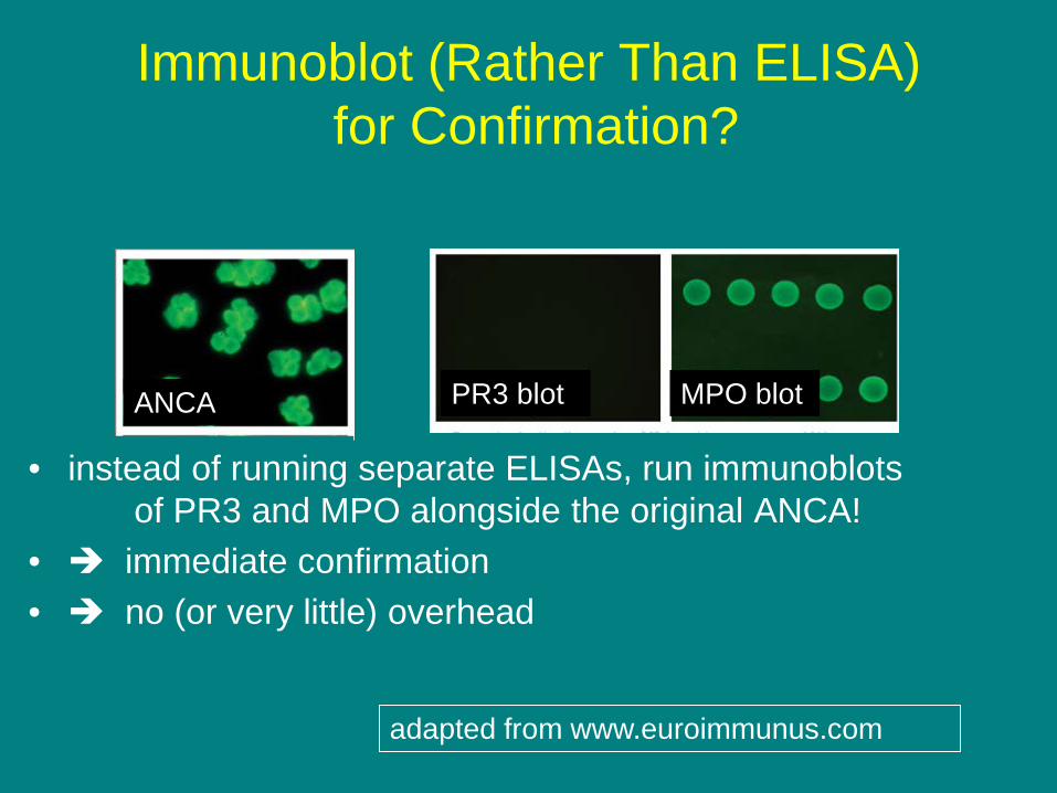
Immunoblot (Rather Than ELISA) for Confirmation?
• instead of running separate ELISAs, run immunoblots of PR3 and MPO alongside the original ANCA!
• immediate confirmation • no (or very little) overhead
PR3 blot ANCA MPO blot
adapted from www.euroimmunus.com

Methods in Use
• Indirect Immunofluorescence
• Immunoassays – Standard ELISAs – “Multiplex” Immunoasssays

“Multiplex” Immunoassays
• run many immunoassays simultaneously – for ANCA, run MPO + PR3 – for ANA, run dsDNA, SSA, SSB, Sm, RNP, SCL-70
• instead of standard microtiter plate ELISAs,
use novel technologies: – e.g., Luminex and euroimmun – if any assay is positive, call the overall test positive and
give the specificity (e.g., ANA positive, dsDNA positive)
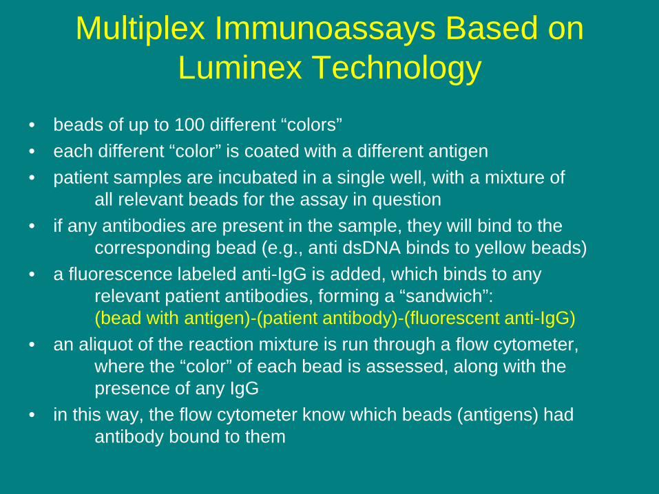
Multiplex Immunoassays Based on Luminex Technology
• beads of up to 100 different “colors” • each different “color” is coated with a different antigen • patient samples are incubated in a single well, with a mixture of
all relevant beads for the assay in question • if any antibodies are present in the sample, they will bind to the
corresponding bead (e.g., anti dsDNA binds to yellow beads) • a fluorescence labeled anti-IgG is added, which binds to any
relevant patient antibodies, forming a “sandwich”: (bead with antigen)-(patient antibody)-(fluorescent anti-IgG)
• an aliquot of the reaction mixture is run through a flow cytometer, where the “color” of each bead is assessed, along with the presence of any IgG
• in this way, the flow cytometer know which beads (antigens) had antibody bound to them
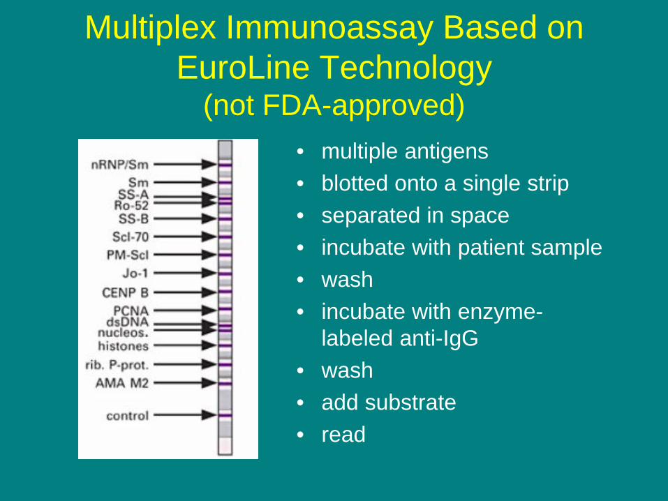
Multiplex Immunoassay Based on EuroLine Technology
(not FDA-approved) • multiple antigens • blotted onto a single strip • separated in space • incubate with patient sample • wash • incubate with enzyme-
labeled anti-IgG • wash • add substrate • read

Potential Downsides of Multiplex Format
• May be expensive: – Multiple standard ELISAs seem expensive – Novel format may be even more expensive,
except that the labor costs are minimal – assumes that every ELISA should be run on every
sample
• May miss “atypical” positives
– samples positive by indirect immunofluorescence but negative by ELISA
– they do exist, but their significance is debated

Celiac Disease
• relatively common (may be as high as 1 in 133 Caucasians) • when susceptible patients eat gluten,
(a protein found in wheat, rye, and barley), they make autoantibodies that attack the villi of the small intestine
• this results in malabsorption diarrhea, gas, bloating inability to absorb nutrients, which can lead to vitamin deficiencies, weight loss, etc.
• also associated with increased risk of several cancers
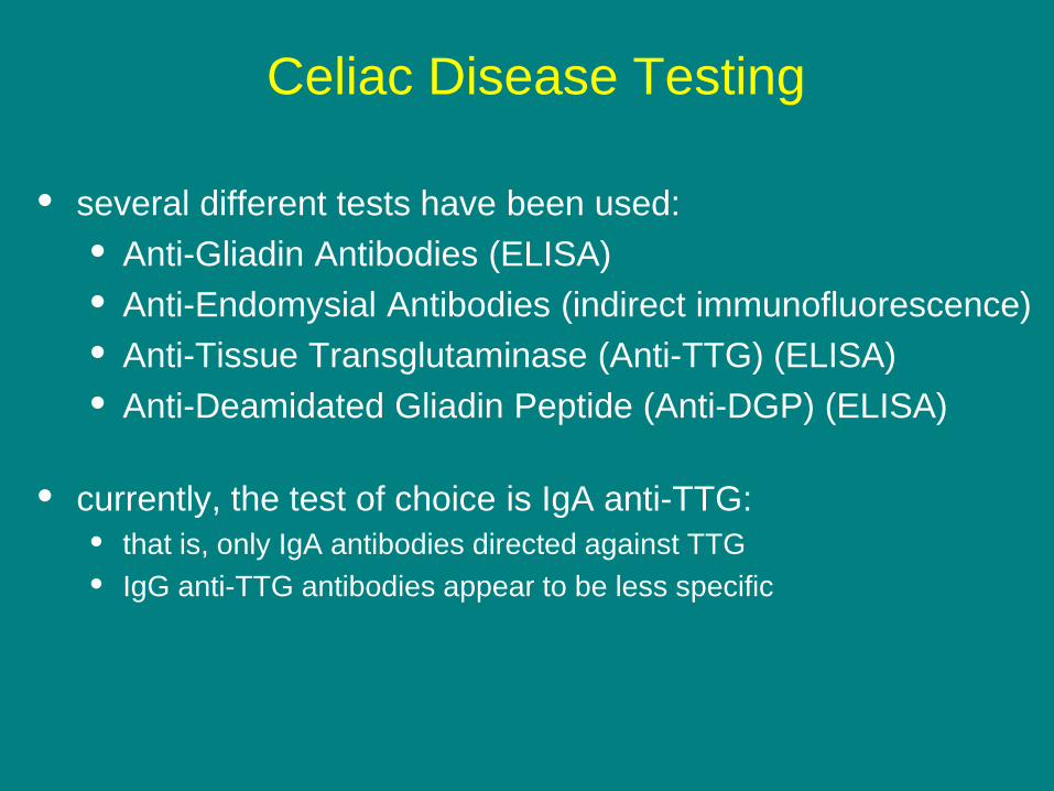
Celiac Disease Testing
• several different tests have been used: • Anti-Gliadin Antibodies (ELISA) • Anti-Endomysial Antibodies (indirect immunofluorescence) • Anti-Tissue Transglutaminase (Anti-TTG) (ELISA) • Anti-Deamidated Gliadin Peptide (Anti-DGP) (ELISA)
• currently, the test of choice is IgA anti-TTG:
• that is, only IgA antibodies directed against TTG • IgG anti-TTG antibodies appear to be less specific

Caveats: Celiac Disease Testing
• false negative IgA anti-TTG results: • patients with IgA deficiency (1 in 400) check IgA levels in patients with negative IgA anti-TTG for IgA deficient patients, retest with an alternative celiac autoantibody test e.g., anti-DGP, which includes IgG and IgA antibodies
• false negative results (all tests) in genuine celiac patients • patients on gluten-free diets if a patient has implemented a gluten-free diet on his own, his autoantibodies may well disappear

Thyroid Antibodies
• Many different tests are classified as “thyroid antibodies”
• The two you should definitely know about are: – Anti-Thyroid Peroxidase (anti-TPO) – Anti-Thyroglobulin (anti-Tg)
• Both are done by ELISA (or automated variants thereof)
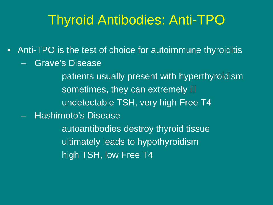
Thyroid Antibodies: Anti-TPO
• Anti-TPO is the test of choice for autoimmune thyroiditis – Grave’s Disease patients usually present with hyperthyroidism sometimes, they can extremely ill undetectable TSH, very high Free T4 – Hashimoto’s Disease autoantibodies destroy thyroid tissue ultimately leads to hypothyroidism high TSH, low Free T4

Thyroid Antibodies: Anti-Tg
• Anti-Tg should only be run to ensure accurate Tg
• Following thyroid gland removal for certain cancers (well differentiated papillary carcinoma), Tg serves as a tumor marker for recurrence
• Since the Tg assay uses anti-Tg in the reagent system, (e.g., capture antibody), the presence of anti-Tg in the patient’s serum will confound the assay
• In the presence of anti-Tg, it is difficult, if not impossible, to interpret the results of a Tg assay (and such results should probably not be reported)
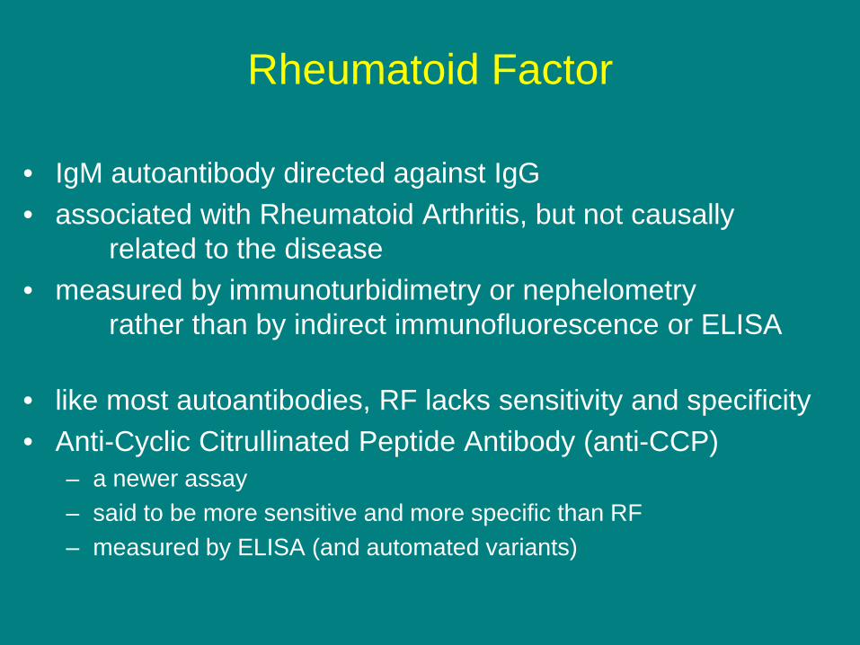
Rheumatoid Factor
• IgM autoantibody directed against IgG • associated with Rheumatoid Arthritis, but not causally
related to the disease • measured by immunoturbidimetry or nephelometry
rather than by indirect immunofluorescence or ELISA
• like most autoantibodies, RF lacks sensitivity and specificity • Anti-Cyclic Citrullinated Peptide Antibody (anti-CCP)
– a newer assay – said to be more sensitive and more specific than RF – measured by ELISA (and automated variants)
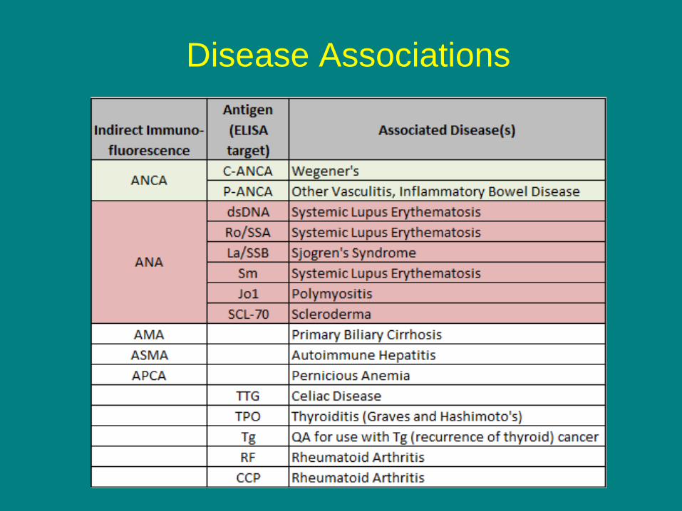
Disease Associations


Self-Assessment Question 1
Which of the following combinations of autoantibody and disease is incorrect? A) ANA: Systemic Lupus Erythematosis B) ANCA: Wegner’s Granulomatosis C) ANA: Primary Biliary Cirrhosis D) anti-TTG: Celiac Disease
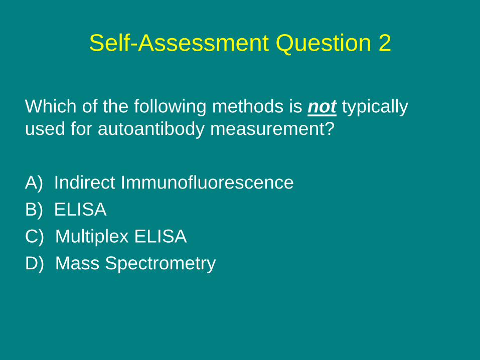
Self-Assessment Question 2
Which of the following methods is not typically used for autoantibody measurement? A) Indirect Immunofluorescence B) ELISA C) Multiplex ELISA D) Mass Spectrometry
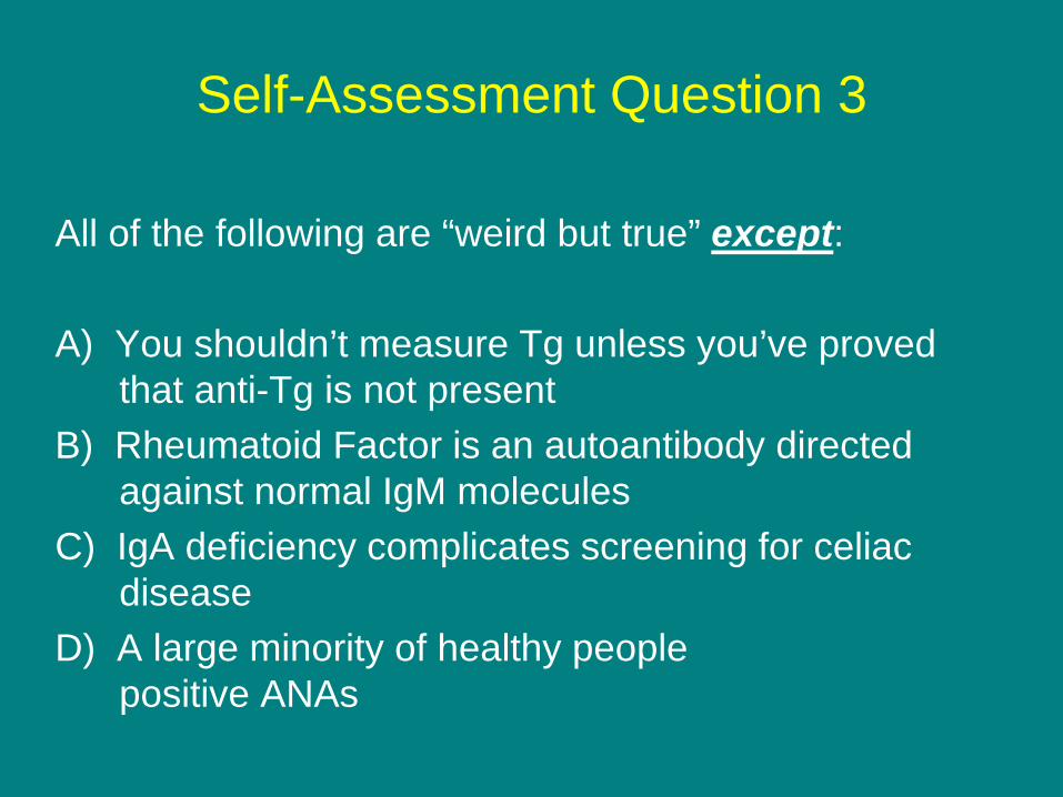
Self-Assessment Question 3
All of the following are “weird but true” except: A) You shouldn’t measure Tg unless you’ve proved that anti-Tg is not present B) Rheumatoid Factor is an autoantibody directed against normal IgM molecules C) IgA deficiency complicates screening for celiac disease D) A large minority of healthy people positive ANAs

Answers
1 (C) ANA: Primary Biliary Cirrhosis 2 (D) Mass Spectrometry 3 (B) Rheumatoid Factor is an autoantibody directed against normal IgM molecules