automatic detection of coronavirus disease (covid-19
TRANSCRIPT

Automatic Detection of Coronavirus Disease (COVID-19) Using X-ray Images and Deep
Convolutional Neural Networks
Ali Narin1, Ceren Kaya2, *, Ziynet Pamuk2
1Department of Electrical and Electronics Engineering, Zonguldak Bulent Ecevit University,
67100, Zonguldak, Turkey. [email protected]
2 Department of Biomedical Engineering, Zonguldak Bulent Ecevit University,
67100, Zonguldak, Turkey. [email protected], [email protected]
*Corresponding author: [email protected], [email protected]
Abstract
The 2019 novel coronavirus disease (COVID-19), with a starting point in China, has spread
rapidly among people living in other countries, and is approaching approximately 34,986,502
cases worldwide according to the statistics of European Centre for Disease Prevention and
Control. There are a limited number of COVID-19 test kits available in hospitals due to the
increasing cases daily. Therefore, it is necessary to implement an automatic detection system
as a quick alternative diagnosis option to prevent COVID-19 spreading among people. In this
study, five pre-trained convolutional neural network based models (ResNet50, ResNet101,
ResNet152, InceptionV3 and Inception-ResNetV2) have been proposed for the detection of
coronavirus pneumonia infected patient using chest X-ray radiographs. We have implemented
three different binary classifications with four classes (COVID-19, normal (healthy), viral
pneumonia and bacterial pneumonia) by using 5-fold cross validation. Considering the
performance results obtained, it has seen that the pre-trained ResNet50 model provides the
highest classification performance (96.1% accuracy for Dataset-1, 99.5% accuracy for Dataset-
2 and 99.7% accuracy for Dataset-3) among other four used models.
Keywords: Coronavirus; Bacterial Pneumonia; Viral Pneumonia; Chest X-ray Radiographs;
Convolutional Neural Network; Deep Transfer Learning

1. Introduction
The coronavirus disease (COVID-19) pandemic emerged in Wuhan, China in December
2019 and became a serious public health problem worldwide [1,2]. Until now, no specific drug
or vaccine has been found against COVID-19 [2]. The virus that causes COVID-19 epidemic
disease is called severe acute respiratory syndrome coronavirus-2 (SARS-CoV-2) [3].
Coronaviruses (CoV) is a large family of viruses that cause diseases such as Middle East
Respiratory Syndrome (MERS-CoV) and Severe Acute Respiratory Syndrome (SARS-CoV).
COVID-19 is a new species discovered in 2019 and has not been previously identified in
humans [4]. COVID-19 causes lighter symptoms in about 99% of cases, according to early data,
while the rest is severe or critical [5]. As of 4th October 2020, the total number of worldwide
cases of Coronavirus is 35,248,330. Of these, 1,039,541 (4%) people were deaths and
26,225,235 (96%) were recovered. The number of active patients is 7,983,554. Of these,
7,917,287 (99%) had mild disease while 66,267 (1%) had more severe disease [6]. Nowadays
the world is struggling with the COVID-19 epidemic. Deaths from pneumonia developing due
to the SARS-CoV-2 virus are increasing day by day.
Chest radiography (X-ray) is one of the most important methods used for the diagnosis of
pneumonia worldwide [7]. Chest X-ray is a fast, cheap [8] and common clinical method [9-11].
The chest X-ray gives the patient a lower radiation dose compared to computed tomography
(CT) and magnetic resonance imaging (MRI) [11]. However, making the correct diagnosis from
X-ray images requires expert knowledge and experience [7]. It is much more difficult to
diagnose using a chest X-ray than other imaging modalities such as CT or MRI [8].
By looking at the chest X-ray, COVID-19 can only be diagnosed by specialist physicians.
The number of specialists who can make this diagnosis is less than the number of normal
doctors. Even in normal times, the number of doctors per person is insufficient in countries
around the world. According to data from 2017, Greece ranks first with 607 doctors per 100,000
people. In other countries, this number is much lower [12].
In case of disasters such as COVID-19 pandemic, demanding health services at the same
time, collapse of the health system is inevitable due to the insufficient number of hospital beds
and health personnel. Also, COVID-19 is a highly contagious disease, and doctors, nurses, and
caregivers are most at risk. Early diagnosis of pneumonia has a vital importance both in terms

of slowing the speed of the spread of the epidemic by quarantining the patient and in the
recovery process of the patient [13].
Doctors can diagnose pneumonia from the chest X-ray more quickly and accurately thanks
to computer-aided diagnosis (CAD) [8]. Use of artificial intelligence methods are increasing
due to its ability to cope with enormous datasets exceeding human potential in the field of
medical services [14]. Integrating CAD methods into radiologist diagnostic systems greatly
reduces the workload of doctors and increases reliability and quantitative analysis [11]. CAD
systems based on deep learning and medical imaging are becoming more and more research
fields [14,15].
2. Related Works
Studies diagnosed with COVID-19 using chest X-rays have binary or multiple
classifications. Some studies use raw data while others have feature extraction process. The
number of data used in studies also varies. Among the studies, the most preferred method is
convolutional neural network (CNN).
Apostolopoulos and Bessiana used a common pneumonia, COVID-19-induced
pneumonia, and an evolutionary neural network for healthy differentiation on automatic
detection of COVID-19. In particular, the procedure called transfer learning has been adopted.
With transfer learning, the detection of various abnormalities in small medical image datasets
is an achievable goal, often with remarkable results [16]. Based on chest X-ray images, Zhang
et al. aimed to develop a deep learning-based model that can detect COVID-19 with high
sensitivity, providing fast and reliable scanning [17]. Singh et al. classified the chest computed
tomography (CT) images from infected people with and without COVID-19 using multi-
objective differential evolution (MODE) based CNN [18]. In the study of Chen et al, they
proposed Residual Attention U-Net for automated multi class segmentation technique to
prepare the ground for the quantitative diagnosis of lung infection on COVID-19 related
pneumonia using CT images [19]. Adhikari's study suggested a network called “Auto
Diagnostic Medical Analysis” trying to find infectious areas to help the doctor better identify
the diseased part, if any. Both X-ray and CT images were used in the study. It has been
recommended DenseNet network to remove and mark infected areas of the lung [20]. In the
study by Alqudah et al., two different methods were used to diagnose COVID-19 using chest
X-ray images. The first one used AOCTNet, MobileNet and ShuffleNet CNNs. Secondly, the
features of their images have been removed and they have been classified using softmax

classifier, K nearest neighbor (kNN), Support vector machine (SVM) and Random forest (RF)
algorithms [21]. Khan et al. classified the chest X-ray images from normal, bacterial and viral
pneumonia cases using the Xception architecture to detect COVID-19 infection [22]. Ghoshal
and Tucker used the dropweights based Bayesian CNN model using chest X-ray images for the
diagnosis of COVID-19 [23]. Hemdan et al. used VGG19 and DenseNet models to diagnose
COVID-19 from X-ray images [24]. Uçar and Korkmaz worked on X-ray images for COVID-
19 diagnosis and supported the SqueezeNet model with Bayesian optimization [25]. In the study
conducted by Apostopolus et al., they performed automatic detection from X-ray images using
CNNs with transfer learning [26]. Sahinbas and Catak used X-ray images for the diagnosis of
COVID-19 and worked on VGG16, VGG19, ResNet, DenseNet and InceptionV3 models [27].
Medhi et al. used X-ray images as feature extraction and segmentation in their study, then
COVID-19 was positively and normally classified using CNN [28]. Barstugan et al. classified
X-ray images for the diagnosis of COVID-19 using five different feature extraction methods
that are Grey Level Cooccurrence Matrix (GLCM), Local Directional Patterns (LDP), Grey
Level Run Length Matrix (GLRLM), Grey Level Size Zone Matrix (GLSZM), and Discrete
Wavelet Transform (DWT). The obtained features were classified by SVM. During the
classification process, 2-fold, 5-fold, and 10-fold cross-validation methods were used [29].
Punn and Agarwal worked on X-ray images and used ResNet, InceptionV3, InceptionResNet
models to diagnose COVID-19 [30]. Afshar et al. developed deep neural network (DNN) based
diagnostic solutions and offered an alternative modeling framework based on Capsule
Networks that can process on small data sets [31].
In our previous study in March 2020, we used ResNet50, InceptionV3 and Inception-
ResNetV2 models for the diagnosis of COVID-19 using chest X-ray images. However, since
there was not enough data on COVID-19, we were only able to train through 50 normal and 50
COVID-19 positive cases [32].
In this study, we have proposed an automatic prediction of COVID-19 using a deep
convolution neural network based pre-trained transfer models and chest X-ray images. For this
purpose, we have used ResNet50, ResNet101, ResNet152, InceptionV3 and Inception-
ResNetV2 pre-trained models to obtain higher prediction accuracies for three different binary
datasets including X-ray images of normal (healthy), COVID-19, bacterial and viral pneumonia
patients.

The novelty and originality of proposed study is summarized as follows:
i) The proposed models have end-to-end structure without manual feature extraction, selection
and classification.
ii) The performances of the COVID-19 data across normal, viral pneumonia and bacterial
pneumonia classes were significantly higher.
iii) It has been studied with more data than many studies in the literature.
iv) It has been studied and compared with 5 different CNN models.
v) A high-accuracy decision support system has been proposed to radiologists for the automatic
diagnosis and detection of patients with suspected COVID-19 and follow-up.
The flow of the manuscript is organized as follows: Dataset is expressed in detail in Section
3.1. Deep transfer learning architecture, pre-trained models and experimental setup parameters
are described in Section 3.2 and 3.3, respectively. Performance metrics are given in detail in
Section 3.4. Obtained experimental results from proposed models and discussion are presented
in Section 4 and 5, respectively. Finally, in Section 6, the conclusion and future works are
summarized.
3. Materials and Methods
3.1 Dataset
In this study, chest X-ray images of 341 COVID-19 patients have been obtained from
the open source GitHub repository shared by Dr. Joseph Cohen et al. [33]. This repository is
consisting chest X-ray / computed tomography (CT) images of mainly patients with acute
respiratory distress syndrome (ARDS), COVID-19, Middle East respiratory syndrome
(MERS), pneumonia, severe acute respiratory syndrome (SARS). 2800 normal (healthy) chest
X-ray images were selected from “ChestX-ray8” database [34]. In addition, 2772 bacterial and
1493 viral pneumonia chest X-ray images were used from Kaggle repository called “Chest X-
Ray Images (Pneumonia)” [35].
Our experiments have been based on three binary created datasets (Dataset-1, Dataset-
2 and Dataset-3) with chest X-ray images. Distribution of images per class in created datasets
are given Table 1.

Table 1. Number of images per class for each dataset.
Bacterial
Pneumonia
COVID-19 Normal Viral
Pneumonia
Dataset-1 - 341 2800 -
Dataset-2 - 341 - 1493
Dataset-3 2772 341 - -
All images were resized to 224x224 pixel size in the datasets. In Figure 1, representative
chest X-ray images of normal (healthy), COVID-19, bacterial and viral pneumonia patients are
given, respectively.
Classes
Datasets

Norm
al
CO
VID
-19
Bact
eria
l P
neu
mon
ia
Vir
al
Pn
eum
on
ia
Figure 1. Representative chest X-ray images of normal (healthy) (first row), COVID-19
(second row), bacterial (third row) and viral pneumonia (fourth row) patients.
3.2 Architecture of Deep Transfer Learning
Deep learning is a sub-branch of the machine learning field, inspired by the structure of
the brain. Deep learning techniques used in recent years continue to show an impressive
performance in the field of medical image processing, as in many fields. By applying deep
learning techniques to medical data, it is tried to draw meaningful results from medical data.

Deep learning models have been used successfully in many areas such as classification,
segmentation and lesion detection of medical data. Analysis of image and signal data obtained
with medical imaging techniques such as Magnetic Resonance Imaging (MRI), Computed
Tomography (CT) and X-ray with the help of deep learning models. As a result of these
analyzes, detection and diagnosis of diseases such as diabetes mellitus, brain tumor, skin cancer
and breast cancer are provided with convenience [36-41].
A convolutional neural network (CNN) is a class of deep neural networks used in image
recognition problems [42]. Coming to how CNN works, the images given as input must be
recognized by computers and converted into a format that can be processed. For this reason,
images are first converted to matrix format. The system determines which image belongs to which
label based on the differences in images and therefore in matrices. It learns the effects of these
differences on the label during the training phase and then makes predictions for new images
using them. CNN consists of three different layers that are a convolutional layer, pooling layer,
and fully connected layer to perform these operations effectively. The feature extraction process
takes place in both convolutional and pooling layers. On the other hand, the classification process
occurs in fully connected layer. These layers are examined sequentially in the following.
3.2.1 Convolutional Layer
Convolutional layer is the base layer of CNN. It is responsible for determining the features of
the pattern. In this layer, the input image is passed through a filter. The values resulting from
filtering consist of the feature map. This layer applies some kernels that slide through the pattern
to extract low- and high-level features in the pattern [43]. The kernel is a 3x3 or 5x5 shaped matrix
to be transformed with the input pattern matrix. Stride parameter is the number of steps tuned for
shifting over input matrix. The output of convolutional layer can be given as:
1 1
1
*N
l l l l
j j a j
a
x f w y b− −
=
= +
(1)
where xjl is the j-th feature map in layer l, wj
l-1 indicates j-th kernels in layer l-1, yal-1 represents
the a-th feature map in layer l-1, bjl indicates the bias of the j-th feature map in layer l, N is number
of total features in layer l-1, and (*) represents vector convolution process.
3.2.2 Pooling Layer
The second layer after the convolutional layer is the pooling layer. Pooling layer is usually
applied to the created feature maps for reducing the number of feature maps and network

parameters by applying corresponding mathematical computation. In this study, we used max-
pooling and global average pooling. The max-pooling process selects only the maximum value
by using the matrix size specified in each feature map, resulting in reduced output neurons. There
is also a global average pooling layer that is only used before the fully connected layer, reducing
data to a single dimension. It is connected to the fully connected layer after global average pooling
layer. The other intermediate layer used is the dropout layer. The main purpose of this layer is to
prevent network overfitting and divergence [44].
3.2.3 Fully Connected Layer
Fully connected layer is the last and most important layer of CNN. This layer functions like a
multi-layer perceptron. Rectified Linear Unit (ReLU) activation function is commonly used on
fully connected layer, while Softmax activation function is used to predict output images in the
last layer of fully connected layer. Mathematical computation of these two activation functions
are as follow:
𝑅𝑒𝐿𝑈 (𝑥) = {0, 𝑥 < 0𝑥, 𝑥 ≥ 0
(2)
1
max( )i
y
x
i mx
y
eSoft x
e=
=
(3)
where xi and m represent input data and the number of classes, respectively.
Neurons in a fully connected layer have full connections to all activation functions in previous
layer.
3.2.4. Pre-Trained Models
In the analysis of medical data, one of the biggest difficulties faced by researchers is the
limited number of available datasets. Deep learning models often need a lot of data. Labeling
this data by experts is both costly and time consuming. The biggest advantage of using transfer
learning method is that it allows the training of data with fewer datasets and requires less
calculation costs. With the transfer learning method, which is widely used in the field of deep
learning, the information gained by the pre-trained model on a large dataset is transferred to the
model to be trained.

In this study, we built deep CNN based ResNet50, ResNet101, ResNet152, InceptionV3
and Inception-ResNetV2 models for the classification of COVID-19 Chest X-ray images to
three different binary classes (Binary Class-1 = COVID-19 and normal (healthy), Binary Class-
2 = COVID-19 and viral pneumonia, Binary Class-3 = COVID-19 and bacterial pneumonia).
In addition, we applied transfer learning technique that was realized by using ImageNet data to
overcome the insufficient data and training time. The schematic representation of conventional
CNN including pre-trained ResNet50, ResNet101, ResNet152, InceptionV3 and Inception
ResNetV2 models for the prediction of normal (healthy), COVID-19, bacterial and viral
pneumonia patients were depicted in Figure 2. It is also available publicly for open access at
https://github.com/drcerenkaya/COVID-19-DetectionV2.
Figure 2. Schematic representation of pre-trained models for the prediction of normal (healthy),
COVID-19, bacterial and viral pneumonia patients.
ResNet50
Residual neural network (ResNet) model is an improved version of convolutional neural
network (CNN). ResNet adds shortcuts between layers to solve a problem. Thanks to this, it
prevents the distortion that occurs as the network gets deeper and more complex. In addition,
bottleneck blocks are used to make training faster in the ResNet model [45]. ResNet50 is a 50-
layer network trained on the ImageNet dataset. ImageNet is an image database with more than
14 million images belonging to more than 20 thousand categories created for image recognition
competitions [46].
InceptionV3
InceptionV3 is a kind of convolutional neural network model. It consists of numerous
convolution and maximum pooling steps. In the last stage, it contains a fully connected neural
network [47]. As with the ResNet50 model, the network is trained with ImageNet dataset.

Inception-ResNetV2
The model consists of a deep convolutional network using the Inception-ResNetV2
architecture that was trained on the ImageNet-2012 dataset. The input to the model is a 299×299
image, and the output is a list of estimated class probabilities [48].
ResNet101 & ResNet152
ResNet101 and ResNet152 consist of 101 and 152 layers respectively due to stacked
ResNet building blocks. You can load a pretrained version of the network trained on more than
a million images from the ImageNet database [46]. As a result, the network has learned rich
feature representations for a wide range of images. The network has an image input size of
224x224.
3.3 Experimental Setup
Python programming language was used to train the proposed deep transfer learning
models. All experiments were performed on Google Colaboratory (Colab) Linux server with
the Ubuntu 16.04 operating system using the online cloud service with Central Processing Unit
(CPU), Tesla K80 Graphics Processing Unit (GPU) or Tensor Processing Unit (TPU) hardware
for free. CNN models (ResNet50, ResNet101, ResNet152, InceptionV3 and Inception-
ResNetV2) were pre-trained with random initialization weights by optimizing the cross-entropy
function with adaptive moment estimation (ADAM) optimizer (β1 = 0.9 and β2 = 0.999). The
batch size, learning rate and number of epochs were experimentally set to 3, 1e-5 and 30,
respectively for all experiments. The dataset used was randomly split into two independent
datasets with 80% and 20% for training and testing respectively. As cross validation method,
k-fold was chosen and results were obtained according to 5 different k values (k=1-5) as shown
in Figure 3.

Figure 3. Visual display of testing and training datasets for 5-fold cross validation.
3.4 Performance Metrics
5 criteria were used for the performances of deep transfer learning models. These are:
Accuracy = (TN + TP) / (TN + TP + FN + FP) (4)
Recall = TP / (TP + FN) (5)
Specificity = TN / (TN + FP) (6)
Precision = TP / (TP + FP) (7)
F1-Score = 2x((PrecisionxRecall)/(Precision+Recall)) (8)
TP, FP, TN and FN given in Equation (4) – (8) represent the number of True Positive,
False Positive, True Negative and False Negative, respectively. For Dataset-1; given a test
dataset and model, TP is the proportion of positive (COVID-19) that are correctly labeled as
COVID-19 by the model; FP is the proportion of negative (normal) that are mislabeled as
positive (COVID-19); TN is the proportion of negative (normal) that are correctly labeled as
normal and FN is the proportion of positive (COVID-19) that are mislabeled as negative
(normal) by the model.
4. Experimental Results
In this paper, we performed 3-different binary classifications with 4 different classes
(COVID-19, normal, viral pneumonia and bacterial pneumonia). 5-fold cross validation method

has been used in order to get a robust result in this study performed with 5-different pre-trained
models that are InceptionV3, ResNet50, ResNet101, ResNet152 and Inception-ResNetV2.
While 80% of the data is reserved for training, the remaining 20% is reserved for testing. All
this process continued until each 20% part was tested.
Firstly, the accuracy and loss values in the training process obtained for the models
applied to Dataset-1 that includes Binary Class-1 (COVID-19 / Normal classes) are given in
Figure 4 and Figure 5. It is clear that the performance of the ResNet50 model is better than the
other models. It can be said that the ResNet50 model reaches lower values among the loss
values of other models. Detection performance on test data is shown in Figure 6. While a lot of
oscillation is observed in some models, some models are more stable. The ResNet50 model
appears to have less oscillation after the 15th epoch. Comprehensive performance values for
each fold value of each model are given in Table 2. As seen from Table 2 that the detection of
the ResNet50 model in the COVID-19 class is significantly higher than the other models.
ResNet50 and ResNet101 have the highest overall performance with 96.1%. It is obvious that
the excess of normal data results in higher performance in all models.
Figure 4. Binary Class-1: Comparison of training accuracy of 5 different models for fold-4.

Figure 5. Binary Class-1: Comparison of training loss values of 5 different models for
fold-4.
Figure 6. Binary Class-1: Comparison of testing accuracy of 5 different models for fold-4.
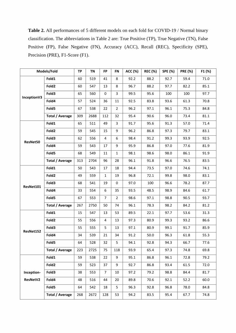
Table 2. All performances of 5 different models on each fold for COVID-19 / Normal binary
classification. The abbreviations in Table 2 are: True Positive (TP), True Negative (TN), False
Positive (FP), False Negative (FN), Accuracy (ACC), Recall (REC), Specificity (SPE),
Precision (PRE), F1-Score (F1).
Models/Fold TP TN FP FN ACC (%) REC (%) SPE (%) PRE (%) F1 (%)
InceptionV3
Fold1 60 519 41 8 92.2 88.2 92.7 59.4 71.0
Fold2 60 547 13 8 96.7 88.2 97.7 82.2 85.1
Fold3 65 560 0 3 99.5 95.6 100 100 97.7
Fold4 57 524 36 11 92.5 83.8 93.6 61.3 70.8
Fold5 67 538 22 2 96.2 97.1 96.1 75.3 84.8
Total / Average 309 2688 112 32 95.4 90.6 96.0 73.4 81.1
ResNet50
Fold1 65 511 49 3 91.7 95.6 91.3 57.0 71.4
Fold2 59 545 15 9 96.2 86.8 97.3 79.7 83.1
Fold3 62 556 4 6 98.4 91.2 99.3 93.9 92.5
Fold4 59 543 17 9 95.9 86.8 97.0 77.6 81.9
Fold5 68 549 11 1 98.1 98.6 98.0 86.1 91.9
Total / Average 313 2704 96 28 96.1 91.8 96.6 76.5 83.5
ResNet101
Fold1 50 543 17 18 94.4 73.5 97.0 74.6 74.1
Fold2 49 559 1 19 96.8 72.1 99.8 98.0 83.1
Fold3 68 541 19 0 97.0 100 96.6 78.2 87.7
Fold4 33 554 6 35 93.5 48.5 98.9 84.6 61.7
Fold5 67 553 7 2 98.6 97.1 98.8 90.5 93.7
Total / Average 267 2750 50 74 96.1 78.3 98.2 84.2 81.2
ResNet152
Fold1 15 547 13 53 89.5 22.1 97.7 53.6 31.3
Fold2 55 556 4 13 97.3 80.9 99.3 93.2 86.6
Fold3 55 555 5 13 97.1 80.9 99.1 91.7 85.9
Fold4 34 539 21 34 91.2 50.0 96.3 61.8 55.3
Fold5 64 528 32 5 94.1 92.8 94.3 66.7 77.6
Total / Average 223 2725 75 118 93.9 65.4 97.3 74.8 69.8
Inception-
ResNetV2
Fold1 59 538 22 9 95.1 86.8 96.1 72.8 79.2
Fold2 59 523 37 9 92.7 86.8 93.4 61.5 72.0
Fold3 38 553 7 10 97.2 79.2 98.8 84.4 81.7
Fold4 48 516 44 20 89.8 70.6 92.1 52.2 60.0
Fold5 64 542 18 5 96.3 92.8 96.8 78.0 84.8
Total / Average 268 2672 128 53 94.2 83.5 95.4 67.7 74.8

Secondly, when the results obtained for the data in Binary Class-2 (COVID-19 / Viral
pneumonia classes) are evaluated, the training performances of the models given in Figure 7
and Figure 8 are quite high. It can be said that the accuracy values and loss values of the
ResNet50 and ResNet101 models perform better than the other models. Performance values
obtained through test data are shown in Figure 9. Here, the models' results on the test data are
generally more stable. There is no oscillation except when there is excessive oscillation only in
the first 3 epochs of the ResNet50 model. Detailed performances of the models are given in
Table 3. It is clearly seen that quite high values are reached for each fold value. While 99.4%
was reached in the detection of COVID-19, it is seen that 99.5% was reached in the detection
of viral pneumonia.
Figure 7. Binary Class-2: Comparison of training accuracy of 5 different models for fold-4.
Figure 8. Binary Class-2: Comparison of training loss values of 5 different models for fold-4.

Figure 9. Binary Class-2: Comparison of testing accuracy of 5 different models for fold-4.

Table 3. All performances of 5 different models on each fold for COVID-19 / Viral pneumonia
binary classification.
Models/Fold TP TN FP FN ACC (%) REC (%) SPE (%) PRE (%) F1 (%)
InceptionV3
Fold1 68 292 6 0 98.4 100 98.0 91.9 95.8
Fold2 68 292 6 0 98.4 100 98.0 91.9 95.8
Fold3 67 295 4 1 98.6 98.5 98.7 94.4 96.4
Fold4 68 290 9 0 97.5 100 97.0 88.3 93.8
Fold5 69 299 0 0 100 100 100 100 100
Total/Average 340 1468 25 1 98.6 99.7 98.3 93.2 96.3
ResNet50
Fold1 68 297 1 0 99.7 100 99.7 98.6 99.3
Fold2 68 293 5 0 98.6 100 98.3 93.2 96.5
Fold3 68 298 1 0 99.7 100 99.7 98.6 99.3
Fold4 66 299 0 2 99.5 97.1 100 100 98.5
Fold5 69 299 0 0 100 100 100 100 100
Total/Average 339 1486 7 2 99.5 99.4 99.5 98.0 98.7
ResNet101
Fold1 61 294 4 7 97.0 89.7 98.7 93.8 91.7
Fold2 62 293 5 6 97.0 91.2 98.3 92.5 91.9
Fold3 68 295 4 0 98.9 100 98.7 94.4 97.1
Fold4 65 298 1 3 98.9 95.6 99.7 98.5 97.0
Fold5 45 299 0 24 93.5 65.2 100 100 78.9
Total/Average 301 1479 14 40 97.1 88.3 99.1 95.6 91.8
ResNet152
Fold1 63 291 7 5 96.7 92.6 97.7 90.0 91.3
Fold2 67 293 5 1 98.4 98.5 98.3 93.1 95.7
Fold3 66 298 1 2 99.2 97.1 99.7 98.5 97.8
Fold4 56 299 0 13 96.5 81.2 100 100 89.6
Fold5 58 298 1 10 97.0 85.3 99.7 98.3 91.3
Total/Average 310 1479 14 31 97.5 90.9 99.1 95.7 93.2
Inception-
ResNetV2
Fold1 68 283 15 0 95.9 100 95.0 81.9 90.1
Fold2 68 267 31 0 91.5 100 89.6 68.7 81.4
Fold3 68 278 21 0 94.3 100 93.0 76.4 86.6
Fold4 41 296 3 27 91.8 60.3 99.0 93.2 73.2
Fold5 69 293 6 0 98.4 100 98.0 92.0 95.8
Total/Average 314 1417 76 27 94.4 92.1 94.9 80.5 85.9

In the last study, the detection success of Binary Class-3 (COVID-19 / Bacterial pneumonia
classes) was investigated. The performances of 5 different models on both training and test data
are given in Figure 10, Figure 11 and Figure 12. As in other studies, it is clearly seen that the
ResNet50 model exhibits higher training performance. InceptionV3 model is seen to exhibit
increasing performance towards the end of the epoch number. When the detailed results given
in Table 4 are evaluated, it can be said that the InceptionV3 model has a performance of 100%
in the detection of COVID-19, while the overall performance is also said to be the ResNet50
model has a high success.
Figure 10. Binary Class-3: Comparison of training accuracy of 5 different models for fold-4.
Figure 11. Binary Class-3: Comparison of training loss values of 5 different models
for fold-4.

Figure 12. Binary Class-3: Comparison of testing accuracy of 5 different models for fold-4.

Table 4. All performances of 5 different models on each fold for COVID-19 / Bacterial
pneumonia binary classification.
Models/Fold TP TN FP FN ACC
(%) REC (%) SPE (%) PRE (%) F1 (%)
InceptionV3
Fold1 68 554 0 0 100 100 100 100 100
Fold2 68 551 3 0 99.5 100 99.5 95.8 97.8
Fold3 68 541 13 0 97.9 100 97.7 84.0 91.3
Fold4 68 532 23 0 96.3 100 95.9 74.7 85.5
Fold5 69 521 34 0 94.6 100 93.9 67.0 80.2
Total/Average 341 2699 73 0 97.7 100 97.4 82.4 90.3
ResNet50
Fold1 68 554 0 0 100 100 100 100 100
Fold2 67 551 3 1 99.4 98.5 99.5 95.7 97.1
Fold3 68 554 0 0 100 100 100 100 100
Fold4 65 555 0 3 99.5 95.6 100 100 97.7
Fold5 69 552 3 0 99.5 100 99.5 95.8 97.9
Total/Average 337 2766 6 4 99.7 98.8 99.8 98.3 98.5
ResNet101
Fold1 42 554 0 26 95.8 61.8 100 100 76.4
Fold2 33 553 1 35 94.2 48.5 99.8 97.1 64.7
Fold3 68 554 0 0 100 100 100 100 100
Fold4 14 554 1 54 91.2 20.6 99.8 93.3 33.7
Fold5 22 555 0 47 92.5 31.9 100 100 48.4
Total/Average 179 2770 2 162 94.7 52.5 99.9 98.9 68.6
ResNet152
Fold1 9 554 0 59 90.5 13.2 100 100 23.4
Fold2 64 552 2 4 99.0 94.1 99.6 97.0 95.5
Fold3 26 554 0 42 93.2 38.2 100 100 55.3
Fold4 28 554 1 40 93.4 41.2 99.8 96.6 57.7
Fold5 47 502 53 22 88.0 68.1 90.5 47.0 55.6
Total/Average 174 2716 56 167 92.8 51.0 98.0 75.7 60.9
Inception-
ResNetV2
Fold1 60 540 14 8 96.5 88.2 97.5 81.1 84.5
Fold2 39 552 2 29 95.0 57.4 99.6 95.1 71.6
Fold3 66 547 7 2 98.6 97.1 98.7 90.4 93.6
Fold4 34 551 4 34 93.9 50.0 99.3 89.5 64.2
Fold5 42 536 19 27 92.6 60.9 96.6 68.9 64.6
Total/Average 241 2726 46 100 95.3 70.7 98.3 84.0 76.8

5. Discussion
The use of artificial intelligence-based systems is very common in detecting those caught in the
COVID-19 epidemic. As given in Table 5, there are many studies on this subject in the
literature. In binary classification, it is common to distinguish COVID-19 positive from
COVID-19 negative. In addition, it is very important to distinguish viral and bacterial
pneumonia patients, which are other types of diseases affecting the lung, from COVID-19
positive patients. There are a limited number of studies in the literature that work with multiple
classes. Das et al. conducted studies for 3 different classes (COVID-19 positive, pneumonia,
and other infection). The researchers used 70% of the data for the training, the remaining 10%
for validation and 20% for the test. As a result, they obtained 94.40% accuracy over test data
with the CNN model they suggested [9]. Singh et al. proposed a two-class study using limited
data. They reported their performances by dividing the dataset at different training and testing
rates. They achieved the highest accuracy of 94.65 ± 2.1 at 70% training - 30% testing rates. In
their study, they set the CNN hyper-parameters using multi-objective adaptive differential
evolution (MADE) [49]. Afshar et al. conducted their studies using a method called COVID-
CAPS with multi-class (Normal, bacterial pneumonia, Non-COVID viral pneumonia and
COVID-19) studies. They achieved 95.7% accuracy with the approach without pre-training and
98.3% accuracy with pre-trained COVID-CAPS. However, although their sensitivity values are
not as high as general accuracy, they detected the without pre-training and 98.3% accuracy with
pre-trained COVID-CAPS as 90% and 80%, respectively [31].
Ucar and Korkmaz carried out multi-class (Normal, pneumonia and COVID-19 cases) work
with deep Bayes-SqueezeNet. They obtained the average accuracy value of 98.26%. They
worked with 76 COVID-19 data [25]. Sahinbas and Catak worked with 5 different pre-trained
models (VGG16, VGG19, ResNet, DenseNet, and InceptionV3). They achieved 80% accuracy
with VGG16 as their binary classifier performances. They worked with 70 COVID-positive
and 70 COVID-negatives in total [27]. Khan et al. worked with normal, pneumonia-bacterial,
pneumonia-viral and COVID-19 chest X-ray images. As a result, they achieved 89.6% overall
performance with the model they named CoroNet. They used 290 COVID-19 data. They
worked with more COVID-19 data than many studies [22]. Medhi et al. achieved 93% overall
performance value in their study using deep CNN. They worked with 150 pieces of COVID-19
data [28].

In another study, Zhang and his colleagues performed binary and multi-class classifications
containing 106 COVID-19 data. They found the detection accuracy of 95.18% with the
confidence-aware anomaly detection (CAAD) model [17]. Apostopolus et al. obtained an
accuracy of 93.48% using a total of 224 COVID-19 data with the VGG-19 CNN model for
their 3-classes (COVID-19 -bacterial-normal) study [26]. Narin et al. used 50 COVID-19 / 50
Normal data in their study, where they achieved 98% accuracy with ResNet50 [32]. In many
studies in the literature, researchers have studied a limited number of COVID-19 data. In this
study, the differentiation performance of 341 COVID-19 data from each other was investigated
with 3 different studies. In the study, 5 different CNN models were compared. The most
important points in the study can be expressed as follows:
• There is no manual feature extraction, feature selection and classification in this method.
It was realized end-to-end directly with raw data.
• The performances of the COVID-19 data across normal, viral pneumonia and bacterial
pneumonia classes were significantly higher.
• It has been studied with more data than many studies in the literature.
• It has been studied and compared with 5 different CNN models.
• A high-accuracy decision support system has been proposed to radiologists for the
automatic diagnosis and detection of patients with suspected COVID-19 and follow-up.
From another point of view, considering that this pandemic period affects the whole world,
there is a serious increase in the work density of radiologists. In these manual diagnoses and
determinations, the expert's tiredness may increase the error rate. It is clear that decision support
systems will be needed in order to eliminate this problem. Thus, a more effective diagnosis can
be made.
The most important issue that restricts this study is to work with limited data. Increasing the
data, testing it with the data in many different centers will enable the creation of more stable
systems.
In future studies, the features will be extracted using image processing methods on X-ray and
CT images. From these extracted features, the features that provide the best separation between
classes will be determined and performance values will be measured with different

classification algorithms. In addition, the results will be compared with deep learning models.
Apart from this, the results of the study will be tested with data from many different centers. In
a future study, studies will be conducted to determine the demographic characteristics of
patients and COVID-19 possibilities with artificial intelligence-based systems.
Table 5. The performance comparison literature about COVID-19 diagnostic methods using
chest X-ray images.
Previous Study Data
Type
Methods / Classifier Number of
Classes
Accuracy
(%)
Das et al. [9] X-ray Xception 3 97.40
Singh et al. [49] X-ray MADE based CNN 2 92.55
Afshar et al. [31] X-ray Capsule Networks 4 95.7
Ucar and
Korkmaz [25]
X-ray Bayes-SqueezeNet 3 98.3
Khan et al. [22] X-ray CoroNet 4 89.60
Sahinbas and
Catak [27]
X-ray VGG16, VGG19, ResNet,
DenseNet, InceptionV3
2 80
Medhi et al. [28] X-ray Deep CNN 2 93
Zhang et al. [17] X-ray CAAD 2 95.18
Apostopolus et.
al. [26]
X-ray VGG-19 3
93.48
Narin et al. [32] X-ray InceptionV3, ResNet50,
Inception-ResNetV2
2 98
This Study X-ray InceptionV3, ResNet50,
ResNet101, ResNet152,
Inception-ResNetV2
2 (COVID-19
/ Normal)
96.1
This Study X-ray InceptionV3, ResNet50,
ResNet101, ResNet152,
Inception-ResNetV2
2 (COVID-19
/ Viral
Pneumonia)
99.5
This Study X-ray InceptionV3, ResNet50,
ResNet101, ResNet152,
Inception-ResNetV2
2 (COVID-19
/ Bacterial
Pneumonia)
99.7

6. Conclusion
Early prediction of COVID-19 patients is vital to prevent the spread of the disease to other
people. In this study, we proposed a deep transfer learning based approach using Chest X-ray
images obtained from normal, COVID-19, bacterial and viral pneumonia patients to predict
COVID-19 patients automatically. Performance results show that ResNet50 pre-trained model
yielded the highest accuracy among five models for used three different datasets (Dataset-1:
96.1%, Dataset-2: 99.5% and Dataset-3: 99.7%) . In the light of our findings, it is believed that
it will help radiologists to make decisions in clinical practice due to the higher performance. In
order to detect COVID-19 at an early stage, this study gives insight on how deep transfer
learning methods can be used. In subsequent studies, the classification performance of different
CNN models can be tested by increasing the number of COVID-19 Chest X-ray images in the
dataset.

References
[1] Roosa K, Lee Y, Luo R, Kirpich A, Rothenberg R, Hyman JM, and et al. Real-time forecasts
of the COVID-19 epidemic in China from February 5th to February 24th, 2020. Infectious
Disease Modelling, 5:256-263, 2020.
[2] Yan L, Zhang H-T, Xiao Y, Wang M, Sun C, Liang J, and et al. Prediction of criticality in
patients with severe COVID-19 infection using three clinical features: a machine learning-
based prognostic model with clinical data in Wuhan. medRxiv 2020.02.27.20028027, 2020.
[3] Stoecklin SB, Rolland P, Silue Y, Mailles A, Campese C, Simondon A, and et al. First cases
of coronavirus disease 2019 (COVID-19) in France: surveillance, investigations and control
measures, January 2020. Eurosurveillance, 25(6):2000094, 2020.
[4] Coronavirus. World Health Organization: https://www.who.int/healthtopics/coronavirus,
2020.
[5] McKeever A. Here's what coronavirus does to the body. National Geographic:
https://www.nationalgeographic.com/science/2020/02/here-is-what-coronavirus-doesto-the-
body/, 2020.
[6] COVID-19 Coronavirus Pandemic. worldometer: https://www.worldometers.info/
coronavirus/, 2020.
[7] Jaiswal AK, Tiwari P, Kumar S, Gupta D, Khanna A, and Rodrigues JJ. Identifying
pneumonia in chest X-rays: A deep learning approach. Measurement, 145:511-518, 2019.
[8] Antin B, Kravitz J, and Martayan E. Detecting Pneumonia in Chest X-Rays with Supervised
Learning. http://cs229.stanford.edu/proj2017/final-reports/5231221.pdf, 1-5, 2017.
[9] Das NN, Kumar N, Kaur M, Kumar V, and Singh D. Automated Deep Transfer Learning-
Based Approach for Detection of COVID-19 Infection in Chest X-rays. IRBM,
https://doi.org/10.1016/j.irbm.2020.07.001, 2020.
[10] Ayan E, and Ünver HM. Diagnosis of Pneumonia from Chest X-Ray Images Using Deep
Learning. Scientific Meeting on Electrical-Electronics Biomedical Engineering and Computer

Science (EBBT), Istanbul, Turkey, pp. 1-5, https://doi.org/10.1109/EBBT.2019.8741582,
2019.
[11] Gaál G, Maga B, and Lukács A. Attention U-Net Based Adversarial Architectures for
Chest X-ray Lung Segmentation. arXiv:2003.10304, 2020.
[12] https://tr.euronews.com Accessed 4 October 2020.
[13] Kadam K, Ahirrao S, Kaur H, Phansalkar S, and Pawar A. Deep Learning Approach for
Prediction of Pneumonia. International Journal of Scientific Technology Research. 8(10):2986-
2989, 2019.
[14] Liang G, and Zheng L. A transfer learning method with deep residual network for pediatric
pneumonia diagnosis. Computer Methods and Programs in Biomedicine, 187:104964, 2020.
[15] Jaiswal A, Gianchandani N, Singh D, Kumar V, and Kaur M. Classification of the COVID-
19 infected patients using DenseNet201 based deep transfer learning. Journal of Biomolecular
Structure and Dynamics, 1-8, https://doi.org/10.1080/07391102.2020.1788642, 2020.
[16] Apostolopoulos ID, and Mpesiana TA. Covid-19: automatic detection from x-ray images
utilizing transfer learning with convolutional neural networks. Physical and Engineering
Sciences in Medicine, 43:635-640, 2020.
[17] Zhang J, Xie Y, Li Y, Shen C, and Xia Y. COVID-19 Screening on Chest X-ray Images
Using Deep Learning based Anomaly Detection. arXiv:2003.12338v1, 2020.
[18] Singh D, Kumar V, and Kaur M. Classification of COVID-19 patients from chest CT
images using multi-objective differential evolution-based convolutional neural networks.
European Journal of Clinical Microbiology Infectious Diseases, 39:1379-1389,
https://doi.org/10.1007/s10096-020-03901-z, 2020.
[19] Chen X, Yao L, and Zhang Y. Residual Attention U-Net for Automated Multi-Class
Segmentation of COVID-19 Chest CT Images. arXiv:2004.05645v1, 2020.
[20] Adhikari NCD. Infection Severity Detection of CoVID19 from X-Rays and CT Scans
Using Artificial Intelligence. International Journal of Computer (IJC), 38(1):73-92, 2020.
[21] Alqudah AM, Qazan S, and Alqudah A. Automated Systems for Detection of COVID-19
Using Chest X-ray Images and Lightweight Convolutional Neural Networks.
https://doi.org/10.21203/rs.3.rs-24305/v1, 2020.
[22] Khan AI, Shah JL, and Bhat MM. Coronet: A deep neural network for detection and
diagnosis of COVID-19 from chest x-ray images. Computer Methods and Programs in
Biomedicine, 196:105581, 2020.
[23] Ghoshal B, and Tucker A. Estimating Uncertainty and Interpretability in Deep Learning
for Coronavirus (COVID-19) Detection. arXiv:2003.10769v1, 2020.
[24] Hemdan EED, Shouman MA, and Karar ME. COVIDX-Net: A Framework of Deep
Learning Classifiers to Diagnose COVID-19 in X-Ray Images. arXiv:2003.11055, 2020.
[25] Ucar F, and Korkmaz D. COVIDiagnosis-Net: Deep Bayes-SqueezeNet based diagnosis
of the coronavirus disease 2019 (COVID-19) from X-ray images. Medical Hypotheses,
140:109761, 2020.
[26] Apostolopoulos ID, Aznaouridis SI, and Tzani MA. Extracting possibly representative
COVID-19 Biomarkers from X-Ray images with Deep Learning approach and image data
related to Pulmonary Diseases. Journal of Medical and Biological Engineering, 40:462-469,
2020.
[27] Sahinbas K, and Catak FO. Transfer Learning Based Convolutional Neural Network for
COVID-19 Detection with X-Ray Images. https://www.ozgurcatak.org/files/papers/covid19-
deep-learning.pdf, 2020.
[28] Jamil M, and Hussain I. Automatic Detection of COVID-19 Infection from Chest X-ray
using Deep Learning. medRxiv, https://doi.org/10.1101/2020.05.10.20097063, 2020.
[29] Barstugan M, Ozkaya U, and Ozturk S. Coronavirus (COVID-19) Classification using CT
Images by Machine Learning Methods. arXiv:2003.09424, 2020.
[30] Punn NS, and Agarwal S. Automated diagnosis of COVID-19 with limited posteroanterior
chest X-ray images using fine-tuned deep neural networks. arXiv:2004.11676v2, 2020.
[31] Afshar P, Heidarian S, Naderkhani F, Oikonomou A, Plataniotis KN, and Mohammadi
A. COVID-CAPS: A Capsule Network-based Framework for Identification of COVID-19
cases from X-ray Images. arXiv:2004.02696v2, 2020.
[32] Narin A, Kaya C, and Pamuk Z. Automatic Detection of Coronavirus Disease (COVID-
19) Using X-ray Images and Deep Convolutional Neural Networks. arXiv:2003.10849v1,
2020.
[33] Cohen JP, Morrison P, and Dao L. COVID-19 Image Data Collection. arXiv:2003.11597,
2020.
[34] Wang X, Peng Y, Lu L, Lu Z, Bagheri M, and Summers RM. ChestX-Ray8: Hospital-
Scale Chest X-Ray Database and Benchmarks on Weakly-Supervised Classification and
Localization of Common Thorax Diseases. IEEE Conference on Computer Vision and Pattern
Recognition (CVPR), Honolulu, HI, pp. 3462-3471, https://doi.org/10.1109/CVPR.2017.369,
2017.
[35] Mooney P. Chest X-ray Images (Pneumonia). Kaggle Repository:
https://www.kaggle.com/paultimothymooney/chest-xray-pnemonia, 2018.
[36] Yildirim O, Talo M, Ay B, Baloglu UB, Aydin G, and Acharya UR. Automated detection
of diabetic subject using pre-trained 2D-CNN models with frequency spectrum images
extracted from heart rate signals. Computers in Biology and Medicine, 113:103387, 2019.
[37] Saba T, Mohamed AS, El-Affendi M, Amin J, and Sharif M. Brain tumor detection using
fusion of hand crafted and deep learning features. Cognitive Systems Research, 59:221-230,
2020.

[38] Dorj UO, Lee KK, Choi JY, and Lee M. The skin cancer classification using deep
convolutional neural network. Multimedia Tools and Applications, 77(8):9909-9924, 2018.
[39] Kassani SH and Kassani PH. A comparative study of deep learning architectures on
melanoma detection. Tissue and Cell, 58:76-83, 2019.
[40] Ribli, D., Horváth, A., Unger, Z., Pollner, P., and Csabai, I. Detecting and classifying
lesions in mammograms with Deep learning. Scientific Reports, 8:4165, 2018.
[41] Celik Y, Talo M, Yildirim O, Karabatak M, and Acharya UR. Automated invasive ductal
carcinoma detection based using deep transfer learning with whole-slide images. Pattern
Recognition Letters, 133:232-239, 2020.
[42] Jmour N, Zayen S, and Abdelkrim A. Convolutional neural networks for image
classification. International Conference on Advanced Systems and Electric Technologies
(IC_ASET), Hammamet, Tunisia, pp. 397-402, 2018.
[43] LeCun Y, Bengio Y, and Hinton G. Deep learning. Nature, 521:436-444,
https://doi.org/10.1038/nature14539, 2015.
[44] Srivastava N, Hinton G, Krizhevsky A, Sutskever I, and Salakhutdinov R. Dropout: A
Simple Way to Prevent Neural Networks from Overfitting. Journal of Machine Learning
Research, 15:1929-1958, 2014.
[45] Wu Z, Shen C, and Van Den Hengel A. Wider or Deeper: Revisiting the ResNet Model
for Visual Recognition. Pattern Recognition, 90:119-133, 2019.
[46] Russakovsky O, Deng J, Su H, Krause J, Satheesh S, Ma S, and et al. ImageNet Large
Scale Visual Recognition Challenge. International Journal of Computer Vision, 115:211-252,
2015.
[47] Ahn JM, Kim S, Ahn KS, Cho SH, Lee KB, and Kim US. A deep learning model for the
detection of both advanced and early glaucoma using fundus photography. PloS ONE,
13(11):e0207982, 2018.
[48] Byra M, Styczynski G, Szmigielski C, Kalinowski P, Micha lowski L, Paluszkiewicz R,

and et al. Transfer learning with deep convolutional neural network for liver steatosis
assessment in ultrasound images. International Journal of Computer Assisted Radiology and
Surgery, 13(12):1895-1903, 2018.
[49] Singh D, Kumar V, Yadav V, and Kaur M. Deep Convolutional Neural Networks based
Classification model for COVID-19 Infected Patients using Chest Xray Images. International
Journal of Pattern Recognition and Artificial Intelligence, https://doi.org/10.1142/
S0218001421510046, 2020.